
Electronic Supplementary Material (ESI) nanoparticles by ... · Electronic Supplementary Material...
20
Electronic Supplementary Material (ESI) Construction of multi-layered white emitting core-shell type nanoparticles by clicking light emitting polymers H. Keita, a, c B. Guzelturk, b J. Pennakalathil, a T. Erdem, b H. V. Demir, b, c D. Tuncel, *, a, c * [email protected] a Department of Chemistry, Bilkent University, 06800 Ankara, Turkey. b Departments of Electrical and Electronics Engineering and Physics, Bilkent University, 06800 Ankara, Turkey. c UNAM−National Nanotechnology Research Center, and Institute of Materials Science and Nanotechnology, Bilkent University, Ankara 06800, Turkey Experimental section All the reagents utilized in the experiments were purchased from Sigma-Aldrich Chemical Co. and were used as received. The purification of monomers were done using column chromatography loaded with silica gel (Kieselgel 60, 0.063-0.200 mm) and thin layer chromatography (TLC) decorated with silica gel (Kieselgel F254, 1 mm) to monitor separation of side products. Morphological characterization was achieved by transmission electron microscopy (TEM, FEI Tecnai G2 F30) and scanning electron microsocopy (SEM). The size of the nanoparticles was measured by dynamic light scattering (DLS, Zetasizer Nano-ZS). Measurements were carried out at 633 nm and the laser, as the light source, was used at room temperature. The time dependent autocorrelation function of the scattered light intensity was measured at an angle Electronic Supplementary Material (ESI) for Journal of Materials Chemistry C. This journal is © The Royal Society of Chemistry 2015
Transcript of Electronic Supplementary Material (ESI) nanoparticles by ... · Electronic Supplementary Material...

Electronic Supplementary Material (ESI)
Construction of multi-layered white emitting core-shell type nanoparticles by clicking light emitting polymers
H. Keita,a, c B. Guzelturk,b J. Pennakalathil,a T. Erdem,b H. V. Demir,b, c D. Tuncel,*, a, c
a Department of Chemistry, Bilkent University, 06800 Ankara, Turkey. b Departments of Electrical and Electronics Engineering and Physics, Bilkent University, 06800
Ankara, Turkey. c UNAM−National Nanotechnology Research Center, and Institute of Materials Science and
Nanotechnology, Bilkent University, Ankara 06800, Turkey
Experimental section
All the reagents utilized in the experiments were purchased from Sigma-Aldrich Chemical Co.
and were used as received. The purification of monomers were done using column
chromatography loaded with silica gel (Kieselgel 60, 0.063-0.200 mm) and thin layer
chromatography (TLC) decorated with silica gel (Kieselgel F254, 1 mm) to monitor separation of
side products. Morphological characterization was achieved by transmission electron microscopy
(TEM, FEI Tecnai G2 F30) and scanning electron microsocopy (SEM). The size of the
nanoparticles was measured by dynamic light scattering (DLS, Zetasizer Nano-ZS). Measurements
were carried out at 633 nm and the laser, as the light source, was used at room temperature. The
time dependent autocorrelation function of the scattered light intensity was measured at an angle
Electronic Supplementary Material (ESI) for Journal of Materials Chemistry C.This journal is © The Royal Society of Chemistry 2015

of 90°. The average particle diameter was calculated by the Marquardt method. The DLS
measurements were usually repeated at least three times and the average values are reported. For
the optical characterization, a UV−vis spectrophotometer (Cary UV−vis) and a fluorescence
spectrophotometer (Cary Eclipse Fluorescent spectrophotometer) equipped with a xenon lamp as
the excitation source were used. For the structural characterization, nuclear magnetic resonance
(NMR, a Bruker Avance III 400 for 1H and a 100 MHz spectrometer for 13C) and FT-IR (Bruker
TENSOR 27) spectra were obtained. The molecular weight of the polymers were determined using
gel permeation chromatography (GPC) on Polymer Laboratories PL-GPC220 system equipped
with a RI detector in THF using a calibration curve of polystyrene standards. The photophysical
properties of nanoparticles including bi-polymer and tandem nanoparticles were investigated using
Time-resolved fluorescence spectroscopy (PicoQuant, FluroTime200) equipped with Time-
correlated single photon counting (TCSPC) electronics which is capable of detecting picoseconds
and slower lifetime values. Deionized water was used to prepare the nanoparticles.
Synthesis:
Synthesis of 2,7-dibromo-9,9-bis(3-bromopropyl)-9H-fluorene,1,2 2-(2,5-dibromothiophen-3-
yl)ethanol,3 poly[9,9-bis(3-bromopropyl)-9H-fluorene-co-benzene], 4 PFBBr, poly[4-(9,9-bis(3-
bromopropyl)-9H-fluoren-7-yl)-co-(1,4-benzo-{2,1,3}-thiadiazole]1,2,5 have reported in our
previous publications.
Synthesis of poly[(9,9-bis{3-azidopropyl}fluorenyl-2,7-diyl)-co-benzene] (PFBN3)
Poly [9,9-bis(3-bromopropyl)-9H-fluorene-co-benzene] (200 mg, 0.357 mmol) and NaN3 (1.07
mmol, 70.0 mg) was dissolved in degassed DMF (15 mL) and then the reaction mixture was heated
up to 50-60 oC for 24 hours while stirring. After the completion of reaction, the solvent was
completely evaporated by rotary evaporator. The crude product was washed with water
thoroughly. The precipitate was dissolved in minimum amount of THF and precipitated into cold
methanol, the precipitated pure polymer was filtered and dried under vacuum for 12 h. Yield: 105
mg, 60%. IR (Solid state, KBr): ν (cm-1): 3030, 1598 (aromatic), 2919, 1458, 1262 (alkyl), 2098

(azide). 1H-NMR (400MHz, CDCl3, 25°C): δ 0.98-1.78 (m, 4H), 2.18-2.36 (m, 4H), 3.02-3.17
(m, 4H), 7.40-7.95 (m, aromatic, 10H).
Fig. S1 1H NMR spectra of poly[9,9-bis(3-bromopropyl)-9H-fluorene-co-benzene], poly [9,9-
bis(3-azidopropyl)-9H-fluorene-co-benzene](PFB-N3), (CDCl3, 400 MHz, 25 C)
Synthesis of poly[(9,9-bis(3-(prop-2 ynyloxy)propyl)fluorenyl-2,7-diyl)-co-(1,4-benzo-
{2,1,3}-thiadiazole)] (PFBT-P)
Poly[4-(9,9-bis(3-bromopropyl)-9H-fluoren-7-yl)-co-(1,4-benzo-{2,1,3}-thiadiazole] PFBT-Br
(250 mg, 0.461 mmol) was dissolved in DMF (8 mL). K2CO3 (5eq) was added to excess propargyl
alcohol (3 mL) in a two-neck flask and stirred for 30 min. PFBT-Br solution was then injected into
the reaction flask and stirred at room temperature for 48 h. Yield: 77%. IR (Solid state, KBr): ν
(cm-1): 3300, (≡C-H stretch), 2119 (C≡C stretch), 2926 (-C-H stretch) 1727 (-C-N stretch. 1H-
NMR (400 MHz, CDCl3, 25°C): δ 1.31 (m, 4H, -CH2), 1.87 (m, 4H, -CH2), 2.32 (s, 1H, ≡CH),
3.41 (m, 2H, -CH2), 4.00 (m, 2H, -CH2), 7.40-8.00 (m, 8H, aromatic).

f
CDCl3 eb
a
NS
N
O O
ab
c
d
e
ff
f
ffff
f
c&d
Fig. S2 1H NMR spectrum of poly[(9,9-bis(3-(prop-2 ynyloxy)propyl)fluorenyl-2,7-diyl)-co-(1,4-
benzo-{2,1,3}-thiadiazole)](PFBT) (CDCl3, 400 MHz, RT).
Synthesis and characterization of poly[(2-azidoethyl)-2-(5-(thiophen-2-yl)thiophen-2-
yl)thiophene (PTN3 )
S SBr Br
OH OH
SBr Br
O SO
O
S
O SO
O
S S
SnS S
Sn
(a) (b) (c)S
N3
S S(d)
Scheme S1. Reaction scheme for PTN3. (a) 2-(thiophen-3-yl)ethanol, NBS, EtOAc, 25 C, 12 h,
60% (b) Toluenesulfonyl chloride, CH2Cl2, pyridine, 0 C, 3 h, 90% (c) THF/DMF (1:1, v/v),
Pd(Ph3)4, 90 C, 72 h, 46% (d) NaN3, DMF, 25 C, 72 h, 75%.
Synhesis of 2-(2,5-dibromothiophen-3-yl)ethyl-4-methylbenzenesulfonate
2-(2,5-dibromothiophen-3-yl)ethanol (1.000 g, 3.496 mmol) was dissolved in 5 mL dry CH2Cl2
and toluenesulfonyl chloride (0.999 mg, 5.244 mmol) was added and followed by pyridine (0.6
mL) . The mixture was stirred at 0 C for 3 h. After the reaction was over, the mixture was
diluted with CH2Cl2 and washed consecutively with water, 1 M HCl, sat.NaHCO3 (aq) and brine
solution. Collected organic phase was dried over Na2SO4. Solvent was removed under reduced
pressure and the residue was purified through Si- column chromatography using the mixture of
cyclohexane/ethyl acetate. Yield: 1.4 g, 90%.
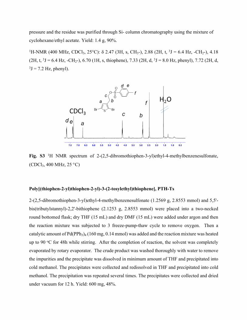
1H-NMR (400 MHz, CDCl3, 25°C): δ 2.47 (3H, s, CH3-), 2.88 (2H, t, 3J = 6.4 Hz, -CH2-), 4.18
(2H, t, 3J = 6.4 Hz, -CH2-), 6.70 (1H, s, thiophene), 7.33 (2H, d, 3J = 8.0 Hz, phenyl), 7.72 (2H, d, 3J = 7.2 Hz, phenyl).
Fig. S3 1H NMR spectrum of 2-(2,5-dibromothiophen-3-yl)ethyl-4-methylbenzenesulfonate,
(CDCl3, 400 MHz, 25 C)
Poly[(thiophen-2-yl)thiophen-2-yl)-3-(2-tosylethyl)thiophene], PTH-Ts
2-(2,5-dibromothiophen-3-yl)ethyl-4-methylbenzenesulfonate (1.2569 g, 2.8553 mmol) and 5,5'-
bis(tributylstannyl)-2,2'-bithiophene (2.1253 g, 2.8553 mmol) were placed into a two-necked
round bottomed flask; dry THF (15 mL) and dry DMF (15 mL) were added under argon and then
the reaction mixture was subjected to 3 freeze-pump-thaw cycle to remove oxygen. Then a
catalytic amount of Pd(PPh3)4 (160 mg, 0.14 mmol) was added and the reaction mixture was heated
up to 90 oC for 48h while stirring. After the completion of reaction, the solvent was completely
evaporated by rotary evaporator. The crude product was washed thoroughly with water to remove
the impurities and the precipitate was dissolved in minimum amount of THF and precipitated into
cold methanol. The precipitates were collected and redissolved in THF and precipitated into cold
methanol. The precipitation was repeated several times. The precipitates were collected and dried
under vacuum for 12 h. Yield: 600 mg, 48%.

Gel-permeation chromatography (GPC): Mn= 20747 g mol-1 , Mw= 23015 g mol-1 (Polystyrene as
standard).
Fig. S4 1H NMR spectrum of Poly[(thiophen-2-yl)thiophen-2-yl)-3-(2-tosylethyl)thiophene],
PTH-Ts, (d6-DMSO, 400 MHz, 25 C).
Synthesis and characterization of poly[(2-azidoethyl)-2-(5-(thiophen-2-yl)thiophen-2-
yl)thiophene (PTH-N3 )
Poly[(thiophen-2-yl)thiophen-2-yl)-3-(2-tosylethyl)thiophene], PTH-Ts (100 mg, 0.232 mmol)
and NaN3 (23 mg, 0.35 mmol,) was dissolved in degassed DMF (15 mL) and then the reaction
mixture was sonicated for a while and allowed to stir at 25 C for 72 h. After the completion of
reaction, the solvent was completely evaporated by rotary evaporator. The crude product was
washed with water thoroughly. The precipitate was dissolved in minimum amount of THF and
precipitated in water, the precipitated pure polymer was filtered and dried. Yield: 53 mg, 75%. IR
(Solid state, KBr): ν (cm-1): 3030, (aromatic), 2919, 2848 (-CH stretching), 2098 (azide). 1H-NMR
(400 MHz, CDCl3, 25°C): 1.78-2.00 (m, 2H), 2.98-(m, 2H), 7.40-7.95 (m, aromatic, 6H).

cb a
S S S
N3a bcccc c
DMSO
H2O
Fig. S1 1H NMR spectrum of poly[(2-azidoethyl)-2-(5-(thiophen-2-yl)thiophen-2-yl)thiophene
(d6-DMSO, 400 MHz, 25 C)
Preparation of nanoparticles:
Synthesis and characterization of PFB-N3 NPs
0.25 mg of PTB-N3 was dissolved in 2 mL THF and ultrasonicated for 10 min. The solution was
then filtered through 0.45 μm syringe filter and injected into 20 mL of Milli-Q water (18.2 MΩ)
and ultrasonicated for 40 min. THF was removed by vacuum evaporator at 40 oC to obtain stable
NPs. Characterization of NPs was done by UV-Vis spectroscopy, fluorescence spectroscopy, DLS
(Zeta sizer), SEM and TEM.
Synthesis and characterization of PFBT-P NPs
0.25 mg of PFBT-P was dissolved in 2 mL THF and ultrasonicated for 10 min. The solution was
then filtered through 0.45 μm syringe filter several times until clear solution was obtained. The
clear solution was injected into 20 mL of Milli-Q water (18.2 MΩ) and ultrasonicated for 40 min.
THF was removed by vacuum evaporator at 40 oC to obtain stable NPs. Characterization of NPs
was done by UV-vis spectroscopy, fluorescence spectroscopy, DLS (Zeta sizer), SEM and TEM.
All the results confirmed formation of stable NPs. However, the emission intensity decreased upon
NP formation.
White-emitting bi-polymer nanoparticles

Four methods were used to design bi-polymer NP dispersion in water. PFB-N3 and PFBT-P were
chosen because of the strong spectral overlap. The amount of donor in each sample was kept
constant.
Method 1
0.5 mg of PFB-N3 polymer was dissolved in 2 mL THF and ultrasonicated for 10 min. The solution
was then filtered through 0.45 μm syringe filter and injected into 20 mL of Milli-Q water (18.2
MΩ) and ultrasonicated for 40 min. THF was removed by vacuum evaporator at 40oC to obtain
stable NPs. Similarly, 0.5 mg (45 mol%) PFBT-P polymer was used to prepare acceptor NPs. The
two NPs were then mix and denoted NP mixed.
Method 2
0.5 mg PFB-N3 and 0.5 mg (45 mol%) PFBT-P were dissolved in 2 mL THF and ultrasonicated
for 10 min. The solution was then filtered through 0.45 μm syringe filter and injected into 20 mL
of Milli-Q water (18.2 MΩ) and ultrasonicated for 1hr. THF was removed by vacuum evaporator
at 40 oC to obtain stable NPs. The resulting NPs was abbreviated as Soln NPs.
Method 3
In this design, bi-polymer NPs were formed sequentially. First, 0.5 mg of PFB-N3 (D) polymer
was dissolved in 2 mL THF and ultrasonicated for 10min. The solution was then filtered through
0.45 μm syringe filter and injected into 20 mL of Milli-Q water (18.2 MΩ) and ultrasonicated for
40min. After 40 min of ultrasonication, a filtered solution of 0.5 mg (45 mol%) PFBT-P (A) in 2
mL THF was added into the preformed PFB-N3 NPs while ultrasonicating. Ultasonication was
continued for another 40 min. THF was removed by vacuum evaporator at 40 oC to obtain stable
NPs. The resulting bi-polymer NPs was denoted DA 45%, donor being core and surrounded with
45 mol% acceptor. Also DA 10% and DA 62% were prepared similarly with the same donor
concentration (0.5 mg) but 10 mol% and 62 mol% of acceptor respectively.
Method 4
In this design, bi-polymer NPs were formed sequentially. First, 0.5 mg (45 mol %) of PFBT-P (A)
polymer was dissolved in 2 mL THF and ultrasonicated for 10 min. The solution was then filtered
through 0.45 μm syringe filter and injected into 20 mL of Milli-Q water (18.2 MΩ) and
ultrasonicated for 40 min. After 40 min. of ultrasonication, a filtered solution of 0.5 mg PFB-N3
(D) in 2 mL THF was added into the preformed PFBT-P NPs while ultrasonicating. Ultasonication
was continued for another 40 min. THF was removed by vacuum evaporator at 40 oC to obtain
stable NPs. The resulting bi-polymer NPs was denoted AD 45%, acceptor being core and
surrounded with donor.
White-emitting tandem nanoparticles
White emitting tandem NPs were obtained using three polymers: PFB-N3, PFBT-P and PTH-N3
polymers. Four methods of nanostructured designs were prepared and investigated.
Method 1
0.5 mg PFB-N3, 0.25 mg PFBT-P and 0.12 mg PTH-N3 polymers were converted separately into
their respective NP form by reprecipitation as described earlier. The resulting NPs of the three
polymers were mixed physically and denoted NP Mixed.
Method 2
0.5 mg PFB-N3, 0.25 mg PFBT-P and 0.12 mg PTH-N3 polymers were dissolved in 2 mL THF
and ultrasonicated for 10 min. The solution was then filtered through 0.45 μm syringe filter and
injected into 20 mL of Milli-Q water (18.2 MΩ) and ultrasonicated for 1hr. THF was removed by
vacuum evaporator at 40 oC to obtain stable NPs. The resulting NPs was donated T.soln NPs.
Method 3
In this design, tandem NPs was formed sequentially. First, 0.5 mg of PFB-N3 (D) polymer was
dissolved in 2 mL THF and ultrasonicated for 10min. The solution was then filtered through 0.45
μm syringe filter and injected into 20 mL of Milli-Q water (18.2 MΩ) and ultrasonicated for 40
min. After 40 min of ultrasonication, a filtered solution of 0.25 mg (24 mol%) PFBT-P (A) in 2
mL THF was added into the preformed PFB-N3 NPs while ultrasonicating. Ultasonication was
continued for 30 min. Finally, 0.12 mg (18 mol %) PTH-N3 solution in 2 mL THF was also added
to the preformed bi-polymer NPs and ultasonicated for another 30 min. THF was removed by
vacuum evaporator at 40 oC to obtain stable NPs. The resulting tandem NPs was denoted DAR.
For DAR 12%, DAR 6% and DAR 4%, the amount of PFBN3 was constant while PFBT-P was

0.06 mg (9 mol %), 0.03 mg (4.5 mol %) and 0.2 mg (3 mol %) and PTN3 was 0.01 mg (2 mol
%), 0.006 mg (1.5 mol %) and 0.0033 mg (1 mol %) respectively.
Method 4
This design is the reverse of the third method; here tandem NPs was also formed sequentially.
First, 0.12 mg (18 mol %) PTN3 polymer was dissolved in 2 mL THF and ultrasonicated for 10
min. The solution was then filtered through 0.45 μm syringe filter and injected into 20 mL of Milli-
Q water (18.2 MΩ) and ultrasonicated for 40 min. After 40 min of ultrasonication, a filtered
solution of 0.25 mg (24 mol %) PFBT-P (A) in 2 mL THF was added into the preformed PTH-N3
NPs while ultrasonicating. Ultasonication was continued for 30 min. Finally, 0.5 mg PFB-N3
solution in 2 mL THF was also added to the preformed bi-polymer NPs and ultrasonicated for
another 30 min. THF was removed by vacuum evaporator at 40 oC to obtain stable NPs. The
resulting tandem NPs was denoted RAD.
Table S1 Summary of DLS data for polymer nanoparticles
Size by DLS
(nm)
SEM TEM PDI
PFB-N3-NP 64 ˅ ˅ 0.190
PFBT-P-NP 63 ˅ ˅ 0.216
PTH-N3-NP 80 0.272
Soln-NP (with catalyst) 74 0.245
D-A (with catalyst) 100 ˅ ˅ 0.204
A-D (with catalyst) 97 0.166
cDA 45 (W/O catalyst) 99 v 0.204
cAD (W/O catalyst) 102 0.253
DAR 12% 116 ˅ ˅ 0.159
DAR 6% 115 ˅ ˅ 0.185
DAR 4% 101 ˅ ˅ 0.129

D
A
DA
D
A
DA
AD
SEM TEM
A
DLSD
A
DA
100nm
100nm
100nm
Fig. S2 DLS, SEM and TEM image of donor (PFB-N3), acceptor (PFBT-P) and bipolymer
nanoparticles, design 3 (DA 45%)
Fig. S7 DLS histograms of nanoparticles of cAD 45% NP prepared without catalyst; AD 45%
NP prepared with catalyst; Soln NP prepared with catalyst Cu(I).

Fig. S8 DLS of polythiophene nanoparticles (PTN3)
300 350 400 450 500 550 600 6500.0
0.1
0.2
0.3
0.4 cDA 45% PL@350nm PL@444nm
Wavelength(nm)
Abso
rban
ce(a
.u)
0
200
400
600
800
PL Intensity(a.u)
300 350 400 450 500 550 600 6500.0
0.1
0.2
0.3 cAD 45% PL@350nm PL@444nm
Wavelength(nm)
Abso
rban
ce(a
.u)
0
100
200
300
400
500
PL Intensity(a.u)
300 350 400 450 500 550 600 650 7000.00
0.04
0.08
0.12 cBipolymer soln NP PL @350nm PL @444nm
Wavelengt(nm)
Abso
rban
ce (a
.u)
0
200
400
600
800
1000
PL Intensity (a.u)535
A B
C D
Fig. S9 Absorbance and emission spectra of catalyst free bi-polymer NPs (a) c-soln NP (NPs
prepared from the solutions of PFB-N3+ PFBT-P without copper catalysts) (b) cDA 45 NPs (c)
cAD 45 NPs (d)The size of cDA 45 NPs from DLS measurements (98.76nm)
350 400 450 500 550 600 650 7000.00
0.02
0.04
0.06 D-A np 62% PL@ 350nm PL@444nm
Wavelength (nm)
Norm
alize
d Ab
s. (a
.u)
0
25
50
75
100
125
150
175
PL Intensity (a.u)
535
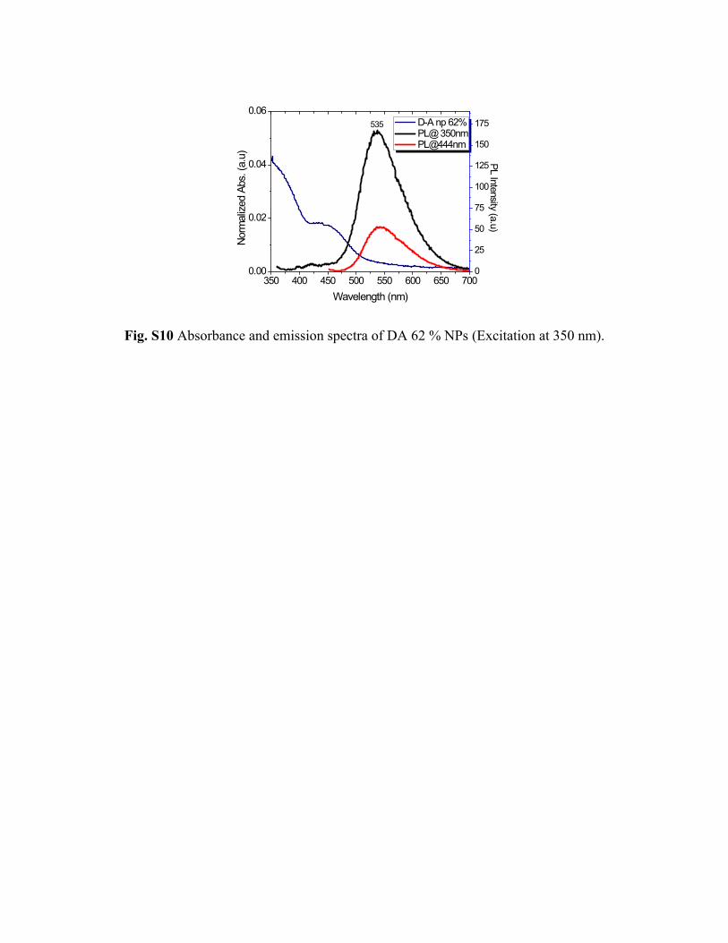
Fig. S10 Absorbance and emission spectra of DA 62 % NPs (Excitation at 350 nm).
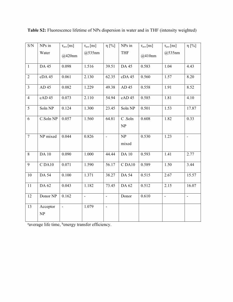
Table S2: Fluorescence lifetime of NPs dispersion in water and in THF (intensity weighted)
aaverage life time, benergy transfer efficiency.
S/N NPs in
Water
τavr [ns]
@420nm
τavr [ns]
@535nm
η [%] NPs in
THF
τavr [ns]
@410nm
τavr [ns]
@535nm
η [%]
1 DA 45 0.098 1.516 39.51 DA 45 0.583 1.04 4.43
2 cDA 45 0.061 2.130 62.35 cDA 45 0.560 1.57 8.20
3 AD 45 0.082 1.229 49.38 AD 45 0.558 1.91 8.52
4 cAD 45 0.073 2.110 54.94 cAD 45 0.585 1.81 4.10
5 Soln NP 0.124 1.300 23.45 Soln NP 0.501 1.53 17.87
6 C.Soln NP 0.057 1.560 64.81 C .Soln
NP
0.608 1.82 0.33
7 NP mixed 0.044 0.826 - NP
mixed
0.530 1.23 -
8 DA 10 0.090 1.000 44.44 DA 10 0.593 1.41 2.77
9 C DA10 0.071 1.590 56.17 C DA10 0.589 1.50 3.44
10 DA 54 0.100 1.371 38.27 DA 54 0.515 2.67 15.57
11 DA 62 0.043 1.182 73.45 DA 62 0.512 2.15 16.07
12 Donor NP 0.162 - - Donor 0.610 - -
13 Acceptor
NP
- 1.079 -
3 6 9 12 15
10
100
1000
Cou
nts
Time(ns)
Nps in THF@ 410nm Donor DA 10 THF DA 54 THF AD 45 THF DA 45 THF DA 62 THF Polym. soln mixed Soln NP THF IRF
3 6 9 12 15
10
100
1000
Cou
nts
Time(ns)
Nps in THF@535nm DA 10 THF DA 54 THF AD 45 THF DA 45 THF Polym. Soln Soln NP THF DA 62 THF Acceptor IRF
2 4 6 8 10
10
100
1000
Cou
nts
Time (ns)
Control NPs in THF@ 410nm cAD 45 THF cDA 10 in THF cDA 45 THF c Soln NP THF Polym. soln mixed Donor IRF
3 6 9 12 15
10
100
1000
Coun
ts
Time(ns)
Control Np in THF@ 535nm CDA 10 THF CAD 45 THF CDA 45 THF C Soln NP THF Polym.soln Acceptor IRF
(a) (b)
(d)(c)
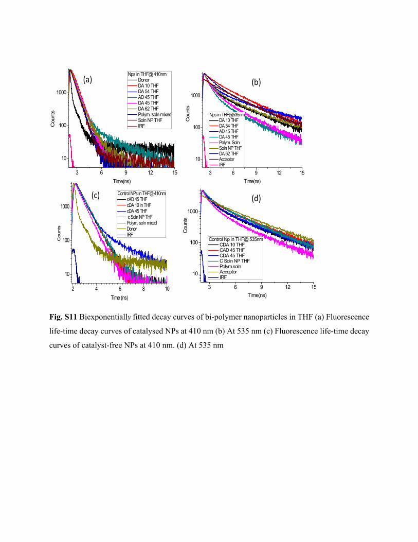
Fig. S11 Biexponentially fitted decay curves of bi-polymer nanoparticles in THF (a) Fluorescence
life-time decay curves of catalysed NPs at 410 nm (b) At 535 nm (c) Fluorescence life-time decay
curves of catalyst-free NPs at 410 nm. (d) At 535 nm

AD Br
AD N3
(a)
(c) (d)
(b)
Fig. S12 (a) DLS: Zeta size of AD Br NP from non-functionalized polymers (115 nm) (b) SEM
micrograph of AD Br (c) DLS: Zeta size of AD N3 formed by click reaction (106 nm) (d) SEM
micrograph of AD N3.
Fig. S13 SEM micrograph of bi-polymer NPs dispersed in THF and emission spectra of re-
dispersed bipolymer NPs in water including ADBr (λex=350 nm).

Table S3 Time resolved-fluorescence life time of tandem nanoparticles in water (intensity
weighted)
aaverage life time, benergy transfer efficiency.
Fig. S14 (a) SEM micrograph of DAR 12% (b) TEM micrograph of DAR 12% (c) SEM
micrograph of DAR 6% (d) TEM micrograph of DAR 6%
S/N NPs in water τavr[ns]
@420nm
τavr[ns]
@535nm
η[%]
1. RAD 0.055 1.692 66
2. T Sol 0.048 1.287 70
3. T.NPs mixed 0.158 0.091 -
4. DAR 12 0.101 1.908 38
5. DAR 6 0.108 2.461 33
6. DAR 4 0.126 2.282 22
7. Donor NP 0.162 -

(a)
(b) (c)
Fig. S15 Size and morphology of DAR 4% NPs (a) DLS (b) SEM and (c) TEM image
Fig. S16 DLS histograms of DAR 6% NPs of DAR 12% NPs.

Fig. S17 The emission spectra of tandem NPs with their corresponding emission colors. (λex=350
nm)
Table S4: x-y Colour coordinates at varying currents.
Current
(mA)
20 30 40 50 60
x 0.3502 0.3511 0.3506 0.3496 0.3504
y 0.3846 0.33857 0.3879 0.3852 0.3878
Fig. S18 (a) The spectra and (b) CRI, CQS, and CCT of the DAR 4% catalyst tandem nanoparticle integrated white LED at varying current levels.

Fig. S19 The chromaticity point of the DAR 4% catalyst tandem nanoparticle integrated LED on CIE 1931 chromaticity coordinate.
References:
1) V. İbrahimova, S. Ekiz, O. Gezici, D. Tuncel, Polym. Chem., 2011, 2, 2818-2824.
2) E.J. Park, T. Erdem, V. Ibrahimova, S. Nizamoglu, H.V. Demir, D. Tuncel, ACS Nano, 2011,
5, 2483–2492.
3) J. Pennakalathil, A. Özgün, I. Durmaz, R. Cetin-Atalay, D. Tuncel, J. Polym. Sci. A Polym.
Chem., 2015, 53, 114-122.
4) B. Baykal, V. Ibrahimova, G. Er, E. Bengü and D. Tuncel, Chem. Comm., 2010, 46, 6762-
6764.
5) T. Erdem, I. Ibrahimova, D. W. Jeon, I. H. Lee, D. Tuncel, H. V. Demir, J. of Phys. Chem. C,
2013, 117, 18613-18619.


![Electronic Supplementary Information (ESI) Supplementary Information (ESI) for Enzyme-Responsive Supramolecular Nanovalves Crafted by Mesoporous Silica Nanoparticles and Choline-Sulfonatocalix[4]arene](https://static.fdocuments.net/doc/165x107/5acfea197f8b9ae2138d1507/electronic-supplementary-information-esi-supplementary-information-esi-for-enzyme-responsive.jpg)