Electron Microscopy
17
HL BIOLOGY CELLS & BIOMOLECULES ELECTRON MICROSCOPY
-
Upload
romy-friedman -
Category
Environment
-
view
246 -
download
1
Transcript of Electron Microscopy
H L B I O L O G Y
C E L L S & B I O M O L E C U L E S
ELECTRON MICROSCOPY
LIMITATIONS OF THE LIGHT
MICROSCOPE
• Light microscopes rely on visible light being refracted to
magnify the image.
• Scientists were unable to study organelles without the
invention of a new, higher powered microscope.
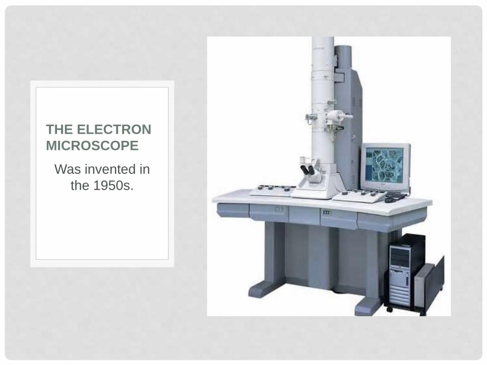
Was invented in
the 1950s.
THE ELECTRON
MICROSCOPE
THE ELECTRON MICROSCOPE
• Rather than light, an electron microscope focuses a
beam of electrons onto or through the specimen.
• Modern electron microscopes are capable of obtaining
resolutions up to 100 times that of standard light
microscopes.
THE ELECTRON MICROSCOPE
• Electron microscopes come in 2 varieties:
• Scanning electron microscopes (SEM)
• Transmission electron microscopes (TEM)
Both are used in Biology for today, but for
different purposes.
SCANNING ELECTRON MICROSCOPES
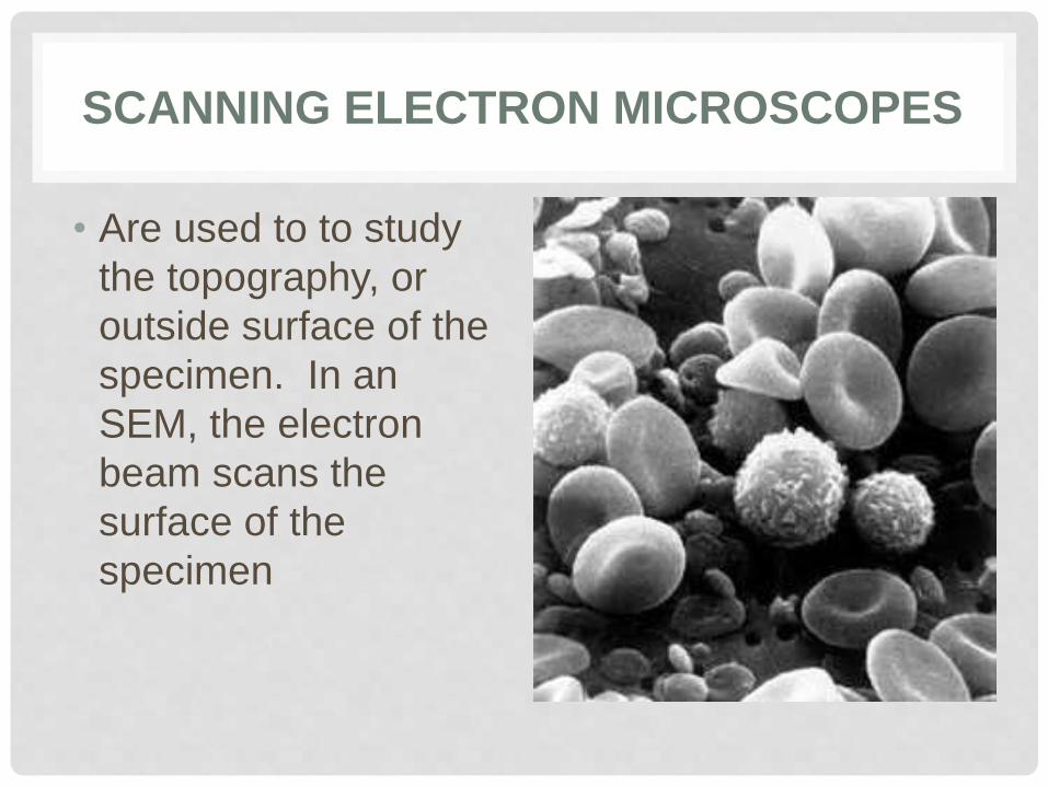
• Are used to to study
the topography, or
outside surface of the
specimen. In an
SEM, the electron
beam scans the
surface of the
specimen
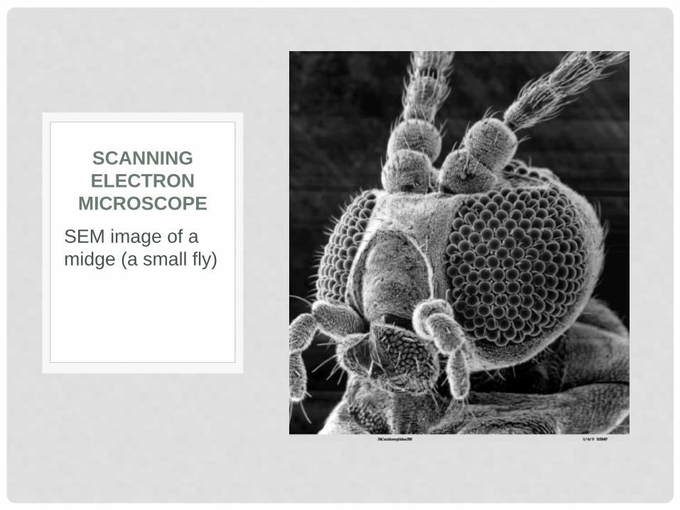
SEM image of a
midge (a small fly)
SCANNING
ELECTRON
MICROSCOPE
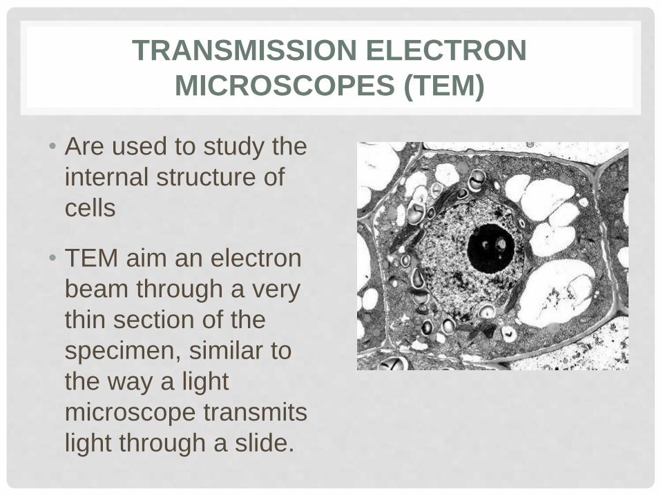
TRANSMISSION ELECTRON
MICROSCOPES (TEM)
• Are used to study the
internal structure of
cells
• TEM aim an electron
beam through a very
thin section of the
specimen, similar to
the way a light
microscope transmits
light through a slide.
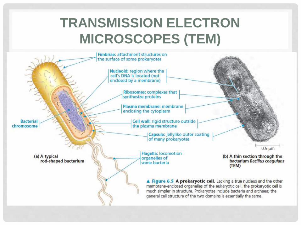
TRANSMISSION ELECTRON
MICROSCOPES (TEM)
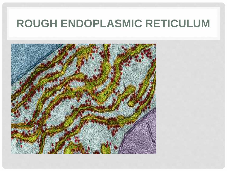
ROUGH ENDOPLASMIC RETICULUM
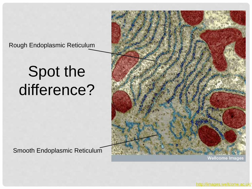
http://images.wellcome.ac.uk/
Rough Endoplasmic Reticulum
Smooth Endoplasmic Reticulum
Spot the
difference?
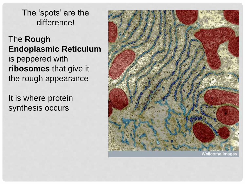
The ‘spots’ are the
difference!
The Rough
Endoplasmic Reticulum
is peppered with
ribosomes that give it
the rough appearance
It is where protein
synthesis occurs
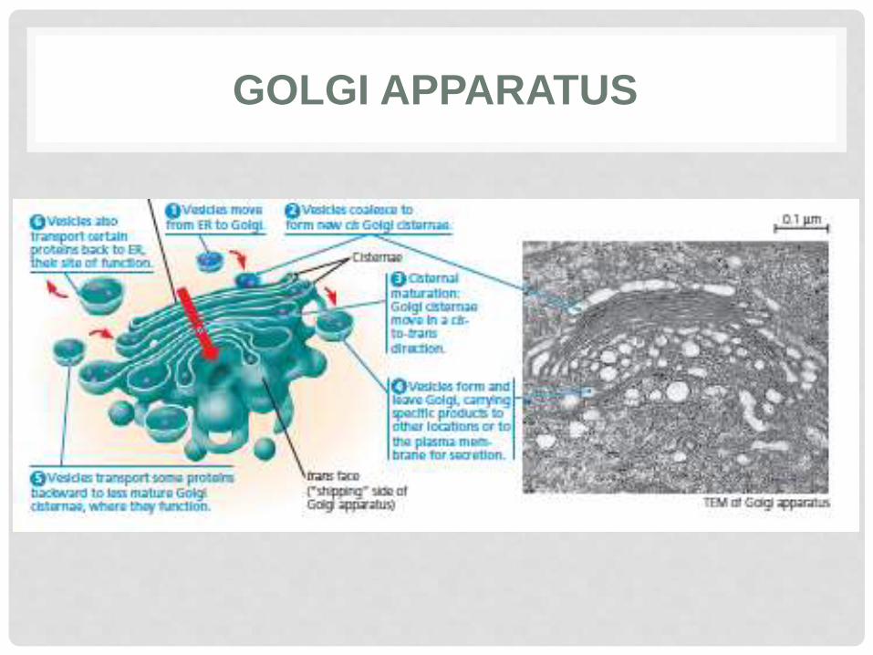
GOLGI APPARATUS
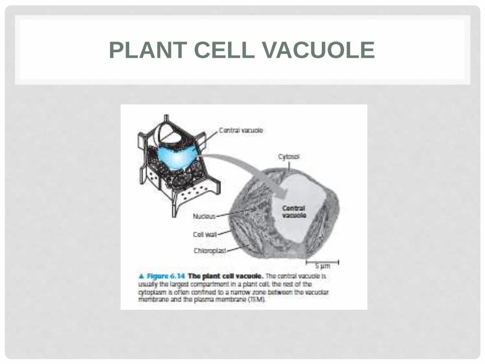
PLANT CELL VACUOLE
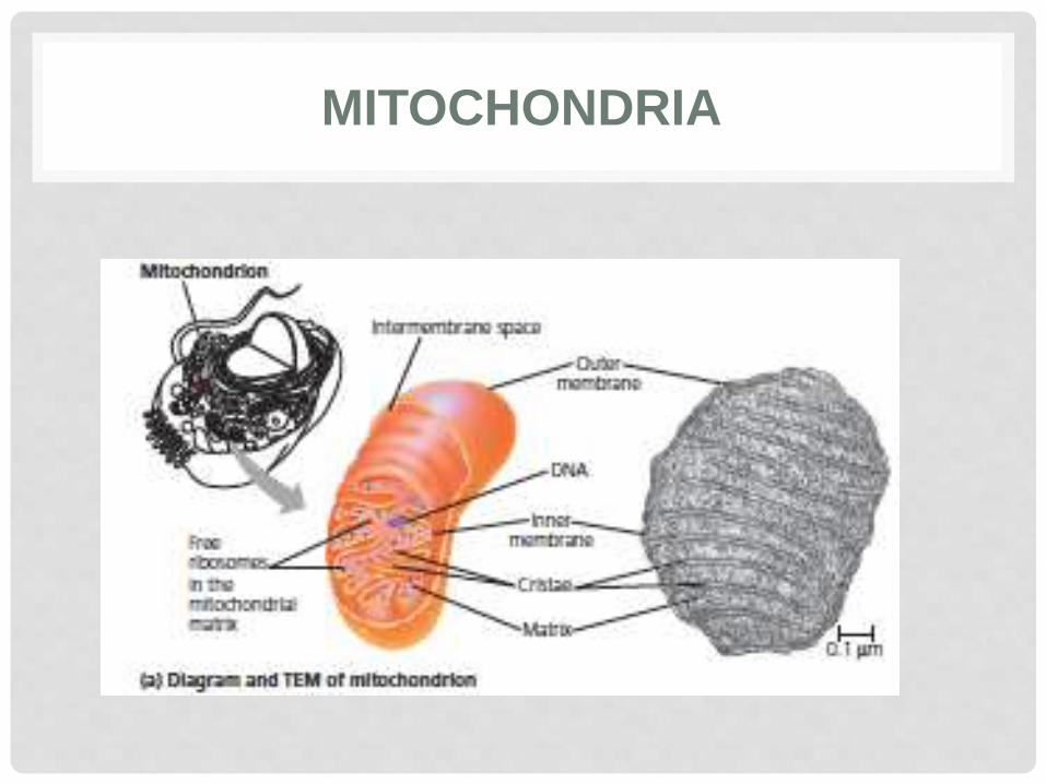
MITOCHONDRIA
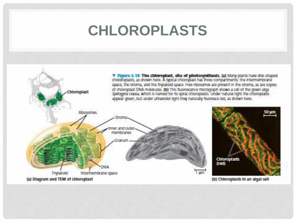
CHLOROPLASTS

What can you
see?