Electromyograms as Physiological Inputs that … as Physiological Inputs that Provide Efficient ......
6
Electromyograms as Physiological Inputs that Provide Efficient Computer Cursor Control Craig Chin and Armando Barreto Electrical and Computer Engineering Department Florida International University 10555 West Flagler St., Miami, FL 33173 USA [email protected] Abstract: - This paper presents a new algorithm to translate electromyogram (EMG) signals, produced during facial muscle contractions, into computer cursor actions. These cursor actions are: left, right, up, down and left-click. The translation is performed in real-time and the classification of the EMG signals is based on features derived from the spectral analysis of the electromyograms. We have sought to improve upon the original EMG-based cursor control system described in [1] [2], and have devised a new system set-up and algorithm for the translation of the EMG signals into cursor actions. The two systems were compared using Matlab simulations and point-and-click trials. The simulation results show a marked improvement of the new system over the previous system in terms of classification accuracy, and the preliminary point-and-click tests indicate the possibility of a faster operation for the new system when compared to the old system. Key-Words: - Electromyogram, EMG, human, computer, interface, spectral, analysis 1 Introduction Typically able-bodied individuals communicate with a computer using standard input devices, such as, a mouse, trackball, touchpad, or keyboard. The motivation for investigating alternative means for communicating with the computer is that there exists a population of individuals who are unable to use such devices due to some form of physical disability. It is estimated that there are 250,000 – 400,000 individuals in the United States living with spinal cord injury or spinal dysfunction [11]. Given the increasing pervasiveness of computer-based systems in most of our daily activities, and the increasing levels of communication and social participation that take place over the Internet, it is clear that facilitating access of these individuals to Graphical User Interface (GUI)-driven computer systems is an important technical goal. With today’s GUI-based PC software, most of the human-to-computer interaction is based on selection operations, which consists of two steps: • Pointing: Positioning the cursor at the desired location of the screen, over the appropriate area or icon. • Clicking: Executing the Mouse Down/Up function that is interpreted by the computer’s operating system as an indicator to complete the selection of the item associated with the icon at the location of the screen cursor. There have been a number of approaches that have attempted to make these operations available to individuals with severe motors disabilities. Several of these devices target motor skills that are still available to some users. One such approach utilizes the user’s ability to focus his line of gaze on an area of interest using his eyes. This approach is called eye-gaze tracking (EGT). EGT techniques seek to determine the user’s visual line of gaze by taking video images of the eye in order to establish a mapping between the geometric properties of the eye and the line of gaze. The most popular EGT technique at present uses the relative position of the bright eye (pupil) center and the center of the glint (corneal reflection) to determine the line of gaze [6] [8] [9] [10] [13]. Once the line of gaze is determined, the point of gaze is found by allowing the line of gaze to intersect with the plane of the scene being viewed (typically the computer screen). This approach has been shown to perform faster than a mouse in object selection tests (Sibert & Jacob, 2000). However, the approach has some disadvantages. One such disadvantage is the so- called “Midas Touch” problem [8] [9]. The problem originates from the use of eye gaze as an object selection technique. Since there may be situations where a user may only desire to stare at an object to examine it, rather than to select it, an eye gaze-based object selection technique may result in unintended selections. Another disadvantage is the limited accuracy inherent to point of gaze estimation. This limitation is rooted in the fact that the eye only Proceedings of the 2006 WSEAS International Conference on Mathematical Biology and Ecology, Miami, Florida, USA, January 18-20, 2006 (pp221-226)
Transcript of Electromyograms as Physiological Inputs that … as Physiological Inputs that Provide Efficient ......
Electromyograms as Physiological Inputs that Provide Efficient Computer Cursor Control
Craig Chin and Armando Barreto
Electrical and Computer Engineering Department Florida International University
10555 West Flagler St., Miami, FL 33173 USA
Abstract: - This paper presents a new algorithm to translate electromyogram (EMG) signals, produced during facial muscle contractions, into computer cursor actions. These cursor actions are: left, right, up, down and left-click. The translation is performed in real-time and the classification of the EMG signals is based on features derived from the spectral analysis of the electromyograms. We have sought to improve upon the original EMG-based cursor control system described in [1] [2], and have devised a new system set-up and algorithm for the translation of the EMG signals into cursor actions. The two systems were compared using Matlab simulations and point-and-click trials. The simulation results show a marked improvement of the new system over the previous system in terms of classification accuracy, and the preliminary point-and-click tests indicate the possibility of a faster operation for the new system when compared to the old system. Key-Words: - Electromyogram, EMG, human, computer, interface, spectral, analysis 1 Introduction Typically able-bodied individuals communicate with a computer using standard input devices, such as, a mouse, trackball, touchpad, or keyboard. The motivation for investigating alternative means for communicating with the computer is that there exists a population of individuals who are unable to use such devices due to some form of physical disability. It is estimated that there are 250,000 – 400,000 individuals in the United States living with spinal cord injury or spinal dysfunction [11]. Given the increasing pervasiveness of computer-based systems in most of our daily activities, and the increasing levels of communication and social participation that take place over the Internet, it is clear that facilitating access of these individuals to Graphical User Interface (GUI)-driven computer systems is an important technical goal.
With today’s GUI-based PC software, most of the human-to-computer interaction is based on selection operations, which consists of two steps:
• Pointing: Positioning the cursor at the desired location of the screen, over the appropriate area or icon.
• Clicking: Executing the Mouse Down/Up function that is interpreted by the computer’s operating system as an indicator to complete the selection of the item associated with the icon at the location of the screen cursor.
There have been a number of approaches that have attempted to make these operations available to individuals with severe motors disabilities. Several of these devices target motor skills that are still available to some users. One such approach utilizes the user’s ability to focus his line of gaze on an area of interest using his eyes. This approach is called eye-gaze tracking (EGT). EGT techniques seek to determine the user’s visual line of gaze by taking video images of the eye in order to establish a mapping between the geometric properties of the eye and the line of gaze. The most popular EGT technique at present uses the relative position of the bright eye (pupil) center and the center of the glint (corneal reflection) to determine the line of gaze [6] [8] [9] [10] [13]. Once the line of gaze is determined, the point of gaze is found by allowing the line of gaze to intersect with the plane of the scene being viewed (typically the computer screen).
This approach has been shown to perform faster than a mouse in object selection tests (Sibert & Jacob, 2000). However, the approach has some disadvantages. One such disadvantage is the so-called “Midas Touch” problem [8] [9]. The problem originates from the use of eye gaze as an object selection technique. Since there may be situations where a user may only desire to stare at an object to examine it, rather than to select it, an eye gaze-based object selection technique may result in unintended selections. Another disadvantage is the limited accuracy inherent to point of gaze estimation. This limitation is rooted in the fact that the eye only
Proceedings of the 2006 WSEAS International Conference on Mathematical Biology and Ecology, Miami, Florida, USA, January 18-20, 2006 (pp221-226)
needs to focus incoming light on a small area of the retina called the fovea, in order to see objects clearly. For an object to be focused on the fovea, it must fall within an area covered by approximately one degree of visual arc [9] [13]. This physical constraint limits the accuracy with which the line of gaze can be estimated.
A different class of alternative cursor control approaches seek to drive the manipulation of the cursor from electrophysiological signals that occur “naturally” in the user, such as the person’s electroencephalogram (EEG) or electromyogram (EMG). Frequently, approaches that monitor and process electrophysiological signals from the brain to communicate messages or commands to a computer are called “Brain-Computer Interfaces” (BCIs). Present day independent BCIs can be classified by the form of physiological signal that they use to determine user intent. These signals include: slow cortical potentials, P300 evoked potentials, mu and beta rhythms, and cortical neuronal activity recorded from electrodes implanted in the scalp [14].
It has been found that movement or preparation for movement is accompanied by a decrease in the mu and beta rhythms, especially in the region of the brain contralateral to the movement. This phenomenon is called “event-related desynchronization” (ERD). In addition, it has been observed that there is mu rhythm increase or “event-related synchronization” (ERS) after a movement and with relaxation. It has also been found that ERD and ERS do not require actual movement, but can accompany imagined movement. These facts make mu/beta rhythms suitable for input into a BCI, and work by Fabiani et al. [5], 2004), and Pfurtscheller et al. [12] has focused on their use as a source of cursor control.
The major advantage of using a BCI system as an assistive technology for individuals with motor disabilities is that it does not require the brain’s normal output pathways to produce its control signals; neither does it require activity in these pathways to generate the control signals. However, present day BCI systems are primarily limited by speed of operation. Current BCIs have maximum information transfer rates of 10 - 25bits/min [14].
Electromyography is the study of muscle function through monitoring of the electrical signals generated by the muscle [3]. When a surface electrode is placed on the skin above a superficial muscle while it is contracting, it will receive electrical signals emanating from several muscle fibers associated with different motor units. The spatio-temporal summation of these electrical
signals results in what is called an EMG signal. Therefore, the EMG signal provides an effective means of monitoring muscle activity.
EMG signals have also been used for cursor control. This approach has been used in [7] [15] and [1] [2], with [1] [2] focused specifically on the use of EMG from cranial muscles. Monitoring the EMG of cranial muscles makes the approach suitable for individuals suffering from severe motor disabilities, who are paralyzed from the neck down.
The advantage of EMG-based cursor control is that it provides the user with the ability to perform small cursor movements, unlike EGT systems. However, it has been shown that this approach performs slowly compared to a mouse-operated system in object selection tests [1] [2].
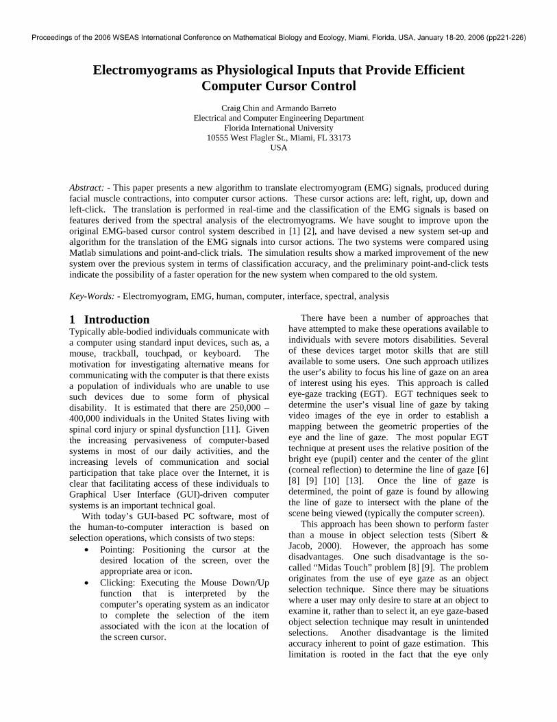
The EMG system used in [1] [2] utilized three electrodes that measured EMG signals from muscles in the head of the user. The EMG signals were classified into cursor actions by performing real-time spectral analysis of these signals. Spectral analysis of the EMG signals from different muscles revealed that they possessed distinguishing frequency characteristics. An example of this is given in Fig. 1.
0 200 400 6000
0.1
0.2
0.3
0.4
0.5
0.6
0.7
0.8
0 200 400 6000
0.5
1
1.5
2
2.5
3
3.5
4
4.5
MAG
NIT
UDE
FREQUENCY (Hz)
MAG
NIT
UDE
FREQUENCY (Hz) Fig. 1 Spectra observed during a right frontalis
contraction (left plot) and left temporalis contraction (right plot)
After a thorough evaluation of the EMG system,
it was found that the three-electrode system was occasionally inaccurate in discriminating between the muscle contractions that command up and down cursor movements (eyebrows up and eyebrows down, respectively). To remedy this problem an additional electrode was added to the forehead region and a new classification algorithm was devised to work with this new input configuration.
Section 2 of this paper details how the new system was implemented and the methodology behind the new classification algorithm. Section 3 describes how the old and new EMG systems were evaluated, firstly with Matlab simulations and then
Proceedings of the 2006 WSEAS International Conference on Mathematical Biology and Ecology, Miami, Florida, USA, January 18-20, 2006 (pp221-226)

with real-time, object-selection tasks. Section 4 provides tabulated results from these tests. Section 5 presents our conclusions and goals for future work.
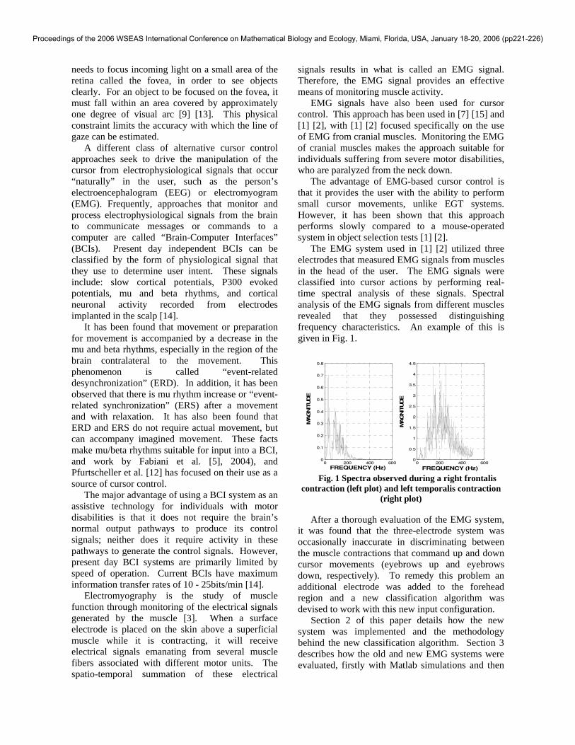
2 System Implementation and Methodology 2.1 Placement of Electrodes for the EMG-
based Cursor Control System Fig. 2 displays the placement of the Ag/AgCl electrodes on the head of the subject. Fig. 2 indicates that electrodes were placed over the right frontalis muscle, the left temporalis muscle, the right temporalis muscle, and the procerus muscle, respectively. An electrode was placed over the right mastoid as a reference.
Right Frontalis
Left Temporalis
Right Temporalis
Procerus
Right Mastoid (Reference)
Fig. 2 Electrode placement for the EMG cursor
control system 2.2 Hardware Components of the EMG-
Based Cursor Control System The hardware components of the cursor control system are presented in Fig. 3. The set of four EMG signals were input into the Grass® P5 Series AC preamplifiers. These preamplifiers were set to preprocess the signals with analog anti-aliasing filters, and with a gain of 10,000 V/V. Each preamplifier also applied a 60Hz notch-filter to each EMG channel. The ADC64TM DSP/AD board (Innovative Integration, Simi Valley, CA) performed analog-to-digital conversion on each signal at a sampling rate of 1.2 kHz, and then applied the classification algorithm to these digitized signals in real-time. The board was connected to the computer through the PCI bus. The output of the board was a series of TTL-compliant binary voltage sequences that were consistent with voltage sequences expected from a serial mouse. The Motorola® MC1488C RS-232C driver converted the TTL sequences into RS-232C format and transmitted these sequences into the serial port of the personal computer (PC). The serial mouse driver of this
computer communicated with the operating system to produce cursor actions consistent with the serial input.
Fig. 3 Block diagram of hardware components of
EMG-based cursor control system 2.3 EMG Processing Algorithm for Muscle
Contraction Identification The desired relations between cursor actions, facial movements, and muscle contractions are given in Table 1.
Table 1 Relations between cursor actions, facial movements and muscle contractions
Cursor Action
Facial Movement
Muscle Contraction
Left Left Jaw Clench Left Temporalis Right Right Jaw
Clench Right Temporalis
Up Eyebrows Up (Right) Frontalis Down Eyebrows Down Procerus Left-Click Left & Right
Jaw Clench Left & Right Temporalis
The purpose of the classification algorithm was
to determine if a facial muscle contraction had occurred and if so, which specific muscle was the source of this contraction. Given the one-to-one correspondence between muscle contraction and cursor action, the output of an effective muscle contraction classification algorithm can be utilized in a real-time implementation for hands-free cursor control.
Both the classification algorithm of [1] [2] and the classification algorithm discussed in this paper, made use of the periodogram estimation of the power spectral density (PSD) of the input EMG signals. In both cases, the PSD indicated how the power of an EMG signal was distributed over a frequency range of 0 Hz – 600 Hz. Periodogram PSD estimations were taken every 256 consecutive samples (every 0.213s) from each of the four EMG channels.
The two classification algorithms differed in the way each utilized the PSD estimates to classify the EMG data. Firstly the algorithm of [1] [2] only
Proceedings of the 2006 WSEAS International Conference on Mathematical Biology and Ecology, Miami, Florida, USA, January 18-20, 2006 (pp221-226)
utilized three electrodes, placed on the left temporalis muscle, the right temporalis muscle, and the right frontalis muscle respectively, to record EMG signals. The classification algorithm adopted for this three-electrode system, calculated partial accumulations over the frequency ranges of 0 Hz – 145 Hz and 145 Hz – 600 Hz of the PSD’s produced from the three EMG channels. These partial accumulations were used to distinguish between the frequency characteristics of a temporalis contraction as opposed to a frontalis contraction. This algorithm also utilized PSD amplitude thresholds to estimate the strength of contraction from each of the three muscles mentioned previously. The partial accumulation and threshold criteria were used to classify the facial movements: left jaw clench, right jaw clench, eyebrows up, and left and right jaw clench. The eyebrows down movement required a divergent set of classification criteria. The eyebrows down movement used a partial accumulation over the frequency range 88 Hz – 250 Hz of the PSD calculated from the frontalis electrode. In addition, it was required that the PSD amplitude thresholds of the three electrodes not be exceeded.
Testing of this algorithm revealed that it did not always classify the eyebrows down movement efficiently. So it was proposed that an additional electrode be placed over the procerus muscle, because it is one of the muscles directly involved in the eyebrows down facial movement. This new four-electrode input configuration required a new classification algorithm, the details of which are described in the following paragraphs.
It was decided that this new classification algorithm would make use of Mean Power Frequency (MPF) values as a means of distinguishing spectral differences associated with each facial muscle contraction, instead of partial PSD accumulations. The MPF is derived from the PSD values as a weighted average frequency in which each frequency component, f, is weighted by its power, P. The equation for the calculation for the MPF is given by:
⎟⎠⎞
⎜⎝⎛
+++
×++×+×=
PnPP
PnfnPfPfMPF
...21
...2211 (1)
n = 1, 2, …, 256
It has been observed previously that the spectral content of the four muscles used in this system are distinct [1] [2]. We have also confirmed this in new observations made on the subjects involved in this research. The frontalis muscle has the majority of
its spectral content below 200 Hz, with an MPF in the range 40 Hz – 165 Hz. The temporalis muscles have a significant portion of their spectral content above 200 Hz, with an MPF in the range 120 Hz – 295 Hz. The procerus muscle has an intermediate spectral content when compared to the frontalis and temporalis muscles, with an MPF in the range 60 Hz – 195 Hz.
For a unidirectional muscle contraction to be correctly classified by the four-electrode algorithm all the following criteria must be satisfied:
i. The maximum PSD amplitude must exceed the threshold set for that electrode.
ii. The sum of the PSD amplitudes for the given electrode must exceed the PSD sums of the other electrodes.
iii. The mean power frequency calculated from the PSD must fall into a range consistent with the muscle associated with the electrode.
For the classification of the bilateral contraction of the left and right temporalis muscles used to trigger the left-click cursor action, all the following conditions must apply:
i. The maximum PSD amplitude thresholds must be exceeded for both electrodes.
ii. The PSD sums for both electrodes must be greater than the other two PSD sums.
iii. The PSD sums for both electrodes must indicate a fairly balanced bilateral contraction, that is, each PSD sum must be greater than 20% of the total of both PSD sums.
iv. The mean power frequencies calculated from both PSDs must fall into a range consistent with the muscles associated with both electrodes.
2.4 Evaluation of EMG detection
Algorithms The two algorithms were evaluated by applying Matlab simulations of the algorithms to recorded data. In addition, they were evaluated using real-time implementations of the algorithms in point-and-click trials.
For the simulations, five subjects (four men and one woman, all able-bodied) were used in the testing of the algorithms. Testing involved recording facial movement sequences for each subject. Each sequence was 190s in duration. During each sequence the subject was given verbal cues to perform specific types of facial movements. There were two unique sequences given to each subject.
Proceedings of the 2006 WSEAS International Conference on Mathematical Biology and Ecology, Miami, Florida, USA, January 18-20, 2006 (pp221-226)

Each sequence was repeated twice. The ordering of facial movements in the two unique sequences is given in Table 2. Further details of these simulations are given in [4].
Table 2 The ordering of facial movement sequences
Time Sequence 1 Facial
Movements
Sequence 2 Facial Movements
0s – 20s No Movement No Movement 20s – 40s Right Clench Right Clench 40s – 50s No Movement No Movement 50s – 70s Eyebrows Up Eyebrows Up 70s – 80s No Movement No Movement 80s – 100s Left/Right
Clench Left/Right Clench
100s – 110s No Movement No Movement 110s – 130s Eyebrows Down Eyebrows Down 130s – 140s No Movement No Movement 140s – 160s Left Clench Left Clench 160s – 170s No Movement No Movement 170s – 190s No Movement Neck Movement
In order to evaluate the real-time
implementations of both algorithms, a test program was created in Visual Basic to compare their point-and-click capabilities. The program was displayed on a 17” color monitor. For each point-and-click trial, a 8.5 x 8.5 mm “Start” button was presented in a corner of the screen and a “Stop” button was presented in the center. The “Stop” button had four possible dimensions: 8.5 x 8.5 mm, 12.5 x 12.5 mm, 17 x 17 mm, 22 x 22 mm. Each subject was instructed to click the “Start” button to begin timing a trial, move the cursor to the “Stop” button, and click on it as quickly as possible. This would record the total task time for the trial. The subject would then click a “Next” button to display another trial layout with the “Start” button located in another corner of the screen. Fig. 4 shows an example layout of a point-and-click trial.
Fig. 4 Point-and-click trial layout
The real-time evaluations were divided into four sessions. During a session only one “Stop” button size was presented. The “Start” button was rotated through the four possible corners in subsequent trials, such that five trials started from each corner. Therefore, there were twenty trials per session and 80 trials per subject.
Six able-bodied, male subjects were used in the real-time evaluations of the old algorithm. Also six able-bodied, male subjects were involved in the real-time evaluations of the new algorithm. 3 Results The results of the simulation evaluations are given as classification percentages for both algorithms in Table 3.
Table 3 Summary of classification percentages on a subject-by-subject basis
Subject No. Classification Percentages (%) Three-Electrode
(“Old”) Algorithm Four-Electrode (“New”) Algorithm
Correct Incorrect Correct Incorrect 1 82.38 17.62 99.52 0.48 2 78.36 21.64 99.01 0.99 3 83.85 16.15 99.08 0.92 4 75.10 24.90 99.01 0.99 5 72.47 27.53 95.49 4.51 Average 78.43 21.57 98.42 1.58
The statistical results for the six test subjects used in the point-and-click evaluations of the two algorithms are shown in Table 4. Table 4 Preliminary statistics for point-and-click trials
Three-Electrode (“Old”) Algorithm
Four-Electrode (“New”) Algorithm
Mean Trial Time (s)
16.36 13.24
Std. Dev. (s) 7.29 3.28 4 Conclusions and Future Work The average results from Table 3 indicate that the four-electrode algorithm has a higher correct classification percentage than the three-electrode algorithm. This would lead one to conclude that the new algorithm is more accurate than the old algorithm in classifying the source of muscle activity.
In addition, the results of Table 4 show that the mean time for point-and-click trials is 3.12 s less for the four-electrode algorithm (13.24 s) when compared with the three-electrode algorithm (16.36 s). The results also show that the standard deviation of the trials for the four-electrode algorithm is about
Proceedings of the 2006 WSEAS International Conference on Mathematical Biology and Ecology, Miami, Florida, USA, January 18-20, 2006 (pp221-226)

half of the standard deviation for the three-electrode algorithm. These facts suggest that the new algorithm produces a system that enables users to complete tasks more quickly and with a higher degree of consistency (which may imply greater ease of use).
Given these favorable preliminary results, testing will continue in order to further validate the assumption that the new four-electrode system provides faster and more accurate cursor control when compared to the three-electrode system. Further testing and data analysis will involve collecting test data from a larger pool of subjects and using a t-statistic to test the significance of the difference in mean trial times between the two algorithms. Additionally, more complex test tasks could be devised to enable the performance of Fitt’s Law analysis for both cursor control approaches. 5 Acknowledgements This work was sponsored by NSF grants IIS-0308155, CNS-0520811, HRD-0317692 and CNS-0426125. References: 1. Barreto, A.B., Scargle, S.D. and Adjouadi,
M., A Practical EMG-based Human-Computer Interface for Users with Motor Disabilities, Journal Of Rehabilitation Research And Development, Vol.37, No.1, pp. 53-63.
2. Barreto, A.B., Scargle, S.D. and Adjouadi, M., A Real-Time Assistive Computer Interface for Users with Motor Disabilities, SIGCAPH Newsletter, 1999, pp. 6-16.
3. Basmajian, J.V. and Deluca, C., Muscles Alive, Williams & Wilkins, Baltimore, 1985.
4. Chin, C., Barreto, A.B., Li, C. and Zhai, J., Hands-Free Human Computer Interaction via an Electromyogram-Based Classification Algorithm, Biomedical Sciences Instrumentation, Vol.41, pp. 31-36.
5. Fabiani, G.E., McFarland, D.J., Wolpaw, J.R. and Pfurtscheller, G., Conversion of EEG activity into cursor movement by a brain-computer interface (BCI), IEEE Transactions on Neural Systems and Rehabilitation Engineering, Vol.12, No.3, pp. 331-338.
6. Hutchinson, T.E., White, K.P.J., Martin, W.N., Reichert, K.C. and Frey, L.A.,
Human-computer interaction using eye-gaze input, IEEE Transactions on Systems, Man and Cybernetics, Vol.19, No.6, pp. 1527-1534.
7. Itou, T., Terao, M., Nagata, J. and Yoshida, M., Mouse cursor control system using EMG, in 2001 Conference Proceedings of the 23rd Annual International Conference of the IEEE Engineering in Medicine and Biology Society, 2001, pp. 1368-1369.
8. Jacob, R.J.K., Hot topics-eye-gaze computer interfaces: what you look at is what you get, Computer, Vol.26, No.7, pp. 65-66.
9. Jacob, R.J.K., The use of eye movements in human-computer interaction techniques: what you look at is what you get, ACM Transactions on Information Systems, Vol.9, No.2, pp. 152-169.
10. Lankford, C., Effective eye-gaze input into Windows, in Proceedings of the symposium on Eye tracking research & applications, 2000, pp. 23-27.
11. National Spinal Cord Injury Association, Fact Sheets: Spinal Cord Injury Statistics, retrieved October 31, 2005, from http://www.spinalcord.org/html/factsheets/spinstat.php.
12. Pfurtscheller, G., Flotzinger, D. and Kalcher, J., Brain-Computer Interface - a new communication device for handicapped persons, Journal of Microcomputer Applications, Vol.16, No.3, pp. 293-299.
13. Sibert, L.E. and Jacob, R.J.K., Evaluation of eye gaze interaction, in CHI 2000 Conference Proceedings. Conference on Human Factors in Computing Systems. CHI 2000. The Future is Here, 2000, pp. 281-288.
14. Wolpaw, J.R., Birbaumer, N., McFarland, D.J., Pfurtscheller, G. and Vaughan, T.M., Brain-computer interfaces for communication and control, Clinical Neurophysiology, Vol.113, No.6, pp. 767-791.
15. Yoshida, M., Itou, T. and Nagata, J., Development of EMG controlled mouse cursor, in Conference Proceedings. Second Joint EMBS-BMES Conference 2002. 24th Annual International Conference of the Engineering in Medicine and Biology Society. Annual Fall Meeting of the Biomedical Engineering Society, 2002, pp. 2436.
Proceedings of the 2006 WSEAS International Conference on Mathematical Biology and Ecology, Miami, Florida, USA, January 18-20, 2006 (pp221-226)