ELECTROENCEPHALOGRAPHY (EEG) · Electroencephalogram EEG is the record of electrical activity of...
39
ELECTROENCEPHALOGRAPHY (EEG)
Transcript of ELECTROENCEPHALOGRAPHY (EEG) · Electroencephalogram EEG is the record of electrical activity of...
ELECTROENCEPHALOGRAPHY(EEG)
EEG
The electroencephalogram (EEG) is a recording of the electrical activity of the brain from the scalp.
The first recordings were made by Hans Berger in 1929
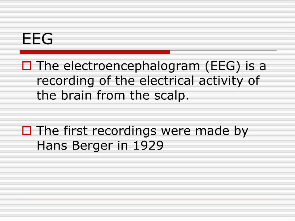
Origin of EEG waves
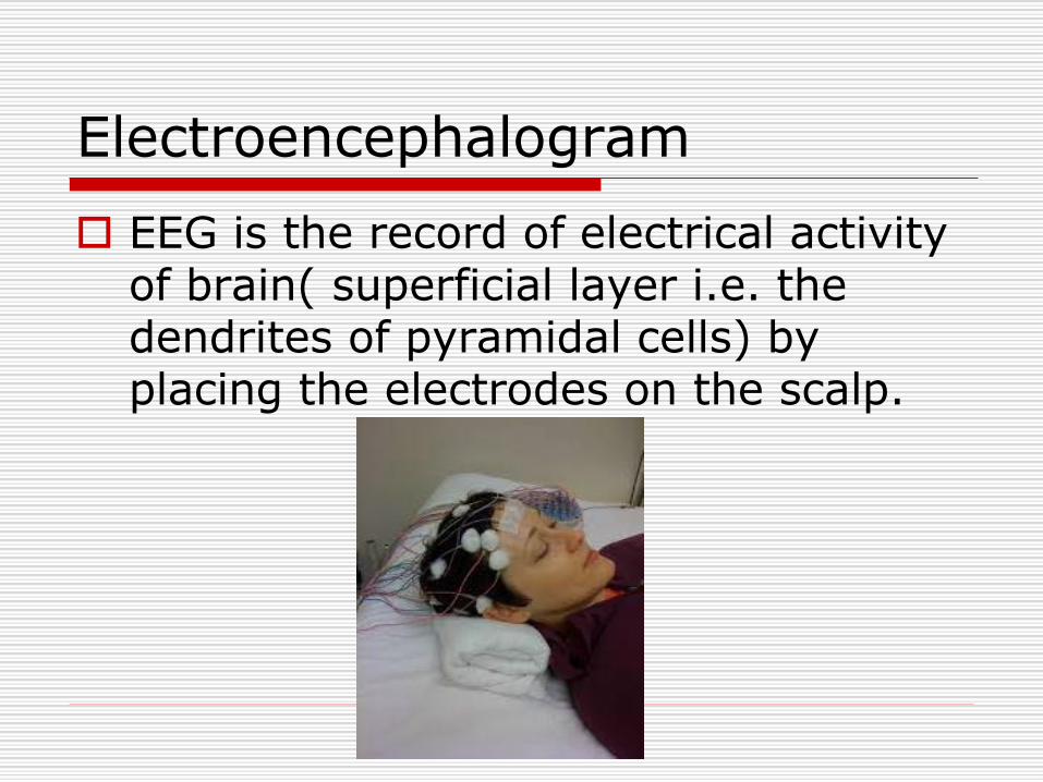
Electroencephalogram
EEG is the record of electrical activity of brain( superficial layer i.e. the dendrites of pyramidal cells) by placing the electrodes on the scalp.
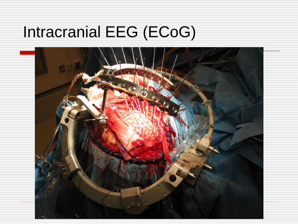
Intracranial EEG (ECoG)
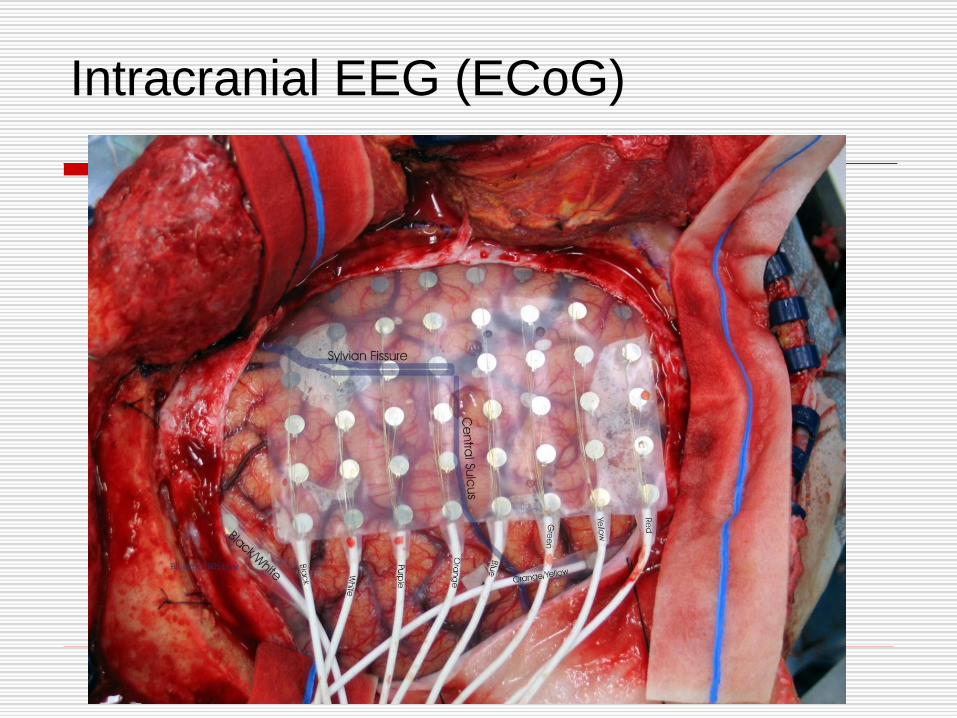
Intracranial EEG (ECoG)
Objectives of EEG practical
Familiarize with the principles of techniques involved
Count frequencies and measure the amplitudes of the record obtained.
Categories the records into appropriate rhythms – α, β, θ,and δ.
Cont…
Objectives of EEG practical
Identify and describe changes produced by provocation tests.
e.g. eye opening & closing, intermittent photic stimulation (IPS) clapping sound, induce thinking & hyperventilation.
Appreciate clinical uses of EEG
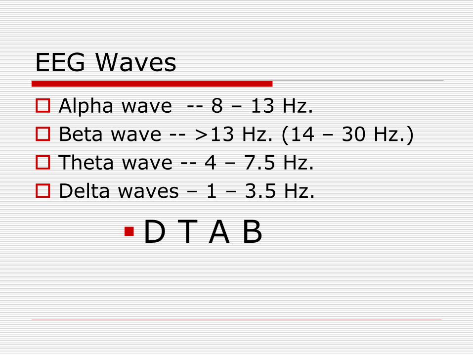
EEG Waves
Alpha wave -- 8 – 13 Hz.
Beta wave -- >13 Hz. (14 – 30 Hz.)
Theta wave -- 4 – 7.5 Hz.
Delta waves – 1 – 3.5 Hz.
▪D T A B
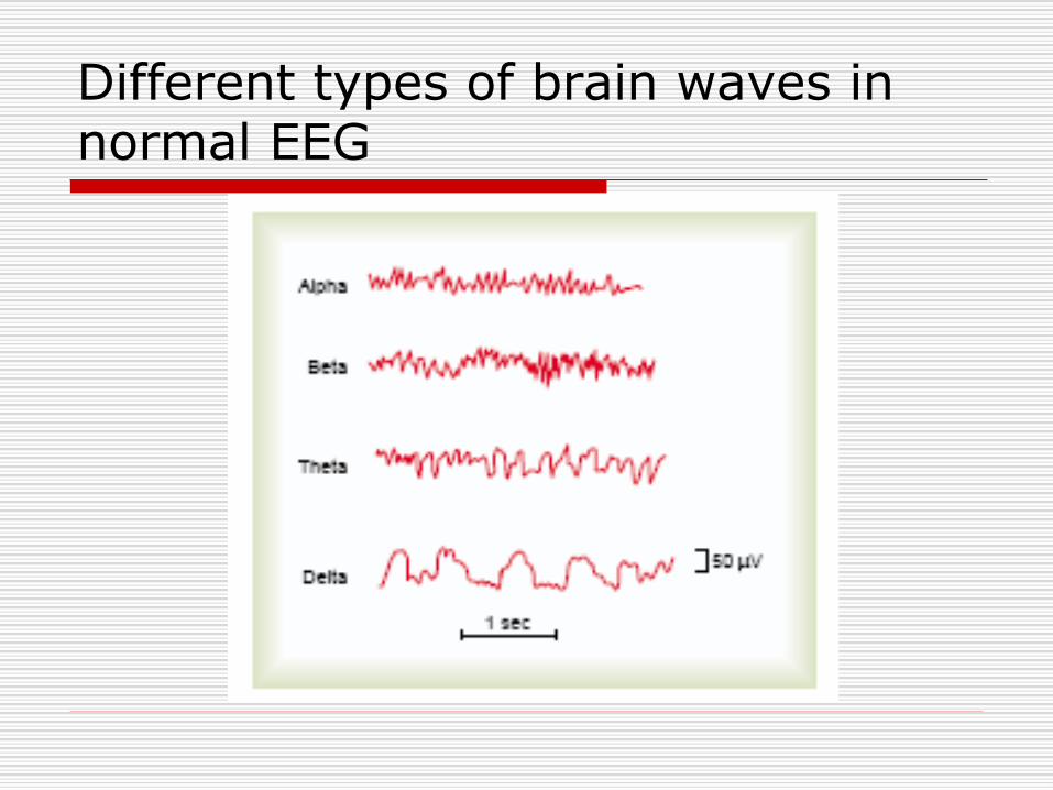
Different types of brain waves in normal EEG
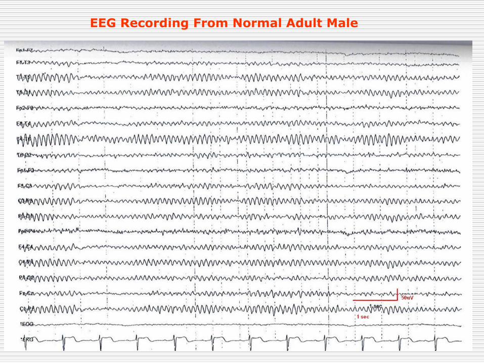
EEG Recording From Normal Adult Male
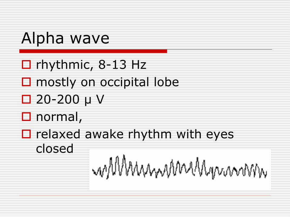
Alpha wave
rhythmic, 8-13 Hz
mostly on occipital lobe
20-200 μ V
normal,
relaxed awake rhythm with eyes closed
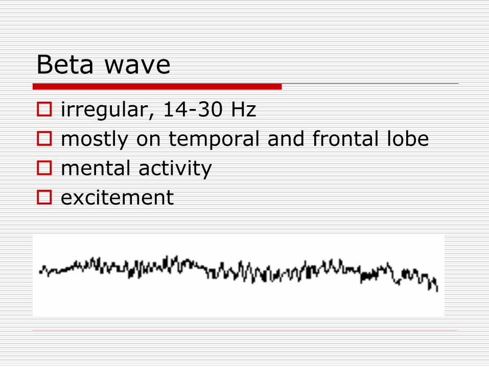
Beta wave
irregular, 14-30 Hz
mostly on temporal and frontal lobe
mental activity
excitement
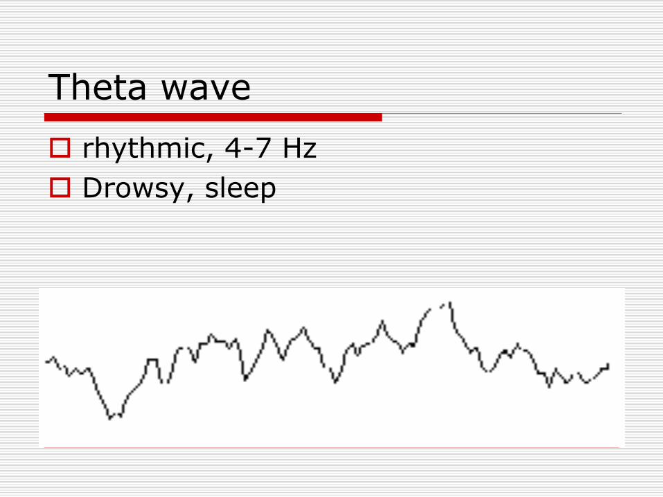
Theta wave
rhythmic, 4-7 Hz
Drowsy, sleep
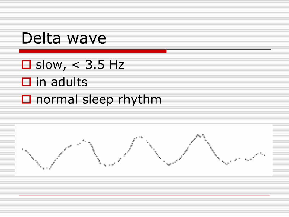
Delta wave
slow, < 3.5 Hz
in adults
normal sleep rhythm

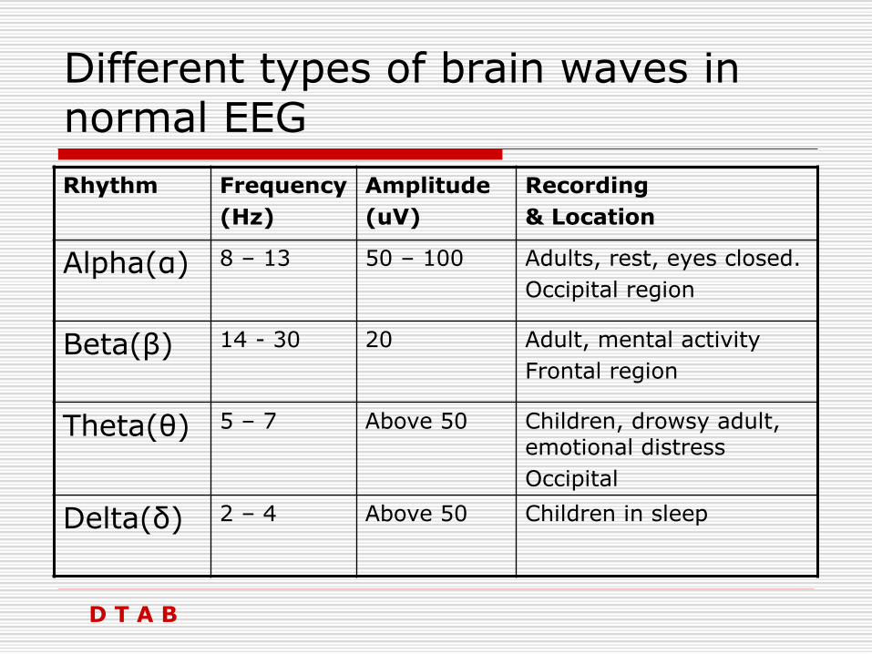
Different types of brain waves in normal EEG
Rhythm Frequency
(Hz)
Amplitude
(uV)
Recording
& Location
Alpha(α) 8 – 13 50 – 100 Adults, rest, eyes closed.
Occipital region
Beta(β) 14 - 30 20 Adult, mental activity
Frontal region
Theta(θ) 5 – 7 Above 50 Children, drowsy adult, emotional distress
Occipital
Delta(δ) 2 – 4 Above 50 Children in sleep
D T A B

Requirements
EEG machine (8/16 channels).
Silver cup electrodes/metallic bridge electrodes.
Electrode jelly.
Rubber cap.
Quiet dark comfortable room.
Skin pencil & measuring tape.
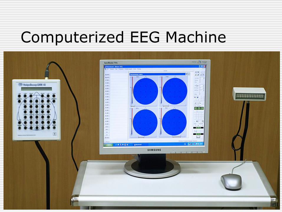
Computerized EEG Machine

EEG Electrodes
Sliver Electrodes Electrodes Cap
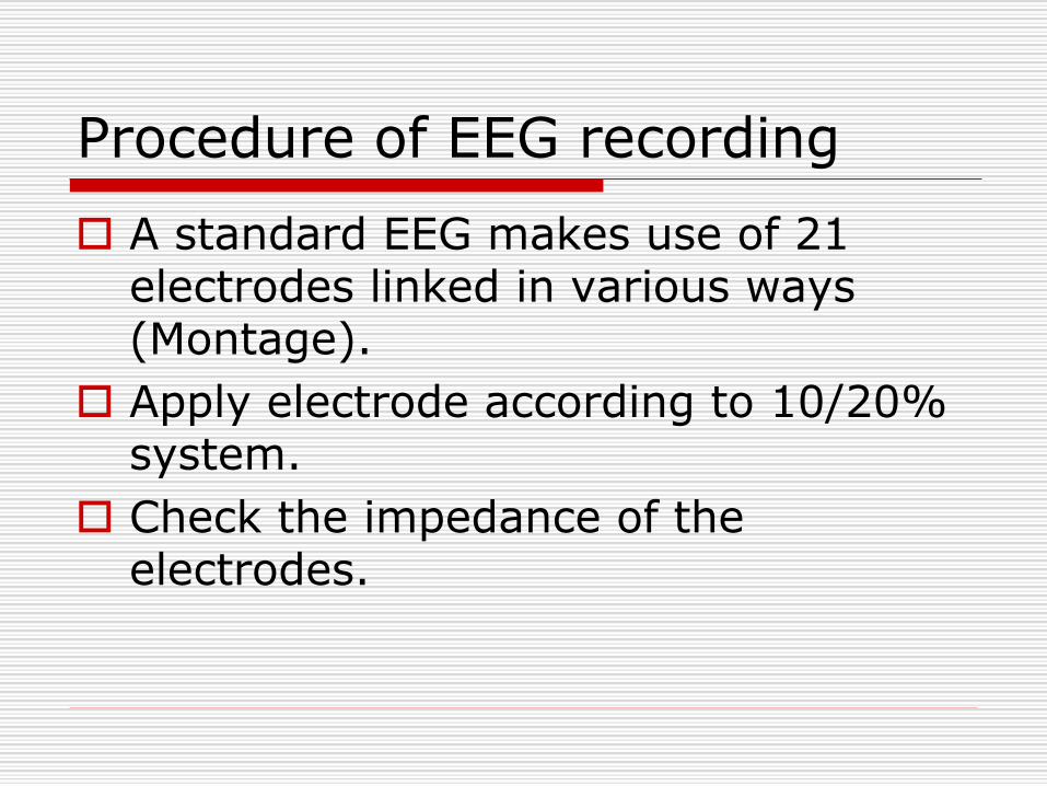
Procedure of EEG recording
A standard EEG makes use of 21 electrodes linked in various ways (Montage).
Apply electrode according to 10/20% system.
Check the impedance of the electrodes.
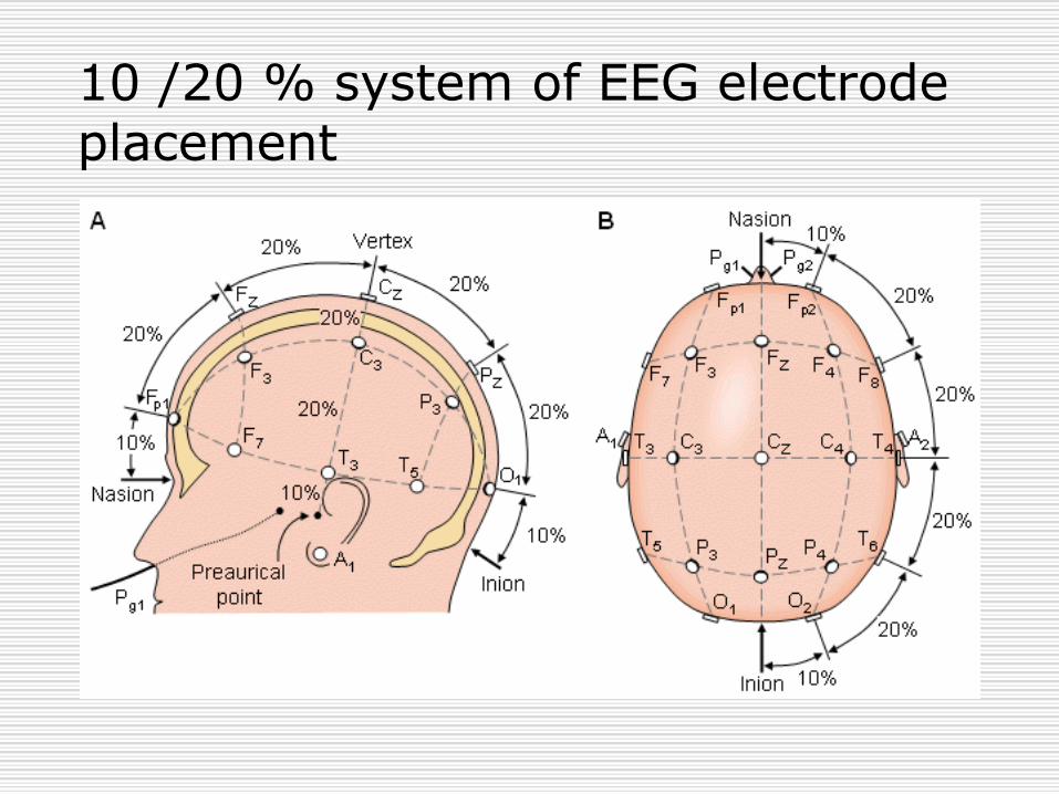
10 /20 % system of EEG electrode placement
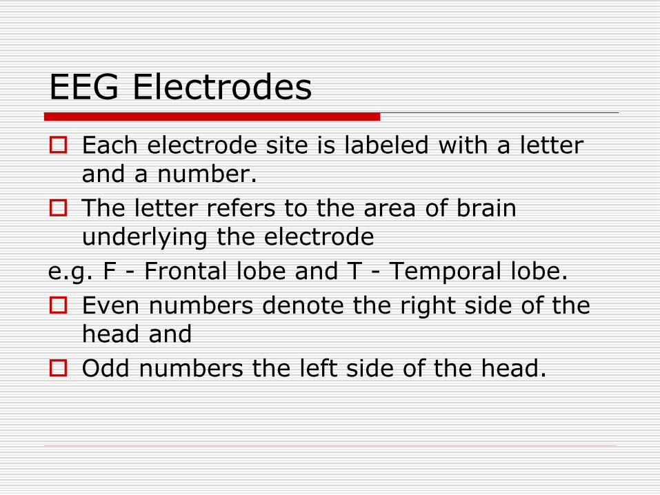
EEG Electrodes
Each electrode site is labeled with a letter and a number.
The letter refers to the area of brain underlying the electrode
e.g. F - Frontal lobe and T - Temporal lobe.
Even numbers denote the right side of the head and
Odd numbers the left side of the head.
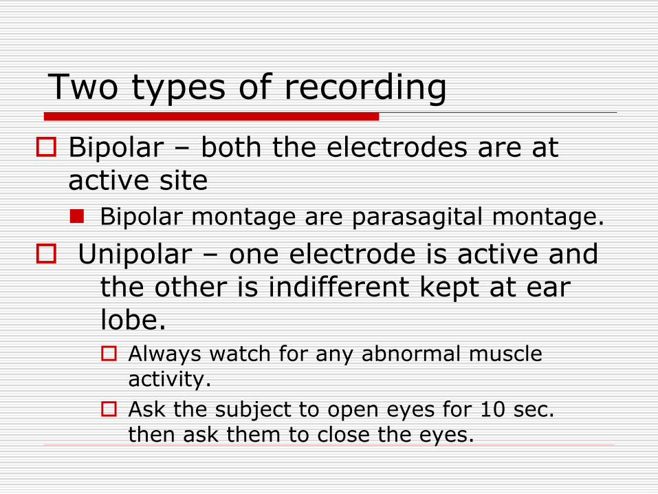
Two types of recording
Bipolar – both the electrodes are at active site
◼ Bipolar montage are parasagital montage.
Unipolar – one electrode is active and the other is indifferent kept at ear lobe. Always watch for any abnormal muscle
activity.
Ask the subject to open eyes for 10 sec. then ask them to close the eyes.


Montage
Different sets of electrode arrangement on the scalp by 10 – 20 system is known as montage.
21 electrodes are attached to give 8 or 16 channels recording.

Analysis
Electrical activity from the brain consist of primarily of rhythms.
They are named according to their frequencies (Hz) and amplitude in micro volt (μv).
Different rhythms at different ages and different conditions (level of consciousness)
Usually one dominant frequency (background rhythm)
Factor influencing EEG
Age
◼ Infant – theta, delta wave
◼ Child – alpha formation.
◼ Adult – all four waves.
Level of consciousness (sleep)
Hypocapnia(hyperventilation) slow & high amplitude waves.
Hypoglycemia
Hypothermia
Low glucocorticoids
Slow waves
NORMAL EEG CHANGES
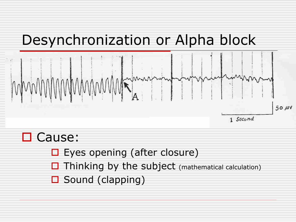
Desynchronization or Alpha block
Cause: Eyes opening (after closure)
Thinking by the subject (mathematical calculation)
Sound (clapping)
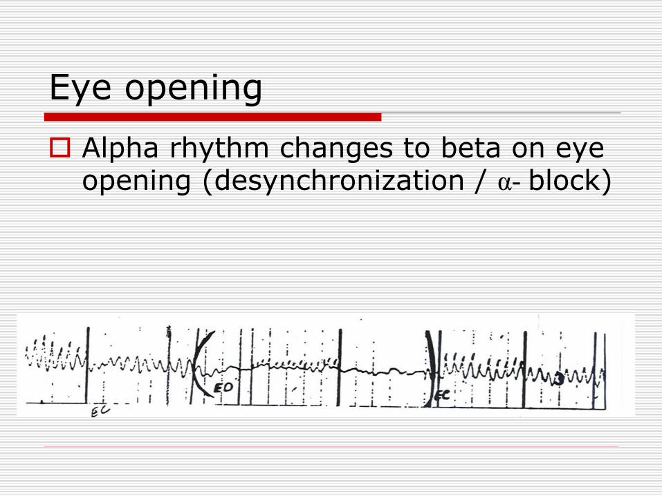
Eye opening
Alpha rhythm changes to beta on eye opening (desynchronization / α- block)
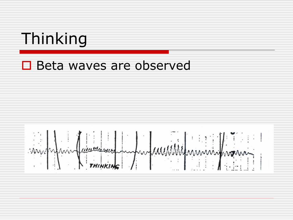
Thinking
Beta waves are observed
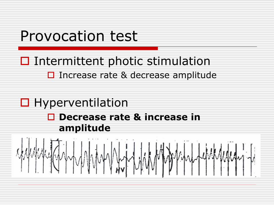
Provocation test
Intermittent photic stimulation Increase rate & decrease amplitude
Hyperventilation Decrease rate & increase in
amplitude
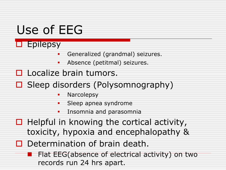
Use of EEG Epilepsy
▪ Generalized (grandmal) seizures.
▪ Absence (petitmal) seizures.
Localize brain tumors.
Sleep disorders (Polysomnography)▪ Narcolepsy
▪ Sleep apnea syndrome
▪ Insomnia and parasomnia
Helpful in knowing the cortical activity, toxicity, hypoxia and encephalopathy &
Determination of brain death.
◼ Flat EEG(absence of electrical activity) on two records run 24 hrs apart.
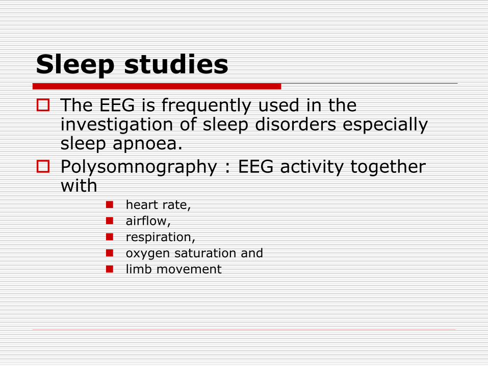
Sleep studies
The EEG is frequently used in the investigation of sleep disorders especially sleep apnoea.
Polysomnography : EEG activity together with
◼ heart rate,
◼ airflow,
◼ respiration,
◼ oxygen saturation and
◼ limb movement
Sleep patterns of EEG
There are two different kinds of sleep: Rapid eye movement sleep (REM-Sleep)
Non-REM sleep (NREM sleep)/ slow wave sleep
NREM sleep is again divided into 4 stages (I to IV). The EEG pattern in sleep is given in the following table:
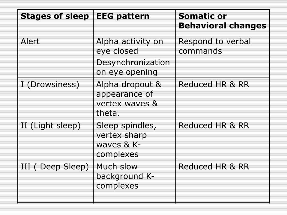
Stages of sleep EEG pattern Somatic or Behavioral changes
Alert Alpha activity on eye closed
Desynchronization on eye opening
Respond to verbal commands
I (Drowsiness) Alpha dropout & appearance of vertex waves & theta.
Reduced HR & RR
II (Light sleep) Sleep spindles, vertex sharp waves & K-complexes
Reduced HR & RR
III ( Deep Sleep) Much slow background K-complexes
Reduced HR & RR
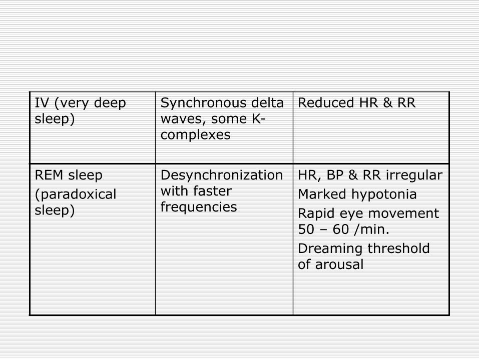
IV (very deep sleep)
Synchronous delta waves, some K-complexes
Reduced HR & RR
REM sleep
(paradoxical sleep)
Desynchronization with faster frequencies
HR, BP & RR irregular
Marked hypotonia
Rapid eye movement 50 – 60 /min.
Dreaming threshold of arousal