Electrocardiography and Electroencephalographymacleod/bioen/be6460/notes/W12-ECG.pdf · ECG/EEG...
20
ECG/EEG Bioengineering 6460 Bioelectricity Electrocardiography and Electroencephalography Bioengineering 6460 Bioelectricity ECG/EEG Components of the Electrocardiogram (ECG) • Source(s) – Potential differences within the heart – Spatially distributed and time varying • Volume conductor – Inhomogeneous and anisotropic – Unique to each individual – Boundary effects • ECG measurement – Lead systems – Bipolar versus unipolar measurements – Mapping procedures • Analysis – Signal analysis – Spatial analysis – Dipole analysis – Simulation and modeling approaches
-
Upload
vuongduong -
Category
Documents
-
view
231 -
download
4
Transcript of Electrocardiography and Electroencephalographymacleod/bioen/be6460/notes/W12-ECG.pdf · ECG/EEG...
ECG/EEG Bioengineering 6460 Bioelectricity
Electrocardiography and Electroencephalography
Bioengineering 6460 BioelectricityECG/EEG
Components of the Electrocardiogram (ECG)
• Source(s)– Potential differences within the heart– Spatially distributed and time varying
• Volume conductor– Inhomogeneous and anisotropic– Unique to each individual– Boundary effects
• ECG measurement– Lead systems– Bipolar versus unipolar measurements– Mapping procedures
• Analysis– Signal analysis– Spatial analysis– Dipole analysis– Simulation and modeling approaches
Bioengineering 6460 BioelectricityECG/EEG
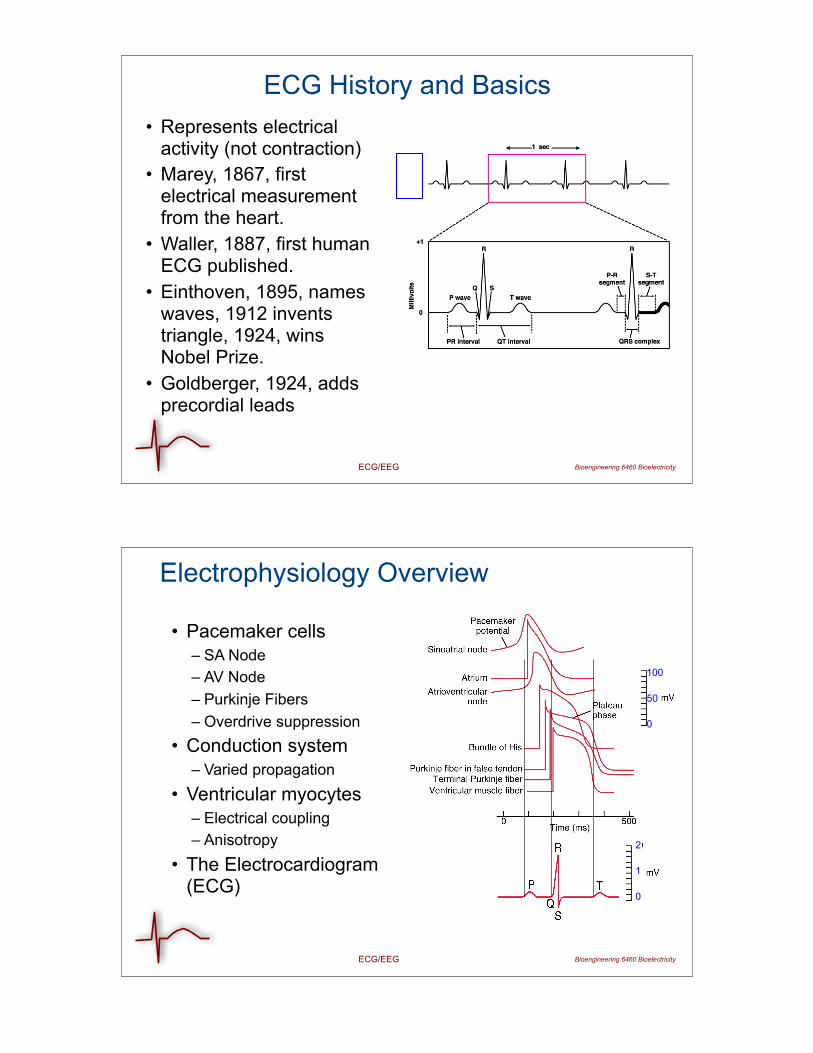
ECG History and Basics• Represents electrical
activity (not contraction)• Marey, 1867, first
electrical measurement from the heart.
• Waller, 1887, first human ECG published.
• Einthoven, 1895, names waves, 1912 invents triangle, 1924, wins Nobel Prize.
• Goldberger, 1924, adds precordial leads
0
1
2
Bioengineering 6460 BioelectricityECG/EEG
Electrophysiology Overview
• Pacemaker cells – SA Node– AV Node– Purkinje Fibers– Overdrive suppression
• Conduction system– Varied propagation
• Ventricular myocytes– Electrical coupling– Anisotropy
• The Electrocardiogram (ECG)
100
0
50
ECG/EEG Bioengineering 6460 Bioelectricity
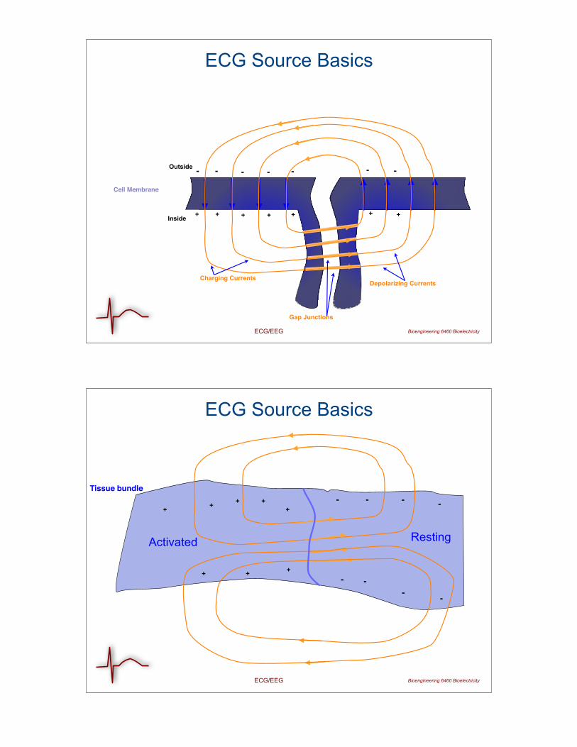
ECG Source Basics
Outside
Inside
Charging Currents
+ + +
-- -
Depolarizing Currents
Cell Membrane
Gap Junctions
+
-
+
-
+
-
+
-
ECG/EEG Bioengineering 6460 Bioelectricity
ECG Source Basics
++
+
-
-
-Tissue bundle
+
-+
-
+
-
+--
+
Activated Resting
ECG/EEG Bioengineering 6460 Bioelectricity
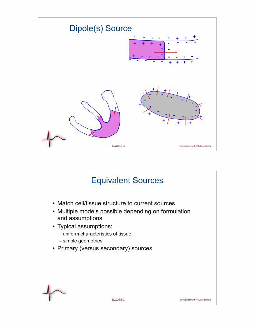
Dipole(s) Source
+ ++
+
++++
+
+
++
- - --
------
--
-
+ + + ++ + + +- - - -- - - -
++++++
--
----- - - -+ + + +
Bioengineering 6460 BioelectricityECG/EEG
Equivalent Sources
• Match cell/tissue structure to current sources• Multiple models possible depending on formulation
and assumptions• Typical assumptions:
– uniform characteristics of tissue– simple geometries
• Primary (versus secondary) sources
Bioengineering 6460 BioelectricityECG/EEG
Cardiac Sources
• Formulation in terms of cells impossible• Dipole(s), multipoles: simple but incomplete• Volume dipole density: hard to describe• Surface dipole density: good compromise in some
problems• All require some model of time dependence
(propagation)
Bioengineering 6460 BioelectricityECG/EEG
Heart Dipole Approaches
• Treat the heart as single dipole• Fixed in space but free to rotate and change amplitude• Einthoven triangle • Vector ECG (Vectorcardiogram)• Lead fields: generalization of heart dipole
Bioengineering 6460 BioelectricityECG/EEG
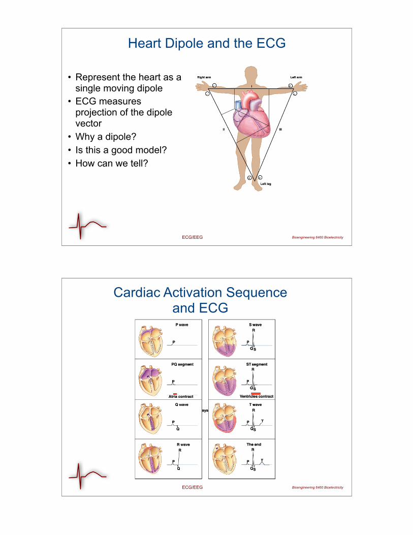
Heart Dipole and the ECG
• Represent the heart as a single moving dipole
• ECG measures projection of the dipole vector
• Why a dipole?• Is this a good model?• How can we tell?
ECG/EEG Bioengineering 6460 Bioelectricity
Cardiac Activation Sequence and ECG
Bioengineering 6460 BioelectricityECG/EEG
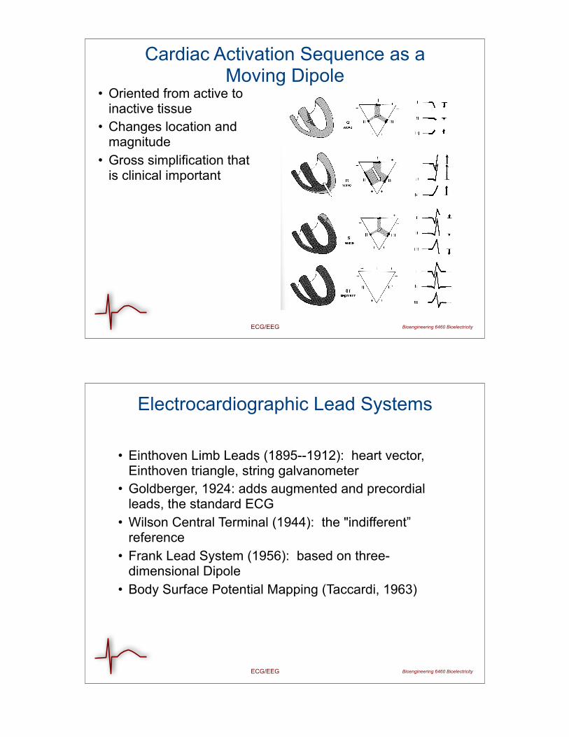
Cardiac Activation Sequence as a Moving Dipole
• Oriented from active to inactive tissue
• Changes location and magnitude
• Gross simplification that is clinical important
Bioengineering 6460 BioelectricityECG/EEG
Electrocardiographic Lead Systems
• Einthoven Limb Leads (1895--1912): heart vector, Einthoven triangle, string galvanometer
• Goldberger, 1924: adds augmented and precordial leads, the standard ECG
• Wilson Central Terminal (1944): the "indifferent” reference
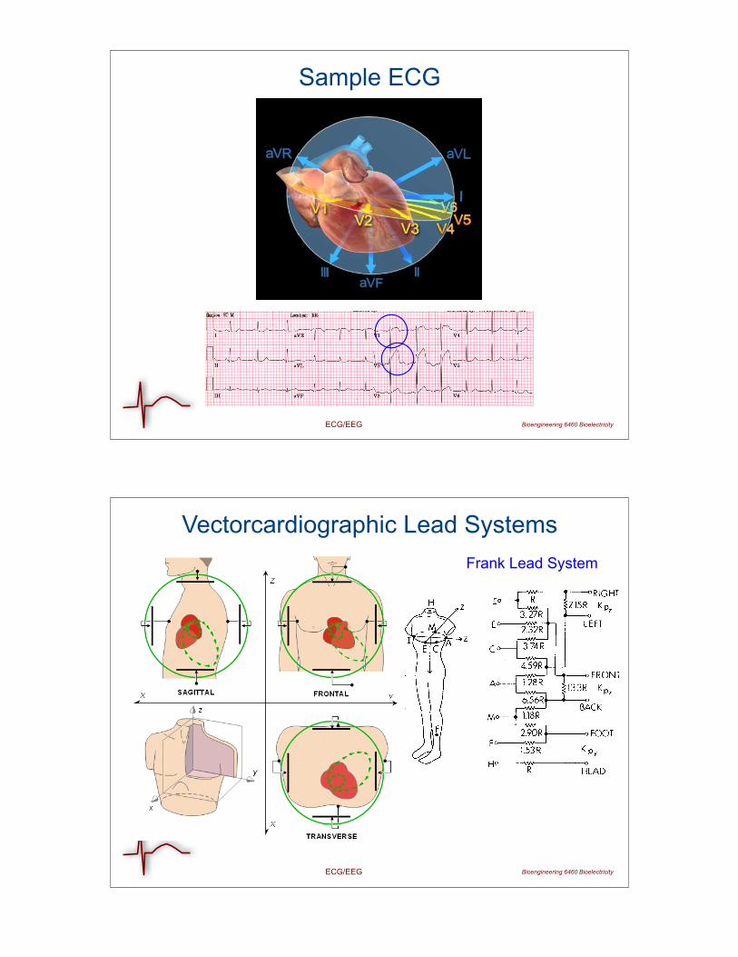
• Frank Lead System (1956): based on three-dimensional Dipole
• Body Surface Potential Mapping (Taccardi, 1963)
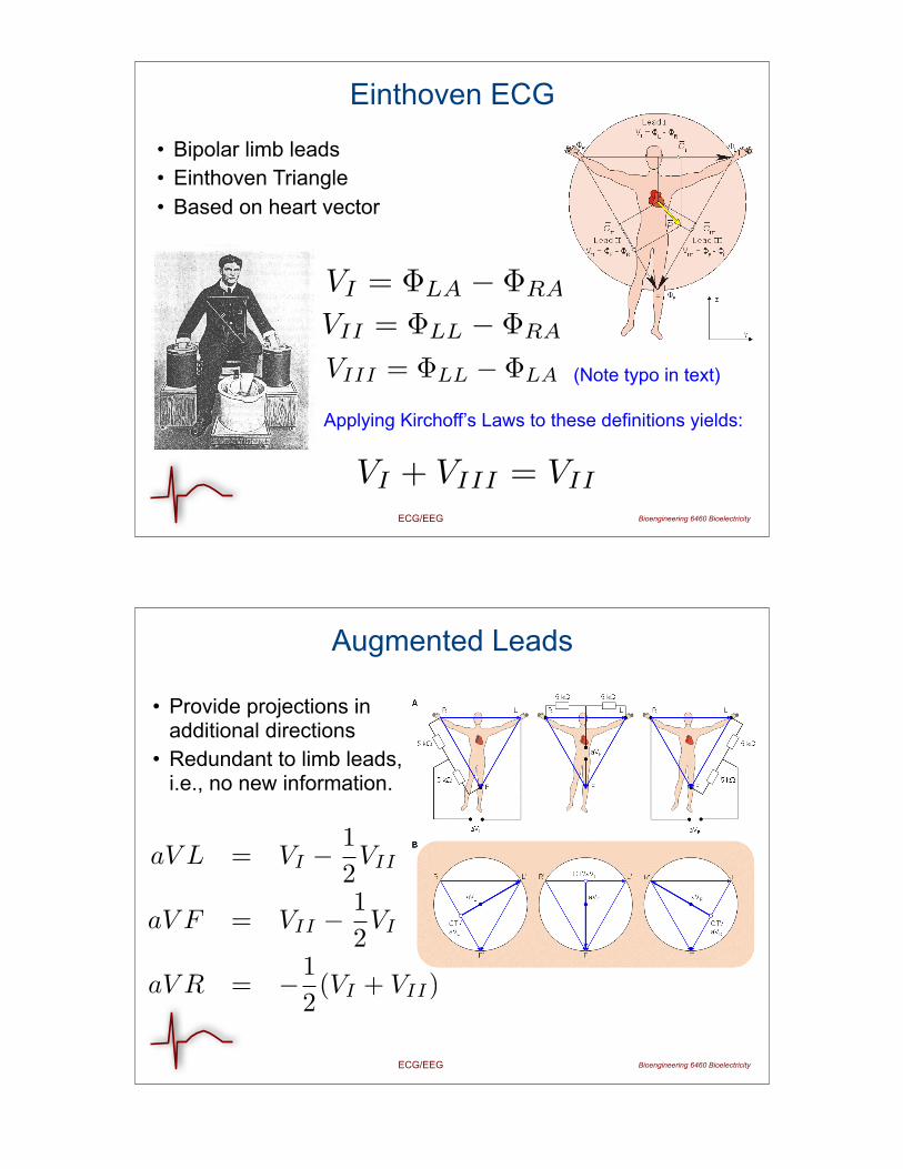
VI = ΦLA − ΦRA
VII = ΦLL − ΦRA
VIII = ΦLL − ΦLA
VI + VIII = VII
Bioengineering 6460 BioelectricityECG/EEG
Einthoven ECG
• Bipolar limb leads• Einthoven Triangle• Based on heart vector
(Note typo in text)
Applying Kirchoff’s Laws to these definitions yields:
Bioengineering 6460 BioelectricityECG/EEG
Augmented Leads
• Provide projections in additional directions
• Redundant to limb leads, i.e., no new information.
aV L = VI −12VII
aV F = VII −12VI
aV R = −12(VI + VII)
IR + IF + IL = 0
Bioengineering 6460 BioelectricityECG/EEG
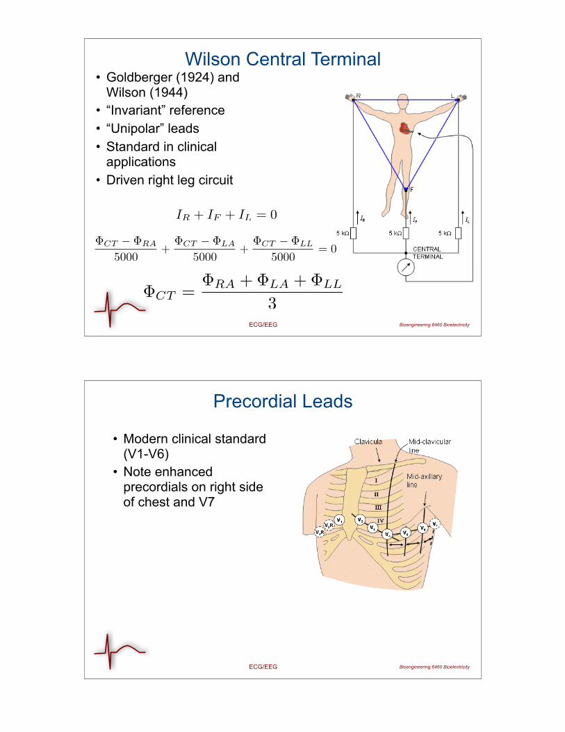
Wilson Central Terminal• Goldberger (1924) and
Wilson (1944)• “Invariant” reference• “Unipolar” leads• Standard in clinical
applications• Driven right leg circuit
ΦCT − ΦRA
5000+
ΦCT − ΦLA
5000+
ΦCT − ΦLL
5000= 0
ΦCT =ΦRA + ΦLA + ΦLL
3
Bioengineering 6460 BioelectricityECG/EEG
Precordial Leads
• Modern clinical standard (V1-V6)
• Note enhanced precordials on right side of chest and V7
Bioengineering 6460 BioelectricityECG/EEG
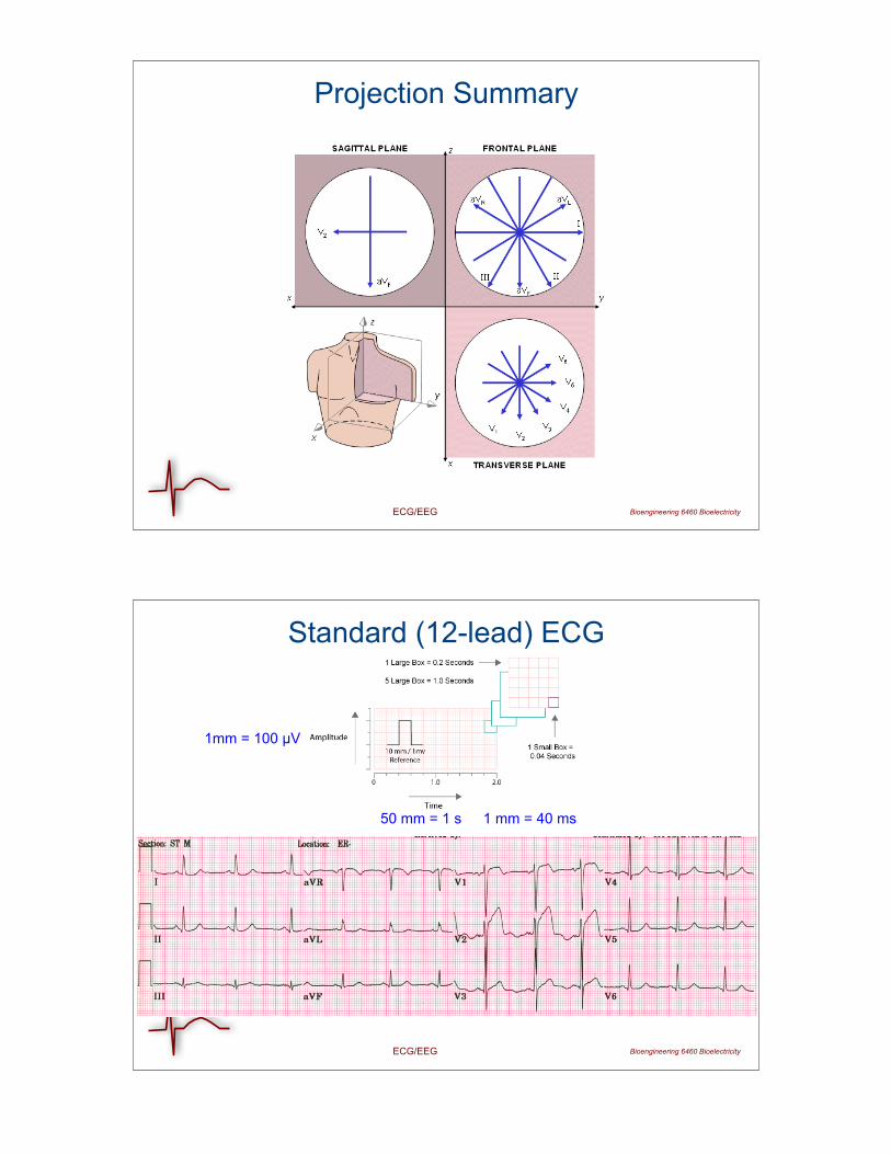
Projection Summary
ECG/EEG Bioengineering 6460 Bioelectricity
Standard (12-lead) ECG
1mm = 100 !V
50 mm = 1 s 1 mm = 40 ms
ECG/EEG Bioengineering 6460 Bioelectricity
Sample ECG
Bioengineering 6460 BioelectricityECG/EEG
Vectorcardiographic Lead SystemsFrank Lead System
ECG/EEG Bioengineering 6460 Bioelectricity
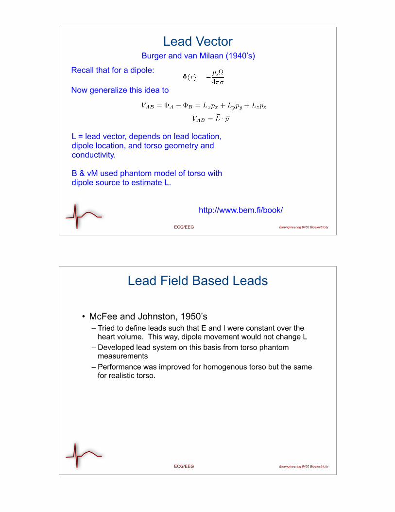
Lead VectorBurger and van Milaan (1940’s)
L = lead vector, depends on lead location, dipole location, and torso geometry and conductivity.
B & vM used phantom model of torso with dipole source to estimate L.
Recall that for a dipole:
Now generalize this idea to
http://www.bem.fi/book/
Bioengineering 6460 BioelectricityECG/EEG
Lead Field Based Leads
• McFee and Johnston, 1950’s– Tried to define leads such that E and I were constant over the
heart volume. This way, dipole movement would not change L – Developed lead system on this basis from torso phantom
measurements– Performance was improved for homogenous torso but the same
for realistic torso.
Bioengineering 6460 BioelectricityECG/EEG
Multipoles
• Higher order expansion of solution to Poisson’s equation
• Monopole, dipole, quadropole, octopole…
• Example: two wavefronts in cardiac tissue
ECG/EEG Bioengineering 6460 Bioelectricity
Multipole Based Models
Bioengineering 6460 BioelectricityECG/EEG
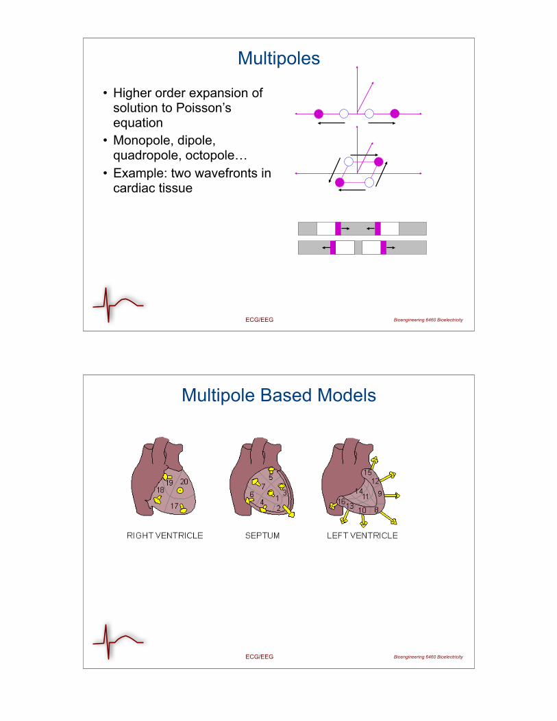
Body Surface Potential Mapping
• Measurements over entire torso
• Showed that resulting pattern was not (always) dipolar
• More complex source model than dipole required
Taccardi et al,Circ., 1963
ECG/EEG Bioengineering 6460 Bioelectricity
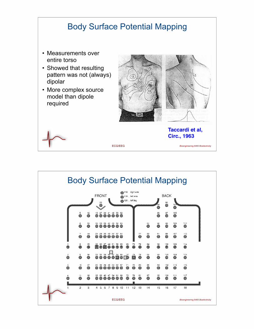
Body Surface Potential Mapping

ECG/EEG Bioengineering 6460 Bioelectricity
BSPM Hisory
http://www.sci.utah.edu/gallery2/v/cibc/taccardi_sm.html
http://www.sci.utah.edu/gallery2/v/cibc/taccardi_lg.html
Small version:
Large version:
ECG/EEG Bioengineering 6460 Bioelectricity
State of the Art
ECG/EEG Bioengineering 6460 Bioelectricity
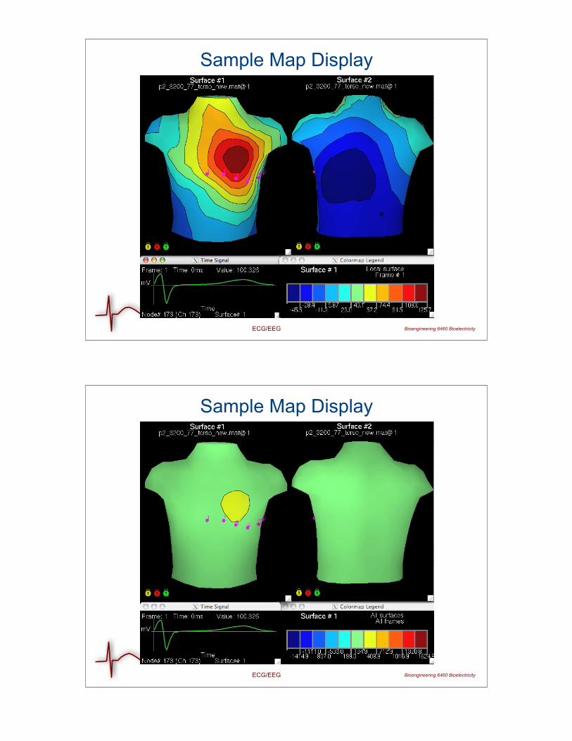
Sample Map Display
ECG/EEG Bioengineering 6460 Bioelectricity
Sample Map Display
Bioengineering 6460 BioelectricityECG/EEG
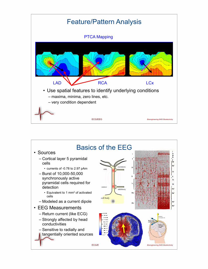
Feature/Pattern Analysis
LAD RCA LCx
PTCA Mapping
• Use spatial features to identify underlying conditions– maxima, minima, zero lines, etc. – very condition dependent
Bioengineering 6460 BioelectricityECG/EEG
Basics of the EEG• Sources
– Cortical layer 5 pyramidal cells
• currents of -0.78 to 2.97 pAm
– Burst of 10,000-50,000 synchronously active pyramidal cells required for detection
• Equivalent to 1 mm2 of activated cells
– Modeled as a current dipole• EEG Measurements
– Return current (like ECG)– Strongly affected by head
conductivities– Sensitive to radially and
tangentially oriented sources
cell body
source
sink
inhibitory
excitatory
Bioengineering 6460 BioelectricityECG/EEG
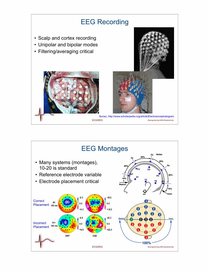
EEG Recording
• Scalp and cortex recording• Unipolar and bipolar modes• Filtering/averaging critical
Nunez, http://www.scholarpedia.org/article/Electroencephalogram
Bioengineering 6460 BioelectricityECG/EEG
EEG Montages
• Many systems (montages), 10-20 is standard
• Reference electrode variable• Electrode placement critical
CorrectPlacement
IncorrectPlacement
Bioengineering 6460 BioelectricityECG/EEG
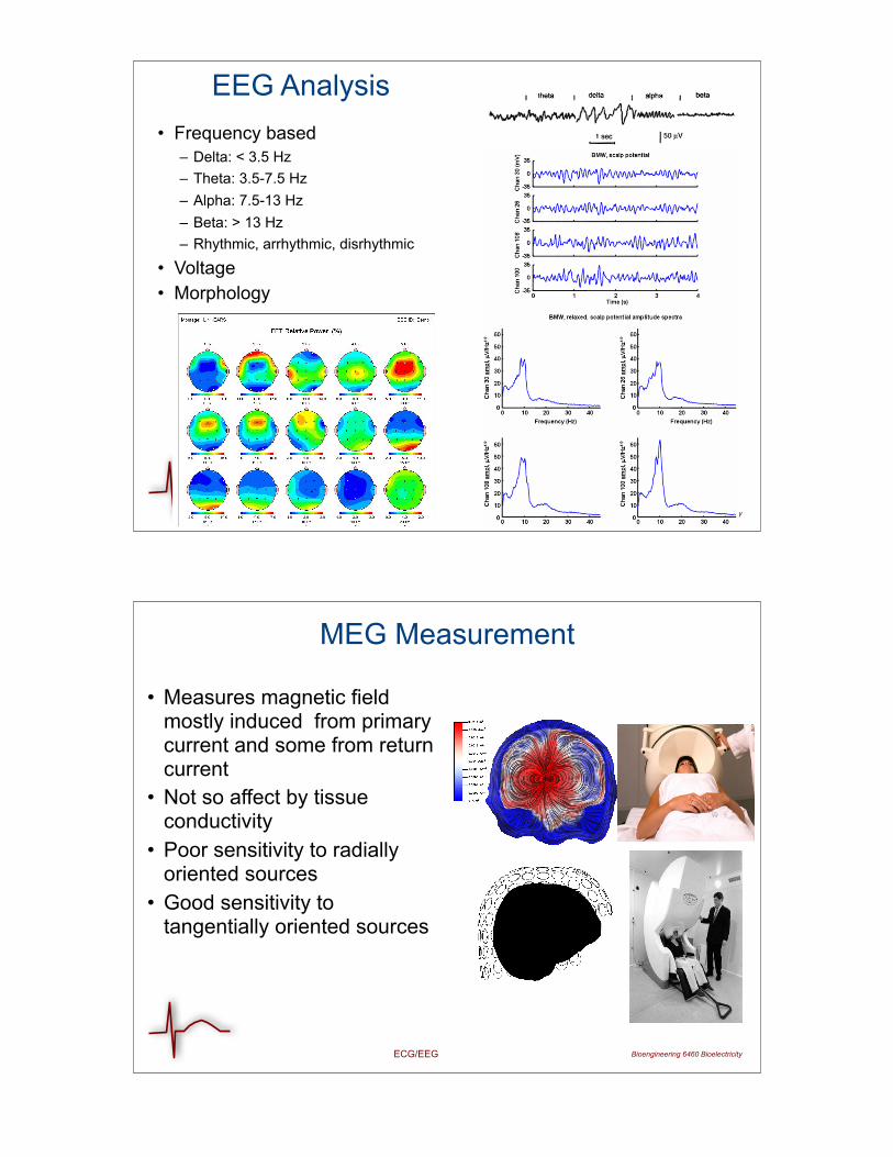
EEG Analysis • Frequency based
– Delta: < 3.5 Hz– Theta: 3.5-7.5 Hz– Alpha: 7.5-13 Hz– Beta: > 13 Hz– Rhythmic, arrhythmic, disrhythmic
• Voltage• Morphology
Bioengineering 6460 BioelectricityECG/EEG
MEG Measurement
• Measures magnetic field mostly induced from primary current and some from return current
• Not so affect by tissue conductivity
• Poor sensitivity to radially oriented sources
• Good sensitivity to tangentially oriented sources
ECG/EEG Bioengineering 6460 Bioelectricity
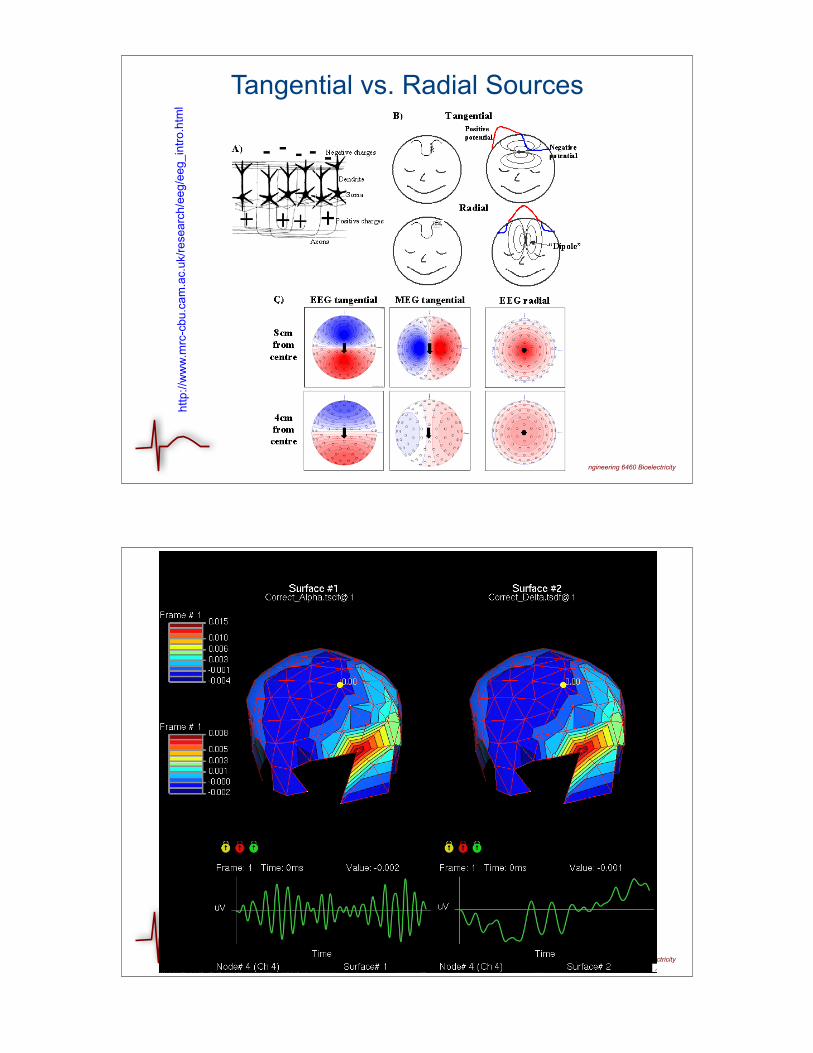
Tangential vs. Radial Sources
http
://w
ww
.mrc
-cbu
.cam
.ac.
uk/re
sear
ch/e
eg/e
eg_i
ntro
.htm
l
ECG/EEG Bioengineering 6460 Bioelectricity