Electrocardiogram manifestations of hyponatraemia
3
CARDIOVASCULAR JOURNAL OF AFRICA • Advance Online Publication, September 2021 AFRICA 1 Electrocardiogram manifestations of hyponatraemia Shi Zou, Qian Zhang, Shicheng Gao, Mingqi Luo, Xuedong Gan, Ke Liang Abstract Electrolytes play a vital role in myocardial electrophysiologi- cal activities in the human body. Electrolyte disturbances can affect depolarisation and repolarisation of myocardial cells and thus result in arrhythmia. The most common electrolyte disturbance among hospitalised patients is hyponatraemia. We report on a case of an acquired immune deficiency syndrome patient with decompensated cirrhosis, who devel- oped sinus arrest from hyponatraemia. The electrocardiogram manifestations at different sodium concentrations were also recorded in subsequent therapeutic processes. Keywords: hyponatraemia, ECG, cirrhosis, sinus arrest Submitted 20/2/21, accepted 12/7/21 Cardiovasc J Afr 2021; 32: online publication www.cvja.co.za DOI: 10.5830/CVJA-2021-036 Hyponatraemia is generally defined as a serum sodium concentration lower than 135 mmol/l, which is associated with increased rates of mortality and morbidity. 1,2 The clinical presentation may be highly variable, ranging from no symptoms to neurological symptoms such as confusion and coma, or even death. 3 Although serum sodium concentration is known to be associated with arrhythmia, 4,5 as far as we know, no study on the various manifestations of arrhythmia caused by different degrees of hyponatraemia has been published. In this case report, we recorded the case of an acquired immune deficiency syndrome (AIDS) patient with decompensated cirrhosis who developed sinus arrest due to hyponatraemia. The changes on electrocardiogram (ECG) at different serum sodium concentrations in subsequent therapeutic process were also documented. Case report A 50-year-old woman was admitted to our hospital on 2 December 2011 with complaints of abdominal distention for two weeks. She had a history of AIDS and chronic hepatitis C for seven years, and had no history of heart disease or hyponatraemia. The patient had been receiving antiretroviral therapy (combination of lamivudine, tenofovir and lopinavir/ ritonavir) for one year. She also received diuretic treatment (furosemide) for cirrhotic ascites. On admission, the patient was alert and orientated, and co-operated with the medical staff. On physical examination, the patient had mild sclerotic jaundice of the skin and sclera, and palmar erythema with grade 2 cirrhotic ascites. Cardiac and other system examinations revealed no abnormalities. Laboratory tests revealed a potassium concentration of 3.68 mmol/l (normal range: 3.5–5.5 mmol/l) and a sodium level of 137.4 mmol/l (normal: 132–146 mmol/l). Total bilirubin concentration was elevated to 83.4 µmol/l (normal: 5–21 µmol/l) and albumin level was reduced to 28.7 g/l (normal: 40–55 g/l). Blood urea nitrogen concentration was 8.97 mmol/l (normal: 2.8–7.6 mmol/l), creatinine level was 156.3 µmol/l (normal: 64–104 µmol/l) and the estimated glomerular filtration rate (eGFR) was 38 ml/min. Myocardial enzymes were normal. Abdominal ultrasound showed liver cirrhosis, portal hypertension, ascites and cholecystolithiasis. The ECG showed normal sinus rhythm on admission. After admission, diuretics (furosemide, spirolactone and hydrochlorothiazide) were started. On 14 December 2011, the patient showed symptoms including chest tightness, palpitation, headache, fatigue and drowsiness, and an ECG showed sinus arrest (the longest R-R interval was 3.8 seconds) and junctional escape beats (Fig. 1A). Electrolyte tests revealed severe hyponatraemia (102 mmol/l). The hyponatraemia was gradually corrected by intravenous 3% hypertonic saline. 6 The patient’s symptoms were improved and the ECG showed various manifestations at different serum sodium concentrations (Fig. 1, Table 1). In the next few days, a 24-hour Holter recording showed sinus rhythm. Department of Infectious Diseases, Zhongnan Hospital, Wuhan University, Hubei, China Shi Zou, MD Shicheng Gao, MD Mingqi Luo, MD Ke Liang, PhD, [email protected] Qingshan District Center for Disease Control and Prevention, Wuhan, China Qian Zhang, MD Department of Cardiology, Zhongnan Hospital, Wuhan University, Hubei, China Xuedong Gan, MD, [email protected] Department of Nosocomial Infection Management; Center of Preventing Mother-to-Child Transmission for Infectious Diseases, Zhongnan Hospital, Wuhan University, Hubei; Wuhan Research Center for Infectious Diseases and Cancer, Chinese Academy of Medical Sciences, Wuhan, China Ke Liang, PhD Case Report
Transcript of Electrocardiogram manifestations of hyponatraemia

CARDIOVASCULAR JOURNAL OF AFRICA • Advance Online Publication, September 2021AFRICA 1
Electrocardiogram manifestations of hyponatraemia Shi Zou, Qian Zhang, Shicheng Gao, Mingqi Luo, Xuedong Gan, Ke Liang
AbstractElectrolytes play a vital role in myocardial electrophysiologi-cal activities in the human body. Electrolyte disturbances can affect depolarisation and repolarisation of myocardial cells and thus result in arrhythmia. The most common electrolyte disturbance among hospitalised patients is hyponatraemia. We report on a case of an acquired immune deficiency syndrome patient with decompensated cirrhosis, who devel-oped sinus arrest from hyponatraemia. The electrocardiogram manifestations at different sodium concentrations were also recorded in subsequent therapeutic processes.
Keywords: hyponatraemia, ECG, cirrhosis, sinus arrest
Submitted 20/2/21, accepted 12/7/21
Cardiovasc J Afr 2021; 32: online publication www.cvja.co.za
DOI: 10.5830/CVJA-2021-036
Hyponatraemia is generally defined as a serum sodium concentration lower than 135 mmol/l, which is associated with increased rates of mortality and morbidity.1,2 The clinical presentation may be highly variable, ranging from no symptoms to neurological symptoms such as confusion and coma, or even death.3
Although serum sodium concentration is known to be associated with arrhythmia,4,5 as far as we know, no study on the various manifestations of arrhythmia caused by different degrees of hyponatraemia has been published. In this case report, we recorded the case of an acquired immune deficiency syndrome (AIDS) patient with decompensated cirrhosis who developed sinus arrest due to hyponatraemia. The changes on electrocardiogram (ECG) at different serum sodium concentrations in subsequent therapeutic process were also documented.
Case reportA 50-year-old woman was admitted to our hospital on 2 December 2011 with complaints of abdominal distention for two weeks. She had a history of AIDS and chronic hepatitis C for seven years, and had no history of heart disease or hyponatraemia. The patient had been receiving antiretroviral therapy (combination of lamivudine, tenofovir and lopinavir/ritonavir) for one year. She also received diuretic treatment (furosemide) for cirrhotic ascites.
On admission, the patient was alert and orientated, and co-operated with the medical staff. On physical examination, the patient had mild sclerotic jaundice of the skin and sclera, and palmar erythema with grade 2 cirrhotic ascites. Cardiac and other system examinations revealed no abnormalities.
Laboratory tests revealed a potassium concentration of 3.68 mmol/l (normal range: 3.5–5.5 mmol/l) and a sodium level of 137.4 mmol/l (normal: 132–146 mmol/l). Total bilirubin concentration was elevated to 83.4 µmol/l (normal: 5–21 µmol/l) and albumin level was reduced to 28.7 g/l (normal: 40–55 g/l). Blood urea nitrogen concentration was 8.97 mmol/l (normal: 2.8–7.6 mmol/l), creatinine level was 156.3 µmol/l (normal: 64–104 µmol/l) and the estimated glomerular filtration rate (eGFR) was 38 ml/min. Myocardial enzymes were normal.
Abdominal ultrasound showed liver cirrhosis, portal hypertension, ascites and cholecystolithiasis. The ECG showed normal sinus rhythm on admission. After admission, diuretics (furosemide, spirolactone and hydrochlorothiazide) were started.
On 14 December 2011, the patient showed symptoms including chest tightness, palpitation, headache, fatigue and drowsiness, and an ECG showed sinus arrest (the longest R-R interval was 3.8 seconds) and junctional escape beats (Fig. 1A). Electrolyte tests revealed severe hyponatraemia (102 mmol/l).
The hyponatraemia was gradually corrected by intravenous 3% hypertonic saline.6 The patient’s symptoms were improved and the ECG showed various manifestations at different serum sodium concentrations (Fig. 1, Table 1). In the next few days, a 24-hour Holter recording showed sinus rhythm.
Department of Infectious Diseases, Zhongnan Hospital, Wuhan University, Hubei, ChinaShi Zou, MDShicheng Gao, MDMingqi Luo, MDKe Liang, PhD, [email protected]
Qingshan District Center for Disease Control and Prevention, Wuhan, China Qian Zhang, MD
Department of Cardiology, Zhongnan Hospital, Wuhan University, Hubei, ChinaXuedong Gan, MD, [email protected]
Department of Nosocomial Infection Management; Center of Preventing Mother-to-Child Transmission for Infectious Diseases, Zhongnan Hospital, Wuhan University, Hubei; Wuhan Research Center for Infectious Diseases and Cancer, Chinese Academy of Medical Sciences, Wuhan, ChinaKe Liang, PhD
Case Report
CARDIOVASCULAR JOURNAL OF AFRICA • Advance Online Publication, September 20212 AFRICA
DiscussionTo our knowledge, sinus arrest associated with hyponatraemia has not been reported in the literature. The pathogenesis of hyponatraemia-induced sinus arrest or cardiac conduction defects is yet to be elucidated, and data in the literature are scarce.
According to the clinical observation, low extracellular sodium concentration may shorten the depolarisation of the cardiac action potential and reduce the amplitude of the action potential in the atrioventricular (AV) node.7,8 It has also been shown that extremely low levels of sodium in the fluid perfusing isolated heart muscle can reduce the number of contractions as well as the excitability and conduction velocity.9
Remarkably, our patient developed various cardiac arrhythmias at different serum sodium concentrations. Clinical studies have suggested that hyponatraemia may cause atrial fibrillation (AF) episodes.10,11 Hyponatraemia due to water retention increases atrial wall stretch, which can increase vulnerability to atrial arrhythmia.12 A study on rabbit hearts demonstrated that hyponatraemia induced genesis of pulmonary vein burst firing, which may contribute to the high occurrence of AF.13 AF might also be due to a combined effect of electrolyte disturbance or induced by bradycardia during sinus arrest. However, AF induced by transient bradycardia is rare and its underlying mechanisms remain unclear.14
The patient was found to have peaked T waves on ECG during severe hyponatraemia (Fig. 1A). T-wave shape reverted to normal after the hyponatraemia was corrected. Although T-wave shape changes in hyperkalaemia are well known, peaked T waves are rare in hyponatraemia. It is recommended that ECG manifestations of peaked T waves should be clinically contextualised and not attributed only to hyperkalaemia.
In the case presented here, the patient was admitted to hospital on foot and had no previous history of heart disease. Treatment of the patient with sinus arrest, AV block and AF was to stop diuresis and supply sodium only. As the patient’s sodium levels returned to normal, the clinical symptoms and ECG also improved and no further arrhythmia was identified at follow ups, which made us attribute the temporary changes in the ECG to low sodium levels.
Our patient experienced hyperkalaemia during the treatment of hyponatraemia. After treatment with hypertonic saline on the first day, her potassium level reached 6.01 mmol/l. Three possible mechanisms could explain this phenomenon. Firstly,
Table 1. Changes in electrolyte levels and ECG manifestations
Time
Serum sodium
(mmol/l)
Serum potassium(mmol/l)
Serum calcium
(mmol/l) ECG
9 December 131 5.6 1.69 Normal sinus rhythm
14 December, 11:20 102 (↓) 5.14 1.71 Sinus arrest;junctional escape beat(Fig. 1A)
14 December, 16:40 114.8 (↓) 6.01 (↑) 1.75 Atrial fibrillation(Fig. 1B)
15 December, 10:38 118.9 (↓) 6.14 (↑) 1.72 Atrial fibrillation with second-degree AV block;ventricular premature beat (Fig. 1C)
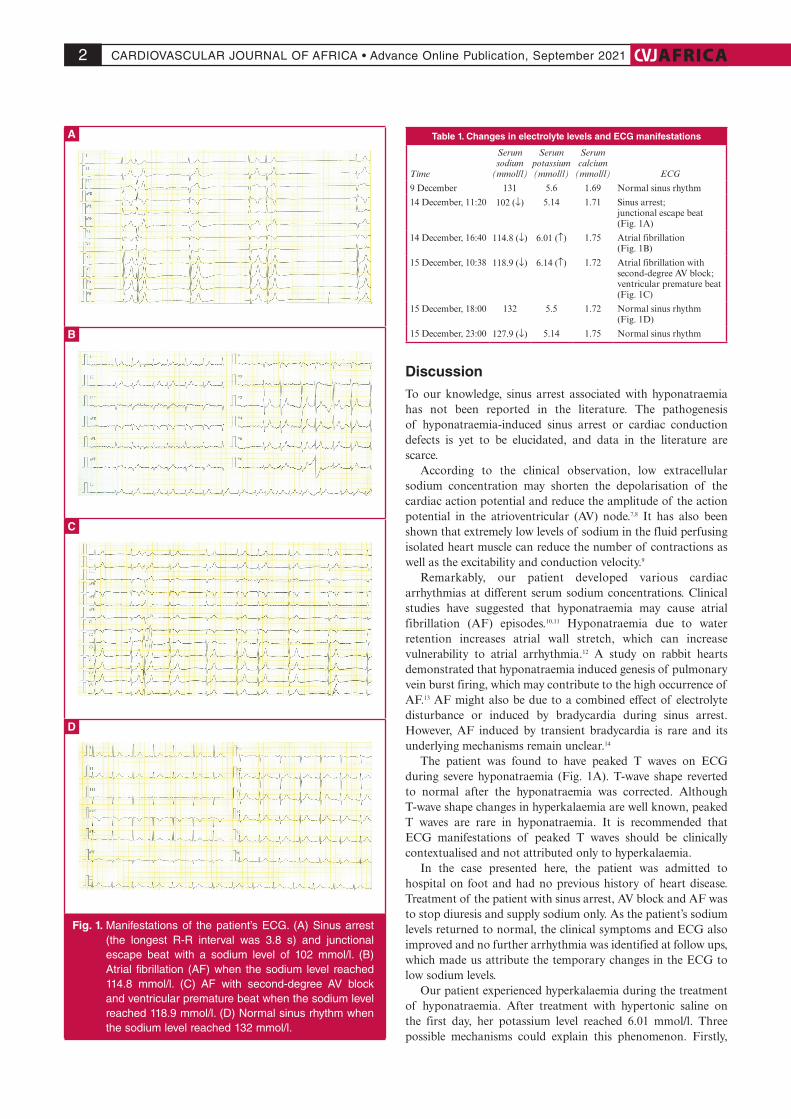
15 December, 18:00 132 5.5 1.72 Normal sinus rhythm(Fig. 1D)
15 December, 23:00 127.9 (↓) 5.14 1.75 Normal sinus rhythm
Fig. 1. Manifestations of the patient’s ECG. (A) Sinus arrest (the longest R-R interval was 3.8 s) and junctional escape beat with a sodium level of 102 mmol/l. (B) Atrial fibrillation (AF) when the sodium level reached 114.8 mmol/l. (C) AF with second-degree AV block and ventricular premature beat when the sodium level reached 118.9 mmol/l. (D) Normal sinus rhythm when the sodium level reached 132 mmol/l.
A
B
C
D

CARDIOVASCULAR JOURNAL OF AFRICA • Advance Online Publication, September 2021AFRICA 3
when the patient received a high dosage of sodium after severe hyponatraemia, there was a reduction in aldosterone secretion, which would tend to decrease the rate of potassium secretion and may consequently reduce urinary excretion of potassium.15
Secondly, the patient’s glomerular filtration rate was low, reducing distal tubular flow rates. The potassium concentration may have built up relatively early in the tubule and thus progressively decreased the electrochemical gradient principal cells and the secretion of potassium.16 Thirdly, considering that the period of discontinuation of spironolactone was too short and the patient’s renal function was also abnormal, there may also have been remaining spironolactone, which contributed to the hyperkalaemia.
ConclusionSerious hyponatraemia can induce sinus arrest. Various arrhythmias shown on ECG may result from different serum sodium concentrations. It is important to raise awareness to give special attention to the ECG and electrolytes in the therapeutic process for a patient with hyponatraemia.
This work was supported by the Medical Science and Technology Innovation
Platform Support Project of Zhongnan Hospital, Wuhan University
(PTXM2020008), Science and Technology Innovation Cultivation Fund of
Zhongnan Hospital, Wuhan University (cxpy2017043) and Medical Science
Advancement Programme (Basic Medical Sciences) of Wuhan University
(TFJC2018004).
References1. Pillai KS, Trivedi TH, Moulick ND. Hyponatremia in ICU. J Assoc
Phys India 2018; 66: 48–52.
2. Hu J, Wang Y, Geng X, Chen R, Zhang P, Lin J, et al. Dysnatremia is
an independent indicator of mortality in hospitalized patients. Med Sci
Monitor Int Med J Exp Clin Res 2017; 23: 2408–2425.
3. Goh KP. Management of hyponatremia. Am Fam Physician 2004; 69:
2387–2394.
4. Kottwitz J, Akdis D, Duru F, Heidecker B. Severe hyponatremia lead-
ing to complete atrioventricular block. Am J Med 2016; 129: e243–244.
5. Karabag T, Kalayci B, Sayin MR, Erten T. Atrioventricular conduc-
tion defect associated with severe hyponatremia. Clujul Med 2018; 91:
342–345.
6. Verbalis JG, Goldsmith SR, Greenberg A, Schrier RW, Sterns RH.
Hyponatremia treatment guidelines 2007: expert panel recommenda-
tions. Am J Med 2007; 120: S1–21.
7. El-Sherif N, Turitto G. Electrolyte disorders and arrhythmogenesis.
Cardiology J 2011; 18: 233–245.
8. Nikolaidou T, Cai XJ, Stephenson RS, Yanni J, Lowe T, Atkinson AJ,
et al. Congestive heart failure leads to prolongation of the PR interval
and atrioventricular junction enlargement and ion channel remodelling
in the rabbit. PLoS One 2015; 10: e0141452.
9. Liamis G, Filippatos TD, Elisaf MS. Thiazide-associated hyponatremia
in the elderly: what the clinician needs to know. J Geriat Cardiol 2016;
13: 175–182.
10. Horio T, Iwashima Y, Kamide K, Tokudome T, Yoshihara F, Nakamura
S, et al. Chronic kidney disease as an independent risk factor for new-
onset atrial fibrillation in hypertensive patients. J Hypertens 2010; 28:
1738–1744.
11. Smith JG, Melander O, Sjogren M, Hedblad B, Engstrom G, Newton-
Cheh C, et al. Genetic polymorphisms confer risk of atrial fibrillation
in patients with heart failure: a population-based study. Eur J Heart Fail
2013; 15: 250–257.
12. Chang SL, Chen YC, Chen YJ, Wangcharoen W, Lee SH, Lin CI, et al.
Mechanoelectrical feedback regulates the arrhythmogenic activity of
pulmonary veins. Heart 2007; 93: 82–88.
13. Lu YY, Cheng CC, Chen YC, Lin YK, Chen SA, Chen YJ. Electrolyte
disturbances differentially regulate sinoatrial node and pulmonary vein
electrical activity: A contribution to hypokalemia- or hyponatremia-
induced atrial fibrillation. Heart Rhythm 2016; 13: 781–788.
14. Altunbas G, Ercan S, Sucu M, Davutoglu V. Atrial fibrillation triggered
by drug-induced bradycardia. J Atrial Fibrillation 2016; 9: 1428.
15. Palmer LG, Frindt G. Aldosterone and potassium secretion by the corti-
cal collecting duct. Kidney Int 2000; 57: 1324–1328.
16. Klevay LM, Bogden JD, Aladjem M, Sandstead HH, Kemp FW, Li W,
et al. Renal and gastrointestinal potassium excretion in humans: new
insight based on new data and review and analysis of published studies.
J Am Coll Nutr 2007; 26: 103–110.