Electroacupuncture Promotes Central Nervous System ... · Electroacupuncture Promotes Central...
13
Electroacupuncture Promotes Central Nervous System-Dependent Release of Mesenchymal Stem Cells T ATIANA E. SALAZAR, a,b,c MATTHEW R. RICHARDSON, d ELENI BELI, b MATTHEW S. RIPSCH, e,f JOHN GEORGE, c YOUNGSOOK KIM, e Y AQIAN DUAN, b LENI MOLDOVAN, b YUANQING Y AN, a ASHAY BHATWADEKAR, b VAISHNAVI JADHAV , e JARED A. SMITH, e SUSAN MCGORRAY , g ALICIA L. BERTONE, h DMITRI O. TRAKTUEV , i,j KEITH L. MARCH, i,j LUIS M. COLON-PEREZ, k KEITH G. AVIN, l EMILY SIMS, m JULIE A. MUND, d,m JAMIE CASE, m,n XIAOLIN DENG, o MIN SU KIM, p BRUCE MCDAVITT , q MICHAEL E. BOULTON, b JEFFREY THINSCHMIDT , r SERGIO LI CALZI, b STEPHANIE D. FITZ, s ROBYN K. FUCHS, l STUART J. WARDEN, l TODD MCKINLEY , t ANANTHA SHEKHAR, s MARCELO FEBO, k PHILLIP L. JOHNSON, u LUNG-JI CHANG, v,w ZHANGUO GAO, x MIKHAIL G. KOLONIN, x SONG LAI, y JINGFENG MA, y XINZHONG DONG, z FLETCHER A. WHITE, e,f HUISHENG XIE, aa MERVIN C. YODER, d,m MARIA B. GRANT b Key Words. Mesenchymal stem cells • Adult stem cells • Nervous system • Neurones ABSTRACT Electroacupuncture (EA) performed in rats and humans using limb acupuncture sites, LI-4 and LI-11, and GV-14 and GV-20 (humans) and Bai-hui (rats) increased functional connectivity between the anterior hypothalamus and the amygdala and mobilized mesenchymal stem cells (MSCs) into the systemic circulation. In human subjects, the source of the MSC was found to be primarily adipose tissue, whereas in rodents the tissue sources were considered more heteroge- neous. Pharmacological disinhibition of rat hypothalamus enhanced sympathetic nervous system (SNS) activation and similarly resulted in a release of MSC into the circulation. EA-mediated SNS activation was further supported by browning of white adipose tissue in rats. EA treatment of rats undergoing partial rupture of the Achilles tendon resulted in reduced mechanical hyperalge- sia, increased serum interleukin-10 levels and tendon remodeling, effects blocked in propranolol-treated rodents. To distinguish the afferent role of the peripheral nervous system, phosphoinositide-interacting regulator of transient receptor potential channels (Pirt)-GCaMP3 (genetically encoded calcium sensor) mice were treated with EA acupuncture points, ST-36 and LIV-3, and GV-14 and Bai-hui and resulted in a rapid activation of primary sensory neurons. EA activated sensory ganglia and SNS centers to mediate the release of MSC that can enhance tis- sue repair, increase anti-inflammatory cytokine production and provide pronounced analgesic relief. STEM CELLS 2017;35:1303–1315 SIGNIFICANCE STATEMENT In this study, we show how the use of electroacupuncture (EA) at specific points stimulates mesenchymal stem cell (MSC) release into peripheral blood through the activation of the ner- vous system. EA could be used to aid tissue repair through increasing the levels of circulating MSC. Moreover, MSC can be harvested directly from the blood of EA-treated humans and ani- mals and expanded ex vivo. Thus, EA may be a low cost, low risk method for MSC harvest for autologous stem cell therapy. INTRODUCTION Acupuncture, one of the oldest medical thera- pies, mediates its therapeutic effect through the insertion of needles into specific points in the body called acupoints [1]. Electroacupuncture (EA) combines traditional acupuncture with modern electrotherapy as a means to enhance the stimulation at the acupoints. Acupoints are located in areas of decreased electrical resis- tance and increased electrical conductivity in the body attributed to both neural and vascular elements in the dermis or hypodermis [2]. His- tological studies reveal that acupoints are locat- ed in areas with high densities of free nerve endings, arterioles, lymphatics, and mast cells a Genetics Institute, c College of Medicine, g Department of Biostatistics, k Department of Psychiatry, McKnight Brain Institute, r Department of Pharmacology and Therapeutics, w Department of Molecular Genetics and Microbiology, y Department of Radiation Oncology, College of Medicine, aa College of Veterinary Medicine, University of Florida, Gainesville, Florida, USA, b Department of Ophthalmology, d Wells Center for Pediatric Research, e Department of Anesthesia, s Department of Psychiatry, t Department of Orthopedic Surgery, u Department of Anatomy and Cell Biology, Indiana University School of Medicine, Indianapolis, Indiana, USA, f Richard L. Roudebush VA Medical Center; i Krannert Institute of Cardiology, j Indiana Center for Vascular Biology and Medicine, l Department of Physical Therapy, School of Health and Rehabilitation Sciences, m Melvin and Bren Simon Cancer Center, Indiana University, Indianapolis, Indiana, USA; h Department of Veterinary Clinical Sciences, The Ohio State University, Columbus, Ohio, USA; n Scripps Clinic Medical Group, Scripps Center for Organ and Cell Transplantation, La Jolla, California, USA; o Mainland Acupuncture Center, Gainesville, Florida, USA; p College of Veterinary Medicine, Chon Buk National University, Jeonju, South Korea; q McDavitt Veterinary Clinic, Zionsville, Indiana, USA; v School of Medicine, University of Electronic Science and Technology of China, Sichuan, China; x The Brown Foundation, Institute of Molecular Medicine, University of Texas Health Science Center, Houston, Texas, USA; z Department of Neuroscience, Center of Sensory Biology, the Johns Hopkins University School of Medicine, Baltimore, Maryland, USA Correspondence: Maria B. Grant, M.D., Department of Ophthalmology, Indiana University School of Medicine, 980 W. Walnut Street, R3-C426D, Indianapolis, Indi- ana 46202, USA. Telephone: 11- 317-274-2628; Fax: 317-274-8046; e-mail: [email protected]; or Mervin C. Yoder, M.D., Wells Center for Pediatric Research, Indiana Uni- versity School of Medicine, 1044 West Walnut Street, R4-W125, Indi- anapolis, Indiana 46202, USA. Tele- phone: 11-317-274-4738; Fax: 317- 274-8679; e-mail: [email protected] Received November 3, 2016; accepted for publication February 11, 2017; first published online in STEM CELLS EXPRESS March 16, 2017. V C AlphaMed Press 1066-5099/2017/$30.00/0 http://dx.doi.org/ 10.1002/stem.2613 REGENERATIVE MEDICINE www.StemCells.com The copyright line for this article was changed on 6 October 2017 after original online publication. V C AlphaMed Press 2017
Transcript of Electroacupuncture Promotes Central Nervous System ... · Electroacupuncture Promotes Central...

Electroacupuncture Promotes Central Nervous
System-Dependent Release of Mesenchymal
Stem Cells
TATIANA E. SALAZAR,a,b,c
MATTHEW R. RICHARDSON,d
ELENI BELI,b
MATTHEW S. RIPSCH,e,f
JOHN GEORGE,c
YOUNGSOOK KIM,e
YAQIAN DUAN,b
LENI MOLDOVAN,b
YUANQING YAN,a
ASHAY BHATWADEKAR,b
VAISHNAVI JADHAV,e
JARED A. SMITH,e
SUSAN MCGORRAY,g
ALICIA L. BERTONE,h
DMITRI O. TRAKTUEV,i,j
KEITH L. MARCH,i,j
LUIS M. COLON-PEREZ,k
KEITH G. AVIN,l
EMILY SIMS,m
JULIE A. MUND,d,m
JAMIE CASE,m,n
XIAOLIN DENG,o
MIN SU KIM,p
BRUCE MCDAVITT,q
MICHAEL E. BOULTON,b
JEFFREY THINSCHMIDT,r
SERGIO LI CALZI,b
STEPHANIE D. FITZ,s
ROBYN K. FUCHS,l
STUART J. WARDEN,l
TODD MCKINLEY,t
ANANTHA SHEKHAR,s
MARCELO FEBO,k
PHILLIP L. JOHNSON,u
LUNG-JI CHANG,v,w
ZHANGUO GAO,x
MIKHAIL G. KOLONIN,x
SONG LAI,y
JINGFENG MA,y
XINZHONG DONG,z
FLETCHER A. WHITE,e,f
HUISHENG XIE,aa
MERVIN C. YODER,d,m
MARIA B. GRANTb
Key Words. Mesenchymal stem cells • Adult stem cells • Nervous system • Neurones
ABSTRACT
Electroacupuncture (EA) performed in rats and humans using limb acupuncture sites, LI-4 and
LI-11, and GV-14 and GV-20 (humans) and Bai-hui (rats) increased functional connectivity
between the anterior hypothalamus and the amygdala and mobilized mesenchymal stem cells
(MSCs) into the systemic circulation. In human subjects, the source of the MSC was found to be
primarily adipose tissue, whereas in rodents the tissue sources were considered more heteroge-
neous. Pharmacological disinhibition of rat hypothalamus enhanced sympathetic nervous system
(SNS) activation and similarly resulted in a release of MSC into the circulation. EA-mediated SNS
activation was further supported by browning of white adipose tissue in rats. EA treatment of
rats undergoing partial rupture of the Achilles tendon resulted in reduced mechanical hyperalge-
sia, increased serum interleukin-10 levels and tendon remodeling, effects blocked in
propranolol-treated rodents. To distinguish the afferent role of the peripheral nervous system,
phosphoinositide-interacting regulator of transient receptor potential channels (Pirt)-GCaMP3
(genetically encoded calcium sensor) mice were treated with EA acupuncture points, ST-36 and
LIV-3, and GV-14 and Bai-hui and resulted in a rapid activation of primary sensory neurons. EA
activated sensory ganglia and SNS centers to mediate the release of MSC that can enhance tis-
sue repair, increase anti-inflammatory cytokine production and provide pronounced analgesic
relief. STEM CELLS 2017;35:1303–1315
SIGNIFICANCE STATEMENT
In this study, we show how the use of electroacupuncture (EA) at specific points stimulatesmesenchymal stem cell (MSC) release into peripheral blood through the activation of the ner-vous system. EA could be used to aid tissue repair through increasing the levels of circulatingMSC. Moreover, MSC can be harvested directly from the blood of EA-treated humans and ani-mals and expanded ex vivo. Thus, EA may be a low cost, low risk method for MSC harvest forautologous stem cell therapy.
INTRODUCTION
Acupuncture, one of the oldest medical thera-pies, mediates its therapeutic effect through theinsertion of needles into specific points in thebody called acupoints [1]. Electroacupuncture(EA) combines traditional acupuncture withmodern electrotherapy as a means to enhance
the stimulation at the acupoints. Acupoints arelocated in areas of decreased electrical resis-tance and increased electrical conductivity inthe body attributed to both neural and vascularelements in the dermis or hypodermis [2]. His-tological studies reveal that acupoints are locat-ed in areas with high densities of free nerveendings, arterioles, lymphatics, and mast cells
aGenetics Institute, cCollege of Medicine,gDepartment of Biostatistics,kDepartment of Psychiatry, McKnightBrain Institute, rDepartment ofPharmacology and Therapeutics,wDepartment of Molecular Genetics andMicrobiology, yDepartment of RadiationOncology, College of Medicine, aaCollegeof Veterinary Medicine, University ofFlorida, Gainesville, Florida, USA,bDepartment of Ophthalmology, dWellsCenter for Pediatric Research,eDepartment of Anesthesia,sDepartment of Psychiatry, tDepartmentof Orthopedic Surgery, uDepartment ofAnatomy and Cell Biology, IndianaUniversity School of Medicine,Indianapolis, Indiana, USA, fRichard L.Roudebush VA Medical Center; iKrannertInstitute of Cardiology, jIndiana Centerfor Vascular Biology and Medicine,lDepartment of Physical Therapy, Schoolof Health and Rehabilitation Sciences,mMelvin and Bren Simon Cancer Center,Indiana University, Indianapolis, Indiana,USA; hDepartment of Veterinary ClinicalSciences, The Ohio State University,Columbus, Ohio, USA; nScripps ClinicMedical Group, Scripps Center for Organand Cell Transplantation, La Jolla,California, USA; oMainland AcupunctureCenter, Gainesville, Florida, USA; pCollegeof Veterinary Medicine, Chon BukNational University, Jeonju, South Korea;qMcDavitt Veterinary Clinic, Zionsville,Indiana, USA; vSchool of Medicine,University of Electronic Science andTechnology of China, Sichuan, China;xThe Brown Foundation, Institute ofMolecular Medicine, University of TexasHealth Science Center, Houston, Texas,USA; zDepartment of Neuroscience,Center of Sensory Biology, the JohnsHopkins University School of Medicine,Baltimore, Maryland, USA
Correspondence: Maria B. Grant,M.D., Department ofOphthalmology, Indiana UniversitySchool of Medicine, 980 W. WalnutStreet, R3-C426D, Indianapolis, Indi-ana 46202, USA. Telephone: 11-317-274-2628; Fax: 317-274-8046;e-mail: [email protected]; orMervin C. Yoder, M.D., Wells Centerfor Pediatric Research, Indiana Uni-versity School of Medicine, 1044West Walnut Street, R4-W125, Indi-anapolis, Indiana 46202, USA. Tele-phone: 11-317-274-4738; Fax: 317-274-8679; e-mail: [email protected]
Received November 3, 2016;accepted for publication February11, 2017; first published online inSTEM CELLS EXPRESS March 16, 2017.
VC AlphaMed Press1066-5099/2017/$30.00/0
http://dx.doi.org/10.1002/stem.2613
REGENERATIVE MEDICINE
www.StemCells.com
The copyright line for thisarticle was changed on6 October 2017 after originalonline publication.
VC AlphaMed Press 2017
[3]. Certain acupoints have been identified to correspond tospecific neural structures, such as superficial nerves and nerveplexuses [3].
The primary mechanism implicated in the anti-nociceptiveeffect of acupuncture involves release of opioid peptides inthe central nervous system (CNS) in response to the long-lasting activation of ascending sensory tracks during the stim-ulation [1]. Adenosine has also been implicated, and interfer-ing with adenosine metabolism prolonged the clinical benefitof acupuncture [4].
While both anatomical characteristics and local mediatorsof signaling are associated with specific acupoints, the mecha-nism responsible for the beneficial systemic effects and heal-ing associated with acupuncture still lacks understanding. Inthis study, we sought to understand the central and peripher-al nervous system response to EA using two sets of points (1:LI-4, LI-11, GV-14, Bai-hui; 2:ST-36, LIV-3, GV-14 and Bai-hui;Supporting Information Fig. S1) associated with successfultreatment of arthritis and asthma and with modulation ofimmunity [5].
RESULTS
EA Induces Activation of Hypothalamic Regions of the
Brain in Both Rats and Humans
The systemic beneficial effects of EA may be centrally driven;therefore, we sought to determine the relationship betweenthe hypothalamus and other brain structures, which is termed“connectivity.” The hypothalamus plays a critical role as a pri-mary homeostatic center in the brain and contains neuronswith important projections to other limbic sites and sympa-thetic nuclei directly communicating with the periphery. Todetermine whether the hypothalamus was involved in the EAresponse, blood oxygen level-dependent (BOLD) functionalmagnetic resonance imaging (fMRI) was performed in anes-thetized male Sprague-Dawley rats (n 5 6) during a single EAsession. Connectivity was derived from four time points: base-line, 0–8 minutes during EA, 9–22 minutes, and immediatelypost-EA. Seed regions included the anterior, posterior, and lat-eral hypothalamus. EA-stimulation produced changes in rats inthe strength of functional connectivity within the hypothala-mus and between the hypothalamus and adjacent brainregions, such as the amygdala (Fig. 1A), compared with base-line and the post-EA period. A representative example of theincrease in signal (over time) in the PVN of a rat receivingacupuncture is shown in Figure 1B.
Arterial spin labeling fMRI was performed in healthyhuman subjects (n 5 6) during a single EA session. Connectivi-ty was derived from four time points: baseline, 0–8 minutesduring EA, 8–16 minutes during EA, and immediately post-EA.EA stimulation produced changes in humans in the strengthof functional connectivity within the hypothalamus andbetween the hypothalamus and adjacent brain regions (Fig. 2)compared with baseline and the post-EA period. Thus, ratsand humans exhibited a similar response in terms of the con-nectivity observed.
As these regions have been associated with hematopoieticstem cell mobilization [6], peripheral blood was examined inthe rat before and following EA for evidence of mesenchymalstem cell (MSC) mobilization. A 313% increase in the
circulating MSC population, Lin-CD901CD44HI cells, wasdetected in the blood of EA-treated rats 2 hours post-EA com-pared with baseline (Fig. 3A, 3B).
Characterization of Human EA Mobilized MSC as
Adipocyte-Derived MSC
The availability of human antibodies to fully characterize thepopulations of cells mobilized by EA lead us to next examinethe populations of cells mobilized by EA in the human sub-jects that underwent fMRI. Due to limitations regarding thefMRI equipment used, only points GV-14, LI-11, and LI-14were able to be used in this experiment. We observed anincrease in MSC in the peripheral blood 2 hours following EAin all subjects (Fig. 3C), while total lymphocyte numbers didnot change. To further characterize the MSC population andpotentially determine whether they were released from adi-pose stores, the levels of CD341CD45-CD31- cells wereexamined. Two hours following completion of EA, CD341
CD45-CD31- cells were increased in four of six individuals (Fig.3D) and the response was proportional to the body massindex of the subjects as previously noted [7]. Two individualswith body mass index less than 18.5 did not demonstrate anincrease in this population. Human EA mobilized MSC wereexpanded in vitro and underwent adipogenic differentiation.Adipogenesis potential was confirmed by the cells’ ability toform lipid droplets as established through oil red O staining(Fig. 3E–3H).
Because hypothalamic activation (Fig. 1B) preceded themobilization of circulating MSC, it was proposed that the EA-induced connectivity changes contributed to the subsequentrelease of these cells into the peripheral blood. Exogenousadministration of epinephrine or dopamine in rats resulted ina similar increase of Lin-CD90HICD441 cell population into thecirculation (Supporting Information Fig. S2A–S2C), supportingthe role of CNS in mobilization of MSC and in EA activatingthese key CNS centers.
EA-Induced Uncoupling Protein 1 Expression Promotes
the Browning of White Adipose Tissue in Rats
To confirm that EA can activate the sympathetic nervous sys-tem (SNS), we asked whether EA was associated with brow-ning of white adipose tissue (WAT). The “inducible/recruitable” brown-like (beige a.k.a bright) adipocytes arise inWAT and are functionally indistinguishable from adipocytes inthe canonical (constitutive) brown adipose tissue [8, 9]. Thefunction of brown and beige adipocytes is muscle-independent thermogenic energy dissipation, which relies onthe function of uncoupling protein 1 (UCP1) [10]. Following14 days of EA (every other day) in rats, UCP1 immunofluores-cence of inguinal subcutaneous WAT was increased (Fig. 4A),indicating a greater number of brown adipocytes, comparedwith control (sham EA) (Fig. 4B). Consistent with the notionthat SNS signaling mainly activates subcutaneous WAT, nochanges were detected in intraperitoneal WAT.
Pharmacological Disinhibition of the Dorsomedial
Regions of the Tuberal Hypothalamus Mobilizes
Lin-CD90
HICD44
1Cells into the Circulation
To confirm that activation of the SNS can mediate release ofMSC, we performed stereotaxic injections with the GABAAreceptor antagonist bicuculline methiodide (BMI) in the
1304 EA Promotes CNS-Dependent Release of MSC
STEM CELLSVC AlphaMed Press 2017

dorsomedial regions of the tuberal hypothalamus to disinhibitthese regions. Histological verification of injection sites is indi-cated in the illustration (Fig. 4C). The exact location of allinjection sites are shown on coronal sections from a StandardStereotaxic Atlas of the Rat Brain [11] (Fig. 4C) and a photo-micrograph that shows the representative injection site (Fig.4D). In rats, BMI at the dose of 50 pmol increased the per-centage of circulating Lin-CD90HICD441 cells (F (2, 14) 5 6.7,p 5 .027) at 4 hours post injection of BMI (Fig. 4E) in theabsence of EA. These results demonstrate that circulatingLin-CD90HICD441 cells are mobilized during hypothalamic
disinhibition and EA. However, the delay in release (4 hours)compared with EA (2 hours) may suggest an alternative mech-anism of release.
EA-Treated Rodents Exhibit Reduced Mechanical
Hyperalgesia, Increased Serum Interleukin-10 Levels
and Enhanced Tissue Remodeling following Partial
Achilles Tendon Rupture
To address the possible anti-inflammatory and analgesic effectsof EA-mobilization of circulating MSC [7], we analyzed the con-tribution of an EA treatment paradigm on injury-induced
Figure 1. Electroacupuncture (EA) stimulation increases hypothalamic functional connectivity in rats. (A): Rat brains were monitoredthrough functional magnetic resonance imaging during administration of EA. Functional connectivity increased within the hypothalamus andbetween the hypothalamus and adjacent brain region with progression of treatment (n 5 6). (B): A representative example of the increase insignal (over time) in the PVN of a rat receiving acupuncture (n 5 1). Abbreviations: EA, electroacupuncture; PVN, paraventricular nucleus.
Salazar, Richardson, Beli et al. 1305
www.StemCells.com VC AlphaMed Press 2017

hyperalgesia in rats in the presence and absence of a b-blocker, propranolol. Treatments included EA (i.e., applied tothe forelimb LI-4, LI-11, GV-14, and Bai-hui, and hindlimbST36, LIV-3, GV-14 and Bai-hui immune points in horses), shamEA (i.e., applied to skin not associated with an acupoint) or EAplus propranolol (i.e., potential inhibitor for sympatheticeffects of EA). EA or sham EA was administered every otherday for 2 weeks following injury. Using sham EA applied to
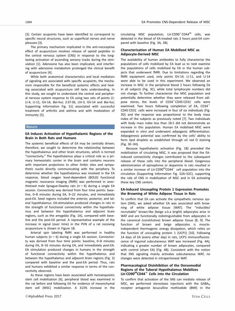
nonimmune acupoints or EA plus propranolol, nociceptivebehavior elicited by von Frey mechanical stimulation did notchange over the time course in the hind paw ipsilateral to theinjury (Supporting Information Fig. S3). In contrast, mechanicalhyperalgesia (assessed at both day 7 and 14) was considerablydecreased (i.e., able to tolerate more pressure) in injuredrodents subjected to EA application at LI-4, LI-11, GV-14, andBai-hui (Fig. 5A). At early stages of tendon repair, the
Figure 2. Electroacupuncture (EA) stimulation increases hypothalamic functional connectivity in humans. Brains from normal subjectswere monitored using functional magnetic resonance imaging before, during, and after EA. Functional connectivity increased within thehypothalamus and between the hypothalamus and adjacent brain regions with progression of treatment (n 5 6).
1306 EA Promotes CNS-Dependent Release of MSC
STEM CELLSVC AlphaMed Press 2017

Figure 3. Electroacupuncture (EA) stimulation induced mesenchymal stem cell (MSC) mobilization. (A): Rat peripheral blood MSC wereincreased (p 5 .0063) after EA. Circulating MSC were defined as Lin-(CD45-CD31-erythroid-CD11b-) cells that were positive for CD44 andCD90. Gated cells increased post treatment (n 5 11 for baseline and 4 hours, n 5 9 for 2 hours). (B): Representative flow charts for rat Lin-
cells are shown at baseline and 2 hours samples. (C): The percentage of human peripheral blood MSC increased in post EA-treatment(p 5 .006, n 5 6). (D): The percentage of circulating MSCs from adipose tissue (AD-MSC) is significantly elevated 2 hours post EA-treatment(p 5 .033, n 5 4). (E, F): EA-mobilized MSCs were expanded in vitro. After undergoing adipogenesis differentiation, EA-mobilized MSCs devel-oped fat deposits as seen by Oil Red staining, which were not seen in the undifferentiated control cells (G, H). Magnification bars5 (E, G):100 mm; (F, H): 50 mm. Abbreviations: ASC, adipose stem cells; EA, electroacupuncture; MNC, mononuclear cell.
Salazar, Richardson, Beli et al. 1307
www.StemCells.com VC AlphaMed Press 2017

granulation tissues mainly synthesize type III collagen, while atlater stages of healing, intrinsic fibroblasts produce type I colla-gen, whose fibers are orientated longitudinally to replace typeIII collagen. At 14 days post-injury, EA sham-treated or EA pluspropranolol did not change type I collagen content, while typeI collagen was significantly enhanced by EA (Fig. 5B); there wasno change in type III collagen across treatments (Fig. 5C). Tak-en together, these data suggest that EA may enhance thereplacement of thinner and immature type III collagen fiberswith mature type I collagen fibers in the injured tendon [12],thereby supporting a better quality of regeneration and tissue
reorganization. Given that EA can attenuate mechanical hyper-algesia and enhance tissue reorganization, we examined possi-ble alterations of the anti-inflammatory cytokine, interleukin(IL)-10, in blood plasma. Previous evidence suggested that EAfollowing surgical trauma contributed to increased levels of IL-10 by T-cells [13] and focal microinjections of IL-10 diminishmechanical hyperalgesia [14]. The results herein demonstratethat tendon injury in rodents subjected to sham EA or EA com-bined with propranolol fail to produce detectable changes inplasma IL-10. However, injured rodents treated with EA signifi-cantly increased IL-10 in plasma. These results indicate that EA
Figure 4. Electroacupuncture (EA) increases sympathetic activation leading to browning of white adipose tissue and the effects of EAcan be duplicated by pharmacological disinhibition of hypothalamus (sympathetic activation). (A, B): UCP1 immunofluorescence (red)detectable in inguinal subcutaneous adipose tissue (blue: adipocytes nuclei) from animals that underwent EA treatment (A) but not incontrol (B). EA resulted in an increase in beige adipocytes (n 5 4). Magnification bars5 50 mm. (C): The effects of EA can be duplicatedby pharmacological disinhibition of hypothalamus. Rats underwent injection of either vehicle, 30 pmol or 50 pmol/100 nL of the GABAAreceptor antagonist bicuculline methiodide. Sites of injections are represented on a coronal section from the tuberal hypothalamus froma Standard Steroetaxic Atlas of the Rat brain [11]. Colored circles indicate injection sites (black, orange, and red represent vehicle, 30pmol and 50 pmol, respectively). (D): Representative photomicrograph showing an injection site from one rat. Magnificationbar5 1 mm. (E): There was a significant increase (p 5 .027) in Lin-CD90HICD441 cells in the plasma 4 hours post injection (n 5 6). Datapresented as means6 SEM. Abbreviations: BMI, bicuculline methiodide; DMN, dorsomedial hypothalamic nucleus; f, fornix; MNC, mono-nuclear cells; mt, mammillothalamic tract; PeF, perifornical hypothalamus; PH, posterior hypothalamic nucleus; VMN, ventromedial hypo-thalamic nucleus; 3V, third ventricle.
1308 EA Promotes CNS-Dependent Release of MSC
STEM CELLSVC AlphaMed Press 2017
increases production of the endogenous anti-inflammatorycytokine IL-10 (Fig. 5D).
EA Performed over Immune Points in Pirt-GCaMP3
Mice Rapidly Activates Primary Sensory Neurons
We next examined mice to confirm that EA stimulation of acupoints(LI-4, LI-11, and GV-14 and Bai-hui) resulted in mobilization of MSC.At 4 hours post EA, murine MSC as defined by Lin-PDGFRa1Sca-11
cells were significantly increased (p 5 .01) in peripheral blood, aresponse that was markedly reduced when mice were pretreatedwith propranolol (Supporting Information Fig. S4). Immune
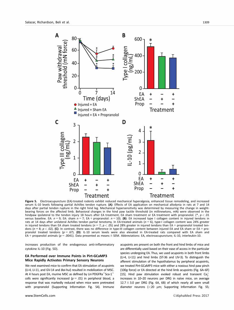
acupoints are present on both the front and hind limbs of mice andare differentially used based on their ease of access in the particularspecies undergoing EA. Thus, we used acupoints in both front limbs(LI-4, LI-11) and hind limbs (ST-36 and LIV-3). To distinguish theafferent stimulation of the hypothalamus by peripheral acupoints,we treated Pirt-GCaMP3 mice with either a noxious hind paw pinch(100g force) or EA directed at the hind limb acupoints (Fig. 6A–6F)[15]. Hind paw stimulation evoked robust and transient Ca1
2
increases in 10–20 neurons per DRG in na€ıve mice, on average12.763.0 per DRG (Fig. 6A, 6B) of which nearly all were smalldiameter neurons (<20 mm; Supporting Information Fig. S5;
Figure 5. Electroacupuncture (EA)-treated rodents exhibit reduced mechanical hyperalgesia, enhanced tissue remodeling, and increasedserum IL-10 levels following partial Achilles tendon rupture. (A): Effects of EA application on mechanical allodynia in rats at 7 and 14days after partial tendon rupture in the right hind leg. Mechanical hypersensitivity was determined by measuring the change in weight-bearing forces on the affected limb. Behavioral changes in the hind paw tactile threshold (in millinewtons, mN) were observed in thehindpaw ipsilateral to the tendon injury 18 hours after EA treatment, EA sham treatment or EA treatment with propranolol. (*, p< .01versus baseline. EA: n 5 9; EA: sham n 5 7; EA1 propranolol: n 5 10). (B): EA increased type I collagen content in injured tendons inrats at 14 days after unilateral Achilles tendon partial tenotomy. In EA-treated animals (n 5 6), type-I collagen content was 24% greaterin injured tendons than EA sham treated tendons (n 5 7; p< .05) and 28% greater in injured tendons than EA1 propranolol treated ten-dons (n 5 8; p< .02). (C): In contrast, there was no difference in type-III collagen content between injured EA and EA sham or EA1 pro-pranolol treated tendons (p 5 .67). (D): IL-10 serum levels were also elevated in EA-treated rats compared with EA sham andEA1 propanolol animals (p 5 .0041). Data presented as means6 SEM. Abbreviations: EA, electroacupuncture; IL-10, interleukin-10.
Salazar, Richardson, Beli et al. 1309
www.StemCells.com VC AlphaMed Press 2017

Figure 6. Small diameter primary afferent sensory neurons show sensitivity to pinch stimulus and medium-large diameter neuronsshow sensitivity to electroacupuncture (EA) in Pirt-GCaMP3 mice with intact dorsal root ganglia (DRG). Representative fluorescentGCaMP3 neuronal imaging response by lumbar DRG in vivo before (A) and after a mechanical press of the hindpaw using a 100g force(B). The mechanical force evokes a robust fluorescent GCaMP3 Ca21 response in numerous small sensory neurons (B; white arrows).Acupuncture needles alone failed to elicit neuronal changes in the same lumbar DRG of the Pirt-GCamp3 mouse (C, D). EA-stimulationof the ST-36 and LIV-3 accupoints produced rapid activation of medium–larger diameter of the lumbar DRG (E, F; white arrows) (n 5 4,p< .0001). Abbreviation: EA, electroacupuncture.
1310 EA Promotes CNS-Dependent Release of MSC
STEM CELLSVC AlphaMed Press 2017
Supporting Information Video 1) suggestive of pain fibers. Place-ment of acupuncture needles alone did not elicit activity in sensoryneurons (Fig. 6C, 6D; Supporting Information Video 2). We noticeda striking pattern of neuronal activation in the DRG following EAstimulation; many activated neurons were medium to large diame-ter neurons (medium [20–25 mm]; large [>25 mm] with an averageof 6.164.0 neurons per ganglia; (Fig. 6E, 6F; Supporting Informa-tion Video 3) suggesting activation of touch fibers.
EA at Immune Points in the Forelimbs Preferentially
Mobilizes MSC Whereas EA of Immune Points in the
Hind Limb Mobilizes Macrophages into the Circulation
Acupuncture of horses and humans is pursued with the samerigor and is not as technically difficult as acupuncture inrodents. We examined the forelimb (LI-4, LI-11, and GV-14and Bai-hui) and hind limb immune points (ST36, LIV-3, andGV-14 and Bai-hui) in horses. Peripheral blood of horses(n 5 30) undergoing EA at LI-4, LI-11, and GV-14 and Bai-hui
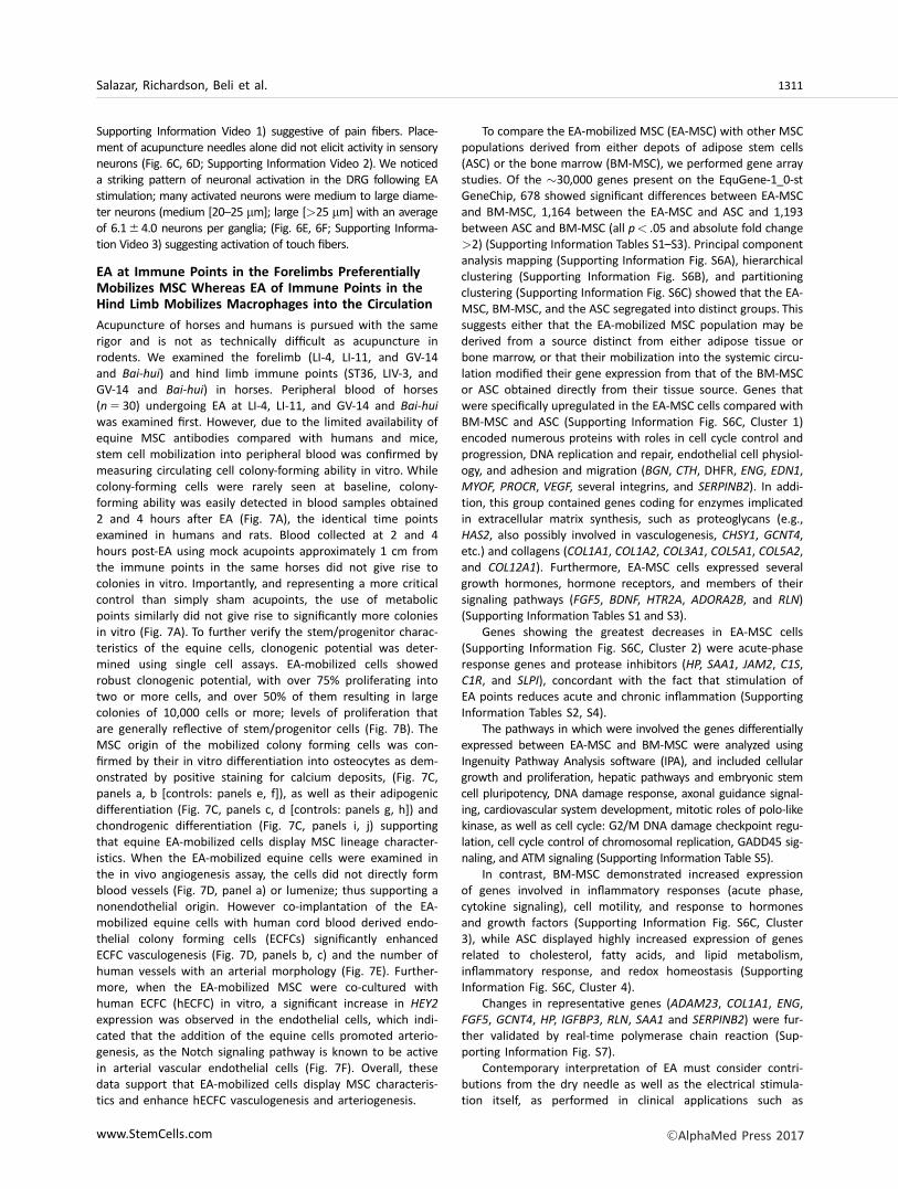
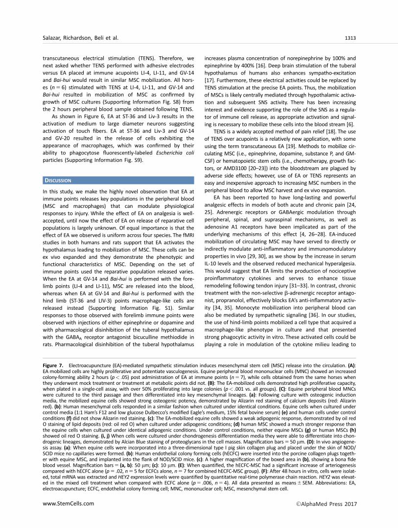
was examined first. However, due to the limited availability ofequine MSC antibodies compared with humans and mice,stem cell mobilization into peripheral blood was confirmed bymeasuring circulating cell colony-forming ability in vitro. Whilecolony-forming cells were rarely seen at baseline, colony-forming ability was easily detected in blood samples obtained2 and 4 hours after EA (Fig. 7A), the identical time pointsexamined in humans and rats. Blood collected at 2 and 4hours post-EA using mock acupoints approximately 1 cm fromthe immune points in the same horses did not give rise tocolonies in vitro. Importantly, and representing a more criticalcontrol than simply sham acupoints, the use of metabolicpoints similarly did not give rise to significantly more coloniesin vitro (Fig. 7A). To further verify the stem/progenitor charac-teristics of the equine cells, clonogenic potential was deter-mined using single cell assays. EA-mobilized cells showedrobust clonogenic potential, with over 75% proliferating intotwo or more cells, and over 50% of them resulting in largecolonies of 10,000 cells or more; levels of proliferation thatare generally reflective of stem/progenitor cells (Fig. 7B). TheMSC origin of the mobilized colony forming cells was con-firmed by their in vitro differentiation into osteocytes as dem-onstrated by positive staining for calcium deposits, (Fig. 7C,panels a, b [controls: panels e, f]), as well as their adipogenicdifferentiation (Fig. 7C, panels c, d [controls: panels g, h]) andchondrogenic differentiation (Fig. 7C, panels i, j) supportingthat equine EA-mobilized cells display MSC lineage character-istics. When the EA-mobilized equine cells were examined inthe in vivo angiogenesis assay, the cells did not directly formblood vessels (Fig. 7D, panel a) or lumenize; thus supporting anonendothelial origin. However co-implantation of the EA-mobilized equine cells with human cord blood derived endo-thelial colony forming cells (ECFCs) significantly enhancedECFC vasculogenesis (Fig. 7D, panels b, c) and the number ofhuman vessels with an arterial morphology (Fig. 7E). Further-more, when the EA-mobilized MSC were co-cultured withhuman ECFC (hECFC) in vitro, a significant increase in HEY2
expression was observed in the endothelial cells, which indi-cated that the addition of the equine cells promoted arterio-genesis, as the Notch signaling pathway is known to be activein arterial vascular endothelial cells (Fig. 7F). Overall, thesedata support that EA-mobilized cells display MSC characteris-tics and enhance hECFC vasculogenesis and arteriogenesis.
To compare the EA-mobilized MSC (EA-MSC) with other MSCpopulations derived from either depots of adipose stem cells(ASC) or the bone marrow (BM-MSC), we performed gene arraystudies. Of the �30,000 genes present on the EquGene-1_0-stGeneChip, 678 showed significant differences between EA-MSCand BM-MSC, 1,164 between the EA-MSC and ASC and 1,193between ASC and BM-MSC (all p< .05 and absolute fold change>2) (Supporting Information Tables S1–S3). Principal componentanalysis mapping (Supporting Information Fig. S6A), hierarchicalclustering (Supporting Information Fig. S6B), and partitioningclustering (Supporting Information Fig. S6C) showed that the EA-MSC, BM-MSC, and the ASC segregated into distinct groups. Thissuggests either that the EA-mobilized MSC population may bederived from a source distinct from either adipose tissue orbone marrow, or that their mobilization into the systemic circu-lation modified their gene expression from that of the BM-MSCor ASC obtained directly from their tissue source. Genes thatwere specifically upregulated in the EA-MSC cells compared withBM-MSC and ASC (Supporting Information Fig. S6C, Cluster 1)encoded numerous proteins with roles in cell cycle control andprogression, DNA replication and repair, endothelial cell physiol-ogy, and adhesion and migration (BGN, CTH, DHFR, ENG, EDN1,MYOF, PROCR, VEGF, several integrins, and SERPINB2). In addi-tion, this group contained genes coding for enzymes implicatedin extracellular matrix synthesis, such as proteoglycans (e.g.,HAS2, also possibly involved in vasculogenesis, CHSY1, GCNT4,etc.) and collagens (COL1A1, COL1A2, COL3A1, COL5A1, COL5A2,and COL12A1). Furthermore, EA-MSC cells expressed severalgrowth hormones, hormone receptors, and members of theirsignaling pathways (FGF5, BDNF, HTR2A, ADORA2B, and RLN)(Supporting Information Tables S1 and S3).
Genes showing the greatest decreases in EA-MSC cells(Supporting Information Fig. S6C, Cluster 2) were acute-phaseresponse genes and protease inhibitors (HP, SAA1, JAM2, C1S,C1R, and SLPI), concordant with the fact that stimulation ofEA points reduces acute and chronic inflammation (SupportingInformation Tables S2, S4).
The pathways in which were involved the genes differentiallyexpressed between EA-MSC and BM-MSC were analyzed usingIngenuity Pathway Analysis software (IPA), and included cellulargrowth and proliferation, hepatic pathways and embryonic stemcell pluripotency, DNA damage response, axonal guidance signal-ing, cardiovascular system development, mitotic roles of polo-likekinase, as well as cell cycle: G2/M DNA damage checkpoint regu-lation, cell cycle control of chromosomal replication, GADD45 sig-naling, and ATM signaling (Supporting Information Table S5).
In contrast, BM-MSC demonstrated increased expressionof genes involved in inflammatory responses (acute phase,cytokine signaling), cell motility, and response to hormonesand growth factors (Supporting Information Fig. S6C, Cluster3), while ASC displayed highly increased expression of genesrelated to cholesterol, fatty acids, and lipid metabolism,inflammatory response, and redox homeostasis (SupportingInformation Fig. S6C, Cluster 4).
Changes in representative genes (ADAM23, COL1A1, ENG,FGF5, GCNT4, HP, IGFBP3, RLN, SAA1 and SERPINB2) were fur-ther validated by real-time polymerase chain reaction (Sup-porting Information Fig. S7).
Contemporary interpretation of EA must consider contri-butions from the dry needle as well as the electrical stimula-tion itself, as performed in clinical applications such as
Salazar, Richardson, Beli et al. 1311
www.StemCells.com VC AlphaMed Press 2017

Figure 7.
1312 EA Promotes CNS-Dependent Release of MSC
STEM CELLSVC AlphaMed Press 2017
transcutaneous electrical stimulation (TENS). Therefore, wenext asked whether TENS performed with adhesive electrodesversus EA placed at immune acupoints LI-4, LI-11, and GV-14and Bai-hui would result in similar MSC mobilization. All hors-es (n 5 6) stimulated with TENS at LI-4, LI-11, and GV-14 andBai-hui resulted in mobilization of MSC as confirmed bygrowth of MSC cultures (Supporting Information Fig. S8) fromthe 2 hours peripheral blood sample obtained following TENS.
As shown in Figure 6, EA at ST-36 and Liv-3 results in theactivation of medium to large diameter neurons suggestingactivation of touch fibers. EA at ST-36 and Liv-3 and GV-14and GV-20 resulted in the release of cells exhibiting theappearance of macrophages, which was confirmed by theirability to phagocytose fluorescently-labeled Escherichia coli
particles (Supporting Information Fig. S9).
DISCUSSION
In this study, we make the highly novel observation that EA atimmune points releases key populations in the peripheral blood(MSC and macrophages) that can modulate physiologicalresponses to injury. While the effect of EA on analgesia is well-accepted, until now the effect of EA on release of reparative cellpopulations is largely unknown. Of equal importance is that theeffect of EA we observed is uniform across four species. The fMRIstudies in both humans and rats support that EA activates thehypothalamus leading to mobilization of MSC. These cells can beex vivo expanded and they demonstrate the phenotypic andfunctional characteristics of MSC. Depending on the set ofimmune points used the reparative population released varies.When the EA at GV-14 and Bai-hui is performed with the fore-limb points (LI-4 and LI-11), MSC are released into the blood,whereas when EA at GV-14 and Bai-hui is performed with thehind limb (ST-36 and LIV-3) points macrophage-like cells arereleased instead (Supporting Information Fig. S1). Similarresponses to those observed with forelimb immune points wereobserved with injections of either epinephrine or dopamine andwith pharmacological disinhibition of the tuberal hypothalamuswith the GABAA receptor antagonist bicuculline methiodide inrats. Pharmacological disinhibition of the tuberal hypothalamus
increases plasma concentration of norepinephrine by 100% andepinephrine by 400% [16]. Deep brain stimulation of the tuberalhypothalamus of humans also enhances sympatho-excitation[17]. Furthermore, these electrical activities could be replaced byTENS stimulation at the precise EA points. Thus, the mobilizationof MSCs is likely centrally mediated through hypothalamic activa-tion and subsequent SNS activity. There has been increasinginterest and evidence supporting the role of the SNS as a regula-tor of immune cell release, as appropriate activation and signal-ing is necessary to mobilize these cells into the blood stream [6].
TENS is a widely accepted method of pain relief [18]. The useof TENS over acupoints is a relatively new application, with someusing the term transcutaneous EA [19]. Methods to mobilize cir-culating MSC (i.e., epinephrine, dopamine, substance P, and GM-CSF) or hematopoietic stem cells (i.e., chemotherapy, growth fac-tors, or AMD3100 [20–23]) into the bloodstream are plagued byadverse side effects; however, use of EA or TENS represents aneasy and inexpensive approach to increasing MSC numbers in theperipheral blood to allow MSC harvest and ex vivo expansion.
EA has been reported to have long-lasting and powerfulanalgesic effects in models of both acute and chronic pain [24,25]. Adrenergic receptors or GABAergic modulation throughperipheral, spinal, and supraspinal mechanisms, as well asadenosine A1 receptors have been implicated as part of theunderlying mechanisms of this effect [4, 26–28]. EA-inducedmobilization of circulating MSC may have served to directly orindirectly modulate anti-inflammatory and immunomodulatoryproperties in vivo [29, 30], as we show by the increase in serumIL-10 levels and the observed reduced mechanical hyperalgesia.This would suggest that EA limits the production of nociceptiveproinflammatory cytokines and serves to enhance tissueremodeling following tendon injury [31–33]. In contrast, chronictreatment with the non-selective b-adrenergic receptor antago-nist, propranolol, effectively blocks EA’s anti-inflammatory activ-ity [34, 35]. Monocyte mobilization into peripheral blood canalso be mediated by sympathetic signaling [36]. In our studies,the use of hind-limb points mobilized a cell type that acquired amacrophage-like phenotype in culture and that presentedstrong phagocytic activity in vitro. These activated cells could beplaying a role in modulation of the cytokine milieu leading to
Figure 7. Electroacupuncture (EA)-mediated sympathetic stimulation induces mesenchymal stem cell (MSC) release into the circulation. (A):EA mobilized cells are highly proliferative and potentiate vasculogenesis. Equine peripheral blood mononuclear cells (MNC) showed an increasedcolony-forming ability 2 hours (p< .05) post administration of EA at immune points (n 5 7), while cells obtained from the same horses whenthey underwent mock treatment or treatment at metabolic points did not. (B): The EA-mobilized cells demonstrated high proliferative capacity,when plated in a single-cell assay, with over 50% proliferating into large colonies (p< .001 vs. all groups). (C): Equine peripheral blood MNCswere cultured to the third passage and then differentiated into key mesenchymal lineages. (a): Following culture with osteogenic inductionmedia, the mobilized equine cells showed strong osteogenic potency, demonstrated by Alizarin red staining of calcium deposits (red: Alizarinred). (b): Human mesenchymal cells responded in a similar fashion when cultured under identical conditions. Equine cells when cultured undercontrol media (1:1 Ham’s F12 and low glucose Dulbecco’s modified Eagle’s medium, 15% fetal bovine serum) (e) and human cells under controlconditions (f) did not show Alizarin red staining. (c): The EA-mobilized equine cells showed a weak adipogenic response, demonstrated by oil redO staining of lipid deposits (red: oil red O) when cultured under adipogenic conditions; (d) human MSC showed a much stronger response thanthe equine cells when cultured under identical adipogenic conditions. Under control conditions, neither equine MSCs (g) or human MSCs (h)showed oil red O staining. (i, j) When cells were cultured under chondrogenesis differentiation media they were able to differentiate into chon-drogenic lineages, demonstrated by Alcian Blue staining of proteoglycans in the cell masses. Magnification bars5 50 mm. (D): In vivo angiogene-sis assay. (a): When equine cells were incorporated into a three-dimensional type I pig skin collagen plug and placed under the skin of NOD/SCID mice no capillaries were formed. (b): Human endothelial colony forming cells (hECFC) were inserted into the porcine collagen plugs togeth-er with equine MSC, and implanted into the flank of NOD/SCID mice. (c): A higher magnification of the boxed area in (b), showing a bona fideblood vessel. Magnification bars5 (a, b): 50 mm; (c): 10 mm. (E): When quantified, the hECFC-MSC had a significant increase of arteriogenesiscompared with hECFC alone (p 5 .02, n 5 5 for ECFCs alone, n 5 7 for combined hECFC-MSC group). (F): After 48 hours in vitro, cells were isolat-ed, total mRNA was extracted and HEY2 expression levels were quantified by quantitative real-time polymerase chain reaction. HEY2 was elevat-ed in the mixed cell treatment when compared with ECFC alone (p 5 .006, n 5 4). All data presented as means6 SEM. Abbreviations: EA,electroacupuncture; ECFC, endothelial colony forming cell; MNC, mononuclear cell; MSC, mesenchymal stem cell.
Salazar, Richardson, Beli et al. 1313
www.StemCells.com VC AlphaMed Press 2017

EA-mediated repair and analgesia (Supporting Information Fig.S10). The specificity of cell mobilization according to the pointswarrants further study.
The activation of the SNS with EA is further supported byour results demonstrating that EA at the specific points LI-11,LI-4, GV-14, and Bai-hui promoted browning of WAT in rats.Brown adipose tissue counter-acts WAT function [37, 38]. Thiseffect was accomplished using an every other day EA protocolfor 14 days. Shen et al. [39] used points typically associatedwith treatment of obesity (acupoints ST-36 and ST-44) sixtimes per week for 5 weeks in DIO mice to accomplish thissame effect. They did not observe weight loss or a decreasein appetite in the EA-treated mice; however, they did observea reduction in the ratio of WAT weight/body weight sugges-ting that EA increased lipolysis in obese mice. WAT“browning” is driven by SNS stimuli, such as cold tempera-ture, and signal transduction cascades triggered by catechol-amines activating b3 adrenergic receptors [37, 40, 41]. UCP11
thermogenic brown-like (beige/bright) adipocytes within WAT[42, 43] activate energy expenditure and can counteract meta-bolic consequences of obesity [44, 45]. Even a mild reductionin lipid content in WAT associated with its browning activatesenergy expenditure and has positive metabolic benefits [46,47]. Fat browning has been considered as a promising avenuein diabetes treatment [48].
CONCLUSION
Acupuncture is among the oldest healing practices in the worldand is currently one of the most rapidly growing complementarytherapies. Our studies provide strong support for the use of EA atspecific immune points to stimulate MSC and macrophage releaseinto peripheral blood through hypothalamic and SNS activation.EA may serve as a way to facilitate tissue repair following injury bysupplying high levels of circulating MSC into the circulation andcould be used to treat acute or chronic conditions associated withinflammation [49, 50]. Furthermore, fMRI and direct neurostimu-lation studies have confirmed SNS activation and the metabolicallybeneficial response of browning of WAT. The importance of adi-posity is demonstrated in individuals with body mass index of>18.5. We observed an increase in the specific subset of MSC,the ASC population only in these individuals. Importantly, EA stim-ulated “browning” of WAT can enhance metabolism and influenceglucose sensitivity. Furthermore, harvesting of MSC from theblood of EA-treated human subjects and ex vivo expansion is feasi-ble and may serve as a practical method to harvest cells for autol-ogous cell therapy, free of the risks and discomfort associatedwith current more invasive and toxic collection methods.
ACKNOWLEDGMENTS
We thank the Angio BioCore at Indiana University School of Med-icine for their work in the human studies, the Flow CytometryResource Facility at Indiana University Simon Cancer Center (par-tially funded by National Cancer Institute Grant P30. CA082709),the OSUCCC Microarray Shared Resource, where the equine Gen-eChips were processed, and Christopher Brown for his originalillustration, the graphical abstract. This work was supported byNIH Grants R01EY012601-15, R01HL11070-03, R01DK090730-04,and R01EY007739-23 (to M.B.G.), U54 DK106846-01 and R01HL109602 (to M.C.Y.), PR151924 (to M.E.B.), and DK100905,R01DK100905 and 101BX002209 (to F.A.W.), by the St. VincentFoundation (to F.A.W.) and by the Cryptic Masons’ MedicalResearch Foundation (to K.L.M. and D.T.).
AUTHOR CONTRIBUTIONS
T.E.S. and M.R.R.: study design, acquisition of data, data analysisand interpretation, manuscript writing, final approval of manu-script; E.B. and J.C.: study design, acquisition of data, data analysisand interpretation, manuscript writing; M.S.R., Y.D., A.B., V.J., J.A.S.,A.L.B., D.O.T., S.D. B.M.D., S.L.C., S.D.F., R.K.F., S.J.W., and T.M.K:acquisition of data; J.G.: study design, acquisition of data; Y.K., Y.Y.,L.M.C.-P., E.S., J.A.M., and P.L.J.: acquisition of data, data analysisand interpretation; L.M. and K.A.: acquisition of data, data analysisand interpretation, manuscript writing; S.M.G.: data analysis andinterpretation; K.L.M.: study design; M.S.K.: study design, acquisi-tion of data, data analysis and interpretation; M.E.B.: manuscriptwriting; J.T.: data acquisition, data analysis and interpretation,manuscript writing; A.S.: data analysis and interpretation; M.F.,M.G.K., and F.A.W.: study design, data acquisition, data analysisand interpretation, manuscript writing; L.J.C., S.L., and X.D.: studydesign, data acquisition, data analysis and interpretation; Z.G.:data acquisition; J.M.: data acquisition and analysis; H.X.: studydesign, data acquisition, manuscript writing; M.C.Y.: study design,data analysis and interpretation, manuscript writing, final approvalof manuscript; M.B.G.: conception of ideas, study design, dataacquisition, data analysis and interpretation, manuscript writing,final approval of manuscript. T.E.S and M.R.R are first authors whocontributed equally to this article. F.A.W., H.X, M.C.Y. and M.B.Gare senior authors who contributed equally to this article.
ACCESSION NUMBERS
Microarray data have been deposited in GEO and given theaccession number GSE53723.
DISCLOSURE OF POTENTIAL CONFLICTS OF INTEREST
The authors indicate no potential conflicts of interest.
REFERENCES
1 Zhao ZQ. Neural mechanism underlyingacupuncture analgesia. Prog Neurobiol 2008;85:355–375.
2 Urano K, Ogasawara S. A fundamentalstudy on acupuncture points phenomena of dogbody. Kitasato Arch Exp Med 1978;51:95–109.
3 Gunn CC, Ditchburn FG, King MH et al.Acupuncture loci: A proposal for their classifi-cation according to their relationship to
known neural structures. Am J Chin Med(Gard City NY) 1976;4:183–195.
4 Goldman N, Chen M, Fujita T et al.Adenosine A1 receptors mediate local anti-nociceptive effects of acupuncture. Nat Neu-rosci 2010;13:883–888.
5 Kim SK, Bae H. Acupuncture and immunemodulation. Auton Neurosci 2010;157:38–41.
6 Katayama Y, Battista M, Kao WM et al.Signals from the sympathetic nervous sys-tem regulate hematopoietic stem cell
egress from bone marrow. Cell 2006;124:407–421.
7 Gil-Ortega M, Garidou L, Barreau Cet al. Native adipose stromal cells egressfrom adipose tissue in vivo: Evidence duringlymph node activation. STEM CELLS 2013;31:1309–1320.
8 Shinoda K, Luijten IH, Hasegawa Y et al.Genetic and functional characterization ofclonally derived adult human brown adipo-cytes. Nat Med 2015;21:389–394.
1314 EA Promotes CNS-Dependent Release of MSC
STEM CELLSVC AlphaMed Press 2017

9 Cypess AM, Haft CR, Laughlin MR et al.Brown fat in humans: Consensus points and exper-imental guidelines. Cell Metab 2014;20:408–415.10 Bartness TJ, Shrestha YB, Vaughan CHet al. Sensory and sympathetic nervous sys-tem control of white adipose tissue lipolysis.Mol Cell Endocrinol 2010;318:34–43.11 Paxinos G,Watson C.The Rat Brain in Stereotax-ic Coordinates. Academic Press, Amsterdam, 2007.12 Liu X, Wu H, Byrne M et al. Type III col-lagen is crucial for collagen I fibrillogenesisand for normal cardiovascular development.Proc Natl Acad Sci U S A 1997;94:1852–1856.13 Wang K,Wu H,Wang G et al. The effects ofelectroacupuncture on TH1/TH2 cytokine mRNAexpression and mitogen-activated protein kinasesignaling pathways in the splenic T cells of trau-matized rats. Anesth Analg 2009;109:1666–1673.14 Shimizu K, Guo W, Wang H et al. Differ-ential involvement of trigeminal transitionzone and laminated subnucleus caudalis inorofacial deep and cutaneous hyperalgesia:The effects of interleukin-10 and glial inhibi-tors. Mol Pain 2009;5:75.15 Kim YS, Anderson M, Park K et al. CoupledActivation of Primary Sensory Neurons Contributesto Chronic Pain. Neuron. 2016;91:1085–1096.16 Wible JH, Jr, DiMicco JA, Luft FC. Hypo-thalamic GABA and sympathetic regulation inspontaneously hypertensive rats. Hyperten-sion 1989;14:623–628.17 Wilent WB, Oh MY, Buetefisch CM et al.Induction of panic attack by stimulation ofthe ventromedial hypothalamus. J Neurosurg2010;112:1295–1298.18 Wall PD, Sweet WH. Temporary abolitionof pain in man. Science 1967;155:108–109.19 Song J, Yin J, Chen J. Needleless transcu-taneous electroacupuncture improves rectaldistension-induced impairment in intestinalmotility and slow waves via vagal mechanismsin dogs. Int J Clin Exp Med 2015;8:4635–4646.20 Kumar S, Ponnazhagan S. Mobilization ofbone marrow mesenchymal stem cells in vivoaugments bone healing in a mouse model of seg-mental bone defect. Bone 2012;50:1012–1018.21 Kassis I, Zangi L, Rivkin R et al. Isolation ofmesenchymal stem cells from G-CSF-mobilizedhuman peripheral blood using fibrin microbeads.Bone Marrow Transplantation 2006;37:967–976.22 Broxmeyer HE, Orschell CM, Clapp DWet al. Rapid mobilization of murine andhuman hematopoietic stem and progenitorcells with AMD3100, a CXCR4 antagonist.J Exp Med 2005;201:1307–1318.
23 Kawada H, Fujita J, Kinjo K et al. Nonhemato-poietic mesenchymal stem cells can be mobilizedand differentiate into cardiomyocytes after myocar-dial infarction. Blood 2004;104:3581–3587.24 Zhang RX, Lao L, Wang X et al. Electroacu-puncture attenuates inflammation in a rat mod-el. J Altern Complement Med 2005;11:135–142.25 Huang C, Hu ZP, Long H et al. Attenuationof mechanical but not thermal hyperalgesia byelectroacupuncture with the involvement ofopioids in rat model of chronic inflammatorypain. Brain Res Bull 2004;63:99–103.26 Zhang Y, Zhang RX, Zhang M et al. Electroa-cupuncture inhibition of hyperalgesia in aninflammatory pain rat model: Involvement of dis-tinct spinal serotonin and norepinephrine recep-tor subtypes. Br J Anaesth 2012;109:245–252.27 Koo ST, Lim KS, Chung K et al. Electroa-cupuncture-induced analgesia in a rat modelof ankle sprain pain is mediated by spinalalpha-adrenoceptors. Pain 2008;135:11–19.28 Silva JR, Silva ML, Prado WA. Analgesiainduced by 2- or 100-Hz electroacupuncturein the rat tail-flick test depends on the acti-vation of different descending pain inhibitorymechanisms. J Pain 2011;12:51–60.29 Newman RE, Yoo D, LeRoux MA et al.Treatment of inflammatory diseases withmesenchymal stem cells. Inflamm AllergyDrug Targets 2009;8:110–123.30 Chamberlain G, Fox J, Ashton B et al.Concise review: Mesenchymal stem cells:Their phenotype, differentiation capacity,immunological features, and potential forhoming. Stem Cells. 2007;25:2739–2749.31 Torres-Rosas R, Yehia G, Pena G et al.Dopamine mediates vagal modulation of theimmune system by electroacupuncture. NatMed 2014;20:291–295.32 Chen XM, Xu J, Song JG et al. Electroacu-puncture inhibits excessive interferon-gammaevoked up-regulation of P2X4 receptor in spi-nal microglia in a CCI rat model for neuropath-ic pain. Br J Anaesth 2015;114:150–157.33 Inoue M, Nakajima M, Oi Y et al. Theeffect of electroacupuncture on tendonrepair in a rat Achilles tendon rupture model.Acupunct Med 2015;33:58–64.34 Choi JW, Kang SY, Choi JG et al. Analge-sic effect of electroacupuncture on paclitaxel-induced neuropathic pain via spinal opioider-gic and adrenergic mechanisms in mice. Am JChin Med 2015;43:57–70.35 Kim HW, Kang SY, Yoon SY et al. Low-fre-quency electroacupuncture suppresseszymosan-induced peripheral inflammation via
activation of sympathetic post-ganglionicneurons. Brain Res 2007;1148:69–75.36 McKim DB, Patterson JM, Wohleb ES et al.Sympathetic release of splenic monocytes pro-motes recurring anxiety following repeatedsocial defeat. Biol Psychiatry 2016;79:803–813.37 Rosen ED, Spiegelman BM. What wetalk about when we talk about fat. Cell 2014;156:20–44.38 Nedergaard J, Cannon B. The browningof white adipose tissue: Some burning issues.Cell Metab 2014;20:396–407.39 Shen W, Wang Y, Lu SF et al. Acupunc-ture promotes white adipose tissue browningby inducing UCP1 expression on DIO mice.BMC Complement Altern Med 2014;14:501.40 Enerback S. The origins of brown adiposetissue. N Engl J Med 2009;360:2021–2023.41 Cinti S. Transdifferentiation properties ofadipocytes in the adipose organ. Am J Phys-iol Endocrinol Metab 2009;297:977–986.42 Wu J, Bostrom P, Sparks LM et al. Beige adi-pocytes are a distinct type of thermogenic fat cellin mouse and human. Cell 2012;150:366–376.43 van Marken Lichtenbelt WD,Vanhommerig JW, Smulders NM et al. Cold-activated brown adipose tissue in healthymen. N Engl J Med 2009;360:1500–1508.44 Tseng YH, Cypess AM, Kahn CR. Cellularbioenergetics as a target for obesity therapy.Nat Rev Drug Discov 2010;9:465–482.45 Lowell BB, S-Susulic V, Hamann A et al.Development of obesity in transgenic miceafter genetic ablation of brown adipose tis-sue. Nature 1993;366:740–742.46 Reilly SM, Chiang SH, Decker SJ et al. Aninhibitor of the protein kinases TBK1 and IKK-varepsilon improves obesity-related metabolicdysfunctions in mice. Nat Med 2013;19:313–321.47 Orci L, Cook WS, Ravazzola M et al. Rap-id transformation of white adipocytes intofat-oxidizing machines. Proc Natl Acad Sci US A 2004;101:2058–2063.48 Yoneshiro T, Aita S, Matsushita M et al.Recruited brown adipose tissue as an anti-obesity agent in humans. J Clin Invest 2013;123:3404–3408.49 Ringden O, Uzunel M, Rasmusson Iet al. Mesenchymal stem cells for treatmentof therapy-resistant graft-versus-host disease.Transplantation 2006;81:1390–1397.50 Le Blanc K, Rasmusson I, Sundberg Bet al. Treatment of severe acute graft-versus-host disease with third party haploidenticalmesenchymal stem cells. Lancet 2004;363:1439–1441.
See www.StemCells.com for supporting information available online.
Salazar, Richardson, Beli et al. 1315
www.StemCells.com VC AlphaMed Press 2017