Electrical properties of the red blood cell membrane and ... · The red blood cell (RBC) membrane...
5
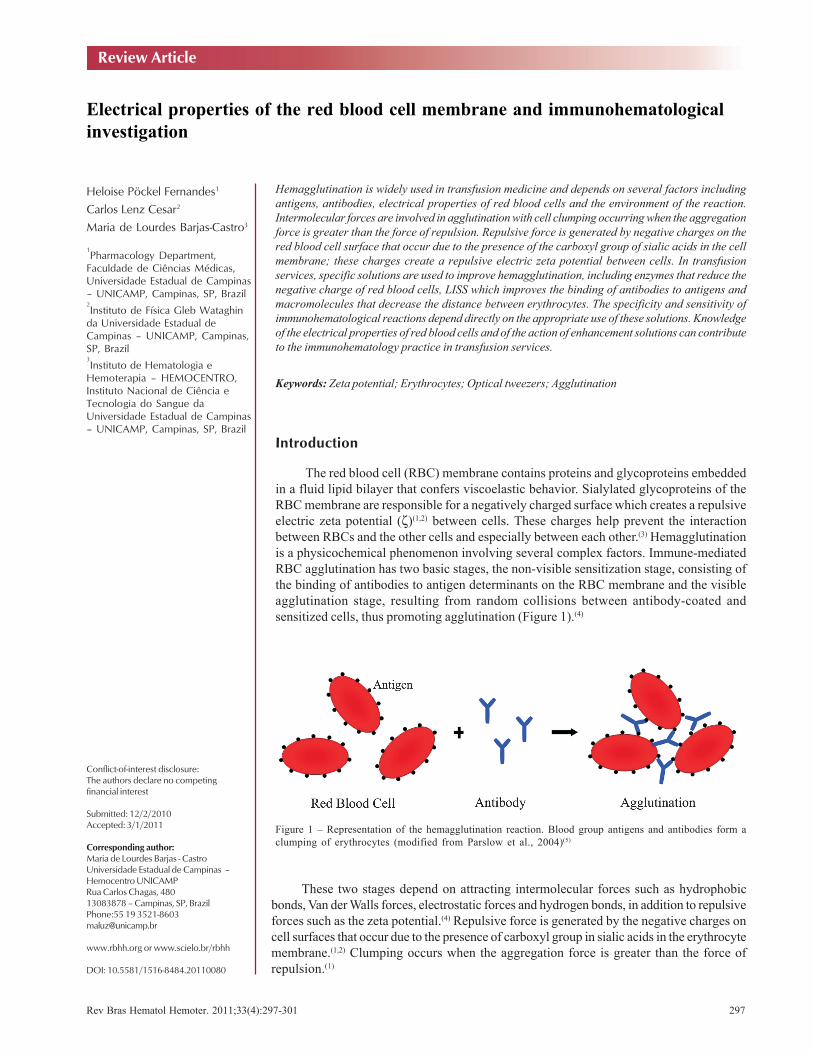
Rev Bras Hematol Hemoter. 2011;33(4):297-301 297 Electrical properties of the red blood cell membrane and immunohematological investigation 1 Pharmacology Department, Faculdade de Ciências Médicas, Universidade Estadual de Campinas – UNICAMP, Campinas, SP, Brazil 2 Instituto de Física Gleb Wataghin da Universidade Estadual de Campinas – UNICAMP, Campinas, SP, Brazil 3 Instituto de Hematologia e Hemoterapia – HEMOCENTRO, Instituto Nacional de Ciência e Tecnologia do Sangue da Universidade Estadual de Campinas – UNICAMP, Campinas, SP, Brazil Heloise Pöckel Fernandes 1 Carlos Lenz Cesar 2 Maria de Lourdes Barjas-Castro 3 Hemagglutination is widely used in transfusion medicine and depends on several factors including antigens, antibodies, electrical properties of red blood cells and the environment of the reaction. Intermolecular forces are involved in agglutination with cell clumping occurring when the aggregation force is greater than the force of repulsion. Repulsive force is generated by negative charges on the red blood cell surface that occur due to the presence of the carboxyl group of sialic acids in the cell membrane; these charges create a repulsive electric zeta potential between cells. In transfusion services, specific solutions are used to improve hemagglutination, including enzymes that reduce the negative charge of red blood cells, LISS which improves the binding of antibodies to antigens and macromolecules that decrease the distance between erythrocytes. The specificity and sensitivity of immunohematological reactions depend directly on the appropriate use of these solutions. Knowledge of the electrical properties of red blood cells and of the action of enhancement solutions can contribute to the immunohematology practice in transfusion services. Keywords: Zeta potential; Erythrocytes; Optical tweezers; Agglutination Introduction The red blood cell (RBC) membrane contains proteins and glycoproteins embedded in a fluid lipid bilayer that confers viscoelastic behavior. Sialylated glycoproteins of the RBC membrane are responsible for a negatively charged surface which creates a repulsive electric zeta potential (ζ) (1,2) between cells. These charges help prevent the interaction between RBCs and the other cells and especially between each other. (3) Hemagglutination is a physicochemical phenomenon involving several complex factors. Immune-mediated RBC agglutination has two basic stages, the non-visible sensitization stage, consisting of the binding of antibodies to antigen determinants on the RBC membrane and the visible agglutination stage, resulting from random collisions between antibody-coated and sensitized cells, thus promoting agglutination (Figure 1). (4) Conflict-of-interest disclosure: The authors declare no competing financial interest Submitted: 12/2/2010 Accepted: 3/1/2011 Corresponding author: Maria de Lourdes Barjas - Castro Universidade Estadual de Campinas – Hemocentro UNICAMP Rua Carlos Chagas, 480 13083878 – Campinas, SP, Brazil Phone:55 19 3521-8603 [email protected] www.rbhh.org or www.scielo.br/rbhh DOI: 10.5581/1516-8484.20110080 Review Article These two stages depend on attracting intermolecular forces such as hydrophobic bonds, Van der Walls forces, electrostatic forces and hydrogen bonds, in addition to repulsive forces such as the zeta potential. (4) Repulsive force is generated by the negative charges on cell surfaces that occur due to the presence of carboxyl group in sialic acids in the erythrocyte membrane. (1,2) Clumping occurs when the aggregation force is greater than the force of repulsion. (1) Figure 1 – Representation of the hemagglutination reaction. Blood group antigens and antibodies form a clumping of erythrocytes (modified from Parslow et al., 2004) (5)
Transcript of Electrical properties of the red blood cell membrane and ... · The red blood cell (RBC) membrane...
Rev Bras Hematol Hemoter. 2011;33(4):297-301 297
Electrical properties of the red blood cell membrane and immunohematologicalinvestigation
1Pharmacology Department,
Faculdade de Ciências Médicas,Universidade Estadual de Campinas– UNICAMP, Campinas, SP, Brazil2Instituto de Física Gleb Wataghin
da Universidade Estadual deCampinas – UNICAMP, Campinas,SP, Brazil3Instituto de Hematologia e
Hemoterapia – HEMOCENTRO,Instituto Nacional de Ciência eTecnologia do Sangue daUniversidade Estadual de Campinas– UNICAMP, Campinas, SP, Brazil
Heloise Pöckel Fernandes1
Carlos Lenz Cesar2
Maria de Lourdes Barjas-Castro3
Hemagglutination is widely used in transfusion medicine and depends on several factors includingantigens, antibodies, electrical properties of red blood cells and the environment of the reaction.Intermolecular forces are involved in agglutination with cell clumping occurring when the aggregationforce is greater than the force of repulsion. Repulsive force is generated by negative charges on thered blood cell surface that occur due to the presence of the carboxyl group of sialic acids in the cellmembrane; these charges create a repulsive electric zeta potential between cells. In transfusionservices, specific solutions are used to improve hemagglutination, including enzymes that reduce thenegative charge of red blood cells, LISS which improves the binding of antibodies to antigens andmacromolecules that decrease the distance between erythrocytes. The specificity and sensitivity ofimmunohematological reactions depend directly on the appropriate use of these solutions. Knowledgeof the electrical properties of red blood cells and of the action of enhancement solutions can contributeto the immunohematology practice in transfusion services.
Keywords: Zeta potential; Erythrocytes; Optical tweezers; Agglutination
Introduction
The red blood cell (RBC) membrane contains proteins and glycoproteins embeddedin a fluid lipid bilayer that confers viscoelastic behavior. Sialylated glycoproteins of theRBC membrane are responsible for a negatively charged surface which creates a repulsiveelectric zeta potential (ζ)(1,2) between cells. These charges help prevent the interactionbetween RBCs and the other cells and especially between each other.(3) Hemagglutinationis a physicochemical phenomenon involving several complex factors. Immune-mediatedRBC agglutination has two basic stages, the non-visible sensitization stage, consisting ofthe binding of antibodies to antigen determinants on the RBC membrane and the visibleagglutination stage, resulting from random collisions between antibody-coated andsensitized cells, thus promoting agglutination (Figure 1).(4)
Conflict-of-interest disclosure:The authors declare no competingfinancial interest
Submitted: 12/2/2010Accepted: 3/1/2011
Corresponding author:Maria de Lourdes Barjas - CastroUniversidade Estadual de Campinas –Hemocentro UNICAMPRua Carlos Chagas, 48013083878 – Campinas, SP, BrazilPhone:55 19 [email protected]
www.rbhh.org or www.scielo.br/rbhh
DOI: 10.5581/1516-8484.20110080
Review Article
These two stages depend on attracting intermolecular forces such as hydrophobicbonds, Van der Walls forces, electrostatic forces and hydrogen bonds, in addition to repulsiveforces such as the zeta potential.(4) Repulsive force is generated by the negative charges oncell surfaces that occur due to the presence of carboxyl group in sialic acids in the erythrocytemembrane.(1,2) Clumping occurs when the aggregation force is greater than the force ofrepulsion.(1)
Figure 1 – Representation of the hemagglutination reaction. Blood group antigens and antibodies form aclumping of erythrocytes (modified from Parslow et al., 2004)(5)
298 Rev Bras Hematol Hemoter. 2011;33(4):297-301
Fernandes HP, Cesar CL, Barjas-Castro ML
First phase of agglutination
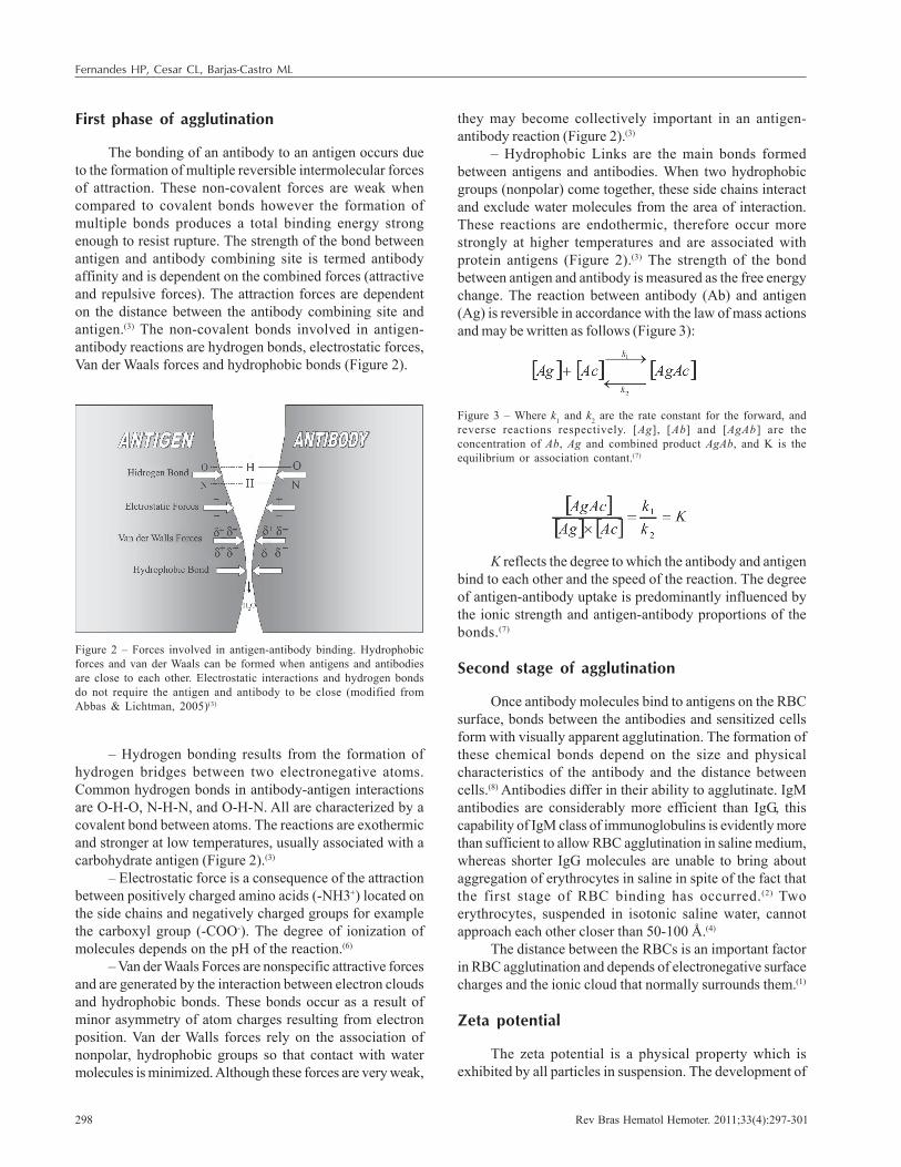
The bonding of an antibody to an antigen occurs dueto the formation of multiple reversible intermolecular forcesof attraction. These non-covalent forces are weak whencompared to covalent bonds however the formation ofmultiple bonds produces a total binding energy strongenough to resist rupture. The strength of the bond betweenantigen and antibody combining site is termed antibodyaffinity and is dependent on the combined forces (attractiveand repulsive forces). The attraction forces are dependenton the distance between the antibody combining site andantigen.(3) The non-covalent bonds involved in antigen-antibody reactions are hydrogen bonds, electrostatic forces,Van der Waals forces and hydrophobic bonds (Figure 2).
they may become collectively important in an antigen-antibody reaction (Figure 2).(3)
– Hydrophobic Links are the main bonds formedbetween antigens and antibodies. When two hydrophobicgroups (nonpolar) come together, these side chains interactand exclude water molecules from the area of interaction.These reactions are endothermic, therefore occur morestrongly at higher temperatures and are associated withprotein antigens (Figure 2).(3) The strength of the bondbetween antigen and antibody is measured as the free energychange. The reaction between antibody (Ab) and antigen(Ag) is reversible in accordance with the law of mass actionsand may be written as follows (Figure 3):
Figure 2 – Forces involved in antigen-antibody binding. Hydrophobicforces and van der Waals can be formed when antigens and antibodiesare close to each other. Electrostatic interactions and hydrogen bondsdo not require the antigen and antibody to be close (modified fromAbbas & Lichtman, 2005)(3)
– Hydrogen bonding results from the formation ofhydrogen bridges between two electronegative atoms.Common hydrogen bonds in antibody-antigen interactionsare O-H-O, N-H-N, and O-H-N. All are characterized by acovalent bond between atoms. The reactions are exothermicand stronger at low temperatures, usually associated with acarbohydrate antigen (Figure 2).(3)
– Electrostatic force is a consequence of the attractionbetween positively charged amino acids (-NH3+) located onthe side chains and negatively charged groups for examplethe carboxyl group (-COO-). The degree of ionization ofmolecules depends on the pH of the reaction.(6)
– Van der Waals Forces are nonspecific attractive forcesand are generated by the interaction between electron cloudsand hydrophobic bonds. These bonds occur as a result ofminor asymmetry of atom charges resulting from electronposition. Van der Walls forces rely on the association ofnonpolar, hydrophobic groups so that contact with watermolecules is minimized. Although these forces are very weak,
Figure 3 – Where k1 and k2 are the rate constant for the forward, andreverse reactions respectively. [Ag], [Ab] and [AgAb] are theconcentration of Ab, Ag and combined product AgAb, and K is theequilibrium or association contant.(7)
K reflects the degree to which the antibody and antigenbind to each other and the speed of the reaction. The degreeof antigen-antibody uptake is predominantly influenced bythe ionic strength and antigen-antibody proportions of thebonds.(7)
Second stage of agglutination
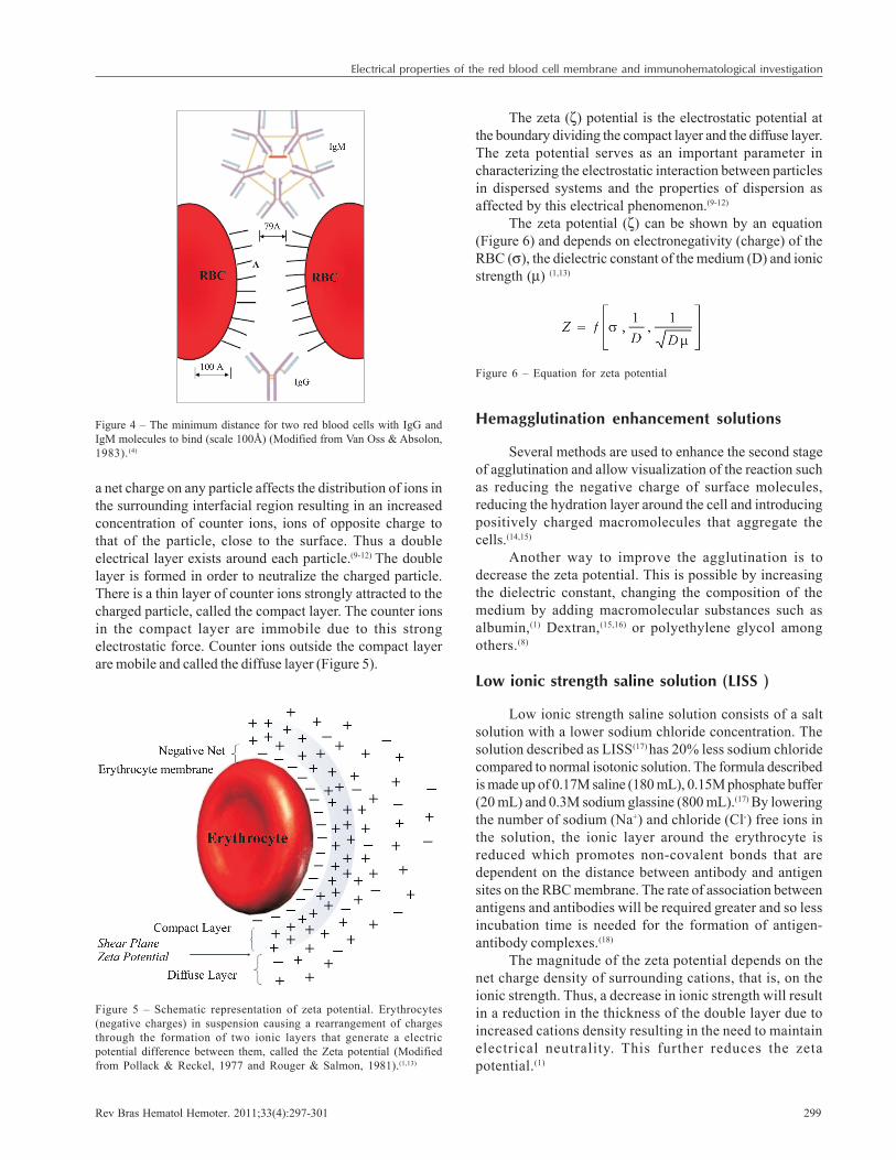
Once antibody molecules bind to antigens on the RBCsurface, bonds between the antibodies and sensitized cellsform with visually apparent agglutination. The formation ofthese chemical bonds depend on the size and physicalcharacteristics of the antibody and the distance betweencells.(8) Antibodies differ in their ability to agglutinate. IgMantibodies are considerably more efficient than IgG, thiscapability of IgM class of immunoglobulins is evidently morethan sufficient to allow RBC agglutination in saline medium,whereas shorter IgG molecules are unable to bring aboutaggregation of erythrocytes in saline in spite of the fact thatthe first stage of RBC binding has occurred.(2) Twoerythrocytes, suspended in isotonic saline water, cannotapproach each other closer than 50-100 Å.(4)
The distance between the RBCs is an important factorin RBC agglutination and depends of electronegative surfacecharges and the ionic cloud that normally surrounds them.(1)
Zeta potential
The zeta potential is a physical property which isexhibited by all particles in suspension. The development of
Rev Bras Hematol Hemoter. 2011;33(4):297-301 299
Electrical properties of the red blood cell membrane and immunohematological investigation
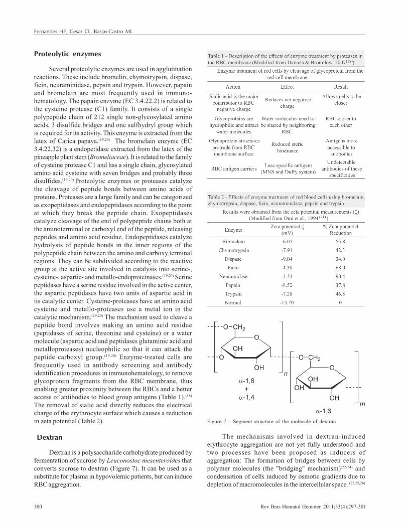
a net charge on any particle affects the distribution of ions inthe surrounding interfacial region resulting in an increasedconcentration of counter ions, ions of opposite charge tothat of the particle, close to the surface. Thus a doubleelectrical layer exists around each particle.(9-12) The doublelayer is formed in order to neutralize the charged particle.There is a thin layer of counter ions strongly attracted to thecharged particle, called the compact layer. The counter ionsin the compact layer are immobile due to this strongelectrostatic force. Counter ions outside the compact layerare mobile and called the diffuse layer (Figure 5).
The zeta (ζ) potential is the electrostatic potential atthe boundary dividing the compact layer and the diffuse layer.The zeta potential serves as an important parameter incharacterizing the electrostatic interaction between particlesin dispersed systems and the properties of dispersion asaffected by this electrical phenomenon.(9-12)
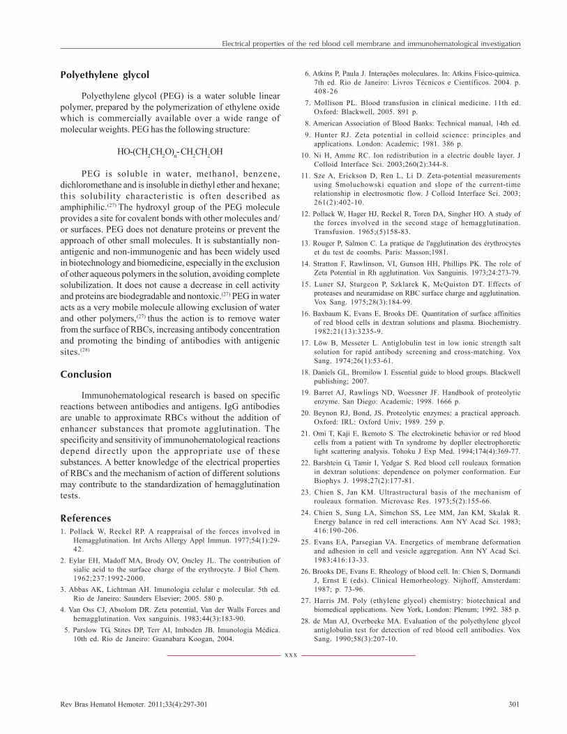
The zeta potential (ζ) can be shown by an equation(Figure 6) and depends on electronegativity (charge) of theRBC (σ), the dielectric constant of the medium (D) and ionicstrength (µ) (1,13)
Figure 4 – The minimum distance for two red blood cells with IgG andIgM molecules to bind (scale 100Å) (Modified from Van Oss & Absolon,1983).(4)
Figure 5 – Schematic representation of zeta potential. Erythrocytes(negative charges) in suspension causing a rearrangement of chargesthrough the formation of two ionic layers that generate a electricpotential difference between them, called the Zeta potential (Modifiedfrom Pollack & Reckel, 1977 and Rouger & Salmon, 1981).(1,13)
Hemagglutination enhancement solutions
Several methods are used to enhance the second stageof agglutination and allow visualization of the reaction suchas reducing the negative charge of surface molecules,reducing the hydration layer around the cell and introducingpositively charged macromolecules that aggregate thecells.(14,15)
Another way to improve the agglutination is todecrease the zeta potential. This is possible by increasingthe dielectric constant, changing the composition of themedium by adding macromolecular substances such asalbumin,(1) Dextran,(15,16) or polyethylene glycol amongothers.(8)
Low ionic strength saline solution (LISS )
Low ionic strength saline solution consists of a saltsolution with a lower sodium chloride concentration. Thesolution described as LISS(17) has 20% less sodium chloridecompared to normal isotonic solution. The formula describedis made up of 0.17M saline (180 mL), 0.15M phosphate buffer(20 mL) and 0.3M sodium glassine (800 mL).(17) By loweringthe number of sodium (Na+) and chloride (Cl-) free ions inthe solution, the ionic layer around the erythrocyte isreduced which promotes non-covalent bonds that aredependent on the distance between antibody and antigensites on the RBC membrane. The rate of association betweenantigens and antibodies will be required greater and so lessincubation time is needed for the formation of antigen-antibody complexes.(18)
The magnitude of the zeta potential depends on thenet charge density of surrounding cations, that is, on theionic strength. Thus, a decrease in ionic strength will resultin a reduction in the thickness of the double layer due toincreased cations density resulting in the need to maintainelectrical neutrality. This further reduces the zetapotential.(1)
Figure 6 – Equation for zeta potential
300 Rev Bras Hematol Hemoter. 2011;33(4):297-301
Proteolytic enzymes
Several proteolytic enzymes are used in agglutinationreactions. These include bromelin, chymotrypsin, dispase,ficin, neuraminidase, pepsin and trypsin. However, papainand bromelain are most frequently used in immuno-hematology. The papain enzyme (EC 3.4.22.2) is related tothe cysteine protease (C1) family. It consists of a singlepolypeptide chain of 212 single non-glycosylated aminoacids, 3 disulfide bridges and one sulfhydryl group whichis required for its activity. This enzyme is extracted from thelatex of Carica papaya.(19,20) The bromelain enzyme (EC3.4.22.32) is a endopetidase extracted from the latex of thepineapple plant stem (Bromeliaceae). It is related to the familyof cysteine protease C1 and has a single chain, glycosylatedamino acid cysteine with seven bridges and probably threedisulfides.(19,20) Proteolytic enzymes or proteases catalyzethe cleavage of peptide bonds between amino acids ofproteins. Proteases are a large family and can be categorizedas exopeptidases and endopeptidases according to the pointat which they break the peptide chain. Exopeptidasescatalyze cleavage of the end of polypeptide chains both atthe aminoterminal or carboxyl end of the peptide, releasingpeptides and amino acid residue. Endopeptidases catalyzehydrolysis of peptide bonds in the inner regions of thepolypeptide chain between the amino and carboxy terminalregions. They can be subdivided according to the reactivegroup at the active site involved in catalysis into serine-,cysteine-, aspartic- and metallo-endoproteinases.(19,20) Serinepeptidases have a serine residue involved in the active center,the aspartic peptidases have two units of aspartic acid inits catalytic center. Cysteine-proteases have an amino acidcysteine and metallo-proteases use a metal ion in thecatalytic mechanism.(19,20) The mechanism used to cleave apeptide bond involves making an amino acid residue(peptidases of serine, threonine and cysteine) or a watermolecule (aspartic acid and peptidases glutaminic acid andmetalloproteases) nucleophilic so that it can attack thepeptide carboxyl group.(19,20) Enzyme-treated cells arefrequently used in antibody screening and antibodyidentification procedures in immunohematology, to removeglycoprotein fragments from the RBC membrane, thusenabling greater proximity between the RBCs and a betteraccess of antibodies to blood group antigens (Table 1).(18)
The removal of sialic acid directly reduces the electricalcharge of the erythrocyte surface which causes a reductionin zeta potential (Table 2).
Dextran
Dextran is a polysaccharide carbohydrate produced byfermentation of sucrose by Leuconostoc mesenteroides thatconverts sucrose to dextran (Figure 7). It can be used as asubstitute for plasma in hypovolemic patients, but can induceRBC aggregation.
Figure 7 – Segment structure of the molecule of dextran
The mechanisms involved in dextran-inducederythrocyte aggregation are not yet fully understood andtwo processes have been proposed as inducers ofaggregation: The formation of bridges between cells bypolymer molecules (the "bridging" mechanism)(22-24) andcondensation of cells induced by osmotic gradients due todepletion of macromolecules in the intercellular space. (22,25,26)
Fernandes HP, Cesar CL, Barjas-Castro ML
Rev Bras Hematol Hemoter. 2011;33(4):297-301 301
Polyethylene glycol
Polyethylene glycol (PEG) is a water soluble linearpolymer, prepared by the polymerization of ethylene oxidewhich is commercially available over a wide range ofmolecular weights. PEG has the following structure:
HO-(CH2CH2O)n- CH2CH2OH
PEG is soluble in water, methanol, benzene,dichloromethane and is insoluble in diethyl ether and hexane;this solubility characteristic is often described asamphiphilic.(27) The hydroxyl group of the PEG moleculeprovides a site for covalent bonds with other molecules and/or surfaces. PEG does not denature proteins or prevent theapproach of other small molecules. It is substantially non-antigenic and non-immunogenic and has been widely usedin biotechnology and biomedicine, especially in the exclusionof other aqueous polymers in the solution, avoiding completesolubilization. It does not cause a decrease in cell activityand proteins are biodegradable and nontoxic.(27) PEG in wateracts as a very mobile molecule allowing exclusion of waterand other polymers,(27) thus the action is to remove waterfrom the surface of RBCs, increasing antibody concentrationand promoting the binding of antibodies with antigenicsites.(28)
Conclusion
Immunohematological research is based on specificreactions between antibodies and antigens. IgG antibodiesare unable to approximate RBCs without the addition ofenhancer substances that promote agglutination. Thespecificity and sensitivity of immunohematological reactionsdepend directly upon the appropriate use of thesesubstances. A better knowledge of the electrical propertiesof RBCs and the mechanism of action of different solutionsmay contribute to the standardization of hemagglutinationtests.
References1. Pollack W, Reckel RP. A reappraisal of the forces involved in
Hemagglutination. Int Archs Allergy Appl Immun. 1977;54(1):29-42.
2. Eylar EH, Madoff MA, Brody OV, Oncley JL. The contribution ofsialic acid to the surface charge of the erythrocyte. J Biol Chem.1962;237:1992-2000.
3. Abbas AK, Lichtman AH. Imunologia celular e molecular. 5th ed.Rio de Janeiro: Saunders Elsevier; 2005. 580 p.
4. Van Oss CJ, Absolom DR. Zeta potential, Van der Walls Forces andhemagglutination. Vox sanguinis. 1983;44(3):183-90.
5. Parslow TG, Stites DP, Terr AI, Imboden JB. Imunologia Médica.10th ed. Rio de Janeiro: Guanabara Koogan, 2004.
6. Atkins P, Paula J. Interações moleculares. In: Atkins Físico-química.7th ed. Rio de Janeiro: Livros Técnicos e Científicos. 2004. p.408-26
7. Mollison PL. Blood transfusion in clinical medicine. 11th ed.Oxford: Blackwell, 2005. 891 p.
8. American Association of Blood Banks: Technical manual, 14th ed.9. Hunter RJ. Zeta potential in colloid science: principles and
applications. London: Academic; 1981. 386 p.10. Ni H, Amme RC. Ion redistribution in a electric double layer. J
Colloid Interface Sci. 2003;260(2):344-8.11. Sze A, Erickson D, Ren L, Li D. Zeta-potential measurements
using Smoluchowski equation and slope of the current-timerelationship in electrosmotic flow. J Colloid Interface Sci. 2003;261(2):402-10.
12. Pollack W, Hager HJ, Reckel R, Toren DA, Singher HO. A study ofthe forces involved in the second stage of hemagglutination.Transfusion. 1965;(5)158-83.
13. Rouger P, Salmon C. La pratique de l'agglutination des érythrocyteset du test de coombs. Paris: Masson;1981.
14. Stratton F, Rawlinson, VI, Gunson HH, Phillips PK. The role ofZeta Potential in Rh agglutination. Vox Sanguinis. 1973;24:273-79.
15. Luner SJ, Sturgeon P, Szklarek K, McQuiston DT. Effects ofproteases and neuramidase on RBC surface charge and agglutination.Vox Sang. 1975;28(3):184-99.
16. Baxbaum K, Evans E, Brooks DE. Quantitation of surface affinitiesof red blood cells in dextran solutions and plasma. Biochemistry.1982;21(13):3235-9.
17. Löw B, Messeter L. Antiglobulin test in low ionic strength saltsolution for rapid antibody screening and cross-matching. VoxSang. 1974;26(1):53-61.
18. Daniels GL, Bromilow I. Essential guide to blood groups. Blackwellpublishing; 2007.
19. Barret AJ, Rawlings ND, Woessner JF. Handbook of proteolyticenzyme. San Diego: Academic; 1998. 1666 p.
20. Beynon RJ, Bond, JS. Proteolytic enzymes: a practical approach.Oxford: IRL: Oxford Univ; 1989. 259 p.
21. Omi T, Kaji E, Ikemoto S. The electrokinetic behavior or red bloodcells from a patient with Tn syndrome by dopller electrophoreticlight scattering analysis. Tohoku J Exp Med. 1994;174(4):369-77.
22. Barshtein G, Tamir I, Yedgar S. Red blood cell rouleaux formationin dextran solutions: dependence on polymer conformation. EurBiophys J. 1998;27(2):177-81.
23. Chien S, Jan KM. Ultrastructural basis of the mechanism ofrouleaux formation. Microvasc Res. 1973;5(2):155-66.
24. Chien S, Sung LA, Simchon SS, Lee MM, Jan KM, Skalak R.Energy balance in red cell interactions. Ann NY Acad Sci. 1983;416:190-206.
25. Evans EA, Parsegian VA. Energetics of membrane deformationand adhesion in cell and vesicle aggregation. Ann NY Acad Sci.1983;416:13-33.
26. Brooks DE, Evans E. Rheology of blood cell. In: Chien S, DormandiJ, Ernst E (eds). Clinical Hemorheology. Nijhoff, Amsterdam:1987; p. 73-96.
27. Harris JM. Poly (ethylene glycol) chemistry: biotechnical andbiomedical applications. New York, London: Plenum; 1992. 385 p.
28. de Man AJ, Overbeeke MA. Evaluation of the polyethylene glycolantiglobulin test for detection of red blood cell antibodies. VoxSang. 1990;58(3):207-10.
xxx
Electrical properties of the red blood cell membrane and immunohematological investigation