
Elastogenesis at the onset of human cardiac valve...
9
RESEARCH ARTICLE 2345 Development 140, 2345-2353 (2013) doi:10.1242/dev.093500 © 2013. Published by The Company of Biologists Ltd INTRODUCTION Despite the fact that elastin and elastic fibers are pivotal for normal cardiac development and function, the majority of research efforts have not been focused on the impact of the extracellular matrix (ECM) on organogenesis or pathological remodeling processes. While recent reports on cardiac ischemia have emphasized the importance of ECM in the cardiovascular system (Barallobre- Barreiro et al., 2012), there has been little focus on the role of the ECM, in particular elastin, in human cardiovascular embryonic development. Elastin is a non-soluble, extremely durable protein. The processes underlying elastic fiber formation are highly complex and require the interplay of various molecules. To date, the mechanisms that drive elastic fiber formation, termed ‘elastogenesis’, are not well defined but are the subject of intense research by many groups (Kielty et al., 2002a; Wagenseil and Mecham, 2007; Wise and Weiss, 2009). Elastic fibers consist of a variety of components, including microfibrillar proteins (fibrillins), linking proteins [fibulins, elastin microfibril interfacers (EMILINs), microfibril- associated glycoproteins (MAGPs)], soluble factors [e.g. transforming growth factor (TGF) beta] and the core protein elastin (Wagenseil and Mecham, 2007; Sherratt, 2009; Wise and Weiss, 2009). In addition, a fibronectin network is needed for the assembly of fibrillins, and consequently of microfibrils, that provides a microenvironment which controls tropoelastin/elastin arrangement and cross-linking processes (Sabatier et al., 2009). Fibulin 5 and MAGP1 (also known as MFAP2) are known to bridge between microfibrils and tropoelastin (Gibson et al., 1986; Nakamura et al., 2002; Yanagisawa et al., 2002; Cirulis et al., 2008). Fibulin 4 (also known as EFEMP2) links the enzyme lysyl oxidase (LOX) to tropoelastin (Horiguchi et al., 2009). Interestingly, in organisms that possess a low-pressure circulation system, microfibrils alone provide sufficient elasticity (Faury, 2001; Piha-Gossack et al., 2012). By contrast, in organisms that feature a higher blood pressure, elastin-containing fibers are required. In such systems it has been suggested that, during embryonic development, an incorporation of elastin into the microfibrillar network takes place when blood pressure increases (Faury, 2001; Wagenseil and Mecham, 2009). As elastin is a very durable protein that can remain functional throughout a lifetime [e.g. elastin turnover in human lung is ~74 years (Shapiro et al., 1991)], gene expression is reduced in aging cardiovascular tissues (Kelleher et al., 2004; Wagenseil and Mecham, 2009). Elastic fibers provide the essential recoil for all tissues that are exposed to constant, repeated strain, such as the larger arteries or heart valve leaflets (Faury, 2001; Kielty et al., 2002b; Wagenseil and Mecham, 2007; Sherratt, 2009). In semilunar valve leaflets, the layer facing the ventricles (the ventricularis) is mainly composed of a sophisticated elastic fiber network, which is vital for physiological leaflet function (Aikawa et al., 2006; Lindsey 1 University Women’s Hospital Tübingen and Inter-University Centre for Medical Technology Stuttgart-Tübingen (IZST), Eberhard Karls University, 72076 Tübingen, Germany. 2 Department of Cell and Tissue Engineering, Fraunhofer Institute for Interfacial Engineering and Biotechnology (IGB), 70569 Stuttgart, Germany. 3 Department of Cardiac, Thoracic, Transplantation and Vascular Surgery, Hannover Medical School, 30625 Hannover, Germany. 4 Department of Anatomy and Cell Biology, and Faculty of Dentistry, Division of Biomedical Sciences, Faculty of Medicine, McGill University, Montreal, Quebec H3A 2B2, Canada. 5 Department of Medicine/Cardiology, Cardiovascular Research Laboratories (CVRL), University of California Los Angeles (UCLA), Los Angeles, CA 90095, USA. 6 Cardiovascular Medicine, Brigham and Women's Hospital, Harvard Medical School, Boston, MA 02115, USA. *Author for correspondence ([email protected]) This is an Open Access article distributed under the terms of the Creative Commons Attribution Non-Commercial Share Alike License (http://creativecommons.org/licenses/by-nc-sa/3.0), which permits unrestricted non-commercial use, distribution and reproduction in any medium provided that the original work is properly cited and all further distributions of the work or adaptation are subject to the same Creative Commons License terms. Accepted 3 April 2013 SUMMARY Semilunar valve leaflets have a well-described trilaminar histoarchitecture, with a sophisticated elastic fiber network. It was previously proposed that elastin-containing fibers play a subordinate role in early human cardiac valve development; however, this assumption was based on data obtained from mouse models and human second and third trimester tissues. Here, we systematically analyzed tissues from human fetal first (4-12 weeks) and second (13-18 weeks) trimester, adolescent (14-19 years) and adult (50-55 years) hearts to monitor the temporal and spatial distribution of elastic fibers, focusing on semilunar valves. Global expression analyses revealed that the transcription of genes essential for elastic fiber formation starts early within the first trimester. These data were confirmed by quantitative PCR and immunohistochemistry employing antibodies that recognize fibronectin, fibrillin 1, 2 and 3, EMILIN1 and fibulin 4 and 5, which were all expressed at the onset of cardiac cushion formation (~week 4 of development). Tropoelastin/elastin protein expression was first detectable in leaflets of 7-week hearts. We revealed that immature elastic fibers are organized in early human cardiovascular development and that mature elastin-containing fibers first evolve in semilunar valves when blood pressure and heartbeat accelerate. Our findings provide a conceptual framework with the potential to offer novel insights into human cardiac valve development and disease. KEY WORDS: Heart, Elastin, Elastic fibers, Extracellular matrix, Heart valves Elastogenesis at the onset of human cardiac valve development Miriam Votteler 1,2 , Daniel A. Carvajal Berrio 2 , Alexander Horke 3 , Laetitia Sabatier 4 , Dieter P. Reinhardt 4 , Ali Nsair 5 , Elena Aikawa 6 and Katja Schenke-Layland 1,2,5, * DEVELOPMENT
Transcript of Elastogenesis at the onset of human cardiac valve...
RESEARCH ARTICLE 2345
Development 140, 2345-2353 (2013) doi:10.1242/dev.093500© 2013. Published by The Company of Biologists Ltd
INTRODUCTIONDespite the fact that elastin and elastic fibers are pivotal for normalcardiac development and function, the majority of research effortshave not been focused on the impact of the extracellular matrix(ECM) on organogenesis or pathological remodeling processes.While recent reports on cardiac ischemia have emphasized theimportance of ECM in the cardiovascular system (Barallobre-Barreiro et al., 2012), there has been little focus on the role of theECM, in particular elastin, in human cardiovascular embryonicdevelopment.
Elastin is a non-soluble, extremely durable protein. The processesunderlying elastic fiber formation are highly complex and requirethe interplay of various molecules. To date, the mechanisms thatdrive elastic fiber formation, termed ‘elastogenesis’, are not welldefined but are the subject of intense research by many groups(Kielty et al., 2002a; Wagenseil and Mecham, 2007; Wise and
Weiss, 2009). Elastic fibers consist of a variety of components,including microfibrillar proteins (fibrillins), linking proteins[fibulins, elastin microfibril interfacers (EMILINs), microfibril-associated glycoproteins (MAGPs)], soluble factors [e.g.transforming growth factor (TGF) beta] and the core protein elastin(Wagenseil and Mecham, 2007; Sherratt, 2009; Wise and Weiss,2009). In addition, a fibronectin network is needed for the assemblyof fibrillins, and consequently of microfibrils, that provides amicroenvironment which controls tropoelastin/elastin arrangementand cross-linking processes (Sabatier et al., 2009). Fibulin 5 andMAGP1 (also known as MFAP2) are known to bridge betweenmicrofibrils and tropoelastin (Gibson et al., 1986; Nakamura et al.,2002; Yanagisawa et al., 2002; Cirulis et al., 2008). Fibulin 4 (alsoknown as EFEMP2) links the enzyme lysyl oxidase (LOX) totropoelastin (Horiguchi et al., 2009). Interestingly, in organisms thatpossess a low-pressure circulation system, microfibrils aloneprovide sufficient elasticity (Faury, 2001; Piha-Gossack et al.,2012). By contrast, in organisms that feature a higher bloodpressure, elastin-containing fibers are required. In such systems ithas been suggested that, during embryonic development, anincorporation of elastin into the microfibrillar network takes placewhen blood pressure increases (Faury, 2001; Wagenseil andMecham, 2009).
As elastin is a very durable protein that can remain functionalthroughout a lifetime [e.g. elastin turnover in human lung is ~74years (Shapiro et al., 1991)], gene expression is reduced in agingcardiovascular tissues (Kelleher et al., 2004; Wagenseil andMecham, 2009). Elastic fibers provide the essential recoil for alltissues that are exposed to constant, repeated strain, such as thelarger arteries or heart valve leaflets (Faury, 2001; Kielty et al.,2002b; Wagenseil and Mecham, 2007; Sherratt, 2009). In semilunarvalve leaflets, the layer facing the ventricles (the ventricularis) ismainly composed of a sophisticated elastic fiber network, which isvital for physiological leaflet function (Aikawa et al., 2006; Lindsey
1University Women’s Hospital Tübingen and Inter-University Centre for MedicalTechnology Stuttgart-Tübingen (IZST), Eberhard Karls University, 72076 Tübingen,Germany. 2Department of Cell and Tissue Engineering, Fraunhofer Institute forInterfacial Engineering and Biotechnology (IGB), 70569 Stuttgart, Germany.3Department of Cardiac, Thoracic, Transplantation and Vascular Surgery, HannoverMedical School, 30625 Hannover, Germany. 4Department of Anatomy and CellBiology, and Faculty of Dentistry, Division of Biomedical Sciences, Faculty ofMedicine, McGill University, Montreal, Quebec H3A 2B2, Canada. 5Department ofMedicine/Cardiology, Cardiovascular Research Laboratories (CVRL), University ofCalifornia Los Angeles (UCLA), Los Angeles, CA 90095, USA. 6CardiovascularMedicine, Brigham and Women's Hospital, Harvard Medical School, Boston, MA02115, USA.
*Author for correspondence ([email protected])
This is an Open Access article distributed under the terms of the Creative Commons AttributionNon-Commercial Share Alike License (http://creativecommons.org/licenses/by-nc-sa/3.0), whichpermits unrestricted non-commercial use, distribution and reproduction in any medium providedthat the original work is properly cited and all further distributions of the work or adaptation aresubject to the same Creative Commons License terms.
Accepted 3 April 2013
SUMMARYSemilunar valve leaflets have a well-described trilaminar histoarchitecture, with a sophisticated elastic fiber network. It was previouslyproposed that elastin-containing fibers play a subordinate role in early human cardiac valve development; however, this assumptionwas based on data obtained from mouse models and human second and third trimester tissues. Here, we systematically analyzedtissues from human fetal first (4-12 weeks) and second (13-18 weeks) trimester, adolescent (14-19 years) and adult (50-55 years) heartsto monitor the temporal and spatial distribution of elastic fibers, focusing on semilunar valves. Global expression analyses revealedthat the transcription of genes essential for elastic fiber formation starts early within the first trimester. These data were confirmedby quantitative PCR and immunohistochemistry employing antibodies that recognize fibronectin, fibrillin 1, 2 and 3, EMILIN1 andfibulin 4 and 5, which were all expressed at the onset of cardiac cushion formation (~week 4 of development). Tropoelastin/elastinprotein expression was first detectable in leaflets of 7-week hearts. We revealed that immature elastic fibers are organized in earlyhuman cardiovascular development and that mature elastin-containing fibers first evolve in semilunar valves when blood pressureand heartbeat accelerate. Our findings provide a conceptual framework with the potential to offer novel insights into human cardiacvalve development and disease.
KEY WORDS: Heart, Elastin, Elastic fibers, Extracellular matrix, Heart valves
Elastogenesis at the onset of human cardiac valvedevelopmentMiriam Votteler1,2, Daniel A. Carvajal Berrio2, Alexander Horke3, Laetitia Sabatier4, Dieter P. Reinhardt4, Ali Nsair5, Elena Aikawa6 and Katja Schenke-Layland1,2,5,*
DEVELO
PMENT

2346
and Butcher, 2011; Schenke-Layland et al., 2004). However, themechanisms underlying this specialized histoarchitectural assemblyare poorly understood and neither the endogenous (e.g. cellular andECM involvement) nor exogenous (e.g. biophysical signals)contribution is sufficiently defined.
It was suggested that elastin-containing fibers are first detectableat the onset of the third trimester in developing human valves(Aikawa et al., 2006). In that study, only routine histologicalanalyses were performed by employing Verhoeff’s elastic stain,which is the elastic fiber-detecting stain within the Russel-Movatpentachrome stain (Movat’s stain). However, Verhoeff’s elastic stainis only suitable to visualize mature elastic fibers (Mulisch andWelsch, 2010) and it does not detect early, non-cross-linked fibersor microfibrils. Therefore, we aimed in this study to employantibody and probe-specific analyses to identify the temporal andspatial distribution of the early elastic fiber network in developinghuman hearts with a focus on the semilunar valves.
MATERIALS AND METHODSHuman heart valve tissue procurement and processingThis study was performed in accordance with institutional guidelines andwas approved by the local research ethics committees at the University ofCalifornia Los Angeles (UCLA) and the University Hospital of theEberhard Karls University (UKT) (UCLA IRB #05-10-093; UKT IRB#356/2008BO2 and #406/2011BO1). First trimester (n=8; 4-12 weeks ofgestation) and second trimester (n=7; 13-18 weeks of gestation) human fetalhearts were obtained from electively aborted fetuses. Cryopreserved adultaortic valves from adolescents (n=5; 14-19 years) and adults (n=5; 50-55years), which were not suitable for transplantation due to extended storagetimes, were obtained from Cell and Tissue Systems (Prof. K. G. Brockbank,Charleston, SC, USA). After either harvest or thawing procedures, alltissues were immediately washed in sterile phosphate-buffered saline (PBS)(Lonza, Cologne, Germany), fixed in 10% buffered formalin for less than12 hours, then rinsed in tap water and transferred to 70% ethanol. Fixedspecimens were embedded in paraffin.
Laser-capture microdissection (LCM) and RNA isolationLCM of formalin-fixed paraffin-embedded (FFPE) samples was performedas previously described (Votteler et al., 2013). Dissected leaflets and largeoutflow tract vessels were collected separately in adhesive caps (Carl Zeiss,Jena, Germany). RNA was then extracted using an isolation kit specific forFFPE samples (#74404 and #73504, RNeasy FFPE Kit, Qiagen, Hilden,Germany). For cryopreserved tissues, we separated leaflets and the aortictrunk by tweezers and extracted RNA using the Microarray RNeasy Kit(#76163, Qiagen). All further processing was performed according to themanufacturer’s protocols. The purified RNA was eluted in RNase-free waterand stored at −80°C until gene expression analyses.
Whole-genome expression analysesMicroarrays were performed by MFTServices (UKT) using the cDNA-mediated annealing, selection, extension and ligation (DASL) assay(Illumina, San Diego, CA, USA) for RNA of FFPE and cryopreservedtissues as previously described (Fan et al., 2004). RNA quality wasmonitored using microfluidics-based electrophoresis (BioAnalyzer 2100,Agilent, Waldbronn, Germany) and spectrophotometric analysis (NanodropND-1000, PEQLAB Biotechnologie, Erlangen, Germany). For dataprocessing and analyses, Genome Studio V2009.1 software (Illumina) wasused. The expression data from all chips were normalized with variancestabilizing normalization (VSN). Heatmaps were created with R(http://www.r-project.org) and vertical scatterplots were designed usingGraphPad Prism 5 software (GraphPad Software, La Jolla, CA, USA). Alldata are displayed as intensities (log2 normalized ratios) ± s.d. Microarraydata have been deposited at GEO with accession number GSE45821.
HistologyMovat’s stain was used to visualize mature elastin-containing elastic fibers(black), nuclei (dark red), collagens (yellow), muscle tissue (red) and
proteoglycans (blue-green) as previously described (Russell, 1972; Aikawaet al., 2006; Schenke-Layland et al., 2008).
Immunofluorescence staining and imagingTissue sections (3 µm) were deparaffinized and all slides were processed aspreviously described (Schenke-Layland et al., 2007). Antigen retrieval wasperformed in a microwave oven for 8 minutes each in 10 mM Tris, 1 mMEDTA, 0.05% Tween 20 (pH 9.0) and in 10 mM citrate solution in PBS (pH6.0). All sections were then incubated for 30 minutes in blocking buffer(2% goat serum, 0.1% Triton X-100, 0.05% Tween 20 in PBS). Antibodieswere diluted in blocking buffer without goat serum. Primary antibodies werepolyclonal rabbit IgG anti-elastin (1:75) and anti-EMILIN1 (1:500) Prestigeantibodies (#HPA018111 and #HPA002822, Sigma-Aldrich, Munich,Germany) and anti-fibronectin (1:500; #A0245, Dako, Eching, Germany).Anti-fibrillin 1, 2 and 3 (each 1:1000) and anti-fibulin 4 and 5 (each 1:500)were produced in rabbits (Lin et al., 2002; Sabatier et al., 2011). Primaryantibodies were incubated overnight at 4°C. Alexa Fluor 594-conjugatedgoat anti-rabbit IgG (H+L) secondary antibodies (Life Technologies,Molecular Probes, Darmstadt, Germany) were diluted 1:250 and appliedfor 30 minutes at room temperature. For co-labeling we employed FITC-labeled α-smooth muscle actin (αSMA) (1:400; #F3777, Sigma-Aldrich).After incubation with secondary antibodies, slides were exposed to 4�,6-diamidino-2-phenylindole (DAPI) solution for 10 minutes followed bymounting using ProLong Gold antifade mounting medium (LifeTechnologies, Molecular Probes). Fluorescence images were acquired usingan Axio Observer Z1 microscope or an LSM710 inverted confocalmicroscope (both Carl Zeiss). Images were processed with Photoshop CS5(Adobe Systems, San Jose, CA, USA).
For semi-quantification of tropoelastin/elastin protein expression inadolescent and adult valve tissue sections, we used identical exposure timesand compared gray value intensity (GVI) signals of antibody-stained tissuesections at 20× magnification as previously described (Aikawa et al., 2006;Schenke-Layland et al., 2010). For each sample group we used threesections of three specimens (n=3) and, within each section, we measuredthree regions of interest (ROIs).
Real-time quantitative PCR (qPCR)qPCR of FFPE RNA was performed using a QuantiFast Probe One-StepAssay (Hs_ELN_1_FAM, Qiagen). We employed 10 ng total RNA and usedthe recommended cycling conditions (95°C for 3 minutes, followed by 45cycles at 95°C for 3 seconds and 60°C for 30 seconds). All data aredisplayed as arbitrary units (a.u.).
Statistical analysesStatistical significance was determined by ANOVA with Tukey’s multiplecomparison tests and Student’s t-test using GraphPad Prism 5 software.P<0.05 was defined as statistically significant.
RESULTSHuman fetal first trimester cardiovascularstructures exhibit elastin-containing fibers at 4 weeks of developmentIt has been reported that elastin-containing fibers and the typicaltrilaminar architecture are first seen in developing human semilunarvalves after 36 weeks of gestation, as detected using Movat’s stain(Aikawa et al., 2006). Our study confirmed these results and alsoshowed no evidence for elastin and elastin-containing fibers inhuman first trimester (4-12 weeks) semilunar valve leaflets usingMovat’s stain (Fig. 1A,D, blue arrow). However, using targetedantibody staining, we detected tropoelastin/elastin-containing fiberstructures in human developing valves as early as 7 weeks ofgestation (Fig. 1B). Interestingly, a rudimentary stratification wasalready visible at this early stage of valve development, astropoelastin/elastin was mainly expressed in the layer facing theventricles (Fig. 1B, arrows).
RESEARCH ARTICLE Development 140 (11)
DEVELO
PMENT
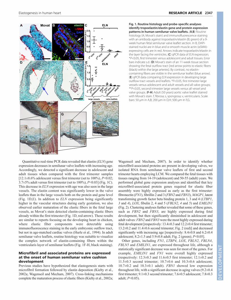
Quantitative real-time PCR data revealed that elastin (ELN) geneexpression decreases in semilunar valve leaflets with increasing age.Accordingly, we detected a significant decrease in adolescent andadult tissues when compared with the first trimester samples[12.1±8.6% adolescent versus first trimester (set to 100%), P<0.02;3.7±5% adult versus first trimester (set to 100%), P<0.03] (Fig. 1C).This decrease in ELN expression with age was also seen in the largevessels. The elastin content was significantly lower in the valveleaflets than in the large vessels both on the protein and gene level(Fig. 1D,E). In addition to ELN expression being significantlyhigher in the vascular structures during early gestation, we alsoobserved earlier maturation of the elastic fibers in the fetal largevessels, as Movat’s stain detected elastin-containing elastic fibersalready within the first trimester (Fig. 1D, red arrow). These resultsare similar to reports focusing on the developing heart in chicken,where elastic fiber components were detectable usingimmunofluorescence staining in the early embryonic outflow tract,but not in age-matched cardiac valves (Hurle et al., 1994). In adultsemilunar valve leaflets, routine histology was suitable to visualizethe complex network of elastin-containing fibers within theventricularis layer of semilunar leaflets (Fig. 1F-H, black staining).
Microfibril and associated proteins are expressedat the onset of human semilunar valve cushiondevelopmentPrevious studies have hypothesized that elastogenesis starts withmicrofibril formation followed by elastin deposition (Kielty et al.,2002a; Wagenseil and Mecham, 2007). Cross-linking mechanismscomplete the maturation process of elastic fibers (Kielty et al., 2002a;
Wagenseil and Mecham, 2007). In order to identify whethermicrofibril-associated proteins are present in developing valves, weisolated RNA from semilunar valve leaflets of first and secondtrimester hearts employing LCM. We compared the fetal tissues withtissues ranging from 14-19 (adolescent) and 50-55 (adult) years. Weperformed global gene expression analyses and identified that keymicrofibril-associated protein genes required for elastic fiberassembly were highly expressed as early as the first trimester:fibronectin (FN1), fibrillin 2 and 3 (FBN2 and FBN3), MAGP1, latenttransforming growth factor beta binding protein 1, 3 and 4 (LTBP1,3 and 4), LOX, fibulin 2, 4 and 5 (FBLN2, 4 and 5) and EMILIN1(Fig. 2). Clustering analyses further revealed that some of these genes,such as FBN2 and FBN3, are highly expressed during fetaldevelopment, but then significantly diminished in adolescent andadult valves: FBN2 and FBN3 were the most highly expressed duringfetal development [respectively: 13.4±0.3 and 12.2±0.4 first trimester;13.2±0.2 and 11.4±0.6 second trimester; Fig. 2 (red)] and decreasedsignificantly with increasing age [respectively: 8.4±0.8 and 6.2±0.4adolescent; 8.2±1.5 and 5.9±0.8 adult; Fig. 2 (green); P<0.05].
Other genes, including FN1, LTBP4, LOX, FBLN2, FBLN4,FBLN5 and EMILIN1, are expressed throughout life, although astatistically significant decrease was seen for most of the genes. Forexample, EMILIN1 and FN1 were overall highly expressed(respectively: 12.3±0.3 and 11.6±0.5 first trimester; 12.1±0.2 and11.5±0.3 second trimester; 10.7±0.6 and 10.3±0.6 adolescent;10.7±0.2 and 10.3±0.1 adult). FBN1 showed low expressionthroughout life, with a significant decrease in aging valves (9.2±0.4first trimester; 9.1±0.3 second trimester; 7.6±0.5 adolescent; 7.8±0.3adult; P<0.05).
2347RESEARCH ARTICLEElastogenesis in human heart
Fig. 1. Routine histology and probe-specific analysesidentify tropoelastin/elastin gene and protein expressionpatterns in human semilunar valve leaflets. (A,B) Routinehistology (A; Movat’s stain) and immunofluorescence stainingwith an antibody against tropoelastin/elastin (B; green) of a 9-week human fetal semilunar valve leaflet section. In B, DAPI-stained nuclei are in blue and α-smooth muscle actin (αSMA)-expressing cells are in red. Arrows indicate tropoelastin/elastin inthe layer facing the ventricles. (C) qPCR data of ELN expression;*P<0.05, first trimester versus adolescent and adult tissues. Errorbars indicate s.d. (D) Movat’s stain of an 11-week tissue sectionshowing the fetal outflow tract [red arrow points to elastic fibers(black) within the large arteries]. By contrast, no elastin-containing fibers are visible in the semilunar leaflet (blue arrow).(E) qPCR data comparing ELN expression in developing largeoutflow tract vessels and leaflets. *P<0.05, first trimester largevessels versus adolescent and adult vessels and all valve groups;**P<0.05, second trimester large vessels versus all vessel andvalve groups. (F-H) Adult (50 years) aortic valve leaflet stainedwith Movat’s stain. f, fibrosa; s, spongiosa; v, ventricularis. Scalebars: 50 μm in A,B; 200 μm in D,H; 500 μm in F,G.
DEVELO
PMENT

2348
Protein expression analyses (Fig. 3; supplementary materialFig. S1) revealed that fibronectin, as well as fibrillin 1, 2 and 3, weredetectable throughout the entire cushion area of the semilunar valvesat embryonic week 4 (Fig. 3A,D,G,J). By contrast, with thebeginning of leaflet elongation within the first trimester, fibronectinand fibrillin 1 and 2 were increasingly detectable within theventricularis layer (Fig. 3B,E,H). Fibrillin 3 was predominantlyvisible within the basement membrane (Fig. 3H), a spatial patternpreviously reported in human epithelia and endothelia (Sabatier etal., 2011). Interestingly, in mature leaflets, fibrillin 1 was notexclusively located in the ventricularis, but also spread into thespongiosa (Fig. 3C). Similar to the FBN2 expression profile, fibrillin2 was not detectable by immunofluorescence staining in adolescentand adult tissues (Fig. 3I).
Differential spatiotemporal expression of elastin-associated proteins and tropoelastin/elastin inhuman fetal, adolescent and adult semilunarvalve leafletsIn addition to microfibrillar proteins, we analyzed proteins that areassociated with tropoelastin/elastin deposition and required forcross-linking processes (Fig. 4; supplementary material Fig. S1).We also explored the expression patterns of the core protein elastin(Figs 5 and 6; supplementary material Fig. S1). Overall, proteinexpression analyses revealed the differential distribution ofEMILIN1, fibulin 4 and 5 in aging valves. EMILIN1 was foundthroughout the 4-week-old cushions (Fig. 4A) and became morerestricted to the ventricularis layer at 9 weeks (Fig. 4B). Inadolescent and adult leaflets, EMILIN1 was detectable in both theventricularis and spongiosa (Fig. 4C). By contrast, fibulin 4 wasexpressed equivalently in developing and aged semilunar valvesand was predominantly located within the ventricularis andspongiosa leaflet layers (Fig. 4D-F). Although fibulin 5 was initiallydetectable throughout the entire leaflet and became spatiallyrestricted to the ventricularis layer, similar to the expression patternobserved for EMILIN1 (Fig. 4G,H), it was highly expressed withinthe ventricularis with increasing age (Fig. 4I). Gene expressionanalyses showed significantly decreasing FBLN4 levels inadolescent and adult leaflets when compared with first and secondtrimester tissues (92±4.5% adolescent and 91.8±0.9% adult;
P<0.001), whereas FBLN5 expression remained constantthroughout the lifetime (Fig. 2).
Tropoelastin/elastin deposition was first seen in the vasculatureof the outflow tract of 4-week-old fetal hearts (Fig. 5A,B, arrow).The frequency of elastin-containing elastic fibers had rapidlyincreased by week 5-6 (Fig. 5C, arrow). In semilunar valves,tropoelastin/elastin expression appeared temporally distinct fromthat in the vascular structures. The earliest elastic fibers weredetectable within the ventricularis layer at week 7 of human fetaldevelopment (Fig. 5D, arrow). Interestingly, at 9 weeks, a networkof elastic fibers was predominantly seen in the ventricularis(Fig. 5E). At 15 weeks of development, elastin-containing elasticfibers were mainly observed in the ventricularis layer, close to thevessel wall (Fig. 5F).
Tropoelastin/elastin was expressed throughout the entireventricularis layer in adolescent and adult valve leaflets (Fig. 6A,B).In addition to the quantitative gene expression analysis (Fig. 1C), weperformed semi-quantification of antibody-stained tissue sections.Here, we identified a significant decrease in tropoelastin/elastinprotein expression with increasing age (a 42.8±7.2% decrease inadult tissues compared with adolescent samples; P=0.0004;Fig. 6C).
DISCUSSIONIn this study, we showed for the first time that tropoelastin/elastinexpression in human developing hearts starts considerably earlier
RESEARCH ARTICLE Development 140 (11)
Fig. 3. Microfibrillar proteins are expressed at the onset of semilunarcushion formation. Immunofluorescence staining of human semilunarvalve tissues of different developmental stages (4 weeks, early firsttrimester; 17 weeks, second trimester; 19 years, adolescent) reveals theexpression patterns of microfibrillar proteins (green) including fibronectin(A-C), fibrillin 1 (D-F), fibrillin 2 (G-I) and fibrillin 3 (J-L). DAPI-stainednuclei are white and αSMA-expressing cells are red. Asterisks indicateerythrocytes. AVV, atrioventricular valves; SV, semilunar valves; OFT,outflow tract; v, ventricularis. Scale bars: 200 μm.
Fig. 2. Genome-wide transcriptional profiling to determine age-related gene expression changes in semilunar valves. Low expression,green; high expression, red.
DEVELO
PMENT

than previously projected (Aikawa et al., 2006). The earliesttropoelastin/elastin patterns were detectable in the outflow tractvasculature of the developing 4-week human heart, at a time whenthe heart first starts to beat, the blood pressure is marginal and thevessel wall is exposed for the first time to shear stress. Duringhuman fetal development, the heartbeat starts at the beginning ofweek 4 with a frequency of ~65 beats per minute (bpm), whichaccelerates in week 7 when it peaks at 180 bpm (Riem Vis et al.,2011). It might be speculated that, for the proper development ofan elastic fiber network, the peak seen at 7 weeks of humandevelopment is the cue for elastin production in semilunar valves;however, this would not explain the early presence oftropoelastin/elastin in the outflow tract vasculature, which raisesquestions as to whether the cues for vascular and valvulartropoelastin/elastin deposition are similar.
It had been hypothesized in mouse models that elastinproduction might be impacted by changes in hemodynamic forces(Wagenseil et al., 2010). The amount of elastin in large arterieswas seen to increase significantly at mouse developmental stageE18, which is approximately equivalent to the third trimester inhumans. The increase in pressure at this stage of development(E18-P1) was reported to be a key signal for fibrillogenesis andelastin-containing fiber assembly. It was further proposed thatthese ontogenetic changes are similar to phylogenetic changes inECM expression patterns in the vasculature of invertebrates (<20-30 mm Hg) as compared with vertebrates (>20 mm Hg) (Faury,2001; Kelleher et al., 2004; Wagenseil et al., 2010; Cheng andWagenseil, 2012). Immunofluorescence analyses in chicken haverevealed that tropoelastin expression starts at embryonic stagesHH21-22 in the outflow tract vessels and that the formation of anelastic layer wall starts between HH22 and HH29, which isequivalent to ~4-6 weeks of gestation in humans; the first signsof elastogenesis in chicken cardiac valves appeared comparativelydelayed, at HH30 (equivalent to ~6 weeks gestation in humans)(Hurle et al., 1994; Little and Rongish, 1995; Lindsey and Butcher,2011).
The major aim of this study was to identify the dynamicexpression patterns of microfibril and associated proteins as well asof elastin and, ultimately, to define the earliest appearance of elastic
fibers in human semilunar valves. Using antibodies and specificprobes, we demonstrated that most of the proteins required for theassembly of an elastic fiber network were present in semilunarcushions as early as 4 weeks of human development.Tropoelastin/elastin deposition was first detectable at 7 weeks,during the period of morphogenic transition from the cushion stageto the elongated leaflet structure. Interestingly, it appeared that thestratified histoarchitecture of the semilunar valve leaflet, with elasticfibers being predominantly located within the ventricularis, wasalready predetermined between weeks 7 and 9 of gestation. Bycontrast, Movat’s stain, with Verhoeff’s stain revealing the elasticfibers, was not appropriate to visualize elastin and these earlyelastin-containing fibers, similar to findings previously describedby others (Aikawa et al., 2006; Lindsey and Butcher, 2011). Indeed,it has been reported that Verhoeff’s elastic stain is not suitable todetect immature elastic fibers (Mulisch and Welsch, 2010). Itappears that the histological dye fails to detect immature elastin-containing microfibrillar structures in early tissues due to the lackof cross-links, which occur later in development (third trimester)when the fibers mature. We conclude that ECM-visualizing stains,such as Movat’s, that contain Verhoeff’s stain are suitable to detectmature ECM components but fail to identify developing, immatureECM structures.
In this study, we present the first comprehensive expressionanalysis of elastogenesis, both on the gene and protein level,providing unique insights into elastogenesis in human developingand aging semilunar valve tissues. Fibrillin 1 showed completelydifferent protein and gene expression patterns to fibrillin 2 and 3.Fibrillin 2 and fibrillin 3 were mainly expressed during fetaldevelopment and decreased significantly in postnatal life, whereasfibrillin 1 protein was detected throughout all developmental stages.These results are consistent with the proposition that fibrillin 2regulates the early process of elastic fiber assembly, whereasfibrillin 1 provides structural and force-bearing support, makingcontinued fibrillin 1 deposition a premise for the maturation of fullyfunctional cardiovascular tissues (Zhang et al., 1995; Carta et al.,2006). In addition, our data suggest that fibrillin 2 is generallyproduced during valve development and remodeling, whereasfibrillin 1 production continues after birth, which is similar to
2349RESEARCH ARTICLEElastogenesis in human heart
Fig. 4. Human semilunar cushion matrix is rich in elastinfiber components EMILIN1, fibulin 4 and fibulin 5.Immunofluorescence imaging of valve leaflet sections at theindicated developmental stages stained for EMILIN1 (A-C),fibulin 4 (D-F) and fibulin 5 (G-I) shows differences in theprotein expression patterns (all in green). DAPI-stained nucleiare white and αSMA-positive cells are red. Asterisks indicateerythrocytes. AVV, atrioventricular valves; SV, semilunarvalves; OFT, outflow tract; v, ventricularis. Scale bars: 200 μm.
DEVELO
PMENT

2350
previous reports in other organ systems (Zhang et al., 1995; Kelleheret al., 2004; Carta et al., 2006). The function of fibrillin 3 has not yetbeen determined (Sabatier et al., 2011); however, our geneexpression analysis confirmed the data of Sabatier et al. that, similarto fibrillin 2, fibrillin 3 is highly expressed during fetal cardiacdevelopment and is significantly decreased in postnatal life.Additionally, we observed that fibrillin 3 protein expression waspredominantly within the basement membrane of semilunar valveleaflets.
In addition to microfibril-associated proteins, we detected thepresence of proteins required for tropoelastin/elastin deposition andintegration into microfibrils, such as fibulin 4 and 5 and EMILIN1,in the early stages of human semilunar cushion formation. Bothfibulin 4 and 5 assist tropoelastin aggregation and transfer to theextracellular space as well as facilitating elastin deposition onmicrofibrils (Nakamura et al., 2002; Yanagisawa et al., 2002; Ciruliset al., 2008; Horiguchi et al., 2009; Yanagisawa and Davis, 2010).Here, we revealed that, in aging valve leaflets, only fibulin 5 wasconstantly expressed in the ventricularis. It had been reported thatEMILIN1 is found on the interface between the amorphous elastincore and microfibrils (Bressan et al., 1993; Wagenseil and Mecham,
2007). Although our results showed that tropoelastin/elastin andEMILIN1 indeed exhibited similar gene and protein expressionpatterns throughout life, with colocalization in the ventricularis andareas of the spongiosa, EMILIN1 was already detectable throughoutthe cushions of 4-week hearts, whereas tropoelastin/elastin was notyet detectable in these early semilunar valves (compare Fig. 4A withFig. 5A,C).
It had been suggested that elastin protein is predominantlyproduced and deposited during fetal and neonatal development andthat it lasts throughout the lifetime without replacement (Kelleher etal., 2004; Sherratt, 2009; Wagenseil and Mecham, 2012). Our dataconfirmed that elastin expression is significantly decreased insemilunar valves with increasing age. We further revealed thattropoelastin/elastin protein expression started at week 7 in semilunarvalve leaflets and that towards the end of the first trimester anetwork of elastin-containing fibers was seen in the developingventricularis layer. An elastic fiber system with fibers branching outto connect to the fibrosa, a pattern that was previously described inthe porcine system (Tseng and Grande-Allen, 2011), was seenthereafter and into postnatal life. However, semi-quantification ofthe antibody-stained tissue sections revealed significantlydecreasing amounts of tropoelastin/elastin protein with increasingage.
When considering the hypothesis that biophysical signals areessential for elastogenesis, it was interesting to identify thattropoelastin/elastin protein was first detectable in the outflow tractvasculature of 4-week fetal human hearts, when the heartbeat beginsand the blood pressure starts to increase (Faury, 2001; Riem Vis etal., 2011). In species with low-pressure circulation systems,microfibrils are known to provide sufficient elasticity and strength
RESEARCH ARTICLE Development 140 (11)
Fig. 6. Tropoelastin/elastin content decreases significantly withinaging valve leaflets. (A,B) Representative immunofluorescence imagesof adolescent (A) and adult (B) aortic heart valve leaflets showingtropoelastin/elastin expression patterns (white). Scale bars: 500 μm. (C) Semi-quantification of tropoelastin/elastin protein expression basedon gray value intensities (GVI). *P=0.0004, adolescent versus adult tissues.Error bars indicate s.d.
Fig. 5. Tropoelastin/elastin protein expression is first seen in theoutflow tract vasculature of 4-week hearts and can be visualizedwith a 3-week delay in semilunar valve leaflets. Immunofluorescenceimages of tropoelastin/elastin (green) expression in fetal (A-F), adolescent(G) and adult (H) cardiac tissues, with focus on the semilunar valves thatare localized in the outflow tract. The boxed region in A is shown athigher magnification in B. DAPI-stained nuclei are white and αSMA-expressing cells are red. Arrows point to elastin-containing fibers.Asterisks indicate erythrocytes. AVV, atrioventricular valves; OFT, outflowtract; SV, semilunar valve; v, ventricularis. Scale bars: 200 μm.
DEVELO
PMENT

within the cardiovascular system. Species with high blood pressurerequire the support of highly resilient proteins such as elastin (Faury,2001). To date, elastin has only been found in vertebrates and somejawless fish [e.g. lamprey (Piha-Gossack et al., 2012)] with highblood pressure and pulsatile blood flow, which indicates that acertain threshold has to be reached to ‘induce’ elastin expression(Faury, 2001; Cheng and Wagenseil, 2012; Piha-Gossack et al.,2012). However, it is unknown whether immature elastin tissuestructures can mature by exposure to defined biophysical signals,what these signals are and how they could be recapitulated (e.g. ina bioreactor system to engineer a cardiac valve). For almost twodecades, scientists have been aiming to deliver a clinically relevantliving tissue-engineered valve that is able to respond to growth andphysiological forces in the same way as a native valve (Vesely,2005), in order that it would be suitable for surgical valvereconstruction in children. To date, the only clinical experience withtissue-engineered valves resulted in a number of early failures andeven patient death (Vesely, 2005). It therefore remains essential toimprove our knowledge of early human cardiac valve developmentand the mechanisms that drive this process. Studying nature’sblueprint, as in this study, the use of computational tools and theutilization of sophisticated human-based in vitro systems will enablegreater progress towards the goal of successfully translatingscientific findings into a fully functional tissue-engineered valvethat can become a clinical reality.
Evidence in recent years has implicated that pathological matrixremodeling plays a key role in cardiovascular disease development,including valve calcification, which is the most common reason forsurgical aortic valve replacement. Accordingly, it had been shownthat ECM structural damage impacts valve durability, leading toallograft degeneration (Schenke-Layland et al., 2009; Lisy et al.,2010). Moreover, it was demonstrated that macrophagemetalloproteinase (MMP12)-mediated degradation of elastic fiberscontributes to valve mineralization by inducing calcium depositiononto fragmented elastin, which was identified as the initial site ofcalcification (Perrotta et al., 2011). Emerging evidence suggests thatelastin degradation contributes to arterial and aortic valvecalcification via the action of macrophage-derived cathepsin S, ahighly potent elastase (Aikawa et al., 2009). Mutations in ELN andmicrofibril-associated protein genes are known to cause a varietyof congenital cardiovascular diseases (Table 1), including Marfan,Beals and Williams-Beuren syndromes, as well as cutis laxa (Curranet al., 1993; Ewart et al., 1993; Loeys et al., 1993; Putnam et al.,1995; Ramirez, 1996; Tassabehji et al., 1998; Loeys et al., 2002;Aikawa et al., 2009; Perrotta et al., 2011).
ConclusionsHere, we demonstrated that elastic fiber formation starts at the onsetof human semilunar valve development. Both gene and proteinexpression of fibronectin, fibrillins, fibulin 4 and 5 and EMILIN1were detectable in developing semilunar valve cushions as early asweek 4 of gestation. In addition, we demonstrated thattropoelastin/elastin expression starts in the outflow tract vasculaturein the 4-week fetal heart and is then followed by deposition in thesemilunar valve leaflets as early as week 7 of gestation, which issubstantially earlier than previously projected based on dataobtained from mouse models and human second and third trimestertissues. Based on previous reports, the hypothesis was raised thatelastin and elastic fibers might not be crucial for earlycardiovascular organ development. By contrast, our study, whichprovided unique insights into the evolution of the humancardiovascular system, demonstrates that elastin and elastic fibersare important for the development of functional semilunar leaflets,providing a conceptual framework to further identify themechanisms that tightly control cardiovascular tissuemorphogenesis and homeostasis.
Because we utilized non-diseased human tissues in this study, wewere limited to descriptive analyses that do not allow functionalinsight into the process of elastogenesis. Although we identifiedinterspecies differences between data obtained from previous mouseexperiments and our findings in the human system, in-depthmechanistic analyses of the semilunar valve tissues of the elastinknockout mouse (Wagenseil et al., 2010) might further elucidategeneral molecular events involved in valvulogenesis. In futurestudies, it will be essential to investigate the impact ofmicroenvironmental cues on the interactions between cells and withthe ECM, soluble factors and biophysical signals that allowelastogenesis. Our findings might provide novel insights intotherapies for acquired and congenital cardiovascular disease.
AcknowledgementsWe thank Simone Liebscher and Susanne Geist (University Women’s HospitalTübingen) for assistance with immunofluorescence staining; Kai Pusch(Fraunhofer IGB Stuttgart) and the MFT Services core facility (UKT) for theperformance of gene expression arrays; and Prof. Dr Kelvin G. Brockbank (Celland Tissue Systems, Charleston, SC, USA) for providing tissues. Specialacknowledgment is given to Shannon Lee Layland (Fraunhofer IGB Stuttgart)for outstanding support in the gene expression analyses and constructivethoughts on the manuscript.
FundingWe are grateful for financial support by the Fraunhofer-Gesellschaft [Attract692263 to K.S.-L.]; the Bundesministerium für Bildung und Forschung (BMBF)
2351RESEARCH ARTICLEElastogenesis in human heart
Table 1. Elastin and elastic fiber protein expression in human developing semilunar valves and the diseases associated with these proteins
Expression in semilunar valves
Protein 4-6 weeks 7+ weeks Adolescent Adult Associated diseases
fibronectin ++ + + + – fibrillin 1 ++ ++ ++ ++ Marfan syndrome (Ramirez, 1996) fibrillin 2 ++ ++ – – Beals syndrome (Putnam et al., 1995) fibrillin 3 + + + + – fibulin 4 + + + + Cutis laxa (Loeys et al., 1993) fibulin 5 ++ ++ ++ ++ Cutis laxa (Loeys et al., 2002) EMILIN1 + ++ ++ ++ – (tropo)elastin – + ++ ++ Supravalvular aortic stenosis (Curran et al., 1993), Williams-
Beuren syndrome (Ewart et al., 1993), cutis laxa (Tassabehji et al., 1998), valve calcification (Aikawa et al., 2009; Perrotta et al., 2011)
–, no expression; +, expression; ++, strong expression.
DEVELO
PMENT

2352
[0316059 to K.S.-L.]; the Ministry of Science, Research and the Arts of Baden-Württemberg [33-729.55-3/214 to K.S.-L.]; and the Canadian Institutes ofHealth Research and the Natural Sciences and Engineering Research Council ofCanada (both to D.P.R.). Deposited in PMC for immediate release.
Competing interests statementThe authors declare no competing financial interests.
Supplementary materialSupplementary material available online athttp://dev.biologists.org/lookup/suppl/doi:10.1242/dev.093500/-/DC1
ReferencesAikawa, E., Whittaker, P., Farber, M., Mendelson, K., Padera, R. F., Aikawa, M.
and Schoen, F. J. (2006). Human semilunar cardiac valve remodeling byactivated cells from fetus to adult: implications for postnatal adaptation,pathology, and tissue engineering. Circulation 113, 1344-1352.
Aikawa, E., Aikawa, M., Libby, P., Figueiredo, J. L., Rusanescu, G., Iwamoto,Y., Fukuda, D., Kohler, R. H., Shi, G. P., Jaffer, F. A. et al. (2009). Arterial andaortic valve calcification abolished by elastolytic cathepsin S deficiency inchronic renal disease. Circulation 119, 1785-1794.
Barallobre-Barreiro, J., Didangelos, A., Schoendube, F. A., Drozdov, I., Yin,X., Fernández-Caggiano, M., Willeit, P., Puntmann, V. O., Aldama-López,G., Shah, A. M. et al. (2012). Proteomics analysis of cardiac extracellular matrixremodeling in a porcine model of ischemia/reperfusion injury. Circulation 125,789-802.
Bressan, G. M., Daga-Gordini, D., Colombatti, A., Castellani, I., Marigo, V.and Volpin, D. (1993). Emilin, a component of elastic fibers preferentiallylocated at the elastin-microfibrils interface. J. Cell Biol. 121, 201-212.
Carta, L., Pereira, L., Arteaga-Solis, E., Lee-Arteaga, S. Y., Lenart, B.,Starcher, B., Merkel, C. A., Sukoyan, M., Kerkis, A., Hazeki, N. et al. (2006).Fibrillins 1 and 2 perform partially overlapping functions during aorticdevelopment. J. Biol. Chem. 281, 8016-8023.
Cheng, J. K. and Wagenseil, J. E. (2012). Extracellular matrix and the mechanicsof large artery development. Biomech. Model. Mechanobiol. 11, 1169-1186.
Cirulis, J. T., Bellingham, C. M., Davis, E. C., Hubmacher, D., Reinhardt, D. P.,Mecham, R. P. and Keeley, F. W. (2008). Fibrillins, fibulins, and matrix-associated glycoprotein modulate the kinetics and morphology of in vitro self-assembly of a recombinant elastin-like polypeptide. Biochemistry 47, 12601-12613.
Curran, M. E., Atkinson, D. L., Ewart, A. K., Morris, C. A., Leppert, M. F. andKeating, M. T. (1993). The elastin gene is disrupted by a translocationassociated with supravalvular aortic stenosis. Cell 73, 159-168.
Ewart, A. K., Morris, C. A., Atkinson, D., Jin, W., Sternes, K., Spallone, P.,Stock, A. D., Leppert, M. and Keating, M. T. (1993). Hemizygosity at theelastin locus in a developmental disorder, Williams syndrome. Nat. Genet. 5, 11-16.
Fan, J. B., Yeakley, J. M., Bibikova, M., Chudin, E., Wickham, E., Chen, J.,Doucet, D., Rigault, P., Zhang, B., Shen, R. et al. (2004). A versatile assay forhigh-throughput gene expression profiling on universal array matrices.Genome Res. 14, 878-885.
Faury, G. (2001). Function-structure relationship of elastic arteries in evolution:from microfibrils to elastin and elastic fibres. Pathol. Biol. (Paris) 49, 310-325.
Gibson, M. A., Hughes, J. L., Fanning, J. C. and Cleary, E. G. (1986). The majorantigen of elastin-associated microfibrils is a 31-kDa glycoprotein. J. Biol. Chem.261, 11429-11436.
Horiguchi, M., Inoue, T., Ohbayashi, T., Hirai, M., Noda, K., Marmorstein, L.Y., Yabe, D., Takagi, K., Akama, T. O., Kita, T. et al. (2009). Fibulin-4 conductsproper elastogenesis via interaction with cross-linking enzyme lysyl oxidase.Proc. Natl. Acad. Sci. USA 106, 19029-19034.
Hurle, J. M., Kitten, G. T., Sakai, L. Y., Volpin, D. and Solursh, M. (1994). Elasticextracellular matrix of the embryonic chick heart: an immunohistologicalstudy using laser confocal microscopy. Dev. Dyn. 200, 321-332.
Kelleher, C. M., McLean, S. E. and Mecham, R. P. (2004). Vascular extracellularmatrix and aortic development. Curr. Top. Dev. Biol. 62, 153-188.
Kielty, C. M., Sherratt, M. J. and Shuttleworth, C. A. (2002a). Elastic fibres. J.Cell Sci. 115, 2817-2828.
Kielty, C. M., Wess, T. J., Haston, L., Ashworth, J. L., Sherratt, M. J. andShuttleworth, C. A. (2002b). Fibrillin-rich microfibrils: elastic biopolymers ofthe extracellular matrix. J. Muscle Res. Cell Motil. 23, 581-596.
Lin, G., Tiedemann, K., Vollbrandt, T., Peters, H., Batge, B., Brinckmann, J.and Reinhardt, D. P. (2002). Homo- and heterotypic fibrillin-1 and -2interactions constitute the basis for the assembly of microfibrils. J. Biol. Chem.277, 50795-50804.
Lindsey, S. E. and Butcher, J. T. (2011). The cycle of form and function in cardiacvalvulogenesis. Aswan Heart Centre Science & Practice Series 2011, doi:10.5339/ahcsps.2011.10.
Lisy, M., Pennecke, J., Brockbank, K. G., Fritze, O., Schleicher, M., Schenke-Layland, K., Kaulitz, R., Riemann, I., Weber, C. N., Braun, J. et al. (2010). The
performance of ice-free cryopreserved heart valve allografts in an orthotopicpulmonary sheep model. Biomaterials 31, 5306-5311.
Little, C. D. and Rongish, B. J. (1995). The extracellular matrix during heartdevelopment. Experientia 51, 873-882.
Loeys, B., De Paepe, A. and Urban, Z. (1993). EFEMP2-related cutis laxa. InGeneReviews (ed. R. A. Pagon, T. D. Bird, C. R. Dolan, K. Stephens and M. P.Adam). Seattle, WA: University of Washington.
Loeys, B., Van Maldergem, L., Mortier, G., Coucke, P., Gerniers, S., Naeyaert,J. M. and De Paepe, A. (2002). Homozygosity for a missense mutation infibulin-5 (FBLN5) results in a severe form of cutis laxa. Hum. Mol. Genet. 11,2113-2118.
Mulisch, M. and Welsch, U. (2010). Romeis Mikroskopische Technik. Heidelberg:Spektrum Akademischer Verlag.
Nakamura, T., Lozano, P. R., Ikeda, Y., Iwanaga, Y., Hinek, A., Minamisawa,S., Cheng, C. F., Kobuke, K., Dalton, N., Takada, Y. et al. (2002). Fibulin-5/DANCE is essential for elastogenesis in vivo. Nature 415, 171-175.
Perrotta, I., Russo, E., Camastra, C., Filice, G., Di Mizio, G., Colosimo, F., Ricci,P., Tripepi, S., Amorosi, A., Triumbari, F. et al. (2011). New evidence for acritical role of elastin in calcification of native heart valves:immunohistochemical and ultrastructural study with literature review.Histopathology 59, 504-513.
Piha-Gossack, A., Sossin, W. and Reinhardt, D. P. (2012). The evolution ofextracellular fibrillins and their functional domains. PLoS ONE 7, e33560.
Putnam, E. A., Zhang, H., Ramirez, F. and Milewicz, D. M. (1995). Fibrillin-2(FBN2) mutations result in the Marfan-like disorder, congenital contracturalarachnodactyly. Nat. Genet. 11, 456-458.
Ramirez, F. (1996). Fibrillln mutations in Marfan syndrome and relatedphenotypes. Curr. Opin. Genet. Dev. 6, 309-315.
Riem Vis, P. W., Kluin, J., Sluijter, J. P., van Herwerden, L. A. and Bouten, C. V.(2011). Environmental regulation of valvulogenesis: implications for tissueengineering. Eur. J. Cardiothorac. Surg. 39, 8-17.
Russell, H. K., Jr (1972). A modification of Movat’s pentachrome stain. Arch.Pathol. 94, 187-191.
Sabatier, L., Chen, D., Fagotto-Kaufmann, C., Hubmacher, D., McKee, M. D.,Annis, D. S., Mosher, D. F. and Reinhardt, D. P. (2009). Fibrillin assemblyrequires fibronectin. Mol. Biol. Cell 20, 846-858.
Sabatier, L., Miosge, N., Hubmacher, D., Lin, G., Davis, E. C. and Reinhardt,D. P. (2011). Fibrillin-3 expression in human development. Matrix Biol. 30, 43-52.
Schenke-Layland, K., Riemann, I., Opitz, F., König, K., Halbhuber, K. J. andStock, U. A. (2004). Comparative study of cellular and extracellular matrixcomposition of native and tissue engineered heart valves. Matrix Biol. 23, 113-125.
Schenke-Layland, K., Angelis, E., Rhodes, K. E., Heydarkhan-Hagvall, S.,Mikkola, H. K. and Maclellan, W. R. (2007). Collagen IV inducestrophoectoderm differentiation of mouse embryonic stem cells. Stem Cells 25,1529-1538.
Schenke-Layland, K., Xie, J., Angelis, E., Starcher, B., Wu, K., Riemann, I.,MacLellan, W. R. and Hamm-Alvarez, S. F. (2008). Increased degradation ofextracellular matrix structures of lacrimal glands implicated in thepathogenesis of Sjögren’s syndrome. Matrix Biol. 27, 53-66.
Schenke-Layland, K., Stock, U. A., Nsair, A., Xie, J., Angelis, E., Fonseca, C.G., Larbig, R., Mahajan, A., Shivkumar, K., Fishbein, M. C. et al. (2009).Cardiomyopathy is associated with structural remodelling of heart valveextracellular matrix. Eur. Heart J. 30, 2254-2265.
Schenke-Layland, K., Xie, J., Magnusson, M., Angelis, E., Li, X., Wu, K.,Reinhardt, D. P., Maclellan, W. R. and Hamm-Alvarez, S. F. (2010).Lymphocytic infiltration leads to degradation of lacrimal gland extracellularmatrix structures in NOD mice exhibiting a Sjögren’s syndrome-likeexocrinopathy. Exp. Eye Res. 90, 223-237.
Shapiro, S. D., Endicott, S. K., Province, M. A., Pierce, J. A. and Campbell, E. J.(1991). Marked longevity of human lung parenchymal elastic fibers deducedfrom prevalence of D-aspartate and nuclear weapons-related radiocarbon. J.Clin. Invest. 87, 1828-1834.
Sherratt, M. J. (2009). Tissue elasticity and the ageing elastic fibre. Age (Dordr.)31, 305-325.
Tassabehji, M., Metcalfe, K., Hurst, J., Ashcroft, G. S., Kielty, C., Wilmot, C.,Donnai, D., Read, A. P. and Jones, C. J. (1998). An elastin gene mutationproducing abnormal tropoelastin and abnormal elastic fibres in a patient withautosomal dominant cutis laxa. Hum. Mol. Genet. 7, 1021-1028.
Tseng, H. and Grande-Allen, K. J. (2011). Elastic fibers in the aortic valvespongiosa: a fresh perspective on its structure and role in overall tissuefunction. Acta Biomater. 7, 2101-2108.
Vesely, I. (2005). Heart valve tissue engineering. Circ. Res. 97, 743-755.Votteler, M., Layland, S. L., Lill, G., Brockbank, K. G. M., Horke, A. and
Schenke-Layland, K. (2013). RNA isolation from fetal and adult human tissuesfor transcriptional profiling. Biotechnol. J. 8, 338-344.
Wagenseil, J. E. and Mecham, R. P. (2007). New insights into elastic fiberassembly. Birth Defects Res. C Embryo Today 81, 229-240.
RESEARCH ARTICLE Development 140 (11)
DEVELO
PMENT

Wagenseil, J. E. and Mecham, R. P. (2009). Vascular extracellular matrix andarterial mechanics. Physiol. Rev. 89, 957-989.
Wagenseil, J. E. and Mecham, R. P. (2012). Elastin in large artery stiffness andhypertension. J. Cardiovasc. Transl. Res. 5, 264-273.
Wagenseil, J. E., Ciliberto, C. H., Knutsen, R. H., Levy, M. A., Kovacs, A. andMecham, R. P. (2010). The importance of elastin to aortic development inmice. Am. J. Physiol. Heart Circ. Physiol. 299, H257-H264.
Wise, S. G. and Weiss, A. S. (2009). Tropoelastin. Int. J. Biochem. Cell Biol. 41, 494-497.
Yanagisawa, H. and Davis, E. C. (2010). Unraveling the mechanism of elasticfiber assembly: The roles of short fibulins. Int. J. Biochem. Cell Biol. 42, 1084-1093.
Yanagisawa, H., Davis, E. C., Starcher, B. C., Ouchi, T., Yanagisawa, M.,Richardson, J. A. and Olson, E. N. (2002). Fibulin-5 is an elastin-bindingprotein essential for elastic fibre development in vivo. Nature 415, 168-171.
Zhang, H., Hu, W. and Ramirez, F. (1995). Developmental expression of fibrillingenes suggests heterogeneity of extracellular microfibrils. J. Cell Biol. 129,1165-1176.
2353RESEARCH ARTICLEElastogenesis in human heart
DEVELO
PMENT


![Media powerpoint[1]2345](https://static.fdocuments.net/doc/165x107/5564d93bd8b42ad3488b480d/media-powerpoint12345.jpg)
