EIGHTH EDITION - GBV · Lubert Stryer W. H. Freeman &Company ... 18 Oxidative Phosphorylation 523...
18
Biochemistr EIGHTH EDITION Jeremy M. Berg John L. Tymoczko Gregory J. Gatto, Jr. Lubert Stryer W. H. Freeman & Company A Macmillan Education Imprint
Transcript of EIGHTH EDITION - GBV · Lubert Stryer W. H. Freeman &Company ... 18 Oxidative Phosphorylation 523...

BiochemistrEIGHTH EDITION
Jeremy M. Berg
John L. Tymoczko
Gregory J. Gatto, Jr.
Lubert Stryer
W. H. Freeman& Company
A Macmillan Education Imprint
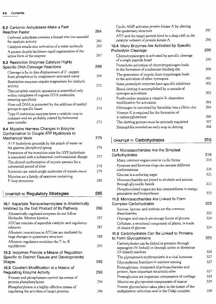
BRIEF CONTENTS CONTENTS
Part I THE MOLECULAR DESIGN OF LIFE
1 Biochemistry: An Evolving Science 1
2 Protein Composition and Structure 27
3 Exploring Proteins and Proteomes 65
4 DNA, RNA, and the Flow of Genetic Information 105
5 Exploring Genes and Genomes 135
6 Exploring Evolution and Bioinformatics 169
7 Hemoglobin: Portrait of a Protein in Action 191
8 Enzymes: Basic Concepts and Kinetics 215
9 Catalytic Strategies 251
10 Regulatory Strategies 285
11 Carbohydrates 315
12 Lipids and Cell Membranes 341
13 Membrane Channels and Pumps 367
14 Signal-Transduction Pathways 397
Part II TRANSDUCING AND STORING ENERGY
15 Metabolism: Basic Concepts and Design 423
16 Glycolysis and Gluconeogenesis 449
17 The Citric Acid Cycle 495
18 Oxidative Phosphorylation 523
19 The Light Reactions of Photosynthesis 565
20 The Calvin Cycle and the Pentose PhosphatePathway 589
21 Glycogen Metabolism 617
22 Fatty Acid Metabolism 643
23 Protein Turnover and Amino Acid Catabolism 681
Part III SYNTHESIZING THE MOLECULES OF LIFE
24 The Biosynthesis of Amino Acids 713
25 Nucleotide Biosynthesis 743
26 The Biosynthesis of Membrane Lipids andSteroids 767
27 The Integration of Metabolism 801
28 DNA Replication, Repair, and Recombination 827
29 RNA Synthesis and Processing 859
30 Protein Synthesis 893
31 The Control of Gene Expression in Prokaryotes 925
32 The Control of Gene Expression in Eukaryotes 941
Part IV RESPONDING TO ENVIRONMENTAL
CHANGES
33 Sensory Systems 961
34 The Immune System 981
35 Molecular Motors 1011
36 Drug Development 1033
Preface v
Part I THE MOLECULAR DESIGN OF LIFE 1
chapter 1 Biochemistry: An Evolving Science 1
1.1 Biochemical Unity Underlies Biological Diversity 1
1.2 DNA Illustrates the Interplay Between Form and
Function 4
DNA is constructed from four building blocks 4
Two single strands of DNA combine to form a double helix 5
DNA structure explains heredity and the storage of
information 5
1.3 Concepts from Chemistry Explain the Propertiesof Biological Molecules 6
The formation of the DNA double helix as a key example 6
The double helix can form from its component strands 6
Covalent and noncovalent bonds are important forthe structure and stability of biological molecules 6
The double helix is an expression ofthe rules ofchemistry 9
The laws of thermodynamics govern the behavior of
biochemical systems 10
Heat is released in the formation of the double helix 12
Acid-base reactions are central in many biochemical
processes 13
Acid-base reactions can disrupt the double helix 14
Buffers regulate pH in organisms and in the laboratory 1S
1.4 The Genomic Revolution Is TransformingBiochemistry, Medicine, and Other Fields 17
Genome sequencing has transformed biochemistryand other fields 17
Environmental factors influence human biochemistry 20
Genome sequences encode proteins and patternsof expression 21
APPENDIX: Visualizing Molecular Structures I:
Small Molecules 22
[ chapter 2 Protein Composition and Structure 27
2.1 Proteins Are Built from a Repertoire of 20 Amino
Acids 29
2.2 Primary Structure: Amino Acids Are Linked byPeptide Bonds to Form Polypeptide Chains 35
Proteins have unique amino acid sequences specifiedby genes 37
Polypeptide chains are flexible yet conformationallyrestricted 38
2.3 Secondary Structure: Polypeptide Chains Can
Fold into Regular Structures Such As the AlphaHelix, the Beta Sheet, and Turns and Loops 40
The alpha helix is a coiled structure stabilized by intrachain
hydrogen bonds 40
Beta sheets are stabilized by hydrogen bonding between
polypeptide strands 42

Contents xvii
Polypeptide chains can change direction by making reverse
turns and loops 44
Fibrous proteins provide structural support for cells
and tissues 44
2.4 Tertiary Structure: Water-Soluble Proteins
Fold into Compact Structures with Nonpolar Cores 46
2.5 Quaternary Structure: Polypeptide Chains Can
Assemble into Multisubunit Structures 48
2.6 The Amino Acid Sequence of a Protein
Determines Its Three-Dimensional Structure 49
Amino acids have different propensities for
forming a helices, P sheets, and turns 51
Protein folding is a highly cooperative process 52
Proteins fold by progressive stabilization of intermediates
rather than by random search 53
Prediction ofthree-dimensional structure from sequence
remains a great challenge 54
Some proteins are inherently unstructured and can exist
in multiple conformations 55
Protein misfolding and aggregation are associated with
some neurological diseases 56
Protein modification and cleavage confer new capabilities 57
APPENDIX: Visualizing Molecular Structures II: Proteins 61
3.3 Mass Spectrometry Is a Powerful Techniquefor the Identification of Peptides and Proteins 85
Peptides can be sequenced by mass spectrometry 87
Proteins can be specifically cleaved into small peptidesto facilitate analysis 88
Genomic and proteomic methods are complementary 89
The amino acid sequence ofa protein provides valuable
information 90
Individual proteins can be identified by mass
spectrometry 91
3.4 Peptides Can Be Synthesized by Automated
Solid-Phase Methods 92
3.5 Three-Dimensional Protein Structure Can Be
Determined by X-ray Crystallography and NMR
Spectroscopy 95
X-ray crystallography reveals three-dimensional
structure in atomic detail 95
Nuclear magnetic resonance spectroscopy can reveal
the structures ofproteins in solution 97
chapter 4 DNA, RNA, and the Flow of
Genetic Information 105
| chapter 3 Exploring Proteins and Proteomes 65
The proteome is the functional representation ofthe genome 66
3.1 The Purification of Proteins Is an Essential
First Step in Understanding Their Function 66
The assay: How do we recognize the protein that we are
looking for? 67
Proteins must be released from the cell to be purified 67
Proteins can be purified according to solubility, size,
charge, and binding affinity 68
Proteins can be separated by gel electrophoresis and
displayed 71
A protein purification scheme can be quantitatively
evaluated 75
Ultracentrifugation is valuable for separatingbiomolecules and determining their masses 76
Protein purification can be made easier with the
use of recombinant DNA technology 78
3.2 Immunology Provides Important Techniques with
Which to Investigate Proteins 79
Antibodies to specific proteins can be generated 79
Monoclonal antibodies with virtually any desired
specificity can be readily prepared 80
Proteins can be detected and quantified by using an
enzyme-linked immunosorbent assay 82
Western blotting permits the detection ofproteinsseparated by gel electrophoresis 83
Fluorescent markers make the visualization ofproteinsin the cell possible 84
4.1 A Nucleic Acid Consists of Four Kinds of
Bases Linked to a Sugar-Phosphate Backbone
RNA and DNA differ in the sugar component and
one ofthe bases
Nucleotides are the monomeric units of nucleic acids
DNA molecules are very long and have directionality
4.2 A Pair of Nucleic Acid Strands with
Complementary Sequences Can Form a
Double-Helical Structure
The double helix is stabilized by hydrogen bonds and
van der Waals interactions
DNA can assume a variety of structural forms
Z-DNA is a left-handed double helix in which
backbone phosphates zigzag
Some DNA molecules are circular and supercoilcd
Single-stranded nucleic acids can adopt elaborate
structures
4.3 The Double Helix Facilitates the Accurate
Transmission of Hereditary Information
Differences in DNA density established the validityof the semiconservative replication hypothesis
The double helix can be reversibly melted
4.4 DNA Is Replicated by Polymerases That Take
Instructions from Templates
DNA polymerase catalyzes phosphodiester-bridge formation
The genes of some viruses are made of RNA
4.5 Gene Expression Is the Transformation
of DNA Information into Functional Molecules
Several kinds of RNA play key roles in gene expression
106
106
107
108
109
109
111
112
113
113
114
115
116
117
117
118
119
119

xviii Contents
All cellular RNA is synthesized by RNA polymerases 120
RNA polymerases take instructions from DNA
templates 121
Transcription begins near promoter sites and ends at
terminator sites 122
Transfer RNAs are the adaptor molecules in protein
synthesis 123
4.6 Amino Acids Are Encoded by Groups of
Three Bases Starting from a Fixed Point 124
Major features of the genetic code 125
Messenger RNA contains start and stop signals for
protein synthesis 126
The genetic code is nearly universal 126
4.7 Most Eukaryotic Genes Are Mosaics of
Introns and Exons 127
RNA processing generates mature RNA 127
Many exons encode protein domains 128
| chapter 5 Exploring Genes and Genomes 135
5.1 The Exploration of Genes Relies on Key Tools 136
Restriction enzymes split DNA into specificfragments 137
Restriction fragments can be separated by gelelectrophoresis and visualized 137
DNA can be sequenced by controlled termination of
replication 138
DNA probes and genes can be synthesized byautomated solid-phase methods 139
Selected DNA sequences can be greatly amplifiedby the polymerase chain reaction 141
PCR is a powerful technique in medical diagnostics,forensics, and studies ofmolecular evolution 142
The tools for recombinant DNA technology have been
used to identify disease-causing mutations 143
5.2 Recombinant DNA Technology Has
Revolutionized All Aspects of Biology 143
Restriction enzymes and DNA ligase are key tools in
forming recombinant DNA molecules 143
Plasmids and \ phage are choice vectors for DNA
cloning in bacteria 144
Bacterial and yeast artificial chromosomes 147
Specific genes can be cloned from digests of
genomic DNA 147
Complementary DNA prepared from mRNA can be
expressed in host cells 149
Proteins with new functions can be created throughdirected changes in DNA 150
Recombinant methods enable the exploration ofthe
functional effects of disease-causing mutations 152
5.3 Complete Genomes Have Been Sequencedand Analyzed 152
The genomes of organisms ranging from bacteria to
multicellular eukaryotes have been sequenced 153
The sequence ofthe human genome has been completed 154
Next-generation sequencing methods enable the rapiddetermination of a complete genome sequence 155
Comparative genomics has become a powerfulresearch tool 156
5.4 Eukaryotic Genes Can Be Quantitated and
Manipulated with Considerable Precision 157
Gene-expression levels can be comprehensivelyexamined 157
New genes inserted into eukaryotic cells can be
efficiently expressed 159
Transgenic animals harbor and express genes introduced
into their germ lines 160
Gene disruption and genome editing provide clues to
gene function and opportunities for new therapies 160
RNA interference provides an additional tool for
disrupting gene expression 162
Tumor-inducing plasmids can be used to introduce
new genes into plant cells 163
Human gene therapy holds great promise for medicine 164
chapter 6 Exploring Evolution and
Bioinformatics 169
6.1 Homologs Are Descended from a Common
Ancestor 170
6.2 Statistical Analysis of Sequence AlignmentsCan Detect Homology 171
The statistical significance of alignments can be
estimated by shuffling 173
Distant evolutionary relationships can be detected
through the use ofsubstitution matrices 174
Databases can be searched to identify homologoussequences 177
6.3 Examination of Three-Dimensional Structure
Enhances Our Understanding of Evolutionary
Relationships 177
Tertiary structure is more conserved than primarystructure 178
Knowledge of three-dimensional structures can aid
in the evaluation of sequence alignments 179
Repeated motifs can be detected by aligning sequences
with themselves 180
Convergent evolution illustrates common solutions to
biochemical challenges 181
Comparison ofRNA sequences can be a source of
insight into RNA secondary structures 182
6.4 Evolutionary Trees Can Be Constructed on the
Basis of Sequence Information 183
Horizontal gene transfer events may explain unexpectedbranches ofthe evolutionary tree 184
6.5 Modern Techniques Make the ExperimentalExploration of Evolution Possible 185
Ancient DNA can sometimes be amplified and
sequenced 185
Molecular evolution can be examined experimentally 185

Contents xix
chapter 7 Hemoglobin: Portrait of a Protein
in Action 191
7.1 Myoglobin and Hemoglobin Bind Oxygenat Iron Atoms in Heme 192
Changes in heme electronic structure upon oxygen
binding are the basis for functional imaging studies 193
The structure of myoglobin prevents the release of
reactive oxygen species 194
Human hemoglobin is an assembly offour myoglobin-likesubunits 195
7.2 Hemoglobin Binds Oxygen Cooperatively 195
Oxygen binding markedly changes the quaternary
structure of hemoglobin 197
Hemoglobin cooperativity can be potentially explained
by several models 198
Structural changes at the heme groups are transmitted
to the u\Pi-ct2P2 interface 200
2,3-Bisphosphoglycerate in red cells is crucial in
determining the oxygen affinity of hemoglobin 200
Carbon monoxide can disrupt oxygen transport byhemoglobin 201
7.3 Hydrogen Ions and Carbon Dioxide Promote
the Release of Oxygen: The Bohr Effect 202
7.4 Mutations in Genes Encoding HemoglobinSubunits Can Result in Disease 204
Sickle-cell anemia results from the aggregation of
mutated deoxyhemoglobin molecules 205
Thalassemia is caused by an imbalanced production
ofhemoglobinchains 207
The accumulation offree alpha-hemoglobin chains is
prevented 207
Additional globins are encoded in the human genome 208
APPENDIX: Binding Models Can Be Formulated
in QuantitativeTerms: The Hill Plot and the
Concerted Model 210
chapter 8 Enzymes: Basic Concepts and
Kinetics 215
8.1 Enzymes are Powerful and Highly Specific
Catalysts 216
Many enzymes require cofactors for activity 217
Enzymes can transform energy from one form
into another 217
8.2 Gibbs Free Energy Is a Useful ThermodynamicFunction for Understanding Enzymes 218
The free-energy change provides information about
the spontaneity but not the rate ofa reaction 218
The standard free-energy change of a reaction is related
to the equilibrium constant 219
Enzymes alter only the reaction rate and not the reaction
equilibrium 220
8.3 Enzymes Accelerate Reactions by Facilitatingthe Formation of the Transition State 221
The formation ofan enzyme-substrate complex is the
first step in enzymatic catalysis 222
The active sites ofenzymes have some common
features 223
The binding energy between enzyme and substrate is
important for catalysis 225
8.4 The Michaelis-Menten Model Accounts for
the Kinetic Properties of Many Enzymes 225
Kinetics is the study of reaction rates 225
The steady-state assumption facilitates a descriptionof enzyme kinetics 226
Variations in K\[ can have physiological consequences 228
K\[ and Vmax values can be determined by several
means 228
K\i and Vmax values are important enzymecharacteristics 229
fecat/KM is a measure of catalytic efficiency 230
Most biochemical reactions include multiple substrates 231
Allosteric enzymes do not obey Michaelis-Menten
kinetics 233
8.5 Enzymes Can Be Inhibited by SpecificMolecules 234
The different types of reversible inhibitors are
kinetically distinguishable 235
Irreversible inhibitors can be used to map the active site 237
Penicillin irreversibly inactivates a key enzyme in
bacterial cell-wall synthesis 239
Transition-state analogs are potent inhibitors of
enzymes 240
Catalytic antibodies demonstrate the importanceof selective binding ofthe transition state to enzymaticactivity 241
8.6 Enzymes Can Be Studied One Molecule
at a Time 242
APPENDIX: Enzymes are Classified on the Basis of the
Types of Reactions That They Catalyze 245
| chapter 9 Catalytic Strategies 251
A few basic catalytic principles are used bymany enzymes 252
9.1 Proteases Facilitate a FundamentallyDifficult Reaction 253
Chymotrypsin possesses a highly reactive serine
residue 253
Chymotrypsin action proceeds in two steps linked
by a covalently bound intermediate 254
Serine is part of a catalytic triad that also includes
histidine and aspartate 255
Catalytic triads are found in other hydrolytic enzymes 258
The catalytic triad has been dissected by site-directed
mutagenesis 260
Cysteine, aspartyl, and metalloproteases are other
major classes of peptide-cleaving enzymes 260
Protease inhibitors are important drugs 263
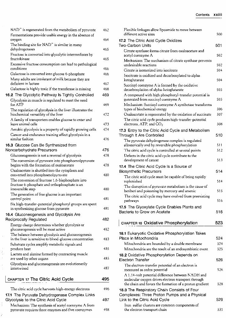
xx Contents
9.2 Carbonic Anhydrases Make a Fast
Reaction Faster 264
Carbonic anhydrase contains a bound zinc ion essential
for catalytic activity 265
Catalysis entails zinc activation of a water molecule 265
A proton shuttle facilitates rapid regeneration ofthe
active form of the enzyme 267
9.3 Restriction Enzymes Catalyze Highly
Specific DNA-Cleavage Reactions 269
Cleavage is by in-line displacement of3'-oxygenfrom phosphorus by magnesium-activated water 269
Restriction enzymes require magnesium for catalytic
activity 271
The complete catalytic apparatus is assembled onlywithin complexes of cognate DNA molecules,
ensuring specificity 272
Host-cell DNA is protected by the addition ofmethyl
groups to specific bases 274
Type II restriction enzymes have a catalytic core in
common and are probably related by horizontal
gene transfer 275
9.4 Myosins Harness Changes in EnzymeConformation to Couple ATP Hydrolysis to
Mechanical Work 275
ATP hydrolysis proceeds by the attack ofwater on
the gamma-phosphoryl group 276
Formation ofthe transition state for ATP hydrolysisis associated with a substantial conformational change 277
The altered conformation of myosin persists for a
substantial period oftime 278
Scientists can watch single molecules of myosin move 279
Myosins are a family ofenzymes containingP-loop structures 280
[ chapter 10 Regulatory Strategies 285
10.1 Aspartate Transcarbamoylase Is AllostericallyInhibited by the End Product of Its Pathway 286
Allosterically regulated enzymes do not follow
Michaelis-Menten kinetics 287
ATCase consists of separable catalytic and regulatorysubunits 287
Allosteric interactions in ATCase are mediated bylarge changes in quaternary structure 288
Allosteric regulators modulate the T-to-R
equilibrium 291
10.2 Isozymes Provide a Means of Regulation
Specific to Distinct Tissues and Developmental
Stages 292
10.3 Covalent Modification Is a Means of
Regulating Enzyme Activity 293
Kinases and phosphatases control the extent of
protein phosphorylation 294
Phosphorylation is a highly effective means of
regulating the activities of target proteins 296
Cyclic AMP activates protein kinase A by alteringthe quaternary structure 297
ATP and the target protein bind to a deep cleft in the
catalytic subunit ofprotein kinase A 298
10.4 Many Enzymes Are Activated by Specific
Proteolytic Cleavage 299
Chymotrypsinogen is activated by specific cleavageofa single peptide bond 299
Proteolytic activation ofchymotrypsinogen leads
to the formation ofa substrate-binding site 300
The generation oftrypsin from trypsinogen leads
to the activation of other zymogens 301
Some proteolytic enzymes have specific inhibitors 302
Blood clotting is accomplished by a cascade of
zymogen activations 303
Prothrombin requires a vitamin K-dependentmodification for activation 304
Fibrinogen is converted by thrombin into a fibrin clot 304
Vitamin K is required for the formation of
7-carboxyglutamate 306
The clotting process must be precisely regulated 307
Hemophilia revealed an early step in clotting 308
| chapter 11 Carbohydrates 315
11.1 Monosaccharides Are the Simplest
Carbohydrates 316
Many common sugars exist in cyclic forms 318
Pyranose and furanose rings can assume different
conformations 320
Glucose is a reducing sugar 321
Monosaccharides are joined to alcohols and amines
through glycosidic bonds 322
Phosphorylated sugars are key intermediates in energy
generation and biosyntheses 322
11.2 Monosaccharides Are Linked to Form
Complex Carbohydrates 323
Sucrose, lactose, and maltose are the common
disaccharides 323
Glycogen and starch are storage forms of glucose 324
Cellulose, a structural component of plants, is made
of chains of glucose 324
11.3 Carbohydrates Can Be Linked to Proteins
to Form Glycoproteins 325
Carbohydrates can be linked to proteins through
asparagine (N-linked) or through serine or threonine
(O-linked) residues 326
The glycoprotein erythropoietin is a vital hormone 327
Glycosylation functions in nutrient sensing 327
Proteoglycans, composed ofpolysaccharides and
protein, have important structural roles 327
Proteoglycans are important components of cartilage 328
Mucins are glycoprotein components of mucus 329
Protein glycosylation takes place in the lumen ofthe
endoplasmic reticulum and in the Golgi complex 330

Contents xxi
Specific enzymes are responsible for oligosaccharideassembly
Blood groups are based on protein glycosylationpatterns
Errors in glycosylation can result in pathologicalconditions
Oligosaccharides can be "sequenced"
11.4 Lectins Are Specific Carbohydrate-BindingProteins
Lectins promote interactions between cells
Lectins are organized into different classes
Influenza virus binds to sialic acid residues
chapter 12 Lipids and Cell Membranes
331
331
332
332
333
334
334
335
341
12.6 Eukaryotic Cells Contain CompartmentsBounded by Internal Membranes 359
Many common features underlie the diversity of
biological membranes
12.1 Fatty Acids Are Key Constituents of Lipids
Fatty acid names are based on their parenthydrocarbons
Fatty acids vary in chain length and degree of
unsaturation
12.2 There Are Three Common Types of
Membrane Lipids
Phospholipids are the major class of membrane lipids
Membrane lipids can include carbohydrate moieties
Cholesterol is a lipid based on a steroid nucleus
Archaeal membranes are built from ether lipids with
branched chains
A membrane lipid is an amphipathic molecule containinga hydrophilic and a hydrophobic moiety
12.3 Phospholipids and Glycolipids Readily Form
Bimolecular Sheets in Aqueous Media
Lipid vesicles can be formed from phospholipids
Lipid bilayers are highly impermeable to ions and most
polar molecules
12.4 Proteins Carry Out Most Membrane
Processes
Proteins associate with the lipid bilayer in a varietyof ways
Proteins interact with membranes in a variety ofways
Some proteins associate with membranes through
covalently attached hydrophobic groups
Transmembrane helices can be accurately predictedfrom amino acid sequences
12.5 Lipids and Many Membrane Proteins Diffuse
Rapidly in the Plane of the Membrane
The fluid mosaic model allows lateral movement but
not rotation through the membrane
Membrane fluidity is controlled by fatty acid
composition and cholesterol content
Lipid rafts are highly dynamic complexes formed
between cholesterol and specific lipidsAll biological membranes are asymmetric
342
342
342
343
344
344
345
346
346
347
348
348
349
350
351
351
354
354
356
357
357
358
358
| chapter 13 Membrane Channels and Pumps 367
The expression oftransporters largely defines the
metabolic activities of a given cell type 368
13.1 The Transport of Molecules Across a
Membrane May Be Active or Passive 368
Many molecules require protein transporters to cross
membranes 368
Free energy stored in concentration gradients can be
quantified 369
13.2 Two Families of Membrane Proteins Use ATP
Hydrolysis to Pump Ions and Molecules Across
Membranes 370
P-type ATPases couple phosphorylation and
conformational changes to pump calcium ions
across membranes 370
Digitalis specifically inhibits the Na+-K+ pump
by blocking its dephosphorylation 373
P-type ATPases are evolutionarily conserved and
play a wide range of roles 374
Multidrug resistance highlights a family of membrane
pumps with ATP-binding cassette domains 374
13.3 Lactose Permease Is an Archetype of
Secondary Transporters That Use One
Concentration Gradient to Power the Formation
of Another 376
13.4 Specific Channels Can Rapidly Transport Ions
Across Membranes 378
Action potentials are mediated by transient changes in
Na+ and K+ permeability 378
Patch-clamp conductance measurements reveal the
activities of single channels 379
The structure of a potassium ion channel is an archetypefor many ion-channel structures 379
The structure of the potassium ion channel reveals the
basis of ion specificity 380
The structure of the potassium ion channel explainsits rapid rate of transport 383
Voltage gating requires substantial conformational
changes in specific ion-channel domains 383
A channel can be inactivated by occlusion of the pore:
the ball-and-chain model 384
The acetylcholine receptor is an archetype for
ligand-gated ion channels 385
Action potentials integrate the activities of several ion
channels working in concert 387
Disruption ofion channels by mutations or chemicals
can be potentially life-threatening 388
13.5 Gap Junctions Allow Ions and Small Molecules
to Flow Between Communicating Cells 389
13.6 Specific Channels Increase the Permeabilityof Some Membranes to Water 390

xx ii Contents
| chapter 14 Signal-Transduction Pathways 397
Signal transduction depends on molecular circuits 398
14.1 Heterotrimeric G Proteins Transmit Signals
and Reset Themselves 399
Ligand binding to 7TM receptors leads to the activation
of heterotrimeric G proteins 400
Activated G proteins transmit signals by binding to
other proteins 402
Cyclic AMP stimulates the phosphorylation ofmany
target proteins by activating protein kinase A 403
G proteins spontaneously reset themselves throughGTP hydrolysis 403
Some 7TM receptors activate the phosphoinositidecascade 404
Calcium ion is a widely used second messenger 405
Calcium ion often activates the regulatory proteincalmodulin 407
14.2 Insulin Signaling: Phosphorylation Cascades
Are Central to Many Signal-Transduction Processes 407
The insulin receptor is a dimer that closes around a
bound insulin molecule 408
Insulin binding results in the cross-phosphorylation and
activation ofthe insulin receptor 408
The activated insulin-receptor kinase initiates a kinase
cascade 409
Insulin signaling is terminated by the action of
phosphatases 411
14.3 EGF Signaling: Signal-Transduction Pathways
Are Poised to Respond 411
EGF binding results in the dimerization of the EGF
receptor 411
The EGF receptor undergoes phosphorylation of its
carboxyl-terminal tail 413
EGF signaling leads to the activation of Ras, a small
G protein 413
Activated Ras initiates a protein kinase cascade 414
EGF signaling is terminated by protein phosphatasesand the intrinsic GTPase activity of Ras 414
14.4 Many Elements Recur with Variation in
Different Signal-Transduction Pathways 415
14.5 Defects in Signal-Transduction PathwaysCan Lead to Cancer and Other Diseases 416
Monoclonal antibodies can be used to inhibit signal -
transduction pathways activated in tumors 416
Protein kinase inhibitors can be effective anticancer drugs 417
Cholera and whooping cough are the result of altered
G- protein activity 417
Part II TRANSDUCING AND STORING ENERGY
chapter 15 Metabolism: Basic Conceptsand Design 423
15.1 Metabolism Is Composed of Many Coupled,
Interconnecting Reactions 424
Metabolism consists of energy-yielding and energy-
requiring reactions 424
A thermodynamically unfavorable reaction can be
driven by a favorable reaction 425
15.2 ATP Is the Universal Currency of Free
Energy in Biological Systems 426
ATP hydrolysis is exergonic 426
ATP hydrolysis drives metabolism by shifting the
equilibrium of coupled reactions 427
The high phosphoryl potential of ATP results from
structural differences between ATP and its hydrolysis
products 429
Phosphoryl-transfer potential is an important form of
cellular energy transformation 430
15.3 The Oxidation of Carbon Fuels Is an
Important Source of Cellular Energy 432
Compounds with high phosphoryl-transfer potentialcan couple carbon oxidation to ATP synthesis 432
Ion gradients across membranes provide an important
form of cellular energy that can be coupled to
ATP synthesis 433
Phosphates play a prominent role in biochemical
processes434
Energy from foodstuffs is extracted in three stages 434
15.4 Metabolic Pathways Contain Many
Recurring Motifs 435
Activated carriers exemplify the modular design and
economy ofmetabolism 435
Many activated carriers are derived from vitamins 438
Key reactions are reiterated throughout metabolism 440
Metabolic processes are regulated in three principal ways 442
Aspects ofmetabolism may have evolved from an
RNA world 444
| chapter 16 Glycolysis and Gluconeogenesis 449
Glucose is generated from dietary carbohydrates 450
Glucose is an important fuel for most organisms 451
16.1 Glycolysis Is an Energy-Conversion Pathway
in Many Organisms 451
Hexokinase traps glucose in the cell and begins
glycolysis 451
Fructose 1,6-bisphosphate is generated from glucose
6-phosphate 453
The six-carbon sugar is cleaved into two three-carbon
fragments 454
Mechanism: Triose phosphate isomerase salvages a
three-carbon fragment 455
The oxidation of an aldehyde to an acid powers the
formation of a compound with high phosphoryl-transferpotential 457
Mechanism: Phosphorylation is coupled to the oxidation
of glyceraldehyde 3-phosphate by a thioester intermediate 458
ATP is formed by phosphoryl transfer from
1,3-bisphosphoglycerate 459
Additional ATP is generated with the formation of
pyruvate 460
Two ATP molecules are formed in the conversion of
glucose into pyruvate 461
Contents xxiii
NAD+ is regenerated from the metabolism of pyruvate 462
Fermentations provide usable energy in the absence of
oxygen 464
The binding site for NAD+ is similar in many
dehydrogenases 465
Fructose is converted into glycolytic intermediates byfructokinase 465
Excessive fructose consumption can lead to pathologicalconditions 466
Galactose is converted into glucose 6-phosphate 466
Many adults are intolerant ofmilk because they are
deficient in lactase 467
Galactose is highly toxic ifthe transferase is missing 468
16.2 The Glycolytic Pathway Is Tightly Controlled 469
Glycolysis in muscle is regulated to meet the need
for ATP 469
The regulation of glycolysis in the liver illustrates the
biochemical versatility ofthe liver 472
A family oftransporters enables glucose to enter and
leave animal cells 473
Aerobic glycolysis is a property ofrapidly growing cells 474
Cancer and endurance training affect glycolysis in a
similar fashion 476
16.3 Glucose Can Be Synthesized from
Noncarbohydrate Precursors 476
Gluconeogenesis is not a reversal of glycolysis 478
The conversion ofpyruvate into phosphoenolpyruvatebegins with the formation ofoxaloacetate 478
Oxaloacetate is shuttled into the cytoplasm and
converted into phosphoenolpyruvate 480
The conversion offructose 1,6-bisphosphate into
fructose 6-phosphate and orthophosphate is an
irreversible step 480
The generation offree glucose is an importantcontrol point 481
Six high-transfer-potential phosphoryl groups are spent
in synthesizing glucose from pyruvate 481
16.4 Gluconeogenesis and Glycolysis Are
Reciprocally Regulated 482
Energy charge determines whether glycolysis or
gluconeogenesis will be most active 482
The balance between glycolysis and gluconeogenesisin the liver is sensitive to blood-glucose concentration 483
Substrate cycles amplify metabolic signals and
produce heat 485
Lactate and alanine formed by contracting muscle
are used by other organs 485
Glycolysis and gluconeogenesis are evolutionarilyintertwined 487
chapter 17 The Citric Acid Cycle 495
The citric acid cycle harvests high-energy electrons
17.1 The Pyruvate Dehydrogenase Complex Links
Glycolysis to the Citric Acid Cycle
Mechanism: The synthesis of acetyl coenzyme A from
pyruvate requires three enzymes and five coenzymes
496
497
498
Flexible linkages allow lipoamide to move between
different active sites 500
17.2 The Citric Acid Cycle Oxidizes
Two-Carbon Units 501
Citrate synthase forms citrate from oxaloacetate and
acetyl coenzyme A 502
Mechanism: The mechanism of citrate synthase preventsundesirable reactions 502
Citrate is isomerized into isocitrate 504
Isocitrate is oxidized and decarboxylated to alpha-ketoglutarate 504
Succinyl coenzyme A is formed by the oxidative
decarboxylation ofalpha-ketoglutarate 505
A compound with high phosphoryl-transfer potential is
generated from succinyl coenzyme A 505
Mechanism: Succinyl coenzyme A synthetase transforms
types ofbiochemical energy 506
Oxaloacetate is regenerated by the oxidation ofsuccinate 507
The citric acid cycle produces high-transfer-potentialelectrons, ATP, and C02 508
17.3 Entry to the Citric Acid Cycle and Metabolism
Through It Are Controlled 510
The pyruvate dehydrogenase complex is regulatedallosterically and by reversible phosphorylation 511
The citric acid cycle is controlled at several points 512
Defects in the citric acid cycle contribute to the
development ofcancer 513
17.4 The Citric Acid Cycle Is a Source of
Biosynthetic Precursors 514
The citric acid cycle must be capable of being rapidlyreplenished 514
The disruption of pyruvate metabolism is the cause of
beriberi and poisoning by mercury and arsenic 515
The citric acid cycle may have evolved from preexisting
pathways 516
17.5 The Glyoxylate Cycle Enables Plants and
Bacteria to Grow on Acetate 516
| chapter 18 Oxidative Phosphorylation 523
18.1 Eukaryotic Oxidative Phosphorylation Takes
Place in Mitochondria 524
Mitochondria are bounded by a double membrane 524
Mitochondria are the result of an endosymbiotic event 525
18.2 Oxidative Phosphorylation Depends on
Electron Transfer 526
The electron-transfer potential ofan electron is
measured as redox potential 526
A 1.14-volt potential difference between NADH and
molecular oxygen drives electron transport throughthe chain and favors the formation ofa proton gradient 528
18.3 The Respiratory Chain Consists of Four
Complexes: Three Proton Pumps and a PhysicalLink to the Citric Acid Cycle 529
Iron-sulfur clusters are common components of
the electron transport chain 531

xx iv Contents
The high-potential electrons of NADH enter the
respiratory chain at NADH-Qoxidoreductase 532
Ubiquinol is the entry point for electrons from
FADH2 of flavoproteins 533
Electrons flow from ubiquinol to cytochrome c through
Q-cytochrome c oxidoreductase 533
The Q cycle funnels electrons from a two-electron
carrier to a one-electron carrier and pumps protons 535
Cytochrome c oxidase catalyzes the reduction of
molecular oxygen to water 535
Toxic derivatives ofmolecular oxygen such as superoxide
radicals are scavenged by protective enzymes 538
Electrons can be transferred between groups that are
not in contact 540
The conformation ofcytochrome c has remained
essentially constant for more than a billion years 541
18.4 A Proton Gradient Powers the Synthesis
of ATP 541
ATP synthase is composed of a proton-conductingunit and a catalytic unit 543
Proton flow through ATP synthase leads to the release
oftightly bound ATP: The binding-change mechanism 544
Rotational catalysis is the world's smallest molecular
motor 546
Proton flow around the c ring powers ATP synthesis 546
ATP synthase and G proteins have several common
features 548
18.5 Many Shuttles Allow Movement Across
Mitochondrial Membranes 549
Electrons from cytoplasmic NADH enter mitochondria
by shuttles 549
The entry of ADP into mitochondria is coupled to the
exit of ATP by ATP-ADP translocase 550
Mitochondrial transporters for metabolites have a
common tripartite structure 551
18.6 The Regulation of Cellular Respiration Is
Governed Primarily by the Need for ATP 552
The complete oxidation of glucose yields about30 molecules ofATP 552
The rate of oxidative phosphorylation is determined
by the need for ATP 553
ATP synthase can be regulated 554
Regulated uncoupling leads to the generation of heat 554
Oxidative phosphorylation can be inhibited at many stages 556
Mitochondrial diseases are being discovered 557
Mitochondria play a key role in apoptosis 557
Power transmission by proton gradients is a central
motif of bioenergetics 558
chapter 19 The Light Reactions of
Photosynthesis 565
Chloroplasts arose from an endosymbiotic event 568
19.2 Light Absorption by Chlorophyll Induces
Electron Transfer 568
A special pair of chlorophylls initiate charge separation 569
Cyclic electron flow reduces the cytochrome of the
reaction center 572
19.3 Two Photosystems Generate a Proton Gradient
and NADPH in Oxygenic Photosynthesis 572
Photosystem II transfers electrons from water to
plastoquinone and generates a proton gradient 572
Cytochrome bf links photosystem II to photosystem I 575
Photosystem I uses light energy to generate reduced
ferredoxin, a powerful reductant 575
Ferredoxin-NADP+ reductase converts NADP+ into
NADPH 576
19.4 A Proton Gradient across the Thylakoid
Membrane Drives ATP Synthesis 578
The ATP synthase of chloroplasts closely resembles
those ofmitochondria and prokaryotes 578
The activity ofchloroplast ATP synthase is regulated 579
Cyclic electron flow through photosystem I leads to the
production ofATP instead of NADPH 580
The absorption of eight photons yields one O2, two
NADPH, and three ATP molecules 581
19.5 Accessory Pigments Funnel Energy into
Reaction Centers 581
Resonance energy transfer allows energy to move from
the site of initial absorbance to the reaction center 582
The components of photosynthesis are highly organized 583
Many herbicides inhibit the light reactions of
photosynthesis 584
19.6 The Ability to Convert Light into Chemical
Energy Is Ancient 584
Artificial photosynthetic systems may provide clean,
renewable energy 585
chapter 20 The Calvin Cycle and the
Pentose Phosphate Pathway 589
Photosynthesis converts light energy into chemical energy 566
19.1 Photosynthesis Takes Place in Chloroplasts 567
The primary events of photosynthesis take place in
thylakoid membranes 567
20.1 The Calvin Cycle Synthesizes Hexoses
from Carbon Dioxide and Water 590
Carbon dioxide reacts with ribulose 1,5-bisphosphateto form two molecules of 3-phosphoglycerate 591
Rubisco activity depends on magnesium and carbamate 592
Rubisco activase is essential for rubisco activity 593
Rubisco also catalyzes a wasteful oxygenase reaction:
Catalytic imperfection 593
Hexose phosphates are made from phosphoglycerate,and ribulose 1,5 -bisphosphate is regenerated 594
Three ATP and two NADPH molecules are used to
bring carbon dioxide to the level of a hexose 597
Starch and sucrose are the major carbohydrate stores
in plants 597
20.2 The Activity of the Calvin Cycle Depends on
Environmental Conditions 598

Contents xxv
Rubisco is activated by light-driven changes in
proton and magnesium ion concentrations 598
Thioredoxin plays a key role in regulating the
Calvin cycle 599
The C4 pathway of tropical plants accelerates
photosynthesis by concentrating carbon dioxide 599
Crassulacean acid metabolism permits growth in arid
ecosystems 601
20.3 The Pentose Phosphate Pathway Generates
NADPH and Synthesizes Five-Carbon Sugars 601
Two molecules of NADPH are generated in the
conversion of glucose 6-phosphate into ribulose
5-phosphate 602
The pentose phosphate pathway and glycolysis are
linked by transketolase and transaldolase 602
Mechanism: Transketolase and transaldolase stabilize
carbanionic intermediates by different mechanisms 605
20.4 The Metabolism of Glucose 6-Phosphate bythe Pentose Phosphate Pathway Is Coordinated
with Glycolysis 607
The rate ofthe pentose phosphate pathway is controlled
by the level of NADP4" 607
The flow ofglucose 6-phosphate depends on the need
for NADPH, ribose 5-phosphate, and ATP 608
The pentose phosphate pathway is required for rapidcell growth 610
Through the looking-glass: The Calvin cycle and the
pentose phosphate pathway are mirror images 610
20.5 Glucose 6-Phosphate Dehydrogenase
Plays a Key Role in Protection Against Reactive
Oxygen Species 610
Glucose 6-phosphate dehydrogenase deficiency
causes a drug-induced hemolytic anemia 610
A deficiency ofglucose 6-phosphate dehydrogenaseconfers an evolutionary advantage in some circumstances 612
Muscle phosphorylase is regulated by the intracellular
energy charge 625
Biochemical characteristics of muscle fiber types differ 625
Phosphorylation promotes the conversion of
phosphorylase b to phosphorylase a 626
Phosphorylase kinase is activated by phosphorylationand calcium ions 626
21.3 Epinephrine and Glucagon Signal the Need
for Glycogen Breakdown 627
G proteins transmit the signal for the initiation of
glycogen breakdown 627
Glycogen breakdown must be rapidly turned off when
necessary 629
The regulation of glycogen phosphorylase became more
sophisticated as the enzyme evolved 629
21.4 Glycogen Is Synthesized and Degraded byDifferent Pathways 630
UDP-glucose is an activated form ofglucose 630
Glycogen synthase catalyzes the transfer of glucosefrom UDP-glucose to a growing chain 6.30
A branching enzyme forms a-1,6 linkages 631
Glycogen synthase is the key regulatory enzyme in
glycogen synthesis 632
Glycogen is an efficient storage form of glucose 632
21.5 Glycogen Breakdown and Synthesis Are
Reciprocally Regulated 632
Protein phosphatase 1 reverses the regulatory effects
of kinases on glycogen metabolism 633
Insulin stimulates glycogen synthesis by inactivating
glycogen synthase kinase 635
Glycogen metabolism in the liver regulates the
blood-glucose level 635
A biochemical understanding of glycogen-storage
diseases is possible 637
chapter 21 Glycogen Metabolism 617 chapter 22 Fatty Acid Metabolism 643
Glycogen metabolism is the regulated release and
storage of glucose 618
21.1 Glycogen Breakdown Requires the Interplay of
Several Enzymes 619
Phosphorylase catalyzes the phosphorolytic cleavageofglycogen to release glucose 1 -phosphate 619
Mechanism: Pyridoxal phosphate participates in the
phosphorolytic cleavage of glycogen 620
A debranching enzyme also is needed for the breakdown
ofglycogen 621
Phosphoglucomutase converts glucose 1 -phosphate
into glucose 6-phosphate 622
The liver contains glucose 6-phosphatase, a hydrolytic
enzyme absent from muscle 622
21.2 Phosphorylase Is Regulated by Allosteric
Interactions and Reversible Phosphorylation 623
Liver phosphorylase produces glucose for use byother tissues 623
Fatty acid degradation and synthesis mirror each other
in their chemical reactions 644
22.1 Triacylglycerols Are Highly Concentrated
Energy Stores 645
Dietary lipids are digested by pancreatic lipases 645
Dietary lipids are transported in chylomicrons 646
22.2 The Use of Fatty Acids as Fuel RequiresThree Stages of Processing 647
Triacylglycerols are hydrolyzed by hormone-
stimulated lipases 647
Free fatty acids and glycerol are released
into the blood 648
Fatty acids are linked to coenzyme A before they are
oxidized 648
Carnitine carries long-chain activated fatty acids into the
mitochondrial matrix 649
Acetyl CoA, NADH, and FADHi are generated in
each round offatty acid oxidation 650

xx vi Contents
The complete oxidation ofpalmitate yields106 molecules ofATP 652
22.3 Unsaturated and Odd-Chain Fatty Acids
Require Additional Steps for Degradation 652
An isomerase and a reductase are required for the
oxidation ofunsaturated fatty acids 652
Odd-chain fatty acids yield propionyl CoA in the final
thiolysis step 654
Vitamin contains a corrin ring and a cobalt atom 654
Mechanism: Methylmalonyl CoA mutase catalyzes a
rearrangement to form succinyl CoA 655
Fatty acids are also oxidized in peroxisomes 656
Ketone bodies are formed from acetyl CoA when fat
breakdown predominates 657
Ketone bodies are a major fuel in some tissues 658
Animals cannot convert fatty acids into glucose 660
Some fatty acids may contribute to the developmentof pathological conditions 661
22.4 Fatty Acids Are Synthesized by Fatty Acid
Synthase 661
Fatty acids are synthesized and degraded by different
pathways 661
The formation of malonyl CoA is the committed step in
fatty acid synthesis 662
Intermediates in fatty acid synthesis are attached to an
acyl carrier protein 662
Fatty acid synthesis consists of a series ofcondensation,reduction, dehydration, and reduction reactions 662
Fatty acids are synthesized by a multifunctional enzyme
complex in animals 664
The synthesis of palmitate requires 8 molecules ofacetylCoA, 14 molecules of NADPH, and 7 molecules of ATP 666
Citrate carries acetyl groups from mitochondria to the
cytoplasm for fatty acid synthesis 666
Several sources supply NADPH for fatty acid synthesis 667
Fatty acid metabolism is altered in tumor cells 667
22.5 The Elongation and Unsaturation of Fatty Acids
are Accomplished by Accessory Enzyme Systems 668
Membrane-bound enzymes generate unsaturated fatty acids 668
Eicosanoid hormones are derived from polyunsaturatedfatty acids 669
Variations on a theme: Polyketide and nonribosomal
peptide synthetases resemble fatty acid synthase 670
22.6 Acetyl CoA Carboxylase Plays a Key Role in
Controlling Fatty Acid Metabolism 670
Acetyl CoA carboxylase is regulated by conditions
in the cell 671
Acetyl CoA carboxylase is regulated by a variety ofhormones 671
chapter 23 Protein Turnover and
Amino Acid Catabolism 681
23.1 Proteins are Degraded to Amino Acids 682
The digestion ofdietary proteins begins in the stomach
and is completed in the intestine 682
Cellular proteins are degraded at different rates 682
23.2 Protein Turnover Is Tightly Regulated 683
Ubiquitin tags proteins for destruction 683
The proteasome digests the ubiquitin-tagged proteins 685
The ubiquitin pathway and the proteasome have
prokaryotic counterparts 686
Protein degradation can be used to regulate biologicalfunction 687
23.3 The First Step in Amino Acid DegradationIs the Removal of Nitrogen 687
Alpha-amino groups are converted into ammonium
ions by the oxidative deamination ofglutamate 687
Mechanism: Pyridoxal phosphate forms Schiff-base
intermediates in aminotransferases 689
Aspartate aminotransferase is an archetypal pyridoxal-dependent transaminase 690
Blood levels of aminotransferases serve a diagnosticfunction 691
Pyridoxal phosphate enzymes catalyze a wide array
ofreactions 691
Serine and threonine can be directly deaminated 692
Peripheral tissues transport nitrogen to the liver 692
23.4 Ammonium Ion Is Converted into Urea in
Most Terrestrial Vertebrates 693
The urea cycle begins with the formation ofcarbamoylphosphate 693
Carbamoyl phosphate synthetase is the key regulatoryenzyme for urea synthesis 694
Carbamoyl phosphate reacts with ornithine to beginthe urea cycle 694
The urea cycle is linked to gluconeogenesis 696
Urea-cycle enzymes are evolutionarily related to
enzymes in other metabolic pathways 696
Inherited defects ofthe urea cycle cause
hyperammonemia and can lead to brain damage 697
Urea is not the only means of disposing ofexcess nitrogen 698
23.5 Carbon Atoms of Degraded Amino Acids
Emerge as Major Metabolic Intermediates 698
Pyruvate is an entry point into metabolism for a
number ofamino acids 699
Oxaloacetate is an entry point into metabolism for
aspartate and asparagine 700
Alpha-ketoglutarate is an entry point into metabolism
for five-carbon amino acids 700
Succinyl coenzyme A is a point of entry for several
nonpolar amino acids 701
Methionine degradation requires the formation of a
key methyl donor, S-adenosylmethionine 701
The branched-chain amino acids yield acetyl CoA,acetoacetate, or propionyl CoA 701
Oxygenases are required for the degradation ofaromatic amino acids 703
23.6 Inborn Errors of Metabolism Can DisruptAmino Acid Degradation 705
Phenylketonuria is one ofthe most common metabolic
disorders 706
Determining the basis of the neurological symptoms of
phenylketonuria is an active area of research 706

Contents xxvii
Part III SYNTHESIZING THE MOLECULES OF LIFE
I chapter 24 The Biosynthesis of Amino Acids 713
Amino acid synthesis requires solutions to three keybiochemical problems 714
24.1 Nitrogen Fixation: Microorganisms Use ATP
and a Powerful Reductant to Reduce AtmosphericNitrogen to Ammonia 714
The iron-molybdenum cofactor ofnitrogenase binds and
reduces atmospheric nitrogen 715
Ammonium ion is assimilated into an amino acid
through glutamate and glutamine 717
24.2 Amino Acids Are Made from Intermediates
of the Citric Acid Cycle and Other Major Pathways 719
Human beings can synthesize some amino acids but
must obtain others from their diet 719
Aspartate, alanine, and glutamate are formed by the
addition of an amino group to an alpha-ketoacid 720
A common step determines the chirality ofall
amino acids 721
The formation of asparagine from aspartate requires an
adenylated intermediate 721
Glutamate is the precursor of glutamine, proline, and
arginine 722
3-Phosphoglycerate is the precursor ofserine, cysteine,and glycine 722
Tetrahydrofolate carries activated one-carbon units at
several oxidation levels 723
S-Adenosylmethionine is the major donor ofmethylgroups 724
Cysteine is synthesized from serine and homocysteine 726
High homocysteine levels correlate with vascular disease 726
Shikimate and chorismate are intermediates in the
biosynthesis ofaromatic amino acids 727
Tryptophan synthase illustrates substrate channeling in
enzymatic catalysis 729
24.3 Feedback Inhibition Regulates Amino Acid
Biosynthesis 730
Branched pathways require sophisticated regulation 731
The sensitivity of glutamine synthetase to allosteric
regulation is altered by covalent modification 732
24.4 Amino Acids Are Precursors of ManyBiomolecules 734
Glutathione, a gamma-glutamyl peptide, serves as a
sulfhydryl buffer and an antioxidant 734
Nitric oxide, a short-lived signal molecule, is formed
from arginine 735
Porphyrins are synthesized from glycine and succinylcoenzyme A 736
Porphyrins accumulate in some inherited disorders of
porphyrin metabolism 737
| chapter 25 Nucleotide Biosynthesis 743
Nucleotides can be synthesized by de novo or
salvage pathways 744
25.1 The Pyrimidine Ring Is Assembled de Novo
or Recovered by Salvage Pathways 744
Bicarbonate and other oxygenated carbon compoundsare activated by phosphorylation 745
The side chain ofglutamine can be hydrolyzed to
generate ammonia 745
Intermediates can move between active sites bychanneling 745
Orotate acquires a ribose ring from PRPP to form
a pyrimidine nucleotide and is converted into
uridylate 746
Nucleotide mono-, di-, and triphosphates are
interconvertible 747
CTP is formed by amination ofUTP 747
Salvage pathways recycle pyrimidine bases 748
25.2 Purine Bases Can Be Synthesized de Novo or
Recycled by Salvage Pathways 748
The purine ring system is assembled on ribose
phosphate 749
The purine ring is assembled by successive steps
of activation by phosphorylation followed bydisplacement 749
AMP and GMP are formed from IMP 751
Enzymes of the purine synthesis pathway associate
with one another in vivo 752
Salvage pathways economize intracellular energy
expenditure 752
25.3 Deoxyribonucleotides Are Synthesized by the
Reduction of Ribonucleotides Through a Radical
Mechanism 753
Mechanism: A tyrosyl radical is critical to the action
of ribonucleotide reductase 753
Stable radicals other than tyrosyl radical are employedby other ribonucleotide reductases 755
Thymidylate is formed by the methylation of
deoxyuridylate 755
Dihydrofolate reductase catalyzes the regeneration of
tetrahydrofolate, a one-carbon carrier 756
Several valuable anticancer drugs block the synthesisof thymidylate 757
25.4 Key Steps in Nucleotide Biosynthesis Are
Regulated by Feedback Inhibition 758
Pyrimidine biosynthesis is regulated by aspartate
transcarbamoylase 758
The synthesis of purine nucleotides is controlled byfeedback inhibition at several sites 758
The synthesis of deoxyribonucleotides is controlled bythe regulation of ribonucleotide reductase 759
25.5 Disruptions in Nucleotide Metabolism
Can Cause Pathological Conditions 760
The loss of adenosine deaminase activity results in
severe combined immunodeficiency 760
Gout is induced by high serum levels of urate 761
Lesch-Nyhan syndrome is a dramatic consequence of
mutations in a salvage-pathway enzyme 761
Folic acid deficiency promotes birth defects such as
spina bifida 762

xxviii Contents
chapter 26 The Biosynthesis of Membrane
Lipids and Steroids 767
26.1 Phosphatidate Is a Common Intermediate
in the Synthesis of Phospholipids and
Triacylglycerols 768
The synthesis of phospholipids requires an activated
intermediate 769
Some phospholipids are synthesized from an activated
alcohol 770
Phosphatidylcholine is an abundant phospholipid 770
Excess choline is implicated in the development of
heart disease 771
Base-exchange reactions can generate phospholipids 771
Sphingolipids are synthesized from ceramide 772
Gangliosides are carbohydrate-rich sphingolipids that
contain acidic sugars 772
Sphingolipids confer diversity on lipid structure andfunction 773
Respiratory distress syndrome and Tay-Sachs disease
result from the disruption of lipid metabolism 774
Ceramide metabolism stimulates tumor growth 774
Phosphatide acid phosphatase is a key regulatory
enzyme in lipid metabolism 775
26.2 Cholesterol Is Synthesized from AcetylCoenzyme A in Three Stages 776
The synthesis ofmevalonate, which is activated as
isopentenyl pyrophosphate, initiates the synthesisofcholesterol 776
Squalene (C30) is synthesized from six molecules of
isopentenyl pyrophosphate (C5) 777
Squalene cyclizes to form cholesterol 778
26.3 The Complex Regulation of Cholesterol
Biosynthesis Takes Place at Several Levels 779
Lipoproteins transport cholesterol and triacylglycerolsthroughout the organism 782
Low-density lipoproteins play a central role in
cholesterol metabolism 784
The absence of the LDL receptor leads to
hypercholesterolemia and atherosclerosis 784
Mutations in the LDL receptor prevent LDL release
and result in receptor destruction 785
Cycling of the LDL receptor is regulated 787
HDL appears to protect against atherosclerosis 787
The clinical management of cholesterol levels can be
understood at a biochemical level 788
26.4 Important Derivatives of Cholesterol Include
Bile Salts and Steroid Hormones 788
Letters identify the steroid rings and numbers identifythe carbon atoms 790
Steroids are hydroxylated by cytochrome P450
monooxygenases that use NADPH and Ot 790
The cytochrome P450 system is widespread and
performs a protective function 791
Pregnenolone, a precursor of many other steroids,is formed from cholesterol by cleavage of its side chain 792
Progesterone and corticosteroids are synthesized from
pregnenolone 792
Androgens and estrogens are synthesized from
pregnenolone 792
Vitamin D is derived from cholesterol by the ring-splitting activity of light 794
I chapter 27 The Integration of Metabolism 801
27.1 Caloric Homeostasis Is a Means of RegulatingBody Weight 802
27.2 The Brain Plays a Key Role in Caloric
Homeostasis 804
Signals from the gastrointestinal tract induce feelingsofsatiety 804
Leptin and insulin regulate long-term control over
caloric homeostasis 805
Leptin is one ofseveral hormones secreted byadipose tissue 806
Leptin resistance may be a contributing factor to
obesity 806
Dieting is used to combat obesity 807
27.3 Diabetes Is a Common Metabolic Disease
Often Resulting from Obesity 807
Insulin initiates a complex signal-transduction
pathway in muscle 808
Metabolic syndrome often precedes type 2 diabetes 809
Excess fatty acids in muscle modify metabolism 810
Insulin resistance in muscle facilitates pancreatic failure 810
Metabolic derangements in type 1 diabetes result from
insulin insufficiency and glucagon excess 812
27.4 Exercise Beneficially Alters the Biochemistryof Cells 813
Mitochondrial biogenesis is stimulated by muscular
activity 813
Fuel choice during exercise is determined by the
intensity and duration of activity 813
27.5 Food Intake and Starvation Induce Metabolic
Changes 816
The starved-fed cycle is the physiological response
to a fast 816
Metabolic adaptations in prolonged starvation minimize
protein degradation 818
27.6 Ethanol Alters Energy Metabolism in the
Liver 819
Ethanol metabolism leads to an excess of NADH 820
Excess ethanol consumption disrupts vitamin
metabolism 821
chapter 28 DNA Replication, Repair,and Recombination 827
28.1 DNA Replication Proceeds by the
Polymerization of DeoxyribonucleosideTriphosphates Along a Template 828
Contents xxix
DNA polymerases require a template and a primer 829
All DNA polymerases have structural features in
common 829
Two bound metal ions participate in the polymerasereaction 829
The specificity ofreplication is dictated bycomplementarity of shape between bases 830
An RNA primer synthesized by primase enables
DNA synthesis to begin 831
One strand of DNA is made continuously, whereas the
other strand is synthesized in fragments 831
DNA ligase joins ends ofDNA in duplex regions 832
The separation ofDNA strands requires specifichelicases and ATP hydrolysis 832
28.2 DNA Unwinding and Supercoiling Are
Controlled by Topoisomerases 833
The linking number ofDNA, a topological property,determines the degree of supercoiling 835
Topoisomerases prepare the double helix for
unwinding 836
Type I topoisomerases relax supercoiled structures 836
Type II topoisomerases can introduce negativesupercoils through coupling to ATP hydrolysis 837
28.3 DNA Replication Is Highly Coordinated 839
DNA replication requires highly processive
polymerases 839
The leading and lagging strands are synthesized in a
coordinated fashion 840
DNA replication in Escherichia coli begins at a
unique site 842
DNA synthesis in eukaryotes is initiated at
multiple sites 843
Telomeres are unique structures at the ends of linear
chromosomes 844
Telomeres are replicated by telomerase, a specializedpolymerase that carries its own RNA template 845
28.4 Many Types of DNA Damage Can Be
Repaired 845
Errors can arise in DNA replication 846
Bases can be damaged by oxidizing agents, alkylatingagents, and light 846
DNA damage can be detected and repaired by a varietyof systems 847
The presence of thymine instead of uracil in DNA
permits the repair ofdeaminated cytosine 849
Some genetic diseases are caused by the expansion of
repeats of three nucleotides 850
Many cancers are caused by the defective repair of DNA 850
Many potential carcinogens can be detected by their
mutagenic action on bacteria 852
28.5 DNA Recombination Plays Important Roles in
Replication, Repair, and Other Processes 852
RecA can initiate recombination by promotingstrand invasion 853
Some recombination reactions proceed throughHolliday-junction intermediates 854
| chapter 29 RNA Synthesis and Processing 859
RNA synthesis comprises three stages: Initiation,elongation, and termination 860
29.1 RNA Polymerases Catalyze Transcription 861
RNA chains are formed de novo and grow in the
5'-to-3'direction 862
RNA polymerases backtrack and correct errors 863
RNA polymerase binds to promoter sites on the
DNA template to initiate transcription 864
Sigma subunits of RNA polymerase recognize
promoter sites 865
RNA polymerases must unwind the templatedouble helix for transcription to take place 865
Elongation takes place at transcription bubbles that
move along the DNA template 866
Sequences within the newly transcribed RNA signaltermination 866
Some messenger RNAs directly sense metaboliteconcentrations 867
The rho protein helps to terminate the transcriptionofsome genes 868
Some antibiotics inhibit transcription 869
Precursors of transfer and ribosomal RNA are cleaved
and chemically modified after transcription in prokaryotes H70
29.2 Transcription in Eukaryotes Is Highly Regulated 871
Three types of RNA polymerase synthesize RNA in
eukaryotic cells 872
Three common elements can be found in the RNA
polymerase II promoter region 874
The TFIID protein complex initiates the assemblyof the active transcription complex 874
Multiple transcription factors interact with eukaryoticpromoters 875
Enhancer sequences can stimulate transcription at
start sites thousands of bases away 876
29.3 The Transcription Products of EukaryoticPolymerases Are Processed 876
RNA polymerase I produces three ribosomal RNAs 877
RNA polymerase III produces transfer RNA 877
The product of RNA polymerase II, the pre-mRNAtranscript, acquires a 5' cap and a 3' poly(A) tail 878
Small regulatory RNAs are cleaved from larger precursors 879
RNA editing changes the proteins encoded by mRNA 879
Sequences at the ends of introns specify splice sites
in mRNA precursors 880
Splicing consists of two sequential transesterification
reactions 881
Small nuclear RNAs in spliceosomes catalyze the
splicing of mRNA precursors 882
Transcription and processing of mRNA are coupled 883
Mutations that affect pre-mRNA splicing cause disease H84
Most human pre-mRNAS can be spliced in alternative
ways to yield different proteins 885
29.4 The Discovery of Catalytic RNA was Revealingin Regard to Both Mechanism and Evolution 886
xxx Contents
chapter 30 Protein Synthesis 893
30.1 Protein Synthesis Requires the Translation of
Nucleotide Sequences into Amino Acid Sequences 894
The synthesis oflong proteins requires a low error
frequency 894
Transfer RNA molecules have a common design 895
Some transfer RNA molecules recognize more than
one codon because ofwobble in base-pairing 897
30.2 Aminoacyl Transfer RNA SynthetasesRead the Genetic Code 898
Amino acids are first activated by adenylation 898
Aminoacyl-tRNA synthetases have highlydiscriminating amino acid activation sites 899
Proofreading by aminoacyl-tRNA synthetasesincreases the fidelity ofprotein synthesis 900
Synthetases recognize various features oftransfer
RNA molecules 901
Aminoacyl-tRNA synthetases can be divided into
two classes 901
30.3 The Ribosome Is the Site of Protein Synthesis 902
Ribosomal RNAs (5S, 16S, and 23S rRNA) play a
central role in protein synthesis 903
Ribosomes have three tRNA-binding sites that bridgethe 30s and 50s subunits 905
The start signal is usually AUG preceded by several
bases that pair with 16S rRNA 905
Bacterial protein synthesis is initiated byformylmethionyl transfer RNA 906
Formylmethionyl-tRNAf is placed in the P site ofthe
ribosome in the formation of the 70S initiation complex 907
Elongation factors deliver aminoacyl-tRNA to the
ribosome 907
Peptidyl transferase catalyzes peptide-bond synthesis 908
The formation of a peptide bond is followed by the GTP-
driven translocation oftRNAs and mRNA 909
Protein synthesis is terminated by release factors that
read stop codons 910
30.4 Eukaryotic Protein Synthesis Differs from
Bacterial Protein Synthesis Primarily in Translation
Initiation 911
Mutations in initiation factor 2 cause a curious
pathological condition 913
30.5 A Variety of Antibiotics and Toxins Can
Inhibit Protein Synthesis 913
Some antibiotics inhibit protein synthesis 914
Diphtheria toxin blocks protein synthesis in
eukaryotes by inhibiting translocation 914
Ricin fatally modifies 28S ribosomal RNA 915
30.6 Ribosomes Bound to the EndoplasmicReticulum Manufacture Secretory and Membrane
Proteins 915
Protein synthesis begins on ribosomes that are free in
the cytoplasm 916
Signal sequences mark proteins for translocation
across the endoplasmic reticulum membrane 916
Transport vesicles carry cargo proteins to their final
destination 918
chapter 31 The Control of Gene Expressionin Prokaryotes 925
31.1 Many DNA-Binding Proteins RecognizeSpecific DNA Sequences 926
The helix-turn-helix motif is common to many
prokaryotic DNA-binding proteins 927
31.2 Prokaryotic DNA-Binding Proteins Bind
Specifically to Regulatory Sites in Operons 927
An operon consists of regulatory elements and
protein-encoding genes 928
The lac repressor protein in the absence oflactose
binds to the operator and blocks transcription 929
Ligand binding can induce structural changes in
regulatory proteins 930
The operon is a common regulatory unit in prokaryotes 930
Transcription can be stimulated by proteins that contact
RNA polymerase 931
31.3 Regulatory Circuits Can Result in SwitchingBetween Patterns of Gene Expression 932
The A. repressor regulates its own expression 932
A circuit based on the \ repressor and Cro forms
a genetic switch 933
Many prokaryotic cells release chemical signals that
regulate gene expression in other cells 933
Biofilms are complex communities of prokaryotes 934
31.4 Gene Expression Can Be Controlled at
Posttranscriptional Levels 935
Attenuation is a prokaryotic mechanism for regulating
transcription through the modulation of nascent
RNA secondary structure 935
chapter 32 The Control of Gene Expressionin Eukaryotes 941
32.1 Eukaryotic DNA Is Organized into Chromatin 943
Nucleosomes are complexes of DNA and histones 943
DNA wraps around histone octamers to form
nucleosomes 943
32.2 Transcription Factors Bind DNA and RegulateTranscription Initiation 945
A range ofDNA-binding structures are employed byeukaryotic DNA-binding proteins 945
Activation domains interact with other proteins 946
Multiple transcription factors interact with eukaryoticregulatory regions 946
Enhancers can stimulate transcription in specificcell types 946
Induced pluripotent stem cells can be generated byintroducing four transcription factors into
differentiated cells 947
32.3 The Control of Gene Expression Can RequireChromatin Remodeling 948

Contents xxxi
The methylation of DNA can alter patterns of gene
expression 949
Steroids and related hydrophobic molecules pass throughmembranes and bind to DNA-binding receptors 949
Nuclear hormone receptors regulate transcription byrecruiting coactivators to the transcription complex 950
Steroid-hormone receptors are targets for drugs 951
Chromatin structure is modulated through covalent
modifications of histone tails 952
Histone deacetylases contribute to transcriptional
repression 953
32.4 Eukaryotic Gene Expression Can Be
Controlled at Posttranscriptional Levels 954
Genes associated with iron metabolism are translationallyregulated in animals 954
Small RNAs regulate the expression of many
eukaryotic genes 956
Part IV RESPONDING TO ENVIRONMENTAL
CHANGES
chapter 33 Sensory Systems 961
33.1 A Wide Variety of Organic Compounds Are
Detected by Olfaction 962
Olfaction is mediated by an enormous family of
seven-transmembrane-helix receptors 962
Odorants are decoded by a combinatorial mechanism 964
33.2 Taste Is a Combination of Senses That
Function by Different Mechanisms 966
Sequencing ofthe human genome led to the discoveryof a large family of 7TM bitter receptors 967
A heterodimeric 7TM receptor responds to sweet
compounds 968
Umami, the taste ofglutamate and aspartate, is mediated
by a heterodimeric receptor related to the sweet receptor 969
Salty tastes are detected primarily by the passage of
sodium ions through channels 969
Sour tastes arise from the effects ofhydrogen ions
(acids) on channels 969
33.3 Photoreceptor Molecules in the Eye Detect
Visible Light 970
Rhodopsin, a specialized 7TM receptor, absorbs
visible light 970
Light absorption induces a specific isomerization of
bound 11 -cis-retinal 971
Light-induced lowering ofthe calcium level coordinates
; recovery 972
Color vision is mediated by three cone receptors that
are homologs ofrhodopsin 973
Rearrangements in the genes for the green and red
pigments lead to "color blindness" 974
33.4 Hearing Depends on the Speedy Detection of
Mechanical Stimuli 975
Hair cells use a connected bundle ofstereocilia to
detect tiny motions 975
Mechanosensory channels have been identified in
Drosophila and vertebrates 976
33.5 Touch Includes the Sensing of Pressure,
Temperature, and Other Factors 977
Studies ofcapsaicin reveal a receptor for sensinghigh temperatures and other painful stimuli 977
| chapter 34 The Immune System 981
Innate immunity is an evolutionarily ancient defense
system 982
The adaptive immune system responds by using the
principles ofevolution 984
34.1 Antibodies Possess Distinct Antigen-Bindingand Effector Units 985
34.2 Antibodies Bind Specific Molecules ThroughHypervariable Loops 988
The immunoglobulin fold consists of a beta-sandwich
framework with hypervariable loops 988
X-ray analyses have revealed how antibodies bind
antigens 989
Large antigens bind antibodies with numerous
interactions 990
34.3 Diversity Is Generated by Gene
Rearrangements 991
J (joining) genes and D (diversity) genes increase
antibody diversity 991
More than 108 antibodies can be formed bycombinatorial association and somatic mutation 992
The oligomerization of antibodies expressed on the
surfaces of immature B cells triggers antibody secretion 993
Different classes of antibodies are formed by the
hopping ofVh genes 994
34.4 Major-Histocompatibility-Complex Proteins
Present Peptide Antigens on Cell Surfaces for
Recognition by T-Cell Receptors 995
Peptides presented by MHC proteins occupy a deepgroove flanked by alpha helices 996
T-cell receptors are antibody-like proteins containingvariable and constant regions 998
CD8 on cytotoxic T cells acts in concert with T-cell
receptors 998
Helper T cells stimulate cells that display foreignpeptides bound to class II MHC proteins 1000
Helper T cells rely on the T-cell receptor and CD4 to
recognize foreign peptides on antigen-presenting cells 1000
MHC proteins are highly diverse 1002
Human immunodeficiency viruses subvert the immune
system by destroying helper T cells 1003
34.5 The Immune System Contributes to the
Prevention and the Development of
Human Diseases 1004
T cells are subjected to positive and negative selection
in the thymus 1004
Autoimmune diseases result from the generation of
immune responses against self-antigens 1005
xxxii Contents
The immune system plays a role in cancer prevention 1005
Vaccines are a powerful means to prevent and
eradicate disease 1006
| chapter 35 Molecular Motors 1011
35.1 Most Molecular-Motor Proteins Are Members
of the P-Loop NTPase Superfamily 1012
Molecular motors are generally oligomeric proteinswith an ATPase core and an extended structure 1012
ATP binding and hydrolysis induce changes in the
conformation and binding affinity of motor proteins 1014
35.2 Myosins Move Along Actin Filaments 1016
Actin is a polar, self-assembling, dynamic polymer 1016
Myosin head domains bind to actin filaments 1018
Motions of single motor proteins can be directlyobserved 1018
Phosphate release triggers the myosin power stroke 1019
Muscle is a complex of myosin and actin 1019
The length of the lever arm determines motor velocity 1022
35.3 Kinesin and Dynein Move Along Microtubules 1022
Microtubules are hollow cylindrical polymers 1022
Kinesin motion is highly processive 1024
35.4 A Rotary Motor Drives Bacterial Motion 1026
Bacteria swim by rotating their flagella 1026
Proton flow drives bacterial flagellar rotation 1026
Bacterial chemotaxis depends on reversal of the
direction of flagellar rotation 1028
Drugs must have suitable properties to reach
their targets 1036
Toxicity can limit drug effectiveness 1040
36.2 Drug Candidates Can Be Discovered by
Serendipity, Screening, or Design 1041
Serendipitous observations can drive drugdevelopment 1041
Natural products are a valuable source of drugs and
drug leads 1043
Screening libraries of synthetic compounds expandsthe opportunity for identification ofdrug leads 1044
Drugs can be designed on the basis of three-dimensional
structural information about their targets 1046
36.3 Analyses of Genomes Hold Great Promise
for Drug Discovery 1048
Potential targets can be identified in the human
proteome 1048
Animal models can be developed to test the validity of
potential drug targets 1049
Potential targets can be identified in the genomes of
pathogens 1050
Genetic differences influence individual responses
to drugs 1050
36.4 The Clinical Development of Drugs Proceeds
Through Several Phases 1051
Clinical trials are time consuming and
expensive 1052
The evolution of drug resistance can limit the utilityofdrugs for infectious agents and cancer 1053
chapter 36 Drug Development 1033 Answers to Problems A1
36.1 The Development of Drugs Presents Huge Selected Readings B1
Challenges 1034
Drug candidates must be potent and selective Index C1modulators of their targets 1035