Ehlers-Danlos Syndrome, Hypermobility Type - … · Ehlers-Danlos Syndrome, Hypermobility Type -...
22
27/02/2013 15:13 Ehlers-Danlos Syndrome, Hypermobility Type - GeneReviews™ - NCBI Bookshelf Page 1 of 22 http://www.ncbi.nlm.nih.gov/books/NBK1279/ NCBI Bookshelf. A service of the National Library of Medicine, National Institutes of Health. Pagon RA, Bird TD, Dolan CR, et al., editors. GeneReviews™ [Internet]. Seattle (WA): University of Washington, Seattle; 1993-. Bookshelf ID: NBK1279 PMID: 20301456 Ehlers-Danlos Syndrome, Hypermobility Type Synonyms: EDS Hypermobility Type, EDS Type III, Ehlers-Danlos Syndrome Type III Howard P Levy, MD, PhD Department of Medicine, Division of General Internal Medicine McKusick-Nathans Institute of Genetic Medicine Johns Hopkins University School of Medicine Baltimore, Maryland Initial Posting: October 22, 2004; Last Update: September 13, 2012. Summary Disease characteristics. Ehlers-Danlos syndrome (EDS), hypermobility type is generally considered the least severe type of EDS, although significant complications, primarily musculoskeletal, can and do occur. The skin is often soft or velvety and may be mildly hyperextensible. Subluxations and dislocations are common; they may occur spontaneously or with minimal trauma and can be acutely painful. Degenerative joint disease is common. Chronic pain, distinct from that associated with acute dislocations, is a serious complication of the condition and can be both physically and psychologically disabling. Easy bruising is common. Functional bowel disorders are likely underrecognized. Autonomic dysfunction, such as orthostatic intolerance, may also be seen. Aortic root dilation is typically of a mild degree with no increased risk of dissection in the absence of significant dilation. Psychological dysfunction, psychosocial impairment, and emotional problems are common. Diagnosis/testing. The diagnosis of EDS, hypermobility type is based entirely on clinical evaluation and family history. In most individuals with EDS, hypermobility type, the gene in which mutation is causative is unknown and unmapped. Haploinsufficiency of tenascin-X (encoded by TNXB) has been associated with EDS, hypermobility type in a small subset of affected individuals. Testing for TNXB mutations is available on a limited clinical basis. Management. Treatment of manifestations: Physical therapy tailored to the individual; assistive devices (braces to improve joint stability; wheelchair or scooter to offload stress on lower-extremity joints; suitable mattress to improve sleep quality); pain medication tailored to symptoms; appropriate therapy for gastritis/reflux /delayed gastric emptying/irritable bowel syndrome; possible beta-blockade for progressive aortic enlargement; psychological and/or pain-oriented counseling. Prevention of primary manifestations: Low-resistance exercise to increase both core and extremity muscle tone for improved joint stability; appropriate writing utensils to reduce finger and hand strain. Prevention of secondary complications: Calcium, vitamin D, low-impact weight-bearing exercise to maximize bone density. Surveillance: DEXA every other year if bone loss is confirmed. Pregnancy management: Labor and delivery may progress very rapidly, even in primigravid women. There is no clear advantage to vaginal vs cesarean delivery. Pregnant women with known aortic root dilation should have an echocardiogram in each trimester. Agents/circumstances to avoid: Joint hyperextension; resistance/isometric exercise can exacerbate joint instability and pain; high-impact activity increases the risk of acute subluxation/dislocation, chronic pain, and osteoarthritis; crutches, canes, and walkers, which put increased stress on the upper extremities, should be used with caution. Genetic counseling. EDS, hypermobility type is inherited in an autosomal dominant manner. Most individuals diagnosed with the syndrome have an affected parent. The proportion of cases caused by de novo mutations is unknown. Each child of an individual with EDS, hypermobility type has a 50% chance of inheriting the disorder. Prenatal testing is available on a limited basis if the disease-causing mutation has been identified in the family. Diagnosis
Transcript of Ehlers-Danlos Syndrome, Hypermobility Type - … · Ehlers-Danlos Syndrome, Hypermobility Type -...
27/02/2013 15:13Ehlers-Danlos Syndrome, Hypermobility Type - GeneReviews™ - NCBI Bookshelf
Page 1 of 22http://www.ncbi.nlm.nih.gov/books/NBK1279/
NCBI Bookshelf. A service of the National Library of Medicine, National Institutes of Health.
Pagon RA, Bird TD, Dolan CR, et al., editors. GeneReviews™ [Internet]. Seattle (WA): University ofWashington, Seattle; 1993-.
Bookshelf ID: NBK1279 PMID: 20301456
Ehlers-Danlos Syndrome, Hypermobility TypeSynonyms: EDS Hypermobility Type, EDS Type III, Ehlers-Danlos Syndrome Type III
Howard P Levy, MD, PhDDepartment of Medicine, Division of General Internal MedicineMcKusick-Nathans Institute of Genetic MedicineJohns Hopkins University School of MedicineBaltimore, Maryland
Initial Posting: October 22, 2004; Last Update: September 13, 2012.
SummaryDisease characteristics. Ehlers-Danlos syndrome (EDS), hypermobility type is generally considered the leastsevere type of EDS, although significant complications, primarily musculoskeletal, can and do occur. The skinis often soft or velvety and may be mildly hyperextensible. Subluxations and dislocations are common; theymay occur spontaneously or with minimal trauma and can be acutely painful. Degenerative joint disease iscommon. Chronic pain, distinct from that associated with acute dislocations, is a serious complication of thecondition and can be both physically and psychologically disabling. Easy bruising is common. Functional boweldisorders are likely underrecognized. Autonomic dysfunction, such as orthostatic intolerance, may also beseen. Aortic root dilation is typically of a mild degree with no increased risk of dissection in the absence ofsignificant dilation. Psychological dysfunction, psychosocial impairment, and emotional problems are common.
Diagnosis/testing. The diagnosis of EDS, hypermobility type is based entirely on clinical evaluation and familyhistory. In most individuals with EDS, hypermobility type, the gene in which mutation is causative is unknownand unmapped. Haploinsufficiency of tenascin-X (encoded by TNXB) has been associated with EDS,hypermobility type in a small subset of affected individuals. Testing for TNXB mutations is available on alimited clinical basis.
Management. Treatment of manifestations: Physical therapy tailored to the individual; assistive devices(braces to improve joint stability; wheelchair or scooter to offload stress on lower-extremity joints; suitablemattress to improve sleep quality); pain medication tailored to symptoms; appropriate therapy forgastritis/reflux /delayed gastric emptying/irritable bowel syndrome; possible beta-blockade for progressiveaortic enlargement; psychological and/or pain-oriented counseling.
Prevention of primary manifestations: Low-resistance exercise to increase both core and extremity muscletone for improved joint stability; appropriate writing utensils to reduce finger and hand strain.
Prevention of secondary complications: Calcium, vitamin D, low-impact weight-bearing exercise to maximizebone density.
Surveillance: DEXA every other year if bone loss is confirmed.
Pregnancy management: Labor and delivery may progress very rapidly, even in primigravid women. There isno clear advantage to vaginal vs cesarean delivery. Pregnant women with known aortic root dilation shouldhave an echocardiogram in each trimester.
Agents/circumstances to avoid: Joint hyperextension; resistance/isometric exercise can exacerbate jointinstability and pain; high-impact activity increases the risk of acute subluxation/dislocation, chronic pain, andosteoarthritis; crutches, canes, and walkers, which put increased stress on the upper extremities, should beused with caution.
Genetic counseling. EDS, hypermobility type is inherited in an autosomal dominant manner. Most individualsdiagnosed with the syndrome have an affected parent. The proportion of cases caused by de novo mutationsis unknown. Each child of an individual with EDS, hypermobility type has a 50% chance of inheriting thedisorder. Prenatal testing is available on a limited basis if the disease-causing mutation has been identified inthe family.
Diagnosis
27/02/2013 15:13Ehlers-Danlos Syndrome, Hypermobility Type - GeneReviews™ - NCBI Bookshelf
Page 2 of 22http://www.ncbi.nlm.nih.gov/books/NBK1279/
Clinical Diagnosis
Clinical diagnostic criteria and a revised nomenclature for all forms of Ehlers-Danlos syndrome (EDS) wereproposed by Beighton et al [1998] (click for full text). EDS, hypermobility type is characterizedchiefly by joint laxity with soft skin and easy bruising, but other organ systems (especially gastrointestinal andcardiovascular) are frequently involved. EDS, classic type has more significant skin and soft tissuemanifestations, but in mild cases may be difficult to clearly distinguish from EDS, hypermobility type.
The diagnosis of EDS, hypermobility type is based entirely on clinical evaluation and family history. The criterialisted below reflect those proposed by Beighton et al [1998] as modified by the author's experience.
Major diagnostic criteria should all be met to establish a diagnosis of EDS, hypermobility type:
Joint hypermobility, which is often confirmed by a score of five or more on the nine-point Beightonscale [Beighton et al 1973], although some individuals with objective joint laxity score fewer than fivepoints (see below for the sensitivity and specificity of examination for joint hypermobility). One point isscored for each of the following:
Passive dorsiflexion of each fifth finger greater than 90°
Passive apposition of each thumb to the flexor surface of the forearm
Hyperextension of each elbow greater than 10°
Hyperextension of each knee greater than 10°
Ability to place the palms on the floor with the knees fully extended
Soft skin with normal or only slightly increased extensibility
Soft skin is subjectively assessed, preferably in an area in which moisturizer has not beenapplied.
Skin hyperextensibility is assessed at a site lacking excess or loose skin and without evidenceof prior trauma by gently pulling until resistance is met. An ideal location is the volar surface ofthe distal forearm or wrist, where the upper limit of normal extensibility is 1-1.5 cm. Extensorsurfaces of joints have excess skin and should not be used.
Absence of fragility or other significant skin or soft tissue abnormalities, which are suggestive ofother types of EDS. Such findings could include:
Spontaneous or easily induced skin cuts or tears
Spontaneous or easily induced tears or ruptures of tendons, ligaments, arteries, or otherinternal organs
Surgical complications, such as arterial rupture or sutures tearing through tissues and failingto hold
Spontaneous wound dehiscence
Recurrent or incisional hernias
Significant skin hyperextensibility (>1.5 cm on the volar wrist)
Thin, translucent skin
Atrophic ("cigarette paper") scars (although mildly atrophic scars are sometimes seen in EDS,hypermobility type, especially in areas subject to physical stress, such as extensor surfacesand the abdominal wall)
Molluscoid pseudotumors
Minor diagnostic criteria are supportive of but not sufficient to establish a diagnosis of EDS, hypermobilitytype:
Positive family history of EDS, hypermobility type (or family history of joint laxity), without significantskin or soft tissue fragility, in a pattern consistent with autosomal dominant inheritance
Recurrent joint dislocations or subluxations
Chronic joint, limb, and/or back pain
27/02/2013 15:13Ehlers-Danlos Syndrome, Hypermobility Type - GeneReviews™ - NCBI Bookshelf
Page 3 of 22http://www.ncbi.nlm.nih.gov/books/NBK1279/
Easy bruising
Functional bowel disorders (functional gastritis, irritable bowel syndrome)
Neurally mediated hypotension or postural orthostatic tachycardia
High, narrow palate
Dental crowding
The sensitivity and specificity of examination for joint hypermobility is dependent in part on the individual's age,gender, and medical history.
Young children (especially age ≤5 years) tend to be very flexible and are therefore difficult to assess.
Women are, on average, more flexible than men.
Older individuals tend to lose flexibility, and post-surgical or arthritic joints often have reduced range ofmotion. A history of former joint laxity or clinical demonstration of substantial laxity in multiple joints issometimes accepted in lieu of a positive Beighton score in such cases, if the family history and otherminor criteria are strongly suggestive.
Testing
The biochemical etiology of EDS, hypermobility type is unknown in most cases.
Molecular Genetic Testing
Genes. Heterozygous mutation in TNXB is associated with EDS, hypermobility type in a small subset ofindividuals. However, skin hyperextensibility, easy bruising and other hematologic manifestations are not partof this phenotype (see Genotype-Phenotype Correlations).
Evidence for locus heterogeneity . A mutation in TNBX is not identified in most individuals with a clinicaldiagnosis of EDS, hypermobility type. Thus, the etiology and genetic locus/loci are unknown in the vastmajority of cases.
Clinical testing. See Table 1.
Research testing. Serum tenascin-X protein testing is available on a research basis only.
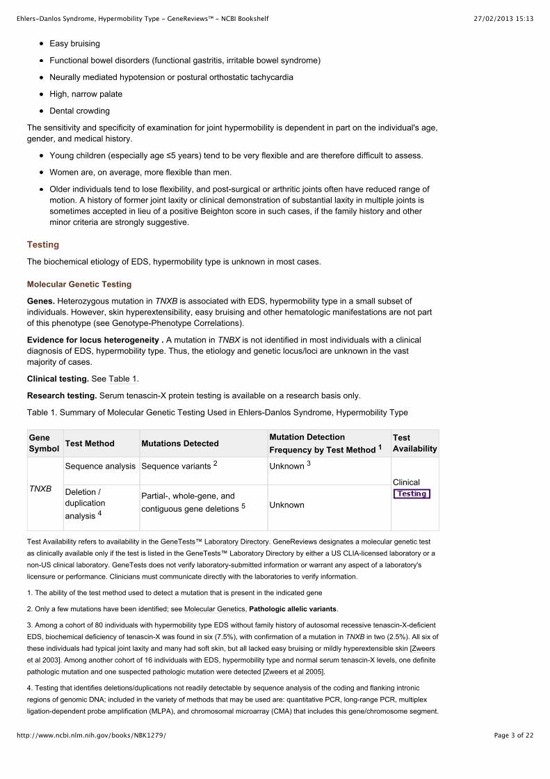
Table 1. Summary of Molecular Genetic Testing Used in Ehlers-Danlos Syndrome, Hypermobility Type
GeneSymbol Test Method Mutations Detected
Mutation DetectionFrequency by Test Method 1
TestAvailability
TNXB
Sequence analysis Sequence variants 2 Unknown 3
ClinicalDeletion /duplicationanalysis 4
Partial-, whole-gene, andcontiguous gene deletions 5 Unknown
Test Availability refers to availability in the GeneTests™ Laboratory Directory. GeneReviews designates a molecular genetic testas clinically available only if the test is listed in the GeneTests™ Laboratory Directory by either a US CLIA-licensed laboratory or anon-US clinical laboratory. GeneTests does not verify laboratory-submitted information or warrant any aspect of a laboratory'slicensure or performance. Clinicians must communicate directly with the laboratories to verify information.
1. The ability of the test method used to detect a mutation that is present in the indicated gene
2. Only a few mutations have been identified; see Molecular Genetics, Pathologic allelic variants.
3. Among a cohort of 80 individuals with hypermobility type EDS without family history of autosomal recessive tenascin-X-deficientEDS, biochemical deficiency of tenascin-X was found in six (7.5%), with confirmation of a mutation in TNXB in two (2.5%). All six ofthese individuals had typical joint laxity and many had soft skin, but all lacked easy bruising or mildly hyperextensible skin [Zweerset al 2003]. Among another cohort of 16 individuals with EDS, hypermobility type and normal serum tenascin-X levels, one definitepathologic mutation and one suspected pathologic mutation were detected [Zweers et al 2005].
4. Testing that identifies deletions/duplications not readily detectable by sequence analysis of the coding and flanking intronicregions of genomic DNA; included in the variety of methods that may be used are: quantitative PCR, long-range PCR, multiplexligation-dependent probe amplification (MLPA), and chromosomal microarray (CMA) that includes this gene/chromosome segment.

27/02/2013 15:13Ehlers-Danlos Syndrome, Hypermobility Type - GeneReviews™ - NCBI Bookshelf
Page 4 of 22http://www.ncbi.nlm.nih.gov/books/NBK1279/
See CMA.
5. Some had congenital adrenal hyperplasia as a result of contiguous gene deletion involving CYP21A2 (see Genetically RelatedDisorders).
Test characteristics. Information on test sensitivity, specificity, and other test characteristics can be found atwww.eurogentest.org [Mayer et al 2012].
Interpretation of test results. For issues to consider in interpretation of sequence analysis results, click here.
Information on specific allelic variants may be available in Molecular Genetics (see Table A. Genes andDatabases and/or Pathologic allelic variants).
Testing Strategy
To confirm/establish the diagnosis in a proband. The diagnosis of EDS, hypermobility type is basedentirely on clinical evaluation and family history (see Clinical Diagnosis). TNXB sequencing or quantitativeserum tenascin-X analysis could be considered in persons with joint laxity in the absence of easy bruising orskin hyperextensibility. Testing of other EDS-associated genes, especially COL3A1, is sometimes useful whenthere is clinical diagnostic uncertainty regarding the specific EDS type (see Differential Diagnosis).
Prenatal diagnosis and preimplantation genetic diagnosis (PGD) for at-risk pregnancies require prioridentification of the disease-causing mutation in the family.
Note: It is the policy of GeneReviews to include in GeneReviews™ chapters any clinical uses of testingavailable from laboratories listed in the GeneTests™ Laboratory Directory; inclusion does not necessarilyreflect the endorsement of such uses by the author(s), editor(s), or reviewer(s).
Genetically Related (Allelic) Disorders
An autosomal recessive form of Ehlers-Danlos syndrome has been described in individuals with tenascin-Xdeficiency resulting from compound heterozygous or homozygous TNXB mutation/deletion [Bristow et al 2005].Clinical features include joint laxity, hyperextensible skin, and easy bruising with normal wound healing andabsence of atrophic scarring. Some, but not all, also have congenital adrenal hyperplasia as a result ofcontiguous gene deletion involving CYP21A2.
Some, but not all, carriers of this autosomal recessive form of EDS have joint laxity and soft skin typical ofEDS, hypermobility type, but do not manifest skin hyperextensibility, easy bruising, or other hematologicmanifestations [Zweers et al 2003].
Clinical Description
Natural History
Ehlers-Danlos syndrome (EDS), hypermobility type is generally considered the least severe type of EDS,although significant complications, primarily musculoskeletal, do occur. Clinical variability is substantial. Mostindividuals who seek medical care are female. Pain and major joint complications are much less commonamong affected males. This bias may result from differences between men and women with respect to painperception and inherent joint stability, as well as the effects of sex hormones [Castori et al 2010b].
Skin
The skin is often soft or velvety and may be mildly hyperextensible.
Piezogenic papules (small herniations of subcutaneous fat through the underlying dermis of the heel occurringonly with weight bearing) are common but rarely painful.
Keratosis pilaris may be more common than in the general population [Castori et al 2010a].
Subcutaneous spheroids and molluscoid pseudotumors are not features of this type.
Clinically significant skin morbidity does not occur, and should prompt consideration of alternative diagnoses.
Musculoskeletal
Joint instability. Joint laxity, instability and excessive joint motion is frequently evident on routine activity,even in the absence of overt subluxation or dislocation.
27/02/2013 15:13Ehlers-Danlos Syndrome, Hypermobility Type - GeneReviews™ - NCBI Bookshelf
Page 5 of 22http://www.ncbi.nlm.nih.gov/books/NBK1279/
Subluxations and dislocations are common. They may occur spontaneously or with minimal traumaand can be acutely painful. Reduction often occurs spontaneously or can be accomplished by theaffected individual or a friend/family member. For most affected individuals, medical intervention for anacute dislocation is not usually necessary, but pain can last for hours or days after an event.
All sites can be involved, including the extremities, vertebral column, costo-vertebral and costo-sternaljoints, clavicular articulations, and temporomandibular joints.
Sprains or twisting of the ankles and buckling or “giving out” of the knees are common.
Iliotibial band syndrome or “snapping hip” is a common symptom, often perceived by the affectedindividual as hip joint instability [Branson et al 2011].
Temporomandibular dysfunction ("TMJ syndrome") is relatively common [Hagberg et al 2004, DeCoster et al 2005], and can be thought of as a specific example of joint degeneration andosteoarthritis.
Females tend to have more substantial laxity than males.
Younger individuals tend to have more substantial laxity than older individuals [Castori et al 2010a].
Tendinitis and bursitis may occur [Rombaut et al 2010b, Rombaut et al 2011a, Rombaut et al 2011b],especially greater trochanteric bursitis in those with iliotibial band syndrome. EDS does not directlycause inflammation, and these problems are likely secondary to joint instability.
Osteoarthritis. Degenerative joint disease occurs at a younger age than in the general population, possiblybecause of chronic joint instability resulting in increased mechanical stress.
Osteoporosis. Bone mineral density in individuals with EDS, hypermobility and classic types may be reducedby up to 0.9 SD compared to healthy controls, even in young adulthood [Dolan et al 1998].
Pain
Chronic pain, distinct from that associated with acute dislocations, is a serious complication of the conditionand can be both physically and psychosocially disabling [Sacheti et al 1997, Hagberg et al 2004, Rombaut etal 2010a, Voermans et al 2010a, Rombaut et al 2011a, Rombaut et al 2011b].
It is variable in age of onset (as early as adolescence or as late as the fifth or sixth decade), number of sites,duration, quality, severity, and response to therapy.
Severity is typically greater than expected based on physical and radiologic examinations.
Severity sometimes correlates with degree of joint instability and with sleep impairment [Voermans et al2010a].
Fatigue and sleep disturbance are frequently associated [Rombaut et al 2010b, Voermans et al 2010a,Voermans et al 2010b, Rombaut et al 2011a, Rombaut et al 2011b, Rombaut et al 2012a]. Affected individualsare often diagnosed with chronic fatigue syndrome, fibromyalgia, depression, hypochondriasis, and/ormalingering prior to recognition of joint laxity and establishment of the correct underlying diagnosis.
Headaches, especially migraine, are common [Rombaut et al 2010b, Rombaut et al 2011a, Rombaut et al2011b]. Cervical muscle tension, temporomandibular dysfunction and stress are some of the likely contributingfactors.
Several recognizable pain syndromes are likely:
Muscular or myofascial pain, localized around or between joints, often described as aching,throbbing, or stiff in quality, may be attributable to myofascial spasm, and palpable spasm with tenderpoints (consistent with fibromyalgia) is often demonstrable, especially in the paravertebral musculature[Castori et al 2010a, Rombaut et al 2010b, Rombaut et al 2011a, Rombaut et al 2011b]. Myofascialspasm possibly occurs in response to chronic joint instability, but this has not been systematicallystudied. Myofascial release often provides temporary relief.
Neuropathic pain, variably described as electric, burning, shooting, numb, tingling, or hot or colddiscomfort, may occur in a radicular or peripheral nerve distribution or may appear to localize to anarea surrounding one or more joints [Camerota et al 2010]. Nerve conduction studies are usually non-diagnostic. Skin biopsy may reveal reduction or absence of small nerve fibers, but may be normal.One hypothesis is that neuropathic pain may result from direct nerve impingement (e.g., by subluxedvertebrae, herniated discs, vertebral osteoarthritis, or peripheral joint subluxations). In addition, theremay be mild-to-moderate nerve compression within areas of myofascial spasm. Neither of these
27/02/2013 15:13Ehlers-Danlos Syndrome, Hypermobility Type - GeneReviews™ - NCBI Bookshelf
Page 6 of 22http://www.ncbi.nlm.nih.gov/books/NBK1279/
possibilities has been evaluated clinically.
Osteoarthritic pain occurs later in life (but earlier than in the general population) and typicallypresents as aching pain in the joints, frequently associated with stiffness. It is often exacerbated bystasis and by resistance and/or highly repetitive activity.
Hematologic
Easy bruising is quite common in all types of EDS, frequently without obvious trauma or injury [Anstey et al1991, De Paepe & Malfait 2004].
Mildly prolonged bleeding, epistaxis, bleeding from the gums (especially after dental extraction) andmenometrorrhagia may also occur.
Bleeding time may or may not be prolonged, but no consistent coagulation factor abnormalities have beenreported [Anstey et al 1991, Mast et al 2009].
Clinically, the hematologic manifestations of EDS mimic von Willebrand disease and respond to similartreatments, but von Willebrand factor and platelet number are almost always normal. It is, however, possiblefor von Willebrand disease, idiopathic thrombocytopenia purpura, or other hemorrhagic conditions to bepresent independent of EDS.
The underlying cause of the hematologic manifestations is unknown.
Capillary fragility and/or impaired soft tissue integrity may play a role [De Paepe & Malfait 2004].
Defects in platelet release or aggregation have been found in some individuals with various types ofEDS [Anstey et al 1991, Yenicesu et al 2000].
Gastrointestinal
Functional bowel disorders are common and underrecognized, affecting 33%-67% of individuals with EDS,hypermobility and classic types [Levy et al 1999, Castori et al 2010a].
Gastroesophageal reflux and gastritis may be symptomatic despite maximal doses of proton pumpinhibitors with additional H2-blockers and acid-neutralizing medications.
Early satiety and delayed gastric emptying may occur and may be exacerbated by opioid (and other)medications.
Irritable bowel syndrome may manifest with diarrhea and/or constipation, associated with abdominalcramping and rectal mucus.
Cardiovascular
Autonomic dysfunction. Many individuals with EDS, hypermobility (and classic) type report atypical chestpain, palpitations at rest or on exertion, and/or orthostatic intolerance with syncope or near syncope [Rowe etal 1999, Gazit et al 2003, Mathias et al 2012]. Holter monitoring usually shows normal sinus rhythm, butsometimes reveals premature atrial complexes or paroxysmal supraventricular tachycardia. Tilt table testingmay reveal neurally mediated hypotension (NMH) and/or postural orthostatic tachycardia syndrome (POTS).
Raynaud syndrome and acrocyanosis occur at an increased frequency, which may be anothermanifestation of autonomic dysfunction [Castori et al 2010a].
Aortic root dilation, usually of a mild degree, occurs in 11%-33% of individuals with EDS, classic andhypermobility types [Wenstrup et al 2002, McDonnell et al 2006, Atzinger et al 2011]. The severity appears tobe much less than occurs in Marfan syndrome, and there is no increased risk of aortic dissection in theabsence of significant dilation. Dilation onset is in childhood and is usually stable over time. It is unlikely toprogress or to develop later in life [Atzinger et al 2011].
Mitral valve prolapse (MVP) was previously considered a manifestation of all types of EDS, but clinicallysignificant MVP has not been confirmed in rigorous evaluations using modern diagnostic criteria [Dolan et al1997, McDonnell et al 2006, Atzinger et al 2011]. It is possible that mild MVP not meeting diagnostic criteria(and therefore not requiring special monitoring or treatment) may also explain some of the atypical chest painand palpitations.
Oral/Dental
High, narrow palate and dental crowding are nonspecific features of most heritable disorders of connective
27/02/2013 15:13Ehlers-Danlos Syndrome, Hypermobility Type - GeneReviews™ - NCBI Bookshelf
Page 7 of 22http://www.ncbi.nlm.nih.gov/books/NBK1279/
tissue. Bifid uvula, submucous cleft palate, and overt cleft palate are not manifestations of EDS, hypermobilitytype, and should prompt consideration of alternative diagnoses (see Differential Diagnosis).
Periodontal disease (friability, gingivitis, gum recession) occurs in some individuals with EDS [Letourneau et al2001, Hagberg et al 2004, De Coster et al 2005, Castori et al 2010a] and is no longer considered a uniquesubtype of EDS [Beighton et al 1998]. The frequency of periodontal manifestations in the hypermobility type isundetermined. De Felice et al [2004] reported an abnormally complex oral microvascular network in 12individuals with classic or hypermobility type EDS; potential correlation of this with periodontal disease has notbeen reported.
Obstetric/Gynecologic
Pregnancy may be complicated by premature rupture of membranes or rapid labor and delivery (<4 hours), butthis is less likely than in the classic type [Volkov et al 2007, Castori et al 2010a].
Joint laxity and pain typically increase throughout gestation, especially in the third trimester, as normally occursduring pregnancy in unaffected women [Volkov et al 2007].
There is no clear advantage to vaginal vs. cesarean delivery. Cesarean delivery may reduce the risk of hipdislocation [Volkov et al 2007, Dutta et al 2011], but carries the same risk for surgical complications as in thegeneral population.
There is no increase in risk for cervical incompetence, and no evidence to support use of prophylactic cerclage[Volkov et al 2007].
No other pregnancy complications are associated with EDS, hypermobility type.
Pelvic prolapse and dyspareunia occur at increased frequency in at least the classic and hypermobility types ofEDS [Mcintosh et al 1995, Carley & Schaffer 2000].
Psychiatric
Psychological dysfunction, psychosocial impairment, and emotional problems are common [Hagberg et al2004, Rombaut et al 2011a].
Specific manifestations may include depression, anxiety, affective disorder, low self-confidence, negativethinking, hopelessness, and desperation [Hagberg et al 2004, Castori et al 2010a, Baeza-Velasco et al 2011,Branson et al 2011, Rombaut et al 2011a].
Fatigue [Voermans et al 2010b] and pain [Rombaut et al 2011a] exacerbate the psychological dysfunction.
Psychological distress exacerbates pain [Baeza-Velasco et al 2011, Branson et al 2011].
Fear of pain and/or joint instability may lead to avoidance behavior and exacerbate dysfunction and disability[Baeza-Velasco et al 2011, Branson et al 2011].
Affected individuals may feel misunderstood, disbelieved, marginalized, and alone [Baeza-Velasco et al 2011].
Resentment, distrust, and hostility may develop between the affected individual/family and the healthcare team(in both directions), adversely affecting the therapeutic relationship [Branson et al 2011].
Ocular
Detailed and systematic evaluation of ocular findings in 44 eyes of 22 individuals with EDS, hypermobility typewas compared to age- and gender matched controls [Gharbiya et al 2012]. Findings included the following:
Subjective and objective measures of xerophthalmia were rare, but more common in EDS,hypermobility type than controls. It is unknown whether this represents an intrinsic feature of EDS orpossibly an indirect association (e.g., a side effect of medication).
Clinically insignificant minor lens opacities were found in 13% of EDS eyes, compared to none amongcontrols.
High myopia (more than -6.0 diopters) and vitreous degeneration were found in 16% of EDS eyes andnone of the controls. There was no difference in frequency of mild or moderate myopia between EDSand control eyes.
There was no significant difference in axial length of the globe between EDS and control eyes.
EDS eyes averaged slightly increased corneal curvature compared to controls, but there was no overtkeratoconus.
27/02/2013 15:13Ehlers-Danlos Syndrome, Hypermobility Type - GeneReviews™ - NCBI Bookshelf
Page 8 of 22http://www.ncbi.nlm.nih.gov/books/NBK1279/
Neurologic & Neuromuscular
Delayed onset and/or resistance to local anesthesia is a frequent complaint [Hakim et al 2005, Castori et al2010a].
Dysautonomia may manifest as functional bowel disorders, cardiovascular autonomic dysfunction, and/orRaynaud syndrome/acrocyanosis.
Poor balance is common, with increased incidence of falls and occasionally fear of falling [Rombaut et al2011c].
Diminished joint position sense has been reported at the knees, but not the shoulders. Vibration sensation isnormal [Rombaut et al 2010a].
It is unclear whether or not weakness is an associated feature. Some studies suggest normal muscle strength[Castori et al 2010a, Rombaut et al 2010b], while others have demonstrated decreased ankle power [Galli et al2011], reduced passive muscle tension and increased Achilles tendon compliance [Rombaut et al 2012b]. Onestudy reported lower extremity weakness, but the findings could also be explained by reduced motor effortsecondary to pain and/or fatigue [Rombaut et al 2012a].
Individuals with EDS, hypermobility type tend to have a slower-than-normal gait with shorter gait length[Cimolin et al 2011, Galli et al 2011, Rombaut et al 2011c].
Kinematic studies are normal at the hips and knees [Galli et al 2011], but the ankles demonstrate excessplantar flexion at ground contact and decreased dorsiflexion during motion [Cimolin et al 2011, Galli et al 2011,Rigoldi et al 2012].
In a series of 2813 individuals with Chiari malformation type 1, 12.7% were felt to also have a hereditarydisorder of connective tissue, including many with EDS, hypermobility type [Milhorat et al 2007]. The incidenceof Chiari malformation among individuals with EDS has not been systematically studied, and the clinicalrelevance of this association is uncertain.
Disability
Functional and psychosocial impairment are common, manifesting with decreased sport-related physicalactivity, diminished health-related quality of life, and significant impact on daily function [Rombaut et al 2010b,Voermans et al 2010a, Rombaut et al 2011a, Rombaut et al 2011b].
Pain, fatigue, and sleep disturbance all may contribute to disability and functional impairment [Rombaut et al2010b, Voermans et al 2010a, Rombaut et al 2011a, Voermans & Knoop 2011].
Other
Fragility of soft tissues with spontaneous ruptures or tears of internal organs is, by definition, not a feature ofEDS, hypermobility type. Such manifestations should prompt consideration of other hereditary connectivetissue disorders (see Differential Diagnosis).
Genotype-Phenotype Correlations
The genetic etiology for most cases is unknown. In the few described individuals with EDS, hypermobility typeresulting from haploinsufficiency of tenascin-X, easy bruisability and mildly hyperextensible skin were not seen[Zweers et al 2003].
Penetrance
Penetrance is believed to be 100%, although expressivity is extremely variable, and careful examination maybe required to demonstrate typical features, especially in adult men who have never experienced a major jointcomplication or significant pain.
Anticipation
Anticipation is not believed to occur.
Nomenclature
The 1997 Villefranche conference [Beighton et al 1998] simplified the classification and nomenclature of theEhlers Danlos syndromes. The former EDS type III was renamed the hypermobility type.
There is disagreement as to whether the "benign familial articular hypermobility syndrome" is identical to EDS,
27/02/2013 15:13Ehlers-Danlos Syndrome, Hypermobility Type - GeneReviews™ - NCBI Bookshelf
Page 9 of 22http://www.ncbi.nlm.nih.gov/books/NBK1279/
hypermobility type or represents a unique condition [Grahame 1999, Tinkle et al 2009]. The distinction is subtleand relates to degree of joint complications and presence or absence of skin manifestations. However, first-degree relatives of probands with hypermobility type EDS often have relatively asymptomatic joint laxity andmild or absent skin manifestations. Therefore, the benign hypermobility syndrome is included as EDS,hypermobility type for this review.
Prevalence
The prevalence of EDS, hypermobility type is unknown. Estimates have ranged between 1:5,000 and1:20,000, and depend in part on whether or not the familial articular hypermobility syndrome is included. Giventhe clinical variability and low probability of affected males being ascertained, the prevalence is likely muchhigher than estimated. EDS, hypermobility type may be the most common heritable disorder of connectivetissue.
Differential DiagnosisFor current information on availability of genetic testing for disorders included in this section, see GeneTestsLaboratory Directory. —ED.
All types of Ehlers-Danlos syndrome (EDS) share some degree of joint laxity and skin/soft tissuemanifestations.
The other forms of EDS are distinguished by additional connective tissue manifestations [Beighton et al 1998]:
EDS, classic type includes skin and soft tissue fragility. Mild presentations of the classic type may bemistaken for the hypermobility type. The diagnosis is sometimes revised from hypermobility to classicwhen the individual or a family member later develops more significant skin and soft tissuemanifestations. Approximately 50% of individuals with classic EDS have an identifiable mutation inCOL5A1 or COL5A2, the two genes encoding type V collagen. The diagnosis is usually established onclinical grounds. Sequence analysis of these genes is available. Demonstration of a pathologic variantconfirms the diagnosis, although failure to identify a mutation does not rule it out.
In EDS, vascular type, the joint laxity is predominantly in small joints, and spontaneous rupture ofhollow organs occurs. Dysfunction and/or deficiency of type III collagen, caused by mutations inCOL3A1, is responsible for all cases of EDS, vascular type. The diagnosis of EDS, vascular type isbased on clinical findings and confirmed by biochemical (protein-based) and/or molecular genetictesting.
EDS, kyphoscoliotic and dermatosparaxis types are autosomal recessive, rare, and distinguishedby more severe skin manifestations and other features. EDS, kyphoscoliotic form is caused bydeficient activity of the enzyme procollagen-lysine, 2-oxoglutarate 5-dioxygenase 1 (PLOD1: lysylhydroxylase 1). The diagnosis of EDS, kyphoscoliotic type can be established by demonstration of anincreased ratio of deoxypyridinoline to pyridinoline crosslinks in urine measured by HPLC, assay oflysyl hydroxylase enzyme activity in skin fibroblasts, or molecular genetic testing of PLOD1.
EDS, arthrochalasia type is autosomal dominant, rare, and distinguished by congenital hipdislocation and more severe skin manifestations. Pathogenic mutations occur in exon 6 of COL1A1and COL1A2, causing impaired cleavage of the amino terminal propeptide of the corresponding type 1procollagen molecules. Clinical testing is available.
Joint laxity is a nonspecific manifestation of dozens of other disorders and syndromes. Some of these aretraditionally thought of as heritable disorders of connective tissue or skeletal dysplasias, but many fall outsidethose general classifications. Most are easily distinguished from EDS by characteristic features and/orinvolvement of systems other than the joints and skin, but mild presentations can sometimes be misdiagnosedas EDS, hypermobility type. Some examples, in order of likelihood and importance, include the following:
Marfan syndrome results in additional skeletal, ocular, cardiovascular, pulmonary, andskin/integument manifestations beyond those seen in EDS. Specific clinical criteria are available toestablish a diagnosis of Marfan syndrome, and it can be confirmed by demonstration of a mutation inFBN1. Joint hypermobility is common in the MASS phenotype (myopia, mitral valve prolapse, mildaortic root dilatation, striae, and minor skeletal manifestations of Marfan syndrome), also caused bymutations in FBN1. Sometimes individuals with hypermobility EDS can have a Marfanoid build and assuch resemble individuals with Marfan syndrome or a Marfan-related disorder. However, application ofthe clinical diagnostic criteria for Marfan syndrome and FBN1 molecular analysis allow differentiationof these conditions.
27/02/2013 15:13Ehlers-Danlos Syndrome, Hypermobility Type - GeneReviews™ - NCBI Bookshelf
Page 10 of 22http://www.ncbi.nlm.nih.gov/books/NBK1279/
Loeys-Dietz syndrome is characterized by multiple arterial aneurysms and tortuosity. Other clinicalfeatures are variable, but may include ocular hypertelorism and bifid uvula. The presentation oftenmimics Marfan syndrome or EDS, vascular type, but prior to detection of the arterial abnormalities,individuals may be misdiagnosed with classic or hypermobility type EDS. The diagnosis is establishedby detection of a mutation in TGFBR1 or TGFBR2.
Stickler syndrome. Distinguishing features include sensorineural hearing loss, vitreoretinalabnormalities, and cleft palate. Mutations affecting one of five genes (COL2A1, COL9A1, COL9A2,COL11A1, and COL11A2) have been associated with Stickler syndrome. However, a few families withfeatures of Stickler syndrome are not linked to any of these loci; thus, mutations in other genes mayalso cause the disorder. Stickler syndrome is diagnosed based on clinical features. In many affectedindividuals and families the diagnosis can be confirmed by molecular genetic testing, although anegative test does not rule out the diagnosis.
Williams syndrome (WS) is a contiguous gene deletion syndrome characterized by cardiovasculardisease (elastin arteriopathy, peripheral pulmonary stenosis, supravalvular aortic stenosis,hypertension), distinctive facies, connective tissue abnormalities, intellectual disability (usually mild), aspecific cognitive profile, unique personality characteristics, growth abnormalities, and endocrineabnormalities (hypercalcemia, hypercalciuria, hypothyroidism, and early puberty). The mainstay fordiagnosis is detection of the contiguous gene deletion of the Williams-Beuren syndrome critical region(WBSCR) that encompasses the elastin gene (ELN). More than 99% of individuals with the clinicaldiagnosis of WS have this contiguous gene deletion, which can be detected using fluorescent in situhybridization (FISH) or targeted mutation analysis. Supravalvular aortic stenosis (SVAS) is caused bymutation (rather than deletion) of ELN. Individuals with either deletion or mutation of ELN have jointlaxity, but the classic elastin arteriopathy is not seen in any type of EDS.
Aarskog-Scott syndrome (faciogenital dysplasia) is an X-linked condition resulting from mutation ofFGD1. The most significant distinguishing feature is shawl scrotum, which may become less obviousin adulthood. Widow's peak, short upturned nose, other dysmorphic features, and the inheritancepattern can be additional diagnostic clues. Intellectual disability, which is not associated with any of thetypes of EDS, is sometimes present.
Fragile X syndrome is not typically confused with EDS, hypermobility type. When a full mutation ofFMR1 is present, fragile X syndrome is characterized by moderate intellectual disability in affectedmales and mild intellectual disability in affected females. Males may have a characteristic appearance(large head, long face, prominent forehead and chin, protruding ears), joint laxity and large testes(post-puberty). However, premutation carriers may have joint laxity and EDS-like skin findings withoutother major manifestations. Family history of intellectual disability, premature ovarian failure and/orfragile X tremor/ataxia syndrome is helpful when present. The frequency of FMR1 premutation amongindividuals diagnosed clinically with EDS, hypermobility type has not been studied, but fragile Xsyndrome has not been reported among offspring of women with EDS, hypermobility type.
Achondroplasia and hypochondroplasia are distinguished by short stature with characteristicskeletal features (marked in achondroplasia, milder in hypochondroplasia). Achondroplasia can bediagnosed by characteristic clinical and radiographic findings in most affected individuals. Moleculargenetic testing reveals a mutation in FGFR3 in 99% of individuals with achondroplasia and about 70%of individuals with hypochondroplasia. However, it is clear that locus heterogeneity exists forhypochondroplasia because mutations in other as-yet unidentified genes can result in similar, if notidentical, phenotypes.
Osteogenesis imperfecta (OI) is distinguished by the presence of fractures and, in some cases,dentinogenesis imperfecta (grey or brown teeth). Biochemical testing (i.e., analysis of the structureand quantity of type I collagen synthesized in vitro by cultured dermal fibroblasts) detectsabnormalities in 98% of individuals with OI type II, about 90% with OI type I, about 84% with OI typeIII, and about 84% with OI type IV. About 90% of individuals with OI types I, II, III, and IV (but nonewith OI types V, VI, and VII) have an identifiable mutation in either COL1A1 or COL1A2.
Aneuploidies, including Down, Turner, and Klinefelter syndromes, are usually easily recognizedbased on dysmorphic features and/or intellectual disability. Small duplications or deletions may be lessclinically obvious, but could be suggested by reduced fertility or recurrent pregnancy loss.
Chronic pain and fatigue are major features of fibromyalgia. A subset of individuals with fibromyalgia and/orchronic fatigue syndrome may have EDS as the underlying etiology.
Note to clinicians: For a patient-specific ‘simultaneous consult’ related to this disorder, go to
27/02/2013 15:13Ehlers-Danlos Syndrome, Hypermobility Type - GeneReviews™ - NCBI Bookshelf
Page 11 of 22http://www.ncbi.nlm.nih.gov/books/NBK1279/
, an interactive diagnostic decision support software tool that provides differentialdiagnoses based on patient findings (registration or institutional access required).
Management
Evaluations Following Initial Diagnosis
To establish the extent of disease in an individual diagnosed with Ehlers-Danlos syndrome (EDS),hypermobility type, the following evaluations are recommended:
Thorough history and physical examination, especially for musculoskeletal, skin, cardiovascular,gastrointestinal, and oral/dental manifestations
Assessment of prior experience with pharmacologic, mechanical, and/or surgical treatment of pain andjoint instability, as well as current degree of pain and disability
Baseline echocardiogram to evaluate aortic root diameter, as adjusted for age and body surface area[Roman et al 1989]. Significant aortic enlargement and/or other cardiac abnormalities should promptconsideration of alternative diagnoses.
The following evaluations should not be routine, but may be appropriate in some situations:
For individuals with significant orthostatic intolerance and/or tachycardia, tilt-table testing to helpconfirm postural orthostatic tachycardia and/or neurally-mediated hypotension. It is sometimesappropriate to rule out adrenocortical insufficiency.
If irritable bowel syndrome is suspected, consideration of formal gastroenterology consultation andpossible endoscopy to rule out other treatable diagnoses. Celiac disease, inflammatory bowel disease,and other causes of malabsorption or bowel dysfunction are not associated with EDS, but may becoexisting diagnoses.
Dual-energy x-ray absorptiometry (DEXA) at any age if height loss greater than one inch isdocumented or x-rays are suggestive of osteopenia. Women should have their first study no later thanmenopause. It is unclear if or at what age men without height loss or abnormal x-rays should have ascreening DEXA.
If a history of severe or prolonged bleeding is present, consideration of hematologic evaluation for vonWillebrand disease, thrombocytopenia, or other bleeding diathesis. Although pathophysiologicallyunrelated, these conditions may coexist with hypermobility type EDS and exacerbate the hematologicmanifestations.
For individuals with significant pain and/or fatigue, screening for and correction of other potentialcauses, including (but not limited to) vitamin D deficiency, vitamin B12 deficiency, folate deficiency,iron deficiency, celiac disease, or hypothyroidism
If there is suspicion of Chiari malformation, consideration of cerebral MRI, possibly with CSF flowstudies
Medical genetics consultation if there is uncertainty about the diagnosis or for assistance in evaluationand management
Treatment of Manifestations
Physical Therapy
Myofascial release (any physical therapy modality that reduces spasm) provides short-term relief of pain,lasting hours to days. While the duration of benefit is short and it must be repeated frequently, this pain reliefmay be critical to facilitate participation in toning exercise for stabilization of the joints. Modalities must betailored to the individual; a partial list includes heat, cold, massage, ultrasound, electrical stimulation,acupuncture, acupressure, biofeedback, and conscious relaxation.
Low-resistance muscle toning exercise can improve joint stability and reduce future subluxations, dislocations,and pain. See Prevention of Primary Manifestations.
Transvaginal pelvic physical therapy and myofascial release (in which massage or ultrasound is applied to thepelvic musculature via a transvaginal approach) may improve dyspareunia, abdominal pain, back pain, andsometimes radicular lower-extremity pain.
27/02/2013 15:13Ehlers-Danlos Syndrome, Hypermobility Type - GeneReviews™ - NCBI Bookshelf
Page 12 of 22http://www.ncbi.nlm.nih.gov/books/NBK1279/
Assistive Devices
Braces are useful to improve joint stability. Orthopedists, rheumatologists, and physical therapists can assist inrecommending appropriate devices for commonly problematic joints such as knees and ankles. Shoulders andhips present more of a challenge for external bracing. Occupational therapists may be consulted for ring splints(to stabilize interphalangeal joints) and wrist or wrist/thumb braces in affected individuals with small jointinstability. A soft neck collar, if tolerated, may help with neck pain and headaches.
A wheelchair or scooter may be necessary to offload stress on lower extremity joints. Special wheelchaircustomizations such as lightweight and/or motorized chairs, seat pads, and specialized wheels and wheelgrasps may be necessary to accommodate pelvic and upper extremity issues. Crutches, canes, and walkersshould be used cautiously as they may put increased stress on the upper extremities.
A waterbed, adjustable air mattress, or viscoelastic foam mattress (and/or pillow) may provide increasedsupport with improved sleep quality and less pain.
Pain Medication
Pain medication is frequently underprescribed, and should be tailored to the individual's subjective symptomsand objective measures of pain, not to physical examination or radiologic findings. Individuals with mild tomoderate pain may get sufficient relief from as-needed use. Those with more significant pain typically requirehigher doses and combinations of multiple medications. Prevention or control of pain with regularly scheduleddosing is often more successful than acute treatment with as-needed dosing. Many clinicians recruit a painmanagement specialist, but pain can be managed by the primary physician if desired.
Note: All of the following dose recommendations are for adults without hepatic or renal disease; adjustmentsmay be necessary for other populations.
Acetaminophen, 4000 mg in three or four divided doses, will not completely alleviate pain but is auseful and well-tolerated adjunct in combination with other agents. Acetaminophen is often present incombination with other analgesic medications and cold/flu preparations, and careful attention shouldbe paid to the total daily dose to avoid exceeding 4000 mg/day.
NSAIDs (nonsteroidal anti-inflammatory drugs) (e.g., ibuprofen, naproxen, meloxicam, nabumetone)should be titrated to the maximum dose or as tolerated by upper gastrointestinal symptoms. NSAIDsare particularly useful for arthralgia, myalgia, and secondary inflammatory conditions (e.g. bursitis,tendinitis, costochodritis, or post-dislocation pain). Bruising is not a contraindication to NSAID therapy,but occasionally requires dose reduction or change to a Cox-2 inhibitor.
Cox-2 inhibitors (e.g. celecoxib) in maximal doses are no stronger than dose-equivalent NSAIDS, butmay be better tolerated and thus more effective.
Tramadol can be added to acetaminophen plus an NSAID or Cox-2 inhibitor before resorting toopioids. Nausea is the most common side effect.
Topical lidocaine as a cream or patch is sometimes useful for localized areas of pain. Topicalcapsaicin is of questionable utility, but is safe.
Skeletal muscle relaxants are useful in combination with all of the above to treat myofascial spasm.They are also sometimes helpful in treating neuropathic pain. Metaxalone may be the least sedating,but all are potentially limited by sedation.
Tricyclic antidepressants are often effective for neuropathic pain, with additional benefits of mildsedation (for those with sleep disturbance) and a little mood elevation. Constipation, a common sideeffect, can be managed with fluids, fiber, stool softeners, and laxatives. For those with diarrhea-predominant irritable bowel syndrome, the constipating effect may be therapeutic. Typical doses arenortriptyline (25-150 mg) or trazadone (50-300 mg) every evening.
Serotonin/norepinephrine receptor inhibitors (SNRIs), such as venlafaxine, desvenlafaxine, duloxetine,and milnacipran also offer combined benefit for depression and neuropathic pain. Venlaxafine mayraise the blood pressure a few points, which potentially could be helpful for individuals with neurallymediated hypotension.
Some anti-seizure medications are also effective for neuropathic pain and can be used in addition totricyclic and/or SNRI antidepressants. All require gradual titration before reaching therapeutic levels.Gabapentin should be titrated as tolerated up to at least 1200 mg three times daily before declaringfailure, but is often limited by sedation and/or gastrointestinal side effects. Pregabalin can be dosedtwice or three times daily up to a total daily dose of at least 300 mg, and tends to be better tolerated
27/02/2013 15:13Ehlers-Danlos Syndrome, Hypermobility Type - GeneReviews™ - NCBI Bookshelf
Page 13 of 22http://www.ncbi.nlm.nih.gov/books/NBK1279/
than gabapentin. Topiramate and lamotrigine have also been used successfully.
Short courses of steroids can be very effective for controlling acute flares of pain associated withsecondary inflammation. EDS is not an intrinsically inflammatory condition, and there is no role forchronic steroid use.
Opioids are effective for both myofascial pain and neuropathic pain, but should be reserved for useafter failing the above medications. They should be administered in conjunction with all of the above,except tramadol, in order to minimize total opioid requirements. Since they are typically usedchronically (or at least several months), the primary formulation should be long acting (e.g., sustained-release oxycodone or morphine or topical fentanyl patch) with short-acting forms of the same drugused as needed for breakthrough pain. Routine use of two or more daily doses of a short-acting formshould prompt an increase in the long-acting dose or another adjustment to the pain regimen.
Supplemental magnesium and/or potassium anecdotally may provide some muscle relaxation andpain relief. Diarrhea, nausea, and sedation are the most common side effects. Specific validated doserecommendations do not exist.
Glucosamine and chondroitin may help to prevent or treat osteoarthritis in the general population.They have not been studied specifically in EDS, but are not contraindicated.
Medication precautions
It is critically important to evaluate all potential sources of acetaminophen and assure that total dailyuse does not exceed 4000 mg.
Abrupt cessation of anti-seizure medications can precipitate a seizure. When discontinuing, theyshould be tapered gradually.
Chronic opioid use can result in dependency with escalating dose requirements and diminishing effect.Narcotic bowel syndrome can also develop, and may be confused with irritable bowel syndrome.
Serotonin syndrome can occur when combining multiple serotonergic medications, such as tramadol,tricyclic antidepressants, SNRI antidepressants, anti-seizure medications, and opiods. Symptoms andsigns may include agitation, restlessness, tremor, hyperreflexia, ataxia, confusion, irritability, nausea,diarrhea, hyperthermia, tachycardia, hyper/hypotension and/or diaphoresis. Severity can range frommild to life-threatening. Many of these manifestations overlap those associated with EDS, makingdiagnosis difficult. Some individuals find mild to moderate serotonergic symptoms acceptable in orderto achieve adequate pain control.
Surgery and Other Procedures
Many individuals will have undergone several orthopedic procedures prior to diagnosis. These often includejoint debridement, tendon relocations, capsulorraphy, and arthroplasty. The degree of stabilization and painreduction, overall patient satisfaction, and duration of improvement are variable, but usually less than inindividuals without EDS [Aldridge et al 2003, Rose et al 2004, Rombaut et al 2011b]. In general, orthopedicsurgery should be delayed in favor of physical therapy and bracing. When surgery is performed, the affectedindividual and physician should cautiously anticipate some improvement but expect less than optimal results.There is one report of long-term improvement in shoulder stability with Achilles tendon allograft reconstructionof the joint capsule in an individual with EDS, hypermobility type [Chaudhury et al 2012]. It is not yet known ifthis approach will be successful in other affected individuals. Unlike the classic and vascular types of EDS, thehypermobility type is not associated with increased risk for perioperative skin and soft tissue complications.
Prolotherapy, in which saline and/or other irritants are injected in tendons or around joints to induce scarformation and increase stability, has not been objectively studied. It is probably safe and probably subject tothe same limitations as orthopedic surgery.
Anesthetic/corticosteroid injections for localized areas of pain and acute inflammation are often helpful, butcannot be repeated indefinitely; "dry needling" without injection of any material sometimes provides similarbenefit.
Anesthetic nerve blocks can provide temporary relief of neuropathic pain. These are sometimes followed bysurgical nerve root destruction and/or implantable stimulators (sensory or motor), with variable results.
Constant intrathecal delivery of anesthetic and/or opioid medication may reduce the need for oral/systemicmedications, but should only be considered as a last resort.
Bone Density
27/02/2013 15:13Ehlers-Danlos Syndrome, Hypermobility Type - GeneReviews™ - NCBI Bookshelf
Page 14 of 22http://www.ncbi.nlm.nih.gov/books/NBK1279/
Therapy is the same as for any other individual with low bone density. Simple weight-bearing exercise, such aswalking or use of an elliptical trainer, should not be overlooked as a means to help maintain bone density aswell as improve resting muscle tone.
Hematologic
Easy and spontaneous bruising does not require treatment, and does not require avoidance of NSAIDs.
For severe bleeding (e.g., epistaxis, menometrorrhagia) or operative prophylaxis, desmopressin acetate(ddAVP) is often beneficial [Stine & Becton 1997, Mast et al 2009].
Gastrointestinal
Gastritis and reflux symptoms may require intensive therapy, including proton pump inhibitor twice daily beforemeals, high-dose H2-blocker at bedtime (e.g., famotidine 20-40 mg or ranitidine 150-300 mg), sucralfate onegram four times daily, and over-the-counter acid-neutralizing agents. Other treatable causes, such as H. pyloriinfection, should be investigated. Upper endoscopy is indicated for resistant symptoms, but frequently isnormal other than chronic gastritis.
Delayed gastric emptying should be identified if present and treated as usual with promotility agents (e.g.,erythromycin, metoclopramide).
Irritable bowel syndrome is treated as usual with antispasmodics, antidiarrheals, and laxatives as needed.Lubiprostone is a motility enhancer that may be helpful for those with constipation only. Tricyclicantidepressants may be especially helpful for persons with both neuropathic pain and diarrhea.
Cardiovascular
Beta-blockade is rarely necessary, but should be considered for progressive aortic enlargement. Rarely,severe enlargement (>4.5-5.0 cm) requires surgical evaluation.
Neurally mediated hypotension and postural orthostatic tachycardia are treated as usual, with avoidance ofsudden postural change, consideration of lower extremity and/or abdominal compression garments, exerciseto increase muscle tone, supplementation of sodium and water to expand the blood volume, and sometimespharmacologic treatment with beta-blockers, midodrine, fludrocortisone, and/or other medications [Mathias etal 2012].
Dental
Orthodontic and palatal corrections may tend to relapse, requiring prolonged use of a retainer.
Periodontal disease should be identified and treated.
Temporomandibular joint laxity and dysfunction are difficult to treat. There are no specific interventions ofproven benefit. Intra-oral devices are sometimes helpful. Oral rest (minimization of chewing and talking), localmyofascial release, and muscle relaxant medications may be beneficial for acute flares. Surgical intervention isoften disappointing and should be considered only as a last resort.
Psychiatric
Validation of the affected individual's symptoms can be immensely helpful, as many with EDS, hypermobilitytype have been accused of malingering or diagnosed with primary psychiatric disorders by previousphysicians.
Establishing trust, rapport and a supportive relationship between patient and provider is important. Emphasisshould be placed on chronic, rather than acute, pain management. Distraction and hypnosis are often helpful[Branson et al 2011].
Depression is a common result of the chronic pain, disability, and other complications. Psychological and/orpain-oriented counseling can improve adaptation to and acceptance of these issues and the necessaryphysical limitations. Cognitive behavioral therapy can be particularly beneficial, but requires active patientparticipation [Baeza-Velasco et al 2011]. Antidepressants are also of great benefit. Many individuals initiallyresist a diagnosis of or therapy for depression because of concern that their problems are being written off aspurely psychiatric.
Consumer support groups are available and can be very beneficial.
Prevention of Primary Manifestations

27/02/2013 15:13Ehlers-Danlos Syndrome, Hypermobility Type - GeneReviews™ - NCBI Bookshelf
Page 15 of 22http://www.ncbi.nlm.nih.gov/books/NBK1279/
Improved joint stability may be achieved by low-resistance exercise to increase muscle tone (subconsciousresting muscle contraction, as opposed to voluntarily recruited muscle strength). Emphasis should be placedon both core and extremity muscle tone. Examples include walking, bicycling, low-impact aerobics, swimmingor water exercise, and simple range-of-motion exercise without added resistance. Core toning activities, suchas balance exercises and repetitive motions focusing on the abdominal, lumbar, and interscapular muscles,are also important. Progress should be made by increasing repetitions, frequency, or duration, not resistance.It often takes months or years for significant progress to be recognized.
Wide grip writing utensils can reduce strain on finger and hand joints. An unconventional grasp of a writingutensil, gently resting the shaft in the web between the thumb and index finger and securing the tip betweenthe distal interphalangeal joints or middle phalanges of the index and third fingers (rather than using the tips ofthe fingers), results in substantially reduced axial stress to the interphalangeal, metacarpophalangeal, andcarpometacarpal joints. These adjustments frequently result in marked reduction of pain in the index finger andat the base of the thumb.
Prevention of Secondary Complications
Calcium (500-600 mg twice daily), vitamin D (400 or more units daily), and low-impact weight bearing exerciseshould be encouraged to maximize bone density.
Surveillance
DEXA should be repeated every other year if bone loss is confirmed.
Annual echocardiography is not necessary in those with a normal initial echocardiogram [Atzinger et al 2011].In children and adolescents with a normal aortic root diameter, it is the author's practice to repeat every two tothree years until young adulthood (age ~25 years). If the aortic root diameter is increased or accelerating fasterthan body surface area, more frequent monitoring is appropriate. In adults with a normal aortic root diameter,no further monitoring is needed.
Agents/Circumstances to Avoid
Joint hyperextension must be avoided. Individuals with EDS, hypermobility type usually need to be educatedabout the normal range of joint extension and cautioned not to exceed it.
Resistance exercise can exacerbate joint instability and pain. Elastic resistance bands should be used withcaution, if at all. In general, it is preferable to increase the number of repetitions of exercise rather than toincrease the resistance.
Isometric exercise can also be problematic if too much force (resistance) is applied.
High-impact activity increases the risk for acute subluxation/dislocation, chronic pain, and osteoarthritis. Somesports, such as football, are therefore contraindicated. However, most sports and activities are acceptable withappropriate precautions.
Chiropractic adjustment is not strictly contraindicated, but must be performed cautiously to avoid iatrogenicsubluxations or dislocations.
Crutches, canes, and walkers should be used cautiously as they may put increased stress on the upperextremities.
Evaluation of Relatives at Risk
All first-degree relatives are at a 50% risk of having EDS, hypermobility type, and may wish to undergo formalclinical assessment. Those without significant clinical manifestations may not benefit directly from knowing thatthey are affected, but may benefit from knowing that their children are at risk. Evaluation of children under ageten years (especially age <5 years) is difficult because of the normal joint laxity in that age group.
See Genetic Counseling for issues related to testing of at-risk relatives for genetic counseling purposes.
Pregnancy Management
Labor and delivery may progress very rapidly. Even in primigravid women, any perception of labor should betaken seriously and she should promptly report to her intended place of delivery.
There is no clear advantage to vaginal vs. cesarean delivery. Cesarean delivery may reduce the risk of hipdislocation [Volkov et al 2007, Dutta et al 2011], but carries the same risk for surgical complications as in thegeneral population.
27/02/2013 15:13Ehlers-Danlos Syndrome, Hypermobility Type - GeneReviews™ - NCBI Bookshelf
Page 16 of 22http://www.ncbi.nlm.nih.gov/books/NBK1279/
There is no increase in risk for cervical incompetence, and no evidence to support use of prophylactic cerclage[Volkov et al 2007].
Pregnant women with known aortic dilation should have an echocardiogram in each trimester.Echocardiography is not needed if the aortic root is normal prior to pregnancy.
Therapies Under Investigation
Search ClinicalTrials.gov for access to information on clinical studies for a wide range of diseases andconditions. Note: There may not be clinical trials for this disorder.
Other
Vitamin C is a cofactor for cross-linking of collagen fibrils. Supplementation with up to 500 mg daily mayimprove some of the manifestations, but this has not been studied. Higher doses are likely excreted andprobably offer no additional clinical benefit.
Losartan is under investigation for treatment and prophylaxis of aortic aneurysm in Marfan syndrome andLoeys-Dietz syndrome. If proven safe and effective, it may be reasonable to use it similarly for individuals withEDS, hypermobility type who have aortic enlargement.
Genetics clinics, staffed by genetics professionals, provide information for individuals and families regardingthe natural history, treatment, mode of inheritance, and genetic risks to other family members as well asinformation about available consumer-oriented resources. See the GeneTests Clinic Directory.
Genetic CounselingGenetic counseling is the process of providing individuals and families with information on the nature,inheritance, and implications of genetic disorders to help them make informed medical and personal decisions.The following section deals with genetic risk assessment and the use of family history and genetic testing toclarify genetic status for family members. This section is not meant to address all personal, cultural, or ethicalissues that individuals may face or to substitute for consultation with a genetics professional. To find a geneticsor prenatal diagnosis clinic, see the GeneTests Clinic Directory.
Mode of Inheritance
Ehlers-Danlos syndrome (EDS), hypermobility type is inherited in an autosomal dominant manner.
Risk to Family Members
Parents of a proband
Most individuals diagnosed with EDS, hypermobility type have an affected parent, although a carefulhistory and examination of the parents is often necessary to recognize that, despite absence ofserious complications, one (and sometimes both) has current or prior history of joint laxity, easybruising, and soft skin.
A proband with EDS, hypermobility type may have the disorder as the result of a de novo genemutation. The proportion of cases caused by de novo mutations is unknown.
Recommendations for the evaluation of parents of a proband with an apparent de novo mutationinclude a careful history and physical examination seeking current or prior history of joint laxity, easybruising, and soft skin.
Note: Although most individuals diagnosed with EDS, hypermobility type have an affected parent, the familyhistory may appear to be negative because of failure to recognize the disorder in family members.
Sibs of a proband. The risk to the sibs of the proband depends on the genetic status of the proband's parents:
If a parent of the proband is affected, the risk to the sibs is 50%.
When the parents are clinically unaffected, the risk to the sibs of a proband appears to be low.
Offspring of a proband. Each child of an individual with EDS, hypermobility type has a 50% chance ofinheriting the same mutation (and thus the same type of EDS). However, because of marked clinical variability,it is difficult to predict severity among affected offspring.
Other family members of a proband. The risk to other family members depends on the genetic status of theproband's parents. If a parent is affected, his or her family members are at risk.
27/02/2013 15:13Ehlers-Danlos Syndrome, Hypermobility Type - GeneReviews™ - NCBI Bookshelf
Page 17 of 22http://www.ncbi.nlm.nih.gov/books/NBK1279/
Related Genetic Counseling Issues
See Management, Evaluation of Relatives at Risk for information on evaluating at-risk relatives for the purposeof early diagnosis and treatment.
It is worthwhile to emphasize to affected individuals and family members that EDS, hypermobility type does notevolve into any of the other types, either in the affected individual or in their offspring, and that thehypermobility type does not confer increased risk for early mortality.
Considerations in families with an apparent de novo mutation. When neither parent of a proband with anautosomal dominant condition has clinical evidence of the disorder, it is likely that the proband has a de novomutation. However, other potential explanations including alternate paternity or maternity (e.g., with assistedreproduction) or undisclosed adoption could also be explored.
Family planning
The optimal time for determination of genetic risk and discussion of the availability of prenatal testingis before pregnancy.
It is appropriate to offer genetic counseling (including discussion of potential risks to offspring andreproductive options) to young adults who are affected or at risk.
DNA banking is the storage of DNA (typically extracted from white blood cells) for possible future use.Because it is likely that testing methodology and our understanding of genes, mutations, and diseases will
improve in the future, consideration should be given to banking DNA of affected individuals. See fora list of laboratories offering DNA banking.
Prenatal Testing
Because the gene(s) and mutation(s) responsible for the majority of cases of Ehlers-Danlos syndrome,hypermobility type have not been identified, prenatal testing for most families is not available.
Prenatal diagnosis for pregnancies at increased risk for EDS, hypermobility type resulting from a TNXBmutation is possible by analysis of DNA extracted from fetal cells obtained by amniocentesis usually performedat approximately 15 to 18 weeks’ gestation or chorionic villus sampling (CVS) at approximately ten to 12weeks’ gestation. The disease-causing mutation of an affected family member must be identified in the familybefore prenatal testing can be performed.
Note: Gestational age is expressed as menstrual weeks calculated either from the first day of the last normalmenstrual period or by ultrasound measurements.
Requests for prenatal testing for conditions which (like Ehlers-Danlos syndrome, hypermobility type) do notaffect intellect and have some treatment available are not common. Differences in perspective may existamong medical professionals and within families regarding the use of prenatal testing, particularly if the testingis being considered for the purpose of pregnancy termination rather than early diagnosis. Although decisionsabout prenatal testing are the choice of the parents, discussion of these issues is appropriate.
Preimplantation genetic diagnosis (PGD) may be available for families in which a TNXB disease-causing
mutation has been identified. For laboratories offering PGD, see .
Note: It is the policy of GeneReviews to include in GeneReviews™ chapters any clinical uses of testingavailable from laboratories listed in the GeneTests™ Laboratory Directory; inclusion does not necessarilyreflect the endorsement of such uses by the author(s), editor(s), or reviewer(s).
ResourcesGeneReviews staff has selected the following disease-specific and/or umbrella support organizations and/orregistries for the benefit of individuals with this disorder and their families. GeneReviews is not responsible forthe information provided by other organizations. For information on selection criteria, click here.
Association Francaise des Syndrome d'Ehlers Danlos
34 rue Léon Joulin
Turns 37 000
France
Email: [email protected]
27/02/2013 15:13Ehlers-Danlos Syndrome, Hypermobility Type - GeneReviews™ - NCBI Bookshelf
Page 18 of 22http://www.ncbi.nlm.nih.gov/books/NBK1279/
www.afsed.com
Canadian Ehlers Danlos Association
88 De Rose Avenue
Bolton Ontario L7E 1A8
Canada
Phone: 905-951-7559
Fax: 905-761-7567
Email: [email protected]
Ehlers-Danlos National Foundation
1760 Old Meadow Road
Suite 500
McLean VA 22102
Phone: 703-506-2892
Email: [email protected]
www.ednf.org
Medline Plus
Ehler-Danlos Syndrome
National Library of Medicine Genetics Home Reference
Ehlers-Danlos syndrome
Ehlers-Danlos Support Group
PO Box 337
Aldershot Surrey GU12 6WZ
United Kingdom
Phone: 01252 690940
Email: [email protected]
www.ehlers-danlos.org
National Registry of Genetically Triggered Thoracic Aortic Aneurysms and CardiovascularConditions (GenTAC)
Phone: 800-334-8571 ext 24640
Email: [email protected]
GenTAC
Molecular GeneticsInformation in the Molecular Genetics and OMIM tables may differ from that elsewhere in the GeneReview:tables may contain more recent information. —ED.
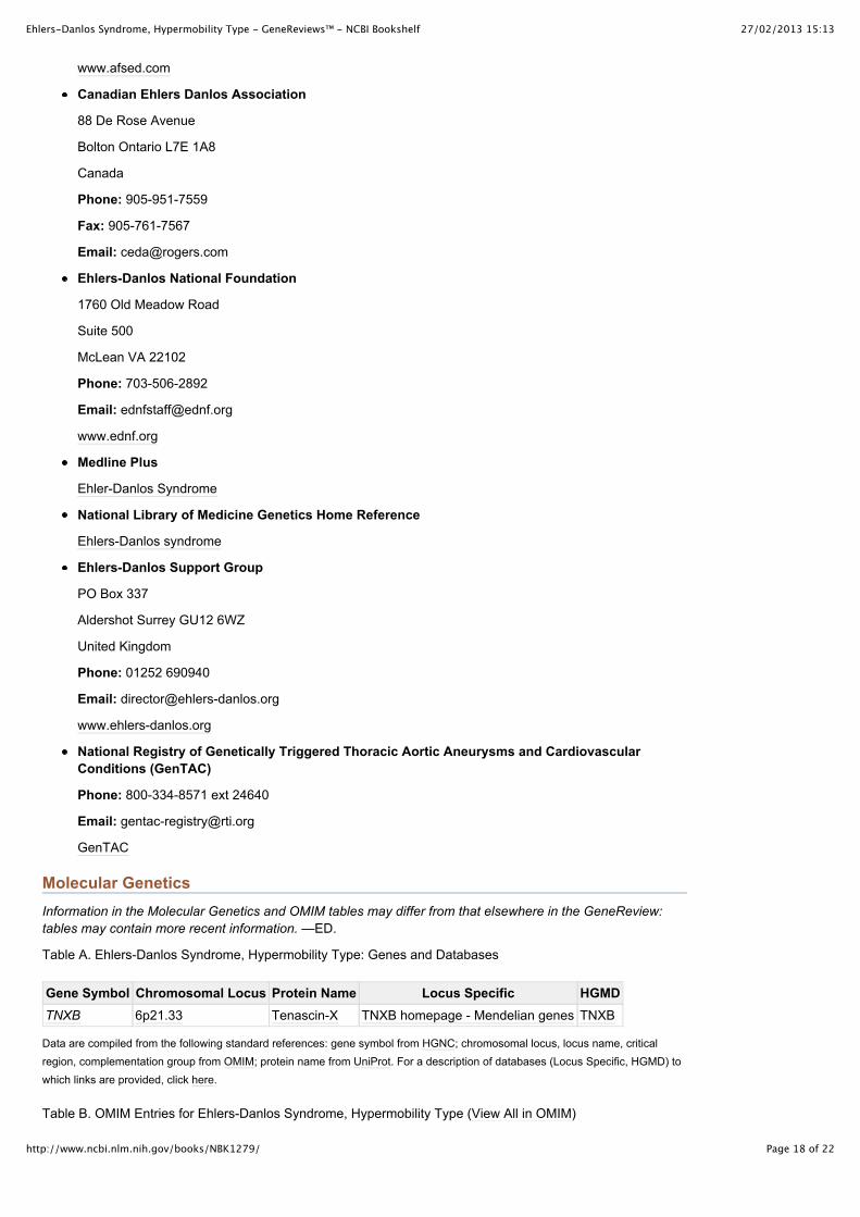
Table A. Ehlers-Danlos Syndrome, Hypermobility Type: Genes and Databases
Gene Symbol Chromosomal Locus Protein Name Locus Specific HGMDTNXB 6p21 .33 Tenascin-X TNXB homepage - Mendelian genes TNXB
Data are compiled from the following standard references: gene symbol from HGNC; chromosomal locus, locus name, criticalregion, complementation group from OMIM; protein name from UniProt. For a description of databases (Locus Specific, HGMD) towhich links are provided, click here.
Table B. OMIM Entries for Ehlers-Danlos Syndrome, Hypermobility Type (View All in OMIM)
27/02/2013 15:13Ehlers-Danlos Syndrome, Hypermobility Type - GeneReviews™ - NCBI Bookshelf
Page 19 of 22http://www.ncbi.nlm.nih.gov/books/NBK1279/
130020 EHLERS-DANLOS SYNDROME, TYPE III
600985 TENASCIN XB; TNXB
Molecular Genetic Pathogenesis
A very small number of individuals with Ehlers-Danlos syndrome (EDS), hypermobility type have a mutation inone allele of TNXB. In most cases the gene(s) in which mutation is causative are unknown.
Normal allelic variants. TNXB is a 39-exon gene spanning 65 kb. It lies in an antisense orientation to andoverlapping the final exon of the steroid 21-hydroxylase gene (CYP21A2), and the entire complex is tandemlyduplicated as a pseudogene (TNXA and CYP21A1P) that predisposes to gene conversion and/or deletionevents. See 21-Hydroxylase-Deficient Congenital Adrenal Hyperplasia and OMIM 600985 for more detail.
Pathologic allelic variants. Three heterozygous mutations resulting in biochemical tenascin-Xhaploinsufficiency have been described: a large (30-kb) deletion, a 2-bp deletion in exon 8 resulting in apremature stop codon, and a 2-bp insertion in exon 3 resulting in a premature stop codon [Zweers et al 2003].In addition, two heterozygous missense mutations have been identified in individuals with EDS, hypermobilitytype and normal tenascin-X serum levels: p.Arg29Trp and p.Val1195Met [Zweers et al 2005].
Normal gene product. Tenascin-X is an extracellular matrix (ECM) glycoprotein that interacts with multipleECM and cell membrane molecules and appears to be important in maintaining connective tissue integrity andelasticity. It is believed to directly or indirectly regulate the rate of production and/or assembly of fibrillarcollagens [Zhuang et at 2010, Hendriks et al 2012].
Abnormal gene product. The p.Arg29Trp mutation has not been studied biochemically but is predicted to bepathogenic. The p.Val1195Met mutation is associated with altered dermal elastic fiber length [Zweers et al2005]. This mutation has been shown to decrease flexibility of the seventh fibronectin type III domain oftenascin-X, which is postulated to affect its interaction with other ECM molecules [Zhuang et al 2010].
ReferencesMedical Genetic Searches: A specialized PubMed search designed for clinicians that is located on the PubMedClinical Queries page
Literature Cited
1. Aldridge JM 3rd, Perry JJ, Osbahr DC, Speer KP. Thermal capsulorraphy of bilateral glenohumeraljoints in a pediatric patient with Ehlers-Danlos syndrome. Arthroscopy. 2003;19:E41. [PubMed:12724665]
2. Anstey A, Mayne K, Winter M, Van de Pette J, Pope FM. Platelet and coagulation studies in Ehlers-Danlos syndrome. Br J Dermatol. 1991;125:155–63. [PubMed: 1911298]
3. Atzinger CL, Meyer RA, Khoury PR, Gao Z, Tinkle BT. Cross-sectional and longitudinal assessment ofaortic root dilation and valvular anomalies in hypermobile and classic Ehlers-Danlos syndrome. JPediatr. 2011;158:826–830. [PubMed: 21193204]
4. Baeza-Velasco C, Gély-Nargeot MC, Bulbena Vilarrasa A, Bravo JF. Joint hypermobility syndrome:problems that require psychological intervention. Rheumatol Int. 2011;31:1131–6. [PubMed:21373784]
5. Beighton P, De Paepe A, Steinmann B, Tsipouras P, Wenstrup RJ. Ehlers-Danlos syndromes: revisednosology, Villefranche, 1997. Ehlers-Danlos National Foundation (USA) and Ehlers-Danlos SupportGroup (UK). Am J Med Genet. 1998;77:31–7. [PubMed: 9557891]
6. Beighton P, Solomon L, Soskolne CL. Articular mobility in an African population. Ann Rheum Dis.1973;32:413–8. [PMC free article: PMC1006136] [PubMed: 4751776]
7. Branson JA, Kozlowska K, Kaczynski KJ, Roesler TA. Managing chronic pain in a young adolescentgirl with Ehlers-Danlos syndrome. Harv Rev Psychiatry. 2011;19:259–70. [PubMed: 21916827]
8. Bristow J, Carey W, Egging D, Schalkwijk J. Tenascin-X, collagen, elastin, and the Ehlers-Danlossyndrome. Am J Med Genet C Semin Med Genet. 2005;139C:24–30. [PubMed: 16278880]
9. Camerota F, Celletti C, Castori M, Grammatico P, Padua L. Neuropathic pain is a common feature inEhlers-Danlos syndrome. J Pain Symptom Manage. 2010 [PubMed: 21145199]
10. Carley ME, Schaffer J. Urinary incontinence and pelvic organ prolapse in women with Marfan orEhlers Danlos syndrome. Am J Obstet Gynecol. 2000;182:1021–3. [PubMed: 10819815]
11. Castori M, Camerota F, Celletti C, Danese C, Santilli V, Saraceni VM, Grammatico P. Natural historyand manifestations of the hypermobility type Ehlers-Danlos syndrome: a pilot study on 21 patients. AmJ Med Genet A. 2010a;152A:556–64. [PubMed: 20140961]
27/02/2013 15:13Ehlers-Danlos Syndrome, Hypermobility Type - GeneReviews™ - NCBI Bookshelf
Page 20 of 22http://www.ncbi.nlm.nih.gov/books/NBK1279/
12. Castori M, Camerota F, Celletti C, Grammatico P, Padua L. Ehlers-Danlos syndrome hypermobilitytype and the excess of affected females: possible mechanisms and perspectives. Am J Med Genet A.2010b;152A:2406–8. [PubMed: 20684008]
13. Chaudhury S, Gasinu S, Rodeo SA. Bilateral anterior and posterior glenohumeral stabilization usingAchilles tendon allograft augmentation in a patient with Ehlers-Danlos syndrome. J Shoulder ElbowSurg. 2012;21:e1–5. [PubMed: 22326542]
14. Cimolin V, Galli M, Vismara L, Grugni G, Camerota F, Celletti C, Albertini G, Rigoldi C, Capodaglio P.Gait pattern in two rare genetic conditions characterized by muscular hypotonia: Ehlers-Danlos andPrader-Willi syndrome. Res Dev Disabil. 2011;32:1722–8. [PubMed: 21454046]
15. De Coster PJ, Martens LC, De Paepe A. Oral health in prevalent types of Ehlers-Danlos syndromes. JOral Pathol Med. 2005;34:298–307. [PubMed: 15817074]
16. De Felice C, Bianciardi G, Dileo L, Latini G, Parrini S. Abnormal oral vascular network geometriccomplexity in Ehlers-Danlos syndrome. Oral Surg Oral Med Oral Pathol Oral Radiol Endod.2004;98:429–34. [PubMed: 15472658]
17. De Paepe A, Malfait F. Bleeding and bruising in patients with Ehlers-Danlos syndrome and othercollagen vascular disorders. Br J Haematol. 2004;127:491–500. [PubMed: 15566352]
18. Dolan AL, Arden NK, Grahame R, Spector TD. Assessment of bone in Ehlers Danlos syndrome byultrasound and densitometry. Ann Rheum Dis. 1998;57:630–3. [PMC free article: PMC1752482][PubMed: 9893576]
19. Dolan AL, Mishra MB, Chambers JB, Grahame R. Clinical and echocardiographic survey of theEhlers-Danlos syndrome. Br J Rheumatol. 1997;36:459–62. [PubMed: 9159540]
20. Dutta I, Wilson H, Oteri O. Pregnancy and delivery in ehlers-danlos syndrome (hypermobility type):review of the literature. Obstet Gynecol Int. 2011;2011:306413. [PMC free article: PMC3134995][PubMed: 21765833]
21. Galli M, Cimolin V, Rigoldi C, Castori M, Celletti C, Albertini G, Camerota F. Gait strategy in patientswith Ehlers-Danlos syndrome hypermobility type: a kinematic and kinetic evaluation using 3D gaitanalysis. Res Dev Disabil. 2011;32:1663–8. [PubMed: 21420276]
22. Gazit Y, Nahir AM, Grahame R, Jacob G. Dysautonomia in the joint hypermobility syndrome. Am JMed. 2003;115:33–40. [PubMed: 12867232]
23. Gharbiya M, Moramarco A, Castori M, Parisi F, Celletti C, Marenco M, Mariani I, Grammatico P,Camerota F. Ocular features in joint hypermobility syndrome/Ehlers-Danlos syndrome hypermobilitytype: a clinical and in vivo confocal microscopy Study. Am J Ophthalmol. 2012;154:593–600.[PubMed: 22633352]
24. Grahame R. Joint hypermobility and genetic collagen disorders: are they related? Arch Dis Child.1999;80:188–91. [PMC free article: PMC1717821] [PubMed: 10325741]
25. Hagberg C, Berglund B, Korpe L, Andersson-Norinder J. Ehlers-Danlos Syndrome (EDS) focusing onoral symptoms: a questionnaire study. Orthod Craniofac Res. 2004;7:178–85. [PubMed: 15359504]
26. Hakim AJ, Grahame R, Norris P, Hopper C. Local anaesthetic failure in joint hypermobility syndrome.J R Soc Med. 2005;98:84–5. [PMC free article: PMC1079398] [PubMed: 15684369]
27. Hendriks AG, Voermans NC, Schalkwijk J, Hamel BC, van Rossum MM. Well-defined clinicalpresentation of Ehlers-Danlos syndrome in patients with tenascin-X deficiency: a report of four cases.Clin Dysmorphol. 2012;21:15–8. [PubMed: 21959861]
28. Letourneau Y, Perusse R, Buithieu H. Oral manifestations of Ehlers-Danlos syndrome. J Can DentAssoc. 2001;67:330–4. [PubMed: 11450296]
29. Levy HP, Mayoral W, Collier K, Tio TL, Fracomano CA. Gastroesophageal reflux and irritable bowelsyndrome in classical and hypermobile Ehlers Danlos syndrome (EDS). Am J Hum Genet.1999;65:A69.
30. Mathias CJ, Low DA, Iodice V, Owens AP, Kirbis M, Grahame R. Postural tachycardia syndrome--current experience and concepts. Nat Rev Neurol. 2011;8(1):22–34. [PubMed: 22143364]
31. Mast KJ, Nunes ME, Ruymann FB, Kerlin BA. Desmopressin responsiveness in children with Ehlers-Danlos syndrome associated bleeding symptoms. Br J Haematol. 2009;144:230–3. [PubMed:19036109]
32. Mayer K, Kennerknecht I, Steinmann B. Clinical utility gene card for: Ehlers–Danlos syndrome types I–VII. Eur J Hum Genet. 2012 [PMC free article: PMC3533317] [PubMed: 22892533]
33. McDonnell NB, Gorman BL, Mandel KW, Schurman SH, Assanah-Carroll A, Mayer SA, Najjar SS,Francomano CA. Echocardiographic findings in classical and hypermobile Ehlers-Danlos syndromes.Am J Med Genet A. 2006;140:129–36. [PubMed: 16353246]
34. McIntosh LJ, Mallett VT, Frahm JD, Richardson DA, Evans MI. Gynecologic disorders in women withEhlers-Danlos syndrome. J Soc Gynecol Investig. 1995;2:559–64. [PubMed: 9420859]
35. Milhorat TH, Bolognese PA, Nishikawa M, McDonnell NB, Francomano CA. Syndrome ofoccipitoatlantoaxial hypermobility, cranial settling, and chiari malformation type I in patients with
27/02/2013 15:13Ehlers-Danlos Syndrome, Hypermobility Type - GeneReviews™ - NCBI Bookshelf
Page 21 of 22http://www.ncbi.nlm.nih.gov/books/NBK1279/
hereditary disorders of connective tissue. J Neurosurg Spine. 2007;7:601–9. [PubMed: 18074684]36. Rigoldi C, Galli M, Cimolin V, Camerota F, Celletti C, Tenore N, Albertini G. Gait strategy in patients
with Ehlers-Danlos syndrome hypermobility type and Down syndrome. Res Dev Disabil.2012;33:1437–42. [PubMed: 22522202]
37. Roman MJ, Devereux RB, Kramer-Fox R, O'Loughlin J. Two-dimensional echocardiographic aorticroot dimensions in normal children and adults. Am J Cardiol. 1989;64:507–12. [PubMed: 2773795]
38. Rombaut L, De Paepe A, Malfait F, Cools A, Calders P. Joint position sense and vibratory perceptionsense in patients with Ehlers-Danlos syndrome type III (hypermobility type). Clin Rheumatol.2010a;29:289–95. [PubMed: 19937459]
39. Rombaut L, Malfait F, Cools A, De Paepe A, Calders P. Musculoskeletal complaints, physical activityand health-related quality of life among patients with the Ehlers-Danlos syndrome hypermobility type.Disabil Rehabil. 2010b;32:1339–45. [PubMed: 20156051]
40. Rombaut L, Malfait F, De Paepe A, Rimbaut S, Verbruggen G, De Wandele I, Calders P. Impairmentand impact of pain in female patients with Ehlers-Danlos syndrome: a comparative study withfibromyalgia and rheumatoid arthritis. Arthritis Rheum. 2011a;63:1979–87. [PubMed: 21391202]
41. Rombaut L, Malfait F, De Wandele I, Cools A, Thijs Y, De Paepe A, Calders P. Medication, surgery,and physiotherapy among patients with the hypermobility type of Ehlers-Danlos syndrome. Arch PhysMed Rehabil. 2011b;92:1106–12. [PubMed: 21636074]
42. Rombaut L, Malfait F, De Wandele I, Mahieu N, Thijs Y, Segers P, De Paepe A, Calders P. Muscle-tendon tissue properties in the hypermobility type of Ehlers-Danlos syndrome. Arthritis Care Res(Hoboken). 2012b;64:766–72. [PubMed: 22232076]
43. Rombaut L, Malfait F, De Wandele I, Taes Y, Thijs Y, De Paepe A, Calders P (2012a) Muscle mass,muscle strength, functional performance,and physical impairment in women with the hypermobilitytype of Ehlers-Danlos syndrome. Arthritis Care Res (Hoboken). doi: 10.1002/acr.21726.
44. Rombaut L, Malfait F, De Wandele I, Thijs Y, Palmans T, De Paepe A, Calders P. Balance, gait, falls,and fear of falling in women with the hypermobility type of Ehlers-Danlos syndrome. Arthritis Care Res(Hoboken). 2011c;63:1432–9. [PubMed: 21793229]
45. Rose PS, Johnson CA, Hungerford DS, McFarland EG. Total knee arthroplasty in Ehlers-Danlossyndrome. J Arthroplasty. 2004;19:190–6. [PubMed: 14973862]
46. Rowe PC, Barron DF, Calkins H, Maumenee IH, Tong PY, Geraghty MT. Orthostatic intolerance andchronic fatigue syndrome associated with Ehlers-Danlos syndrome. J Pediatr. 1999;135:494–9.[PubMed: 10518084]
47. Sacheti A, Szemere J, Bernstein B, Tafas T, Schechter N, Tsipouras P. Chronic pain is amanifestation of the Ehlers-Danlos syndrome. J Pain Symptom Manage. 1997;14:88–93. [PubMed:9262038]
48. Stine KC, Becton DL. DDAVP therapy controls bleeding in Ehlers-Danlos syndrome. J PediatrHematol Oncol. 1997;19:156–8. [PubMed: 9149748]
49. Tinkle BT, Bird HA, Grahame R, Lavallee M, Levy HP, Sillence D. The lack of clinical distinctionbetween the hypermobility type of Ehlers-Danlos syndrome and the joint hypermobility syndrome(a.k.a. hypermobility syndrome). Am J Med Genet A. 2009;149A:2368–70. [PubMed: 19842204]
50. Voermans NC, Knoop H. Both pain and fatigue are important possible determinants of disability inpatients with the Ehlers-Danlos syndrome hypermobility type. Disabil Rehabil. 2011;33:706–7.[PubMed: 21077749]
51. Voermans NC, Knoop H, Bleijenberg G, van Engelen BG. Pain in ehlers-danlos syndrome is common,severe, and associated with functional impairment. J Pain Symptom Manage. 2010a;40:370–8.[PubMed: 20579833]
52. Voermans NC, Knoop H, van de Kamp N, Hamel BC, Bleijenberg G, van Engelen BG. Fatigue is afrequent and clinically relevant problem in Ehlers-Danlos syndrome. Semin Arthritis Rheum.2010b;40:267–74. [PubMed: 19878973]
53. Volkov N, Nisenblat V, Ohel G, Gonen R. Ehlers-Danlos syndrome: insights on obstetric aspects.Obstet Gynecol Surv. 2007;62:51–7. [PubMed: 17176488]
54. Wenstrup RJ, Meyer RA, Lyle JS, Hoechstetter L, Rose PS, Levy HP, Francomano CA. Prevalence ofaortic root dilation in the Ehlers-Danlos syndrome. Genet Med. 2002;4:112–7. [PubMed: 12180144]
55. Yenicesu I, Uçkan D, Soysal A, Büyükaşik Y, Gümrük F. Platelet release defect in a child with Ehlers-Danlos syndrome. Pediatr Hematol Oncol. 2000;17:193–4. [PubMed: 10734665]
56. Zhuang S, Linhananta A, Li H. Phenotypic effects of Ehlers–Danlos syndrome-associated mutation onthe FnIII domain of tenascin-X. Protein Science. 2010;19:2231–9. [PMC free article: PMC3005793][PubMed: 20853426]
57. Zweers MC, Bristow J, Steijlen PM, Dean WB, Hamel BC, Otero M, Kucharekova M, Boezeman JB,Schalkwijk J. Haploinsufficiency of TNXB is associated with hypermobility type of Ehlers-Danlossyndrome. Am J Hum Genet. 2003;73:214–7. [PMC free article: PMC1180584] [PubMed: 12865992]
27/02/2013 15:13Ehlers-Danlos Syndrome, Hypermobility Type - GeneReviews™ - NCBI Bookshelf
Page 22 of 22http://www.ncbi.nlm.nih.gov/books/NBK1279/
58. Zweers M, Dean W, Van Kuppevelt T, Bristow J, Schalkwijk J. Elastic fiber abnormalities inhypermobility type Ehlers–Danlos syndrome patients with tenascin-X mutations. Clinical Genetics.2005;67:330–4. [PubMed: 15733269]
Suggested Reading
1. Byers PH. Disorders of collagen biosynthesis and structure. In: Scriver CR, Beaudet AL, Sly WS, ValleD, Vogelstein B, eds. The Metabolic and Molecular Bases of Inherited Disease (OMMBID). Chap 205.New York, NY: McGraw-Hill. Available at www .ommbid.com. Accessed 9-10-12.
Chapter Notes
Revision History
13 September 2012 (me) Comprehensive update posted live
14 December 2010 (cd) Revision: sequence analysis of the entire coding region available on a limitedbasis for TNXB mutations
27 April 2010 (me) Comprehensive update posted live
1 May 2007 (me) Comprehensive update posted to live Web site
22 October 2004 (me) Review posted to live Web site
1 June 2004 (hpl) Original submission
Copyright © 1993-2013, University of Washington, Seattle. All rights reserved.