Pathophysiology and management of reperfusion injury and ...

This document is downloaded at: 2019-04-11T07:13:14Z
Title Efficancy of a Rinse Solution for Prevention of Reperfusion Injury inCanine 24-hour Cold Preserved Lungs
Author(s) Shingu, Hiroshi
Citation Acta medica Nagasakiensia. 1994, 39(1-3), p.119-125
Issue Date 1994-10-25
URL http://hdl.handle.net/10069/15983
Right
NAOSITE: Nagasaki University's Academic Output SITE
http://naosite.lb.nagasaki-u.ac.jp
Acta Med. Nagasaki 39:119-125
Efficancy of a Rinse Solution for Prevention of Reperfusion Injury
in Canine 24-hour Cold Preserved Lungs
Hiroshi SHINGU
The First Department of Surgery, Nagasaki University School of Medicine
The efficacy of a rinse solution for prevention of ischemia-reperfusion injury in the lung preserved for 24 hours was evaluated by using the canine isolated lung perfusion model. The heart and the lungs were harvested and preserved for 24hours at 4-6℃ in modified:Euro-Collins(EC)solution.
Prostaglandin El (100 p g) was adiministered into the pul-monary trunk prior to flushing canine lungs with preserva-tion solution. The next day, left lungs were isolated with a
perfluorochemical (FC-43) or Carolina rinse II solution. Immediately after rinsing, the lung was reperfused with homologous venous blood (400 ml) for 120 min. in the experi-mental pump circulation system. The lungs were classified into the following six groups: Group 1 (n = 5), without rinsing (control group), Group 2 (n = 6) , rinsing with oxygenated FC-43 solution (250 ml), Group 3 (n = 4), rinsing with non-oxygenated FC-43 solu-tion (250 ml), Group 4 (n = 5), rinsing with pluronic F 68 solution (250 ml), Group 5 (n = 4), rinsing with the Carolina Rinse Solution a (250 ml) and Group 6 (n = 4). rinsing with the Carolina Rinse Solution 11 (250 ml) in addition with hydroxyethyl starch (50 g/l). Pulmonary vascular resistance and lung water volume (wet-dry/wet ratio) were lower and dynamic lung compliance was higher in Group 2 than in other groups. The tissue myeloperioxidase level in Group 2 was significantly lower than those in Groups 4, 5 and 6 (p < 0.05). Histologically, severe marked lung edema was observed in Groups 1, 5 and 6. The lung showed almost normal architec-ture in Group 2. In conclusion, terminal rinsing with oxygen-ated FC-43 solution immediatly before blood reperfusion is useful for prevention of ischemia-reperfusion injury after 24-hour cold ischemic storage in modified Euro-Collins solution.
Introduction
In lung preservation, ischemia-reperfusion injury is dependent on preservation time and preservation solution. Locke et al.') reported the successful 6-hour canine lung
preservation using Euro-Collins solution confirmed the superiority to topical cooling alone, and then Euro-Collins has become the standard pulmonary flush solution in
practice of clinical lung and heart-lung transplantation'). Recently many investigators has studied how the storage interval of the lung preservation could be extended. The
duration of lung allograft preservation has been length-ened but the storage intervals of at least 12 hours are now regarded as a safe limit of lung preservation' 5>
FC-43 solution, one of perfluorochemicals, is an artifi-cial blood substitute which has a high-oxygen-carrying capacity. Further studies have revealed that the solution had an anti-neutrophilic actions and kept microvascular
permeability and resulted in limiting myocardial reper-fusion injury in the experimental models'").
Carolina rinse solution was developed in 1990 to mini-mize reperfusion injury in the liver transplantation') and improved graft function after transplatation. Carolina rinse solution II was a modified solution reducing some components of carolina rinse solution to establish more adequate effects.
In this study, we evaluated the efficacy of a rinse
solution for ischemia-reperfusion injury of canine 24-hour cold ischemic storaged lungs using the canine isolated lung
perfusion model.
Materials and methods
(1) Experimental animals
Twenty-eight adult mongrel dogs weighting 7 to 12 kg, were prepared and provided from the Laboratory Animal Center for Biochemical Research of Nagasaki University School of Medicine.
All animals recieved human care in compliance with the "Guide for the Care and Use of Laboratory Animals of
Nagasaki University". After overnight fast, the animals
were premedicated with intramuscular ketamine hydro-chloride at 10 mg/kg and atropine sulfate at 0.03 mg/kg, and anesthetized with intravenous administration of
sodium pentobarbital (25mg/kg). The animals were intubated with an endotracheal tube and ventilated at a fixed FIO2 of 1.0, a tidal volume (TV) of 35 ml/kg and the respiratory rate (RR) of 14 breaths/min using volume-cycled ventilator (Harvard-animal-ventilator). Median sternotomy was performed, and the both subclavian arteries, the innominate artery and the superior vena cava

were exposed and looped. The right main bronchus was clamped, a tidal volume and ventilatory rate were de-creased to 20 ml/kg and 10 breath/min, and then the airway pressure was measured. 500 units/kg of heparin sodium was injected intravenously and 100 microgram of the prostaglandin E 1 was adiministered through the main
pulmonary artery. A silastic 24 Fr. catheter was inserted through the pulmonary trunk. After ligation of the left subclavian artery, the innominate artery and the superior vena cava, both lungs were flushed with 500 ml cold EC solution at the pressure of 10 mmHg through the catheter
placed in the pulmonary trunk. During the flushing, ventilation was continued. The left atrium was opened to
permit drainage of the perfusate. Immediately after the flushing, the heart and the lungs were harvested and immersed in a sterile vinyl bag filled with the same cold storage solution keeping these distensibility at their inflation of 80 % endotidal volume and stored at 4-6°C for 24 hours in a refrigerator.
The animals were classified into the following six groups according to a rinse solution.
Group 1 (n = 5): not rinsed (control group). Group 2 (n = 6): rinsed with oxgenated perfluoro-
carbon emulsion (FC-43).
Group 3 (n = 4): rinsed with non-oxgenated FC-43. Group 4 (n = 5) : rinsed with Pluronic F68 solution.
Group 5 (n = 4): rinsed with the Carolina Rinse Solution II.
Group 6 (n = 4): rinsed with the Carolina Rinse Solution II containing with HES
(50 g/L). Oxygen pressure of oxygenated and non-oxygenated
FC-43 was 217 ±28 mmHg and 95 ±5 mmHg, respectively. Rinse solutions were warmed at 37°C.
(2) Oxygenation of FC-43
The FC-43 emulsion was mixed in the Erlenmeyer flask and oxygen (5L/min) was blown on the surface of FC-43 emulsion in the Erlenmeyer flask for over 10 minites. The circulation tube was put into the flask and filled with the emulsion, and then rinsing was performed. The non-oxygenated FC-43 was maintained in a sealed vinyl chlo-ride pack without air.
(3) Procedure of rinse and reperfusion
After hypothermic preservation for 24 hours, the left lung was devided. A pulmonary arterial cannula was
connected to the reservior filled with rinse solution which was warmed at 37°C, and pulmonary vein was left open allowing lung perfusate to drain freely into a waste bottle. The isolated left lung was rinsed with 250 mL solution by a roller pump at a flow rate of 10 mL/kg/min for 10 minites. The flow rate was gradually increased for the
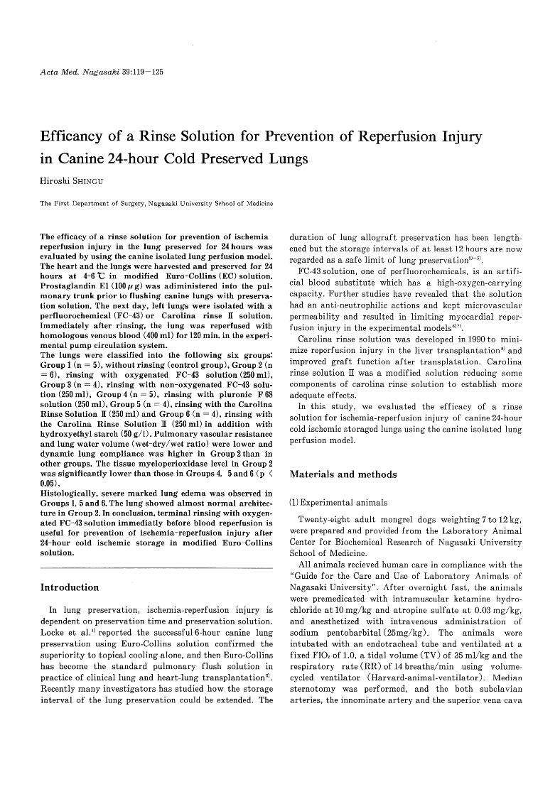
Fig. 1. Isolated lung perfusion model
(PAP: pulmonary artery pressure, AWP: air way pressure)
initial 2 minutes. During rinsing, the lung was ventirated with room air at a tidal volume of 20 mL/kg and at a rate of 10 breath/min, and the same ventilating condition was continued through the subsequential blood reperfusion. After rinsing, the isolated left lung was reperfused with allogenous blood for 120 minutes (Fig.1). The pulmonary artery cannula was connected to the reservior containing allogeneous blood, and the lung perfusate from the pulmo-nary vein was drained into the reservoir.
(4) Monitoring of isolated lung function
The lung perfusate for the initial 5 minutes was collected
and blood gas analysis was performed. When reperfusion was finished, the lungs were reperf used with 100 mL of the second allogeneous venuos blood and blood gas analysis was performed. During the reperfusion, blood samples were taken from the pulmonary vein for the blood gas analysys and count of blood red cells, leukocytes and
platlets at 5, 10, 30, 60 and 120 min. The pulmonary arterial pressure and the airway pressure were monitored, the dynamic and stastic lung compliance and the pulmo-nary vascular resistance were calculated; the dynamic lung compliance: tidal volume/pressure (mL/cmH2O) at end-inspiratory plateau, the static lung compliance: tidal
volume/pressure (mL/cmH2 O) at 1.4 sec of the end-inspiratory plateau, pulmonary vascular resistance:
pulmonary artery pressure/flow rate (mmHg/L/min). Tissue TBA (lipid peroxidation) and myeloperoxidase were measured bef ore and after reperf usion, After reperf usion, lung water volume was measured.

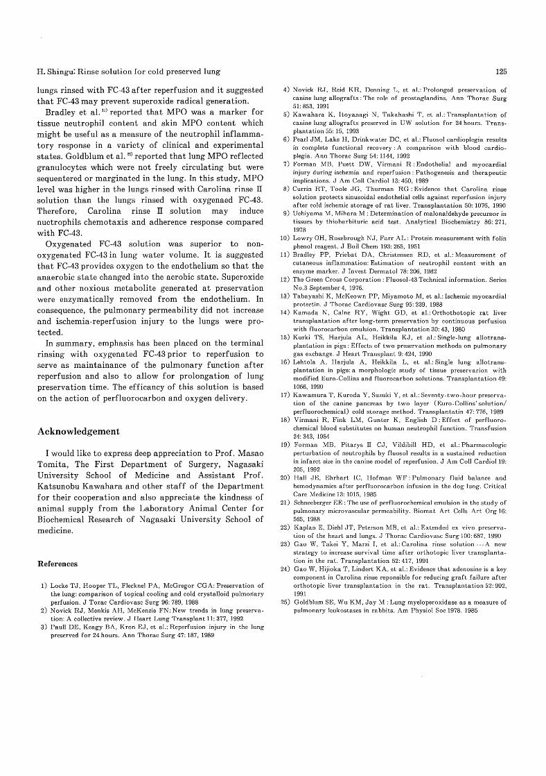
Fig. 2. Photomicrogram of the lung in Group 1 (HE stainning X 16). Severe alveoiar edema with
thickness of the alveolar septa can be seen.
Fig. 3. Photomicrogram of the lung in Group 2 (HE stainning X 16). Histolically, the lung appears
normal architecture.
Fig. 4. Photomicrogram of the lung in Group 3 (HE stainning X 16). Histolically, the lung appears
normal architecture.
Fig. 5. Photomicrogram of the lung in Group 4 (HE stainning X 16). Mild intraalveolar and intraseptal
Weeding can be seen.
Fig. 6. Photomicrogram of the lung in Group 5 (HE stainning X 16). Marked alveolar and perivascular
extravasation and ragged alveolar septa can be seen.
Fig. 7. Photomicrogram of the lung in Group 6 (HE stainning X 16). Marked alveolar and perivascular
extravasation and ragged alveolar septa can be seen.

Lung tissue lipid peroxdation was evaluated as TBA reactive materials). The assey was performed immediately before and after reperfusion. A 0.3 g lung specimen was frozen in liquid nitrogen. The frozen specimen was homoge-nized and added with cold saline to make a 10 % homogen-ate. A 0.2 mL of the homogenate was added and mixed with 0.2 mL of 8.1 % sodium dodecyl sulfate (SDS), 1.5 mL of 20 % acetic acid (pH 3.5), 0.6 mL of distilled water and 1.5 mL of 0.8 mL TBA. The mixture was heated at 95C for 60 minutes. A 1.0 mL of distilled water and 5 mL of n-butanol/pyridine (15:1, vol/vol) were added to the mixture after cooling. The mixture was centrifuged at 3000 rpm for 10 minites. The fluorecent intensity of the supernatant was measured with excitation of 515 nm and emission of 553 nm by using Spectrophotofluorometer RF 5000 (Shimazu Cop, Tokyo, Japan). The lipid peroxide concentration was determined by reference to a standard fluid of 0.5 nmol 1, 1, 3, 3-tetrametoxypropane that yields
0.5 nmol of malondialdehyde (MDA). The protein content of the homogenate was determined by the Lowry's method"). Tissue lipid peroxidation was evaluated as MDA nmol/mg tissue protein.
Lung tissue myeloperoxidase
Myeloperoxidase (MPO) can be used as a marker for tissue neutrophils which was trapping in the tissue during reperfusionll°. Lung tissues were homogenized and sus-
pended by a 0.5 % hexadecyltrimethylammonium bromide (HTAB) (Sigma chemical Co., St. Louis, MO) in 50 mM
potassium phosphate buffer, ph 6.0, to yeild the 5 % homogenate. The suspention Oral) was centrifuged at 40,000 X g for 15 minutes, and then the supernatant (0.1 ml) was mixed with 2.9 ml of 50 mM phosphate buffer, pH 6.0, containing 0.167 mg/mL o-dianisidine dihydrochloride
(Sigma Chemical Co.) and 0.0005 % hydrogen peroxide (WAKO pure chemical industries, LTD. Osaka, Japan). The mixure was assayed by spectrophotometry. The change of absorbance at 460 nm was measured with (Double-beam spectrophotometer, UV-150-02. Shimadzu Seisakusho, Kyoto, Japan). MPO concentration was determined by reference to a standard human MPO. One unit of MPO activity was defined as that degrading one micromole of
peroxide per minute at 25°C.
Lung water volume
The left lower lobe was devided and weighed and placed in a desiccator at 160°C for 48hrs after reperfusion, and then the dry lung was weighed. Lung water volume was determined by the (wet-dry)/wet weight ratio in the left lower lobe.
Histological examination
The reperfused lungs were fixed in 20% formalin, paraf-fin embedded, and standard hematoxylin and eosin-stained slides were prepared.
(5) Statistical analysis
Data were compared using the unpaired Wilcoxon test. A P-value of less than 0.05 was considered statistically singifficant.
Results
There was no significant difference in the rinse pressure among Groups 2, 3, 4, 5 and 6 (Table 1). Lung reperfusion time was 64.2±34.6, 120±0, 97.5±39.0, 52.8 ± 35.2, 85.5 ± 36.2 and 62.4 ± 16.3 minutes in Group 1, 2, 3, 4, 5 and 6 each. Successful reperfusion for the entire 120 minutes was accomplished in all of the lungs in Group 2 (Table 2). There was no significant difference in PaO2 and dynamic lung compliance among the groups. The PVR and the lung (wet-dry)/wet weight ratio were lower in Group 2 than in the other five groups (p <0.01). The t-LPO was higher in Group 3 than in Groups 1, 4, 5 and
6 (p <0.05). The tissue MPO level was lower in Group 2 than in
Groups 4, 5 and 6 (p <0.05). Histologically, severe lung edema was observed in the
lungs in Groups 1, 5 and 6 (Fig.1, 5 and 6), and bleeding
was observed in Group 4 (Fig.4). Microscopic findings of the lung in Groups 2 and 3, showed almost normal lung architecture (Fig.2, 3).
Discussion
Our data demonstrates that the oxygenated FC-43 may be useful for prevention of ischemic reperfusion injury of canine 24-hour cold preserved lung.
FC-43 (The Green Cross Corporation, Osaka, Japan) is an emulsion of perfluorocarbon which has the high oxygen solubility (Table 3). Perfluorocarbons have been initialy
developed as artificial blood substitutes because the emulsion has a high affinity for oxygen and releases oxygen in a linear fasion unaffected by tempraturel2). Pluronic F-68 is the agent that is used to stabilize the emulsion in which the perfluorocarbon is suspended.
The experimental studies with perfluorocarbon emulsion are mainly for the organs' preservation for transplanta-tion. Perfluorochemicals have significantly prolonged the
preservation and prevent the reperfusion injury of the heart')"), the liver'), the lung")") and the pancreatic")

Table 1. Results after 120 minutes perfusion(mean ±SD)
Group 1 Group 2 Group 3 Group 4 Group 5 Group 6 (n=5) (n=6) (n=4) (n=5) (n=4) (n=4)
Rinse P.(mmHg) 6.8±2.4 7.8±2.9 11.8±3.5 7.9±4.1 13.6±5.1
AP02(mmHg) 66.3±43.5 83.0±38.7 39.3±48.1 65.1±12.5 43.6±32.8 34.2±12.4 at initial 5 minute
AP02(mmHg) 37.7±31.4 68.6±32.6 66.6±26.1 61.6±21.5 48.1±34.4 46.9±22.4 at additional venous blood
Dymamic LC 7 .30±1.39 10.33±2.31' 8.92±2.56 5.97±1.896 6.28±1.48` 7.32±1.374 (mL/cmH,O)
PVR (mmHg/ L/ min) 322.30±190.52' 84.26±33.26` 171.19±104.779 357.85±166.52h 474.10±219.10' 430.22±88.74'
WD ratio 0.9055±0.0067k 0.8683±0.0210' 0.9256±0.0230" 0.9133±0.0092" 0.9217±0.0217' 0.9213±0.0108'
t-LPO (nmol/mg protein) 0.507±0.2259 1.776±0.689 1.241±0.142` 0.541±0.326' 0.441±0.119' 0.547±0.261'
t-MPO 358.5±125.9 240.0±94.0' 275.1±42.8 450.0±345.2`" 399.4±106.4' 454.7±215.7Y (unit/g tissue)
abbreviation Rinse P.; Rinse Pressure
APO2; gradient of blood oxygen pressure between pulmonary vein and pulmonary artery LC ; Lung Compliance
PVR ; Pulmonary Vascular Resistance WD ratio ;(wet lung weight-dry lung weight)/ ( wet lung weight)
t-LPO ; tissue lipid peroxidation t-MPO ; tissue myeloperoxidase
p < 0.01; a vs. b and d ; f vs. e, h, i and j ; e vs. i and j ; g vs. i and j; h vs. i and j; l vs. k, m and p ; r vs. u and t. p < 0.05; a vs. c ; I vs. o ; r vs. q and s ; v vs. w, x and y.
Table 2. Lung reperfusion time (minute)
Group 1 Group 2 Group 3 Group 4 Group 5 Group 6 (n=5) (n=6) (n=4) (n=5) (n=4) (n=4)
No.1 50 120 120 120 120 35 No.2 120 120 30 51 93 71 No.3 45 120 120 20 104 77 No.4 21 120 120 43 25 67 No.5 85 120 30
No.6 120
mean ±SD 64.2±34.6' 120±06 97.5±39.0` 52.8±35.2d 85.5±36.2e 62.5±16.3`
p < 0.05 ; b vs. a, b vs. d, b vs. e, b vs. f no significant difference ; b vs. c
grafts. Tabayashi et al.") reported that the oxygenated fluorocarbon solution gave superior myocardial protection during 2 hours of ischemic arrest and postischemic left ventricular hemodynamics, and water content was signifi-cantly lower in the oxygenated fluorocarbon group than in the nonoxygenated crystalloid cardioplegia. Kawamura et al. 17) reported that a 2-layer (Euro-Collins'/Perfluoro-chemical) cold storage method for the preservation of canine prancreas extended the storage interval for 72-hour and that the functional success rate after transplantation was 100 % and biopsies of the graft showed almost normal
architecture in exocrine and endocrine tissues. These papers
showed that one of the perfluorochemical action, an
oxygen-supplying agent, was utilized for the ischemic
organs and the tissues were kept in almost normal
functons and structures. Tuula S. Kurki et al.") reported
that oxygenated FC-43 solution was useful for lung preser-
vation in pigs. After donor lung was preserved with
oxygenated FC-43 for 6 hours, single lung allotrans-
plantaion was performed and they concluded that oxygen-ated FC-43 donor lung preservation was superior in
functional recovery in pulmanary gas exchange during

Table 3. Composition of peservation sollution and rinse solution
Euro-Collins FC-43 F-68 Carolina Rinse II (mm) (mm) (mm) (mm)
FC-43 20 % Pluronic F68 2.56% 2.56%
NaCI 102 102 102 KCl 15 4.6 4.6 4
K2HPO4 42.5 KH2P04 15 NaC03 10 NaHCO3 2.5 2.5
CaC12 2.5 2.5 3 MgC12 2.1 2.1
Sodium lactate 28 Glucose 3.5% 1.8 % 1.8 %
Adenosine 0.1 Alloprinol 1 Desferrioxamine 1 Gluthatione 3 Hydroxyethyl 30g/L 30g/L additional HES
starch C 50g/L Na+ 10 mEg/L 104.5 mEg/L 104.5 mEg/L 102 mEg/L
K+ 115 mEg/L 4.6 mEg/L 4.6 mEg/L 4 mEg/L Osmotic pressure 335 mOsm/L 309 mOsm 309 mOsm 278 mOsm/L
pH (room temp.) 7.2-7.5 7.3-7.5 7.3-7.4 7.4
HES : Hydroxyethyl starch temp.: tempreture
reperfusion compared with the lung function preserved with Euro-Collins solution. Lehtola and colleagues") demonstrated that electron microscopic findings of the lungs preserved with oxygenated FC-43 for 6 hours showed better-preserved alveolar epithelium lining comparing with the findings of the lungs preserved with modified Euro-Collins solution.
The perfluorochemicals have not only high oxygen solubility but beneficial effects on prevention of ischemia-reperfusion injury. Many reports indicated that the
perfluorochemicals reduced neutrophil chemotaxis, adher-ence, degranulation and superoxide production') W 19) and kept the microvascular permeability'-22). Virmani et al. 18) reported oxypherol (perfluorotributylamine) inhibited human neutrophil function in vitro. Neutrophils treated with oxypherol-treated caused nearly 90% suppression of chemotactic response to zymosan-activated serum calcu-lated by comparing the control response. Neutrophils exposed to oxypherol were stimulated by exposure to
phorbol myristate acetate and superoxide release of the neutrophils was reduced to nearly 80% by oxypherol exposure compared with buffer response. Joseph E. Hall et al.' reported that perfluorocarbon-related changes in canine lobar permeability were determined by measuring the pulmonary filtration coefficient (Kf). In the isolated canine right lower lobe model, Kf value was not affected by FC-43 infusion, the mean Kf values in FC-43 infused
group and bovine serum albumin solution group were 0.075
and 0.070 mL/min/torr/100 g each. In the experimental liver trasplantation, Carolina rinse
solution was developed for the use as a rinse after storage to prevent lethal injury to sinusoidal endothelial cells'). Wensi Gao et al.") reported that in the Lewis rat livers which were implanted after 12-hour cold storage in University of Wisconsin solution and rinsed with Carolina rinse solution prior to reperfusion the survival time was improved as compared with the livers rinsed with Ringer's solution. They also reported that adenosine was an essen-tial component of Carolina rinse and they simplified Carolina rinse and formulated Carolina rinse II with a minimum of ingredients z'). It contains electrolytes similar to plasma, antioxidants against oxygen radical and vasodilator to improve micro circulation. In this study, Carolina rinse II solution was not effective to prevent lung edema in canine 24-hour cold preserved lungs.
The tissue lipoperoxidation (MDA) and MPO were significantly lower in the lungs rinsed with oxygenated FC-43 than those in the lungs rinsed with non-oxygenated FC-43 or Carolina rinse II solution. Lipid peroxidation, the oxidative deterioration of polyunsaturated fatty acids, is a widely accepted mechanism for cellular injury, especially ischemia-reperfusion injury. Activated xanthine oxidase, which catalyzes the reactoin of hypoxanthine with oxygen, produces xanthine and superoxide radicals. The hypoxanthine substrate accumulates in ischemic tissue. In
this study, MDA reactants production was reduced in the
lungs rinsed with FC-43 after reperf usion and it suggested that FC-43 may prevent superoxide radical generation.
Bradley et al.") reported that MPO was a marker for tissue neutrophil content and skin MPO content which might be useful as a measure of the neutrophil inflamma-tory response in a variety of clinical and experimental states. Goldblum et al.") reported that lung MPO reflected
granulocytes which were not freely circulating but were sequentered or marginated in the lung. In this study, MPO level was higher in the lungs rinsed with Carolina rinse II solution than the lungs rinsed with oxygenaed FC-43. Therefore, Carolina rinse II solution may induce nuetrophils chemotaxis and adherence response compared with FC-43.
Oxygenated FC-43 solution was superior to non-oxygenated FC-43 in lung water volume. It is suggested that FC-43 provides oxygen to the endothelium so that the anaerobic state changed into the aerobic state. Superoxide and other noxious metabolite generated at preservation were enzymatically removed from the endothelium. In consequence, the pulmonary permeability did not increase
and ischemia-reperfusion injury to the lungs were pro-tected.
In summary, emphasis has been placed on the terminal rinsing with oxygenated FC-43 prior to reperfusion to serve as maintainance of the pulmonary function after reperfusion and also to allow for prolongation of lung
preservation time. The efficancy of this solution is based on the action of perfluorocarbon and oxygen delivery.
Acknowledgement
I would like to express deep appreciation to Prof. Masao Tomita, The First Department of Surgery, Nagasaki University School of Medicine and Assistant Prof. Katsunobu Kawahara and other staff of the Department for their cooperation and also appreciate the kindness of
animal supply from the Laboratory Animal Center for Biochemical Research of Nagasaki University School of medicine.
References
1) Locke TJ, Hooper TL, Flecknel PA, McGregor CGA: Preservation of the lung: comparison of topical cooling and cold crystalloid pulmonary
perfusion. J Torac Cardiovasc Surg 96: 789, 1988 2) Novick RJ, Menkis AH, McKenzie FN: New trends in lung preserva-
tion: A collective review. J Heart Lung Transplant 11: 377, 1992 3) Paull DE, Keagy BA, Kron EJ, et al.: Reperfusion injury in the lung
preserved for 24 hours. Ann Thorac Surg 47: 187, 1989
4) Novick RJ, Reid KR, Denning L, et al.: Prolonged preservation of canine lung allografts : The role of prostaglandins. Ann Thorac Surg
51:853, 1991 5) Kawahara K, Itoyanagi N, Takahashi T, et al.: Transplantation of
canine lung allografts preserved in UW solution for 24 hours. Trans-
plantation 55: 15, 1993 6) Pearl JM, Laks H, Drinkwater DC, et al.: Fluosol cardioplegia results
in complete functional recovery : A comparison with blood cardio-
plegia. Ann Thorac Surg 54: 1144, 1992 7) Forman MB, Puett DW, Virmani R : Endothelial and myocardial
injury during ischemia and reperfusion : Pathogenesis and therapeutic implications. J Am Coll Cardiol 13:450, 1989
8) Currin RT, Toole JG, Thurman RG : Evidence that Carolina rinse solution protects sinusoidal endothelial cells against reperfusion injury
after cold ischemic storage of rat liver. Transplantation 50: 1076, 1990 9) Uchiyama M, Mihara M : Determination of malonaldehyde precursor in
tissues by thiobarbituric acid test. Analytical Biochemistry 86: 271, 1978
10) Lowry OH, Rosebrough NJ, Farr AL: Protein measurement with folin
phenol reagent. J Boil Chem 193: 265, 1951 11) Bradley PP, Priebat DA, Christensen RD, et al.: Measurement of
cutaneous inflammation: Estimation of neutrophil content with an enzyme marker. J Invest Dermatol 78: 206, 1982
12) The Green Cross Corporation : Fluosol-43 Technical information. Series No.3 September 4, 1976.
13) Tabayashi K, McKeown PP, Miyamoto M, et al.: Ischemic myocardial
protectin. J Thorac Cardiovasc Surg 95: 239, 1988 14) Kamada N, Calne RY, Wight GD, et al.: Orthothotopic rat liver
transplantation after long-term preservation by continuous perfusion with fluorocarbon emulsion. Transplantation 30: 43, 1980
15) Kurki TS, Harjula AL, Heikkila KJ, et al.: Single-lung allotrans-
plantation in pigs : Effects of two preservation methods on pulmonary gas exchange. J Heart Transplant 9: 424, 1990
16) Lehtola A, Harjula A, Heikkila L, et al.: Single lung allotrans-
plantation in pigs: a morphologic study of tissue preservarion with modified Euro-Collins and fluorocarbon solutions. Transplantation 49:
1066, 1990 17) Kawamura T, Kuroda Y, Suzuki Y, et al.: Seventy-two-hour preserva-
tion of the canine pancreas by two layer (Euro-Collins' solution/
perfluorochemical) cold storage method. Transplantatin 47: 776, 1989 18) Virmani R, Fink LM, Gunter K, English D : Effect of perfluoro-
chemical blood substitutes on human neutrophil function. Transfusion 24: 343, 1984
19) Forman MB, Pitarys II CJ, Vildibill HD, et al.: Pharmacologic
perturbation of neutrophils by fluosol results in a sustained reduction in infarct size in the canine model of reperfusion. J Am Coll Cardiol 19:
205, 1992 20) Hall JE, Ehrhart IC, Hofman WF : Pulmonary fluid balance and
hemodynamics after perfluorocarbon infusion in the dog lung. Critical Care Medicine 13: 1015, 1985
21) Schneeberger EE : The use of perfluorochemical emulsion in the study of
pulmonary microvascular permeability. Biomat Art Cells Art Org 16: 565, 1988
22) Kaplan E, Diehl JT, Peterson MB, et al.: Extended ex vivo preserva- tion of the heart and lungs. J Thorac Cardiovasc Surg 100: 687, 1990
23) Gao W, Takei Y, Marzi I, et al.: Carolina rinse solution ---A new strategy to increase survival time after orthotopic liver transplanta-
tion in the rat. Transplantation 52:417, 1991 24) Gao W, Hijioka T, Lindert KA, et al.: Evidence that adenosine is a key
component in Carolina rinse reponsible for reducing graft failure after orthotopic liver transplantation in the rat. Transplantation 52: 992,
1991 25) Goldblum SE, Wu KM, Jay M : Lung myeloperoxidase as a measure of
pulmonary leukostases in rabbits. Am Physiol Soc 1978. 1985