Effects of Second Messengers - WordPress.com...Effects of Second Messengers Inositol trisphosphate...
20
3/21/2013 1 Effects of Second Messengers Inositol trisphosphate Opens Calcium Channels Binding to IP 3 -gated Channel Cooperative binding Diacylglycerol Activates Protein Kinase C Ca 2+ is required Phosphorylation of many target proteins
Transcript of Effects of Second Messengers - WordPress.com...Effects of Second Messengers Inositol trisphosphate...

3/21/2013
1
Effects of Second Messengers
Inositol trisphosphate
Opens Calcium Channels
Binding to IP3-gated Channel
Cooperative binding
Diacylglycerol
Activates Protein Kinase C
Ca2+ is requiredPhosphorylation of
many target proteins

3/21/2013
2
The domain structures of protein kinase C isoforms
Interaction with phospholipids
Binds Diacylglycerol
Pseudosubstrate Sequence
• Resemble the substrate sequence• Contains
A-R-K-G-A-L-R-Q-KSubstrate Sequence
X-R-X-X-(S,T)-Hyd-R-X
• Binds to the Enzyme’s Active Site.

3/21/2013
3
Termination of IP3 Signal
Lithium Ions,Used to treat some
psychological disordersInhibits IP3 recycling
IP3 is a Short-LivedMessenger
3/21/2013
4
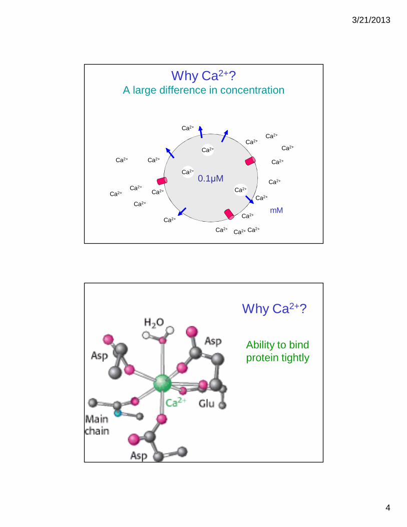
Why Ca2+?A large difference in concentration
0.1μM
Ca2+
Ca2+
Ca2+
Ca2+
Ca2+
Ca2+
Ca2+
Ca2+
Ca2+
Ca2+Ca2+
Ca2+
Ca2+
Ca2+Ca2+
Ca2+
Ca2+
Ca2+Ca2+
Ca2+
mM
Ca2+
Why Ca2+?
Ability to bind protein tightly
3/21/2013
5
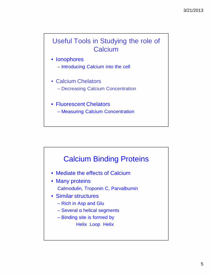
Useful Tools in Studying the role of Calcium
• Ionophores– Introducing Calcium into the cell
• Calcium Chelators– Decreasing Calcium Concentration
• Fluorescent Chelators– Measuring Calcium Concentration
Calcium Binding Proteins
• Mediate the effects of Calcium• Many proteins
Calmodulin, Troponin C, Parvalbumin• Similar structures
– Rich in Asp and Glu– Several α helical segments– Binding site is formed by
Helix Loop Helix

3/21/2013
6
Helix E
Calmodulin
• Found in almost all eukaryotic cells• Consists of two globular regions
– Connected by flexible region– Each contains 2 EF hands– Four Ca2+ binding sites.

3/21/2013
7
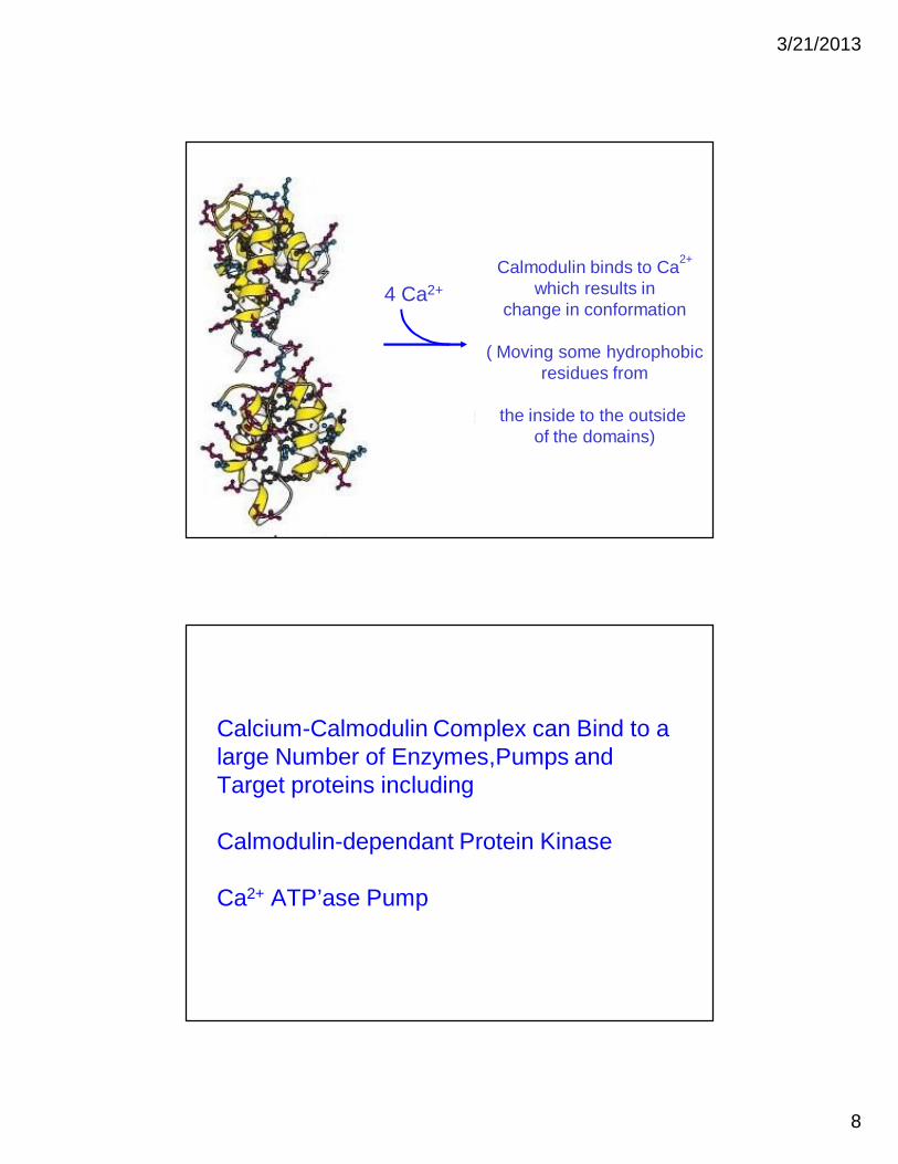
Calmodulin changes conformation upon binding to Calcium
3/21/2013
8
4 Ca2+Calmodulin binds to Ca2+
which results in change in conformation
( Moving some hydrophobicresidues from
the inside to the outside of the domains)
Calcium-Calmodulin Complex can Bind to a large Number of Enzymes,Pumps and Target proteins including
Calmodulin-dependant Protein Kinase
Ca2+ ATP’ase Pump
3/21/2013
9
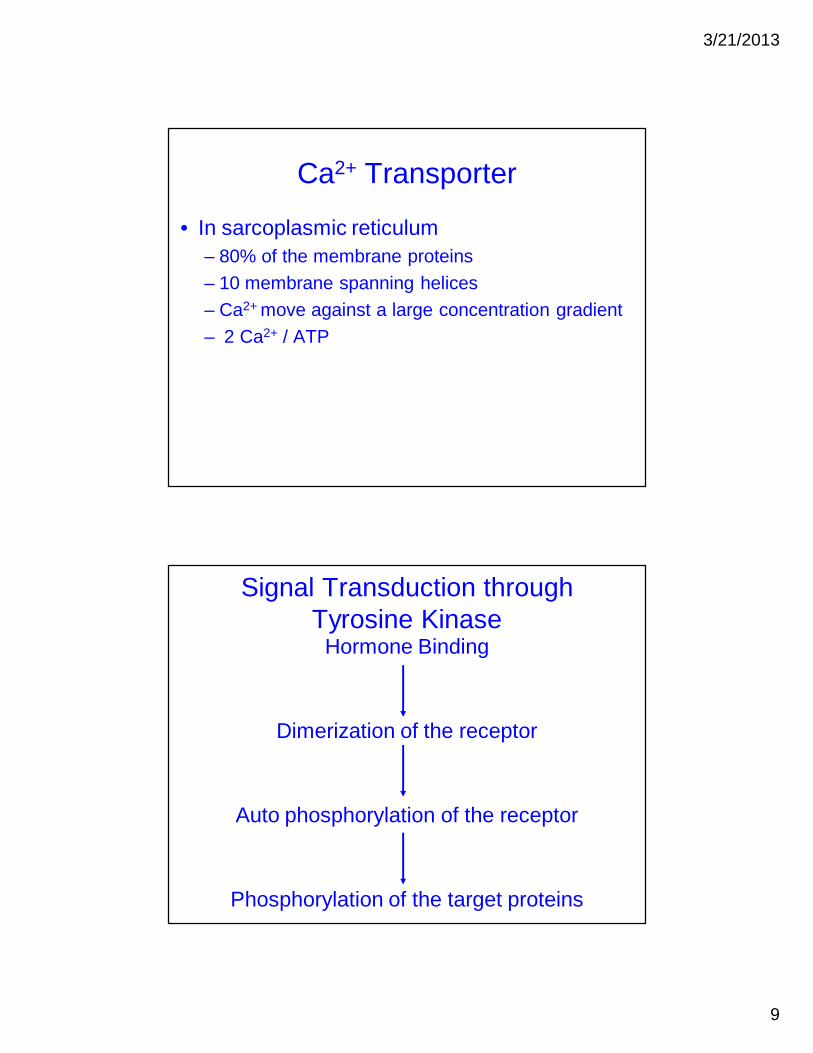
Ca2+ Transporter
• In sarcoplasmic reticulum– 80% of the membrane proteins– 10 membrane spanning helices– Ca2+ move against a large concentration gradient– 2 Ca2+ / ATP
Signal Transduction through Tyrosine Kinase
Hormone Binding
Dimerization of the receptor
Auto phosphorylation of the receptor
Phosphorylation of the target proteins

3/21/2013
10
Some Hormones that use Tyrosine Kinase
• Growth Hormone• Insulin• Epidermal Growth Factor • Platelet-derived growth Factor
Growth Hormone
• Monomeric Protein• 217 Amino Acids• Compact Four-helix Bundle

3/21/2013
11
Growth Hormone Receptor
• 638 A.Acid– Membrane Spanning Protein– Extracellular Domain ≈250 A.A– Single Membrane-Spanning Helix– Intracellular Domain 350 A.A
• Monomeric when not bound to hormone• Dimeric when bound to hormone

3/21/2013
12
GrowthHormone
Binding of one molecule of growth hormone
Dimerization of the receptor
Each Intracellular Domain is associated with a protein kinase called
Janus Kinase 2
Interaction with
membrane
Binds peptides that contain
Phosphotyrosine
protein kinaseprotein kinase-like
Janus

3/21/2013
13
Receptor dimerization brings two JAKs together
Each Phosphorylates key residues on the other
Activated JAK 2 can Phosphorylate other substrates
• STAT 5– Signal transducer and activators of transcription
• Regulator of transcription
• STAT5 Phosphorylation Dimerization
Binding to specific DNA sites

3/21/2013
14
STAT is phosphorylated on a tyrosine residue near the carboxyl terminus
Phosphorylated tyr binds to SH2 domain of another STAT 5 molecule
Activated JAK 2 can Phosphorylate other substrates (cont.)
• Phosphorylation of the Receptor
– Association with JAK 2
– Association with other proteins in the signal transduction pathway

3/21/2013
15
Signal-Transduction Cascades - 3
•Tyrosine kinase and receptor dimerization•Signaling Pathways and Cancer
Receptor dimerization brings two JAKs together
Each Phosphorylates key residues on the other

3/21/2013
16
Tyrosine Kinase is Part of some receptors
• Epidermal Growth Factor Receptor– Monomeric (inactive)– EGF binding Dimerization Cross
Phosphorylation Activation• Insulin Receptor
– Dimer of 2 αβ pairs– Insulin Binding ››› Activation of the Kinase
Do these receptors transfer information across the membrane in the same way?

3/21/2013
17
Is dimerization alone sufficient for activation of EGF receptor?
• Synthesis of a gene that encoded a chemeric receptor– Extracellular (insulin)– Intracellular (EGF)
• EGF receptor and insulin receptor use a common mechanism for transmitting information across membrane
Epidermal growth factor signaling pathway
• Grb-2 binds phosphorylated EGF receptor↓↓
Binding to a protein called Sos↓↓
Activation of Ras↓↓
Activation of specific Protein Kinases
GTPGDP

3/21/2013
18
Ras is a member of small G proteins family
• Monomeric• Exist in two forms
– GDP bound «-----» GTP bound• Smaller than G proteins• Have GTPase activity• Many similarities in structure and mechanism
with Gα
• Include several groups or subfamilies• Major role in growth, differentiation, cellular
transport, motility etc…
SH2

3/21/2013
19
Defects in Signaling Pathways Can Lead to Cancer and Other Diseases
• Cancer, is characterized by uncontrolled or inappropriate cell growth,
• Can be caused by certain viruses• Can be associated with defects in signal-
transduction proteins;failure of signal transduction process
Rous sarcoma virus (in Chicken) carries a gene called v-src
• Oncogene• Encode a tyrosine kinase protein• Similar protein is found in cells called c-Src
• Small differences in the amino acid sequences between the proteins

3/21/2013
20
Impaired GTPase activity can lead to cancer in human
• Mammalian cells contain 3 Ras proteinsMutation
Loss of ability to hydrolyze GTPRas is locked in “ON” position
continuous stimulation of growth
Cholera and Whooping Cough Are Due to Altered G-Protein Activity
• The cholera toxin: protein composed of two functional units– B subunit: binds to GM1 gangliosides of the intestinal
epithelium – A catalytic subunit: enters the cell. – A subunit catalyzes the covalent modification of a Gαs
protein: – Attachment of an ADP-ribose to an arginine residue.– Stabilization of the GTP-bound form of Gαs, – The active G protein, activates protein kinase A.– Openining of chloride channels
• Excessive loss of NaCl and the loss of large amounts of water into the intestine.