Effects of pleiotropic molecules on alpha- crystallin ... - Febbraio/0203 - ITARVO... ·...
27
Effects of pleiotropic molecules on alpha- crystallin aggregation Vincenzo Giuseppe Nicoletti Università di Catania - Scuola “Facoltà di Medicina” Dipartimento di Scienze Bio-mediche sez. Biochimica
Transcript of Effects of pleiotropic molecules on alpha- crystallin ... - Febbraio/0203 - ITARVO... ·...
Effects of pleiotropic molecules on alpha-
crystallin aggregation
Vincenzo Giuseppe Nicoletti
Università di Catania - Scuola “Facoltà di Medicina”
Dipartimento di Scienze Bio-mediche sez. Biochimica
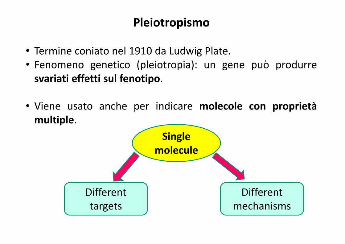
Pleiotropismo
• Termine coniato nel 1910 da Ludwig Plate.
• Fenomeno genetico (pleiotropia): un gene può produrre
svariati effetti sul fenotipo.
• Viene usato anche per indicare molecole con proprietà
multiple.
Different
targets
Different
mechanisms
Single
molecule
Il disaccaride trealosio e il dipeptide carnosina
sono tipiche molecole pleiotropiche:
� stabilizzazione del ripiegamento delle
proteine;
� inibizione dell’aggregazione;
� elevata capacità protettiva contro vari tipi
di stress.
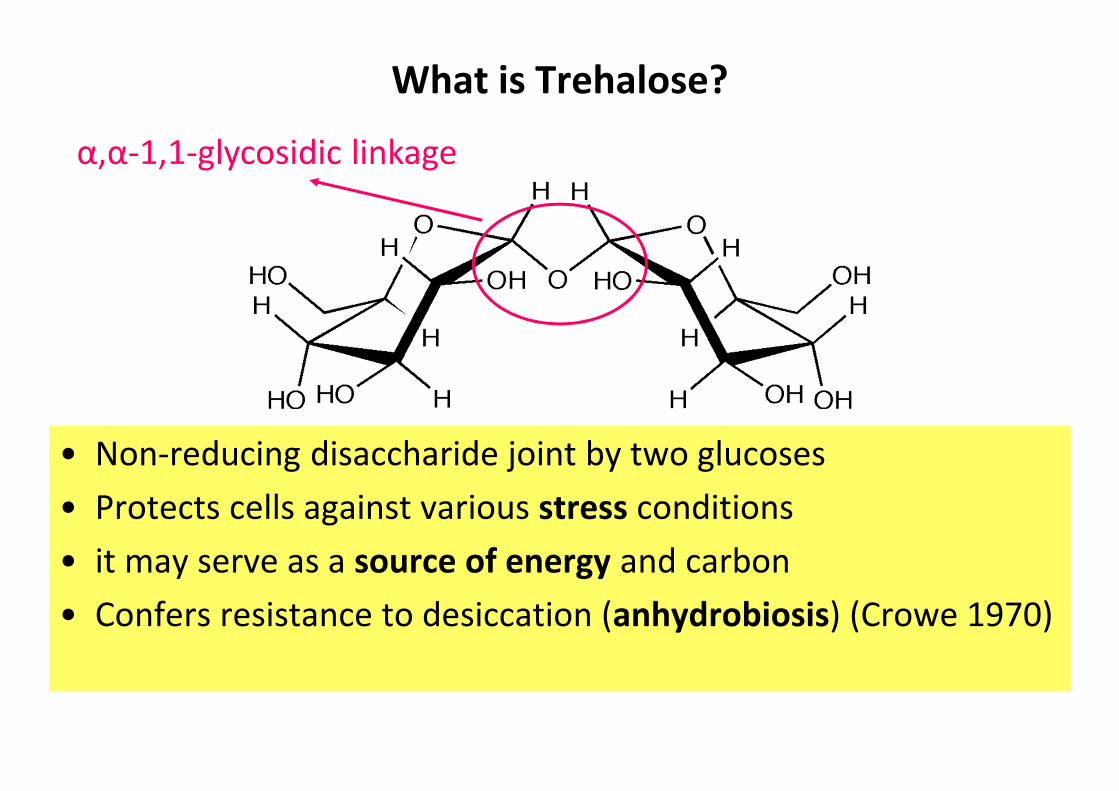
What is Trehalose?
• Non-reducing disaccharide joint by two glucoses
• Protects cells against various stress conditions
• it may serve as a source of energy and carbon
• Confers resistance to desiccation (anhydrobiosis) (Crowe 1970)
α,α-1,1-glycosidic linkage
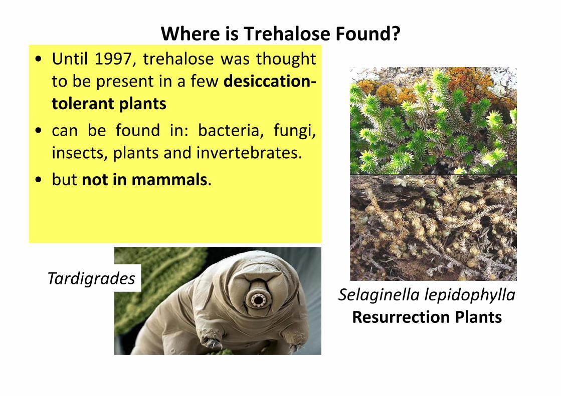
Where is Trehalose Found?
• Until 1997, trehalose was thought
to be present in a few desiccation-
tolerant plants
• can be found in: bacteria, fungi,
insects, plants and invertebrates.
• but not in mammals.
Selaginella lepidophylla
Resurrection Plants
Tardigrades
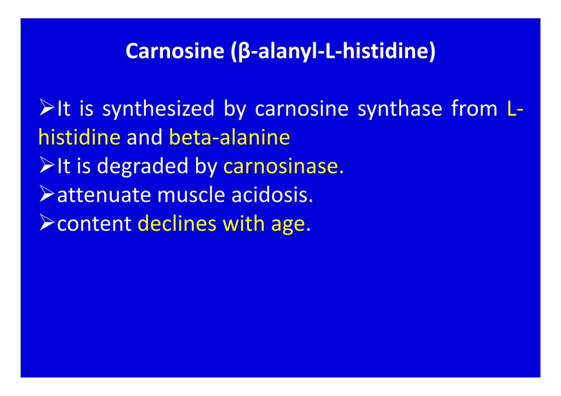
Carnosine (β-alanyl-L-histidine)
�It is synthesized by carnosine synthase from L-
histidine and beta-alanine
�It is degraded by carnosinase.
�attenuate muscle acidosis.
�content declines with age.

� the family is composed of
three classes: α, β and γ.
� predominant proteins within the eye lens.
� α-Crystallin belongs to the heat shock proteins
family;
� prevents mysfolding (aggregation and/or
precipitation) of other proteins, itself included due
to a self-chaperone property.
crystallins

alpha crystallin
α-A20 kDa 173 AA
α-B20 kDa 175 AA
Nel suo stato nativo l’αααα-cristallina forma un grosso
aggregato eterogeneo a basso peso molecolare (LMW)
solubile in acqua

LMW
crystallin
↓800 KdaHMW crystallin
↑1000 Kda Aging
cataract
Folded
crystallin
Aggregation
Postraslational
modification
mysfolded
crystallin
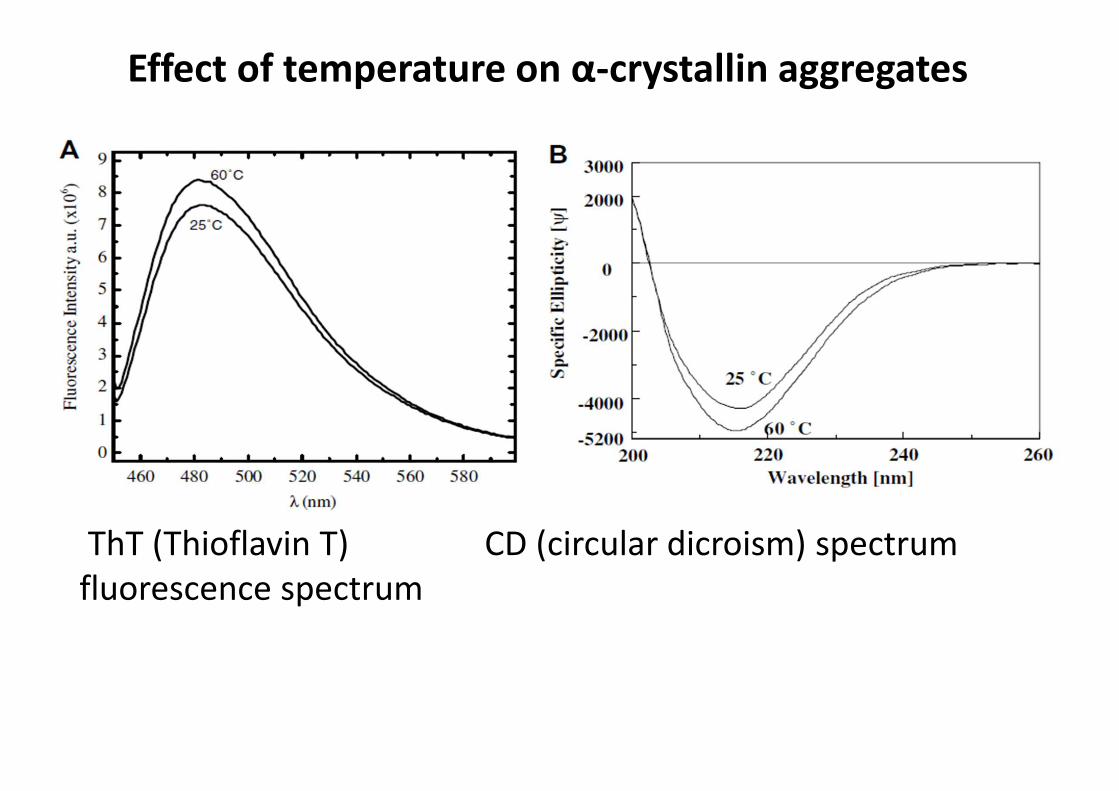
ThT (Thioflavin T) CD (circular dicroism) spectrum
fluorescence spectrum
Effect of temperature on α-crystallin aggregates

460 480 500 520 540 560 580
0
1
2
3
4
5
6
7
8
9
460 480 500 520 540 560 580
λ(nm)
0.75M
0.25M
0.5M
0.1M
1M
Fluorescence Intensity a.u. (x106)
λ(nm)
A0M
1M
0.75M
0.5M
0.25M
0.1M
0M B
25°C 60°C
Fluorescenza di ThT in soluzione con αααα-cristallina a diverse
concentrazioni di trealosio (0M-1M)
ThT alone
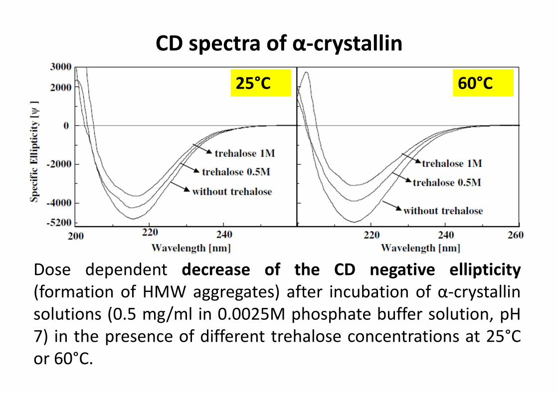
CD spectra of α-crystallin
Dose dependent decrease of the CD negative ellipticity
(formation of HMW aggregates) after incubation of α-crystallin
solutions (0.5 mg/ml in 0.0025M phosphate buffer solution, pH
7) in the presence of different trehalose concentrations at 25°C
or 60°C.
25°C 60°C

63°C +
trealosio 1M
63°C
Effetto antiaggregante del trealosio osservato tramite
microscopia a forza atomica
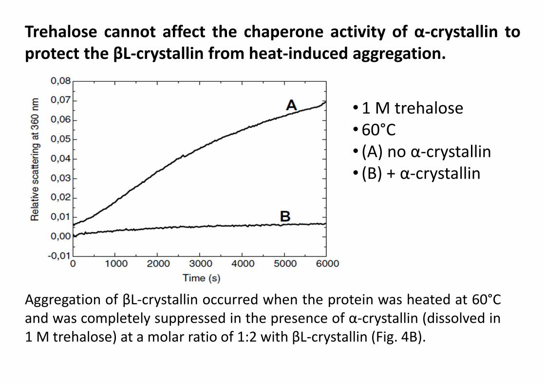
Aggregation of βL-crystallin occurred when the protein was heated at 60°C
and was completely suppressed in the presence of α-crystallin (dissolved in
1 M trehalose) at a molar ratio of 1:2 with βL-crystallin (Fig. 4B).
Trehalose cannot affect the chaperone activity of α-crystallin to
protect the βL-crystallin from heat-induced aggregation.
• 1 M trehalose
• 60°C
• (A) no α-crystallin
• (B) + α-crystallin
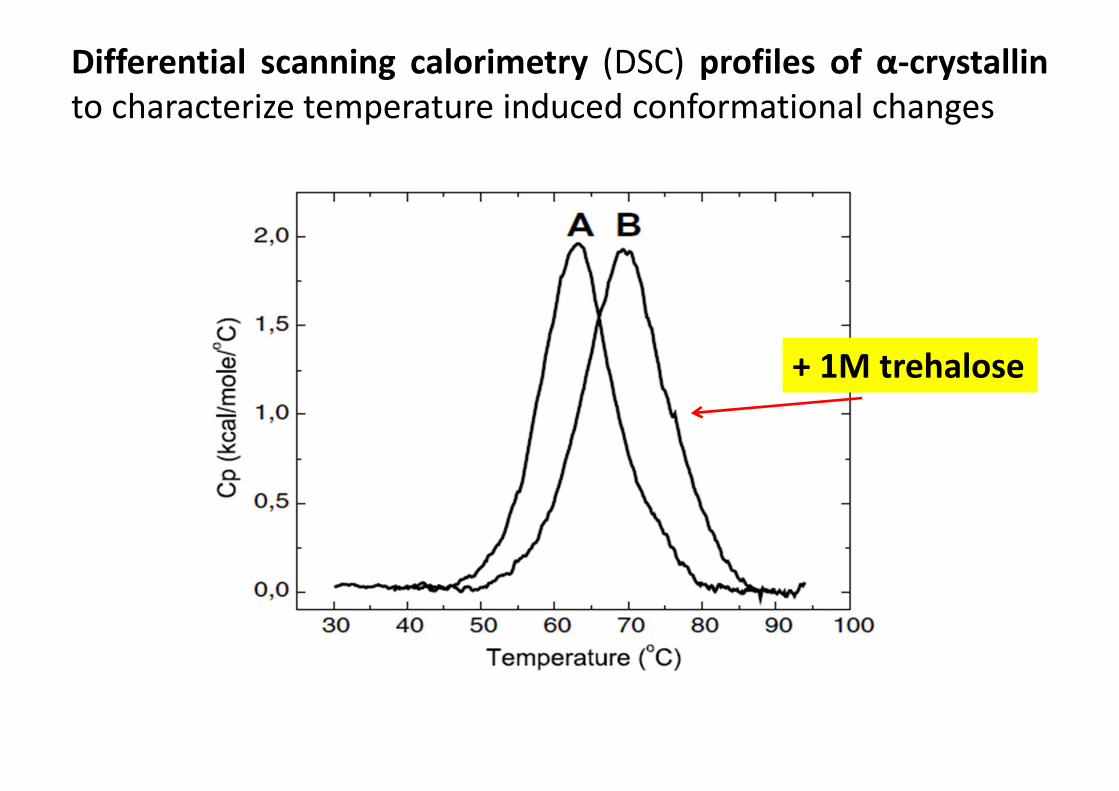
Differential scanning calorimetry (DSC) profiles of α-crystallin
to characterize temperature induced conformational changes
+ 1M trehalose

ββββ-Alanine+L-Histidine N
CH2 CH NH C CH2
HN
HOOC O
CH2
NH2
CH2
carnosine
Carnosine sinthetase
� Skeletal muscle
� brain
� heart
• Physiological intracellular buffer
• Metal ion chelator
• Antioxidant
• an antiglycant agent
• free radicals scavenger
FunctionsDistribution up to 20mM in:

450 500 550 600
0
20
40
60
80
100
120
140
Flu
ore
sce
nce
In
ten
sity (
u.a
.)
λ (nm)
1
2
3
Spettri di fluorescenza di ThT di campioni di α-cristallina in
guanidina 1M (curva 1) e co-incubata con L o D carnosina
+ L carnosina [0.1M]
+ D carnosina [0.1M]

AFM: α-Cristallina (10 mg/ml)
+ guanidina 1M
+ guanidina 1M e
D-carnosina 100 mM
co-incubata (60°C / 24h)
0 20 40 60 80 100-0.0020
-0.0015
-0.0010
-0.0005
1a
C
p(c
al/
oC
)
Temperature (oC)
1
34
2
4a3a 2a
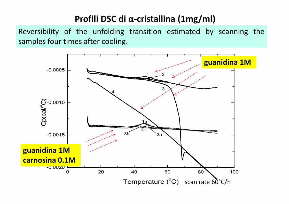
Profili DSC di α-cristallina (1mg/ml)
scan rate 60°C/h
guanidina 1M
guanidina 1M
carnosina 0.1M
Reversibility of the unfolding transition estimated by scanning the
samples four times after cooling.
0 20 40 60 80 100-0.0020
-0.0015
-0.0010
-0.0005
1a
C
p(c
al/
oC
)
Temperature (oC)
1
34
2
4a3a 2a
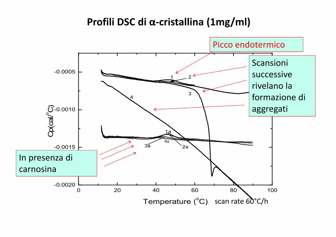
Profili DSC di α-cristallina (1mg/ml)
scan rate 60°C/h
Picco endotermico
Scansioni
successive
rivelano la
formazione di
aggregati
In presenza di
carnosina
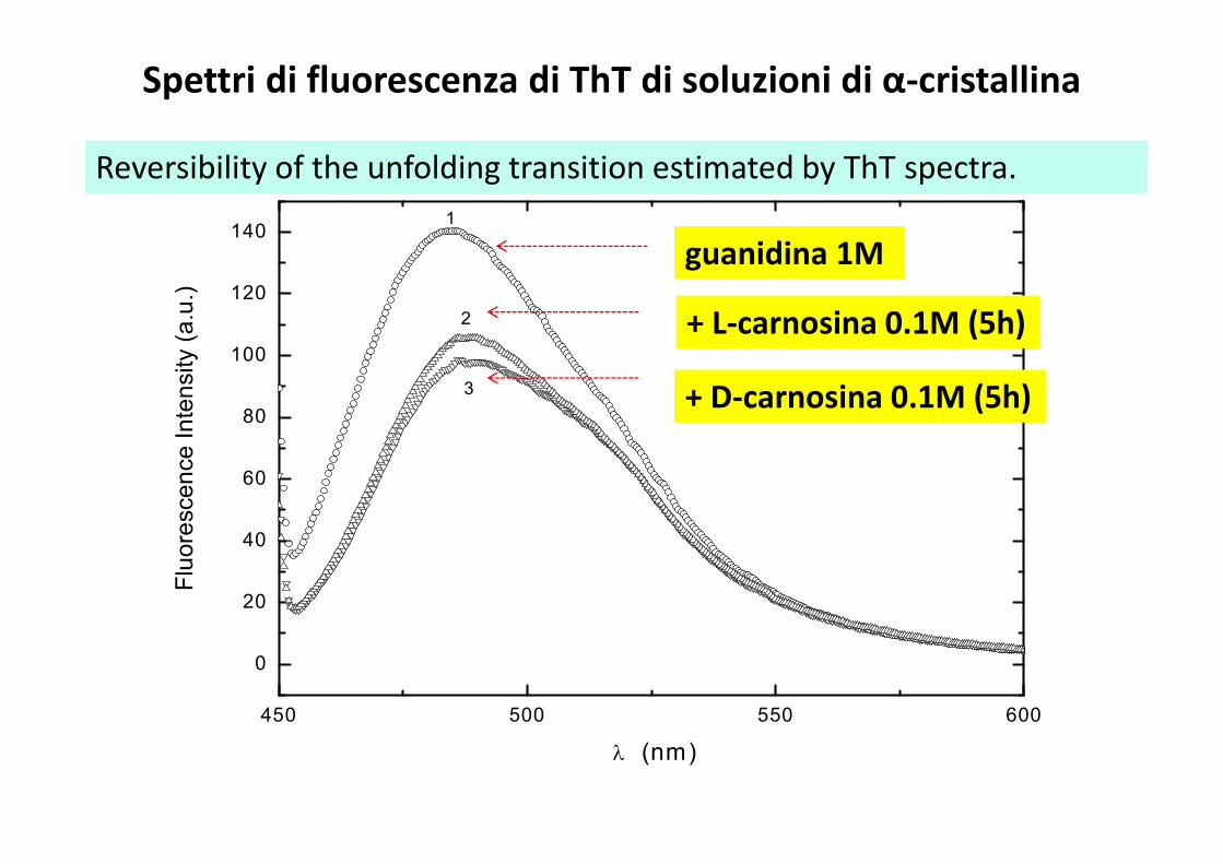
Spettri di fluorescenza di ThT di soluzioni di α-cristallina
450 500 550 600
0
20
40
60
80
100
120
140
λ (nm)
Flu
ore
scen
ce
In
ten
sity (
a.u
.)
1
2
3
guanidina 1M
+ L-carnosina 0.1M (5h)
+ D-carnosina 0.1M (5h)
Reversibility of the unfolding transition estimated by ThT spectra.
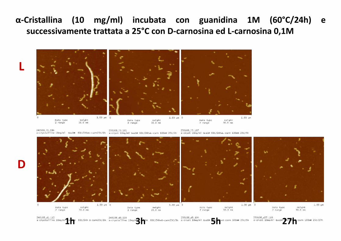
α-Cristallina (10 mg/ml) incubata con guanidina 1M (60°C/24h) e
successivamente trattata a 25°C con D-carnosina ed L-carnosina 0,1M
D
L
1h 3h 5h 27h

D-carnosina 1wkL-carnosina 1wk
D-carnosina x 1 sett.
Particolare della
dissoluzione di una
singola fibrilla
α-Cristallina (10 mg/ml) incubata con guanidina 1M (60°C/24h) e
successivamente trattata con carnosina 0,1M a T°amb per 7gg .
0 200 400 600 800 1000 1200 1400 1600 1800 2000
0,0
0,1
0,2
0,3
0,4
0,5
0,6
0,7
0,8
0,9
1,0
ab
s
Time (s)
a
b
c
d
e
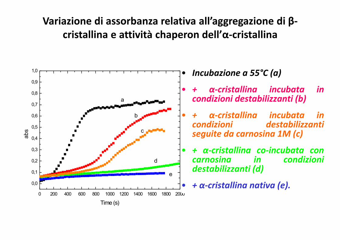
• Incubazione a 55°C (a)
• + α-cristallina incubata incondizioni destabilizzanti (b)
• + α-cristallina incubata incondizioni destabilizzantiseguite da carnosina 1M (c)
• + α-cristallina co-incubata concarnosina in condizionidestabilizzanti (d)
• + α-cristallina nativa (e).
Variazione di assorbanza relativa all’aggregazione di β-
cristallina e attività chaperon dell’α-cristallina
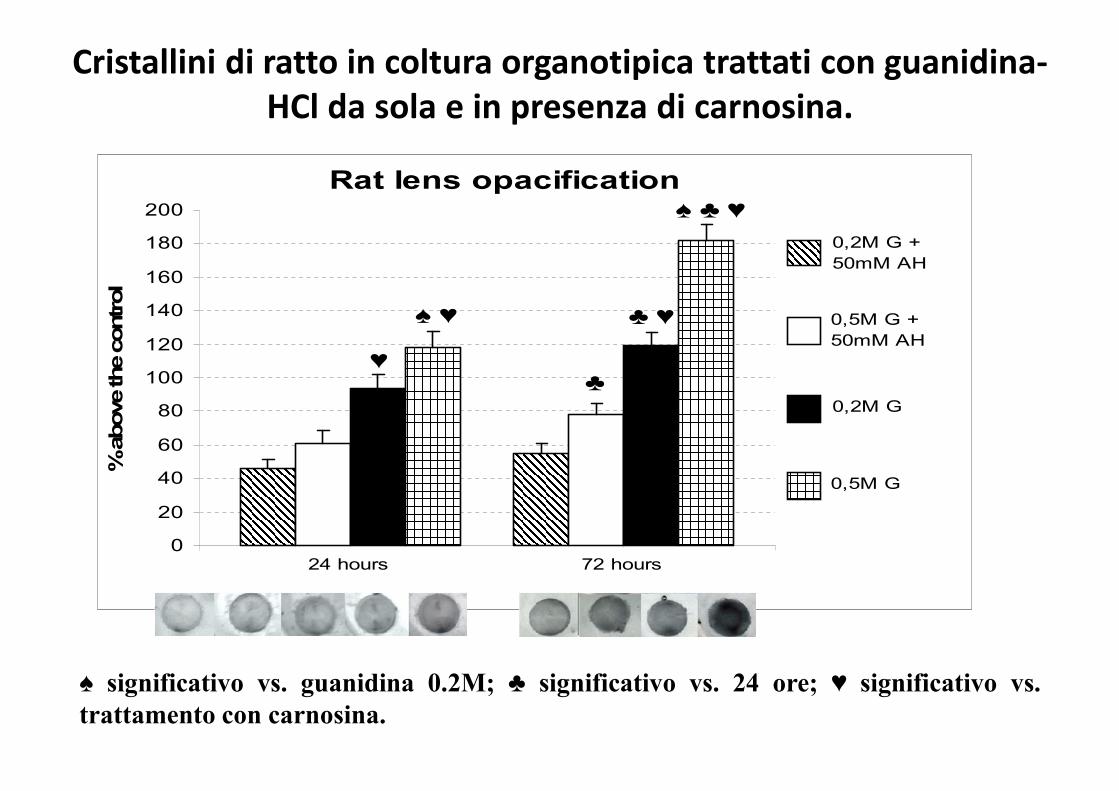
Rat lens opacification
0
20
40
60
80
100
120
140
160
180
200
% above the control
0,2M G +
50mM AH
0,5M G +
50mM AH
0,5M G
0,2M G
24 hours 72 hours
♠ ♣ ♥
♣ ♥♠ ♥
♣♥
♠ significativo vs. guanidina 0.2M; ♣ significativo vs. 24 ore; ♥ significativo vs.
trattamento con carnosina.
Cristallini di ratto in coltura organotipica trattati con guanidina-
HCl da sola e in presenza di carnosina.
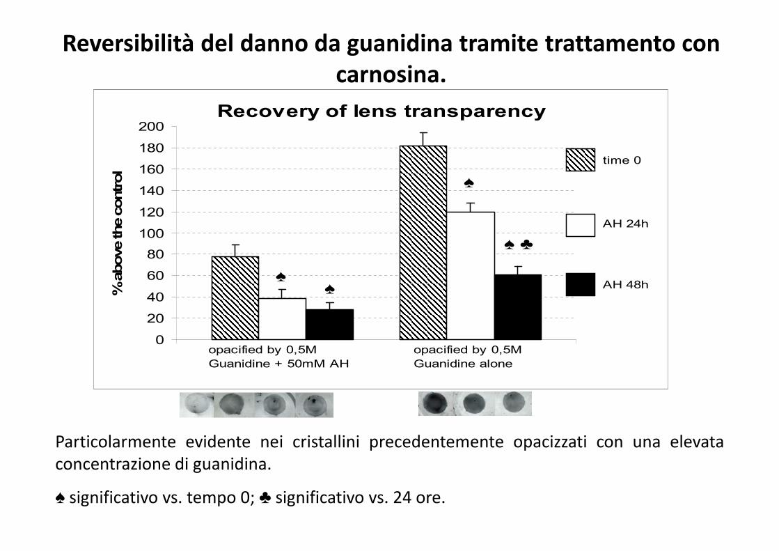
Recovery of lens transparency
0
20
40
60
80
100
120
140
160
180
200% above the control
time 0
opacified by 0,5M
Guanidine + 50mM AH
opacified by 0,5M
Guanidine alone
AH 48h
AH 24h
♠ ♣
♠ ♠
♠
Particolarmente evidente nei cristallini precedentemente opacizzati con una elevata
concentrazione di guanidina.
♠ significativo vs. tempo 0; ♣ significativo vs. 24 ore.
Reversibilità del danno da guanidina tramite trattamento con
carnosina.

Enrico Rizzarelli,
Salvatore Fisichella,
Dipartimento di Scienze Chimiche, Universita` di Catania.
Francesco Attanasio:
Istituto di Biostrutture e Bioimmagini (IBB), CNR, Catania.
Bruno Pignataro,
Sebastiano Cataldo:
Dipartimento di Chimica Fisica, Universita` di Palermo.
Silvia Nicoletti,
Anna Savarino: Dottorato di ricerca in Scienze Chimiche, Catania.