EFFECTS OF HIGH LIGHT INTENSITY AND DESICCATION STRESS …
66
EFFECTS OF HIGH LIGHT INTENSITY AND DESICCATION STRESS ON MOSS SPECIES By Sikhethile Mbatha Submitted in fulfilment of the academic requirements of Master of Science in the Discipline of Biological Sciences School of Life Sciences College of Agriculture, Engineering and Science University of KwaZulu-Natal Pietermaritzburg Campus South Africa July 2021
Transcript of EFFECTS OF HIGH LIGHT INTENSITY AND DESICCATION STRESS …

EFFECTS OF HIGH LIGHT INTENSITY AND
DESICCATION STRESS ON MOSS SPECIES
By
Sikhethile Mbatha
Submitted in fulfilment of the academic requirements of
Master of Science
in the Discipline of Biological Sciences
School of Life Sciences
College of Agriculture, Engineering and Science
University of KwaZulu-Natal
Pietermaritzburg Campus
South Africa
July 2021

iii
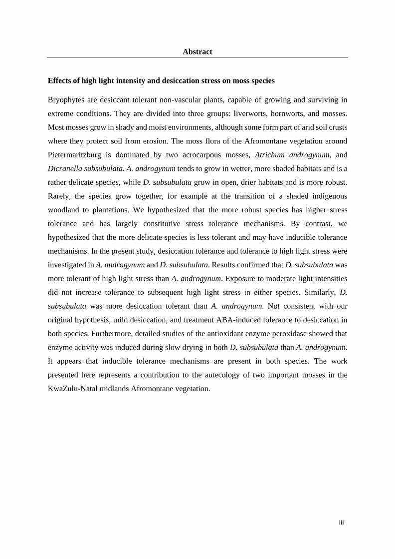
Abstract
Effects of high light intensity and desiccation stress on moss species
Bryophytes are desiccant tolerant non-vascular plants, capable of growing and surviving in
extreme conditions. They are divided into three groups: liverworts, hornworts, and mosses.
Most mosses grow in shady and moist environments, although some form part of arid soil crusts
where they protect soil from erosion. The moss flora of the Afromontane vegetation around
Pietermaritzburg is dominated by two acrocarpous mosses, Atrichum androgynum, and
Dicranella subsubulata. A. androgynum tends to grow in wetter, more shaded habitats and is a
rather delicate species, while D. subsubulata grow in open, drier habitats and is more robust.
Rarely, the species grow together, for example at the transition of a shaded indigenous
woodland to plantations. We hypothesized that the more robust species has higher stress
tolerance and has largely constitutive stress tolerance mechanisms. By contrast, we
hypothesized that the more delicate species is less tolerant and may have inducible tolerance
mechanisms. In the present study, desiccation tolerance and tolerance to high light stress were
investigated in A. androgynum and D. subsubulata. Results confirmed that D. subsubulata was
more tolerant of high light stress than A. androgynum. Exposure to moderate light intensities
did not increase tolerance to subsequent high light stress in either species. Similarly, D.
subsubulata was more desiccation tolerant than A. androgynum. Not consistent with our
original hypothesis, mild desiccation, and treatment ABA-induced tolerance to desiccation in
both species. Furthermore, detailed studies of the antioxidant enzyme peroxidase showed that
enzyme activity was induced during slow drying in both D. subsubulata than A. androgynum.
It appears that inducible tolerance mechanisms are present in both species. The work
presented here represents a contribution to the autecology of two important mosses in the
KwaZulu-Natal midlands Afromontane vegetation.

iv
Acknowledgments
I would like to thank my supervisor Prof R.P Beckett for the support, guidance, and valuable
feedback for the research and writing of the thesis. Most important he provided me with
financial support for 2 years so I could finish my degree. He played an important role to make
sure I stay positive and motivated to complete my studies. I would like to thank my
laboratory mates for words of hope everyday and clarity on using different laboratory
machines. I would like to appreciate and pass my gratitude to my friend and family for
support and prayers. The support I got above made everything possible for me to submit this
thesis.

v
Table of Contents Preface ..................................................................................................................................................... i
Declaration 1: Plagiarism ..................................................................................................................... ii
Abstract ................................................................................................................................................. iii
Acknowledgments ................................................................................................................................ iv
List of tables ......................................................................................................................................... vii
List of figures ...................................................................................................................................... viii
Abbreviations ....................................................................................................................................... xi
Chapter 1: Literature Review .............................................................................................................. 1
1.1. Bryophytes ................................................................................................................................... 1
1.2. Desiccation tolerance in bryophytes ............................................................................................ 3
1.3. Morphological and anatomical changes in bryophytes during desiccation .................................. 4
1.4. Changes in physiology and biochemistry of bryophytes during desiccation ................................ 4
1.4.1. ROS formation during desiccation ........................................................................................ 5
1.4.2. Desiccation mechanism based on enzymes and non-enzymatic antioxidants ........................ 5
1.4.3. Desiccation tolerance mechanisms based on sugar .............................................................. 7
1.4.4. Desiccation tolerance mechanisms based on LEA and other proteins .................................. 7
1.5. Abscisic acid ................................................................................................................................ 7
1.6. Tolerance to light stress by avoiding ROS formation. ................................................................. 8
1.6.1. Photoinhibition in mosses ..................................................................................................... 8
1.6.2. Light screening by flavonoids and phenols in bryophytes ..................................................... 9
1.6.3. Induction of Non-Photochemical quenching (NPQ) by light .............................................. 10
1.7. Class I and Class III Peroxidases in mosses ............................................................................... 10
1.8 Inducibility of tolerance .............................................................................................................. 11
1.9. Introduction to the present study ................................................................................................ 12
Chapter 2: Material and Methods ..................................................................................................... 13
2.1. Plant Material ............................................................................................................................. 13
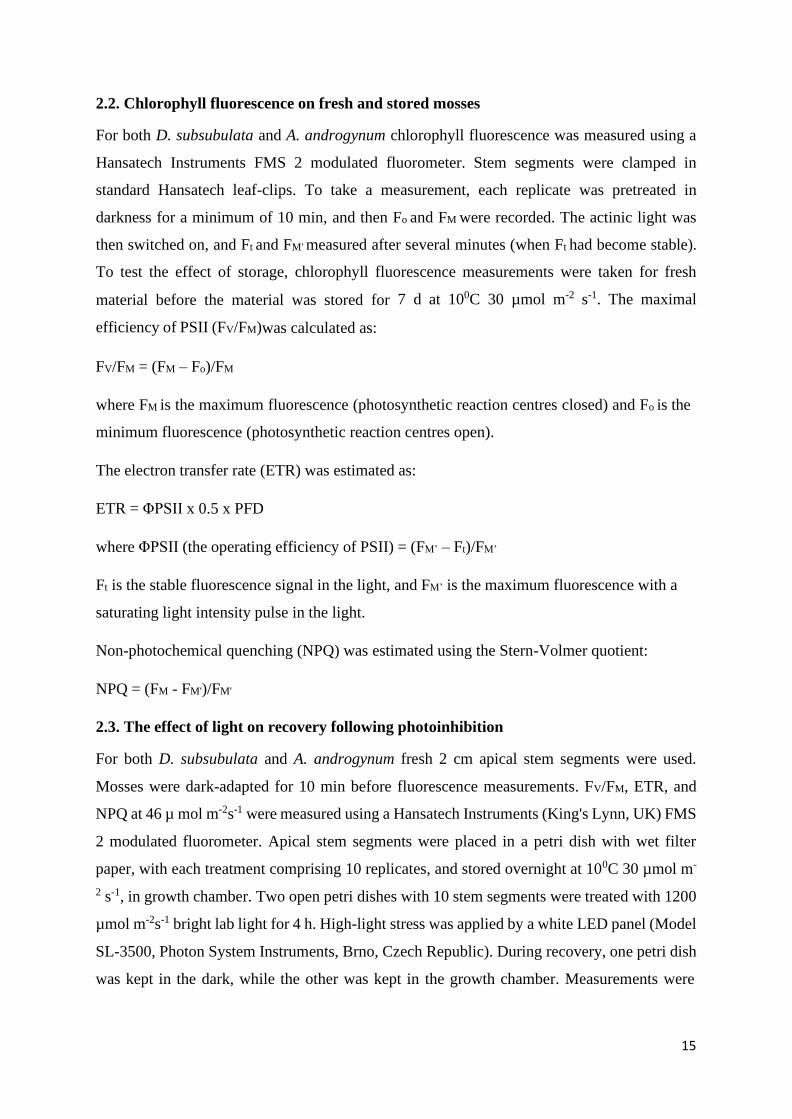
2.2. Chlorophyll fluorescence on fresh and stored mosses................................................................ 15
2.3. The effect of light on recovery following photoinhibition ......................................................... 15
2.4. The effect of slow desiccation using CaCl2 ................................................................................ 16
2.5. Effect of slow and fast desiccation on RWC and chlorophyll fluorescence parameters ............ 16
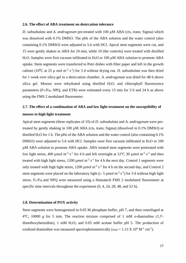
2.6. The effect of ABA treatment on desiccation tolerance .............................................................. 17
2.7. The effect of a combination of ABA and low light treatment on the susceptibility of mosses to
high light treatment ........................................................................................................................... 17
2.8. Determination of POX activity .................................................................................................. 17
2.9. Effect of slow desiccation on enzyme activity ........................................................................... 18
vi
2.10. Changes in RWC during slow and fast desiccation ................................................................. 18
2.11. Gel electrophoresis ................................................................................................................... 18
Chapter 3: Results ............................................................................................................................... 20
3.1 Light response curves.................................................................................................................. 20
3.2 Recovery from photoinhibition in dim light and the dark ........................................................... 21
3.3. Effect of slow desiccation over a saturated solution of CaCl2 ................................................... 23
3.4. Effect of slow and fast desiccation on RWC ............................................................................. 25
3.5. Effect of slow and fast desiccation on photosynthesis ............................................................... 25
3.6 The effect of ABA pretreatment on desiccation tolerance ......................................................... 29
3.7 ABA and light stress ................................................................................................................... 31
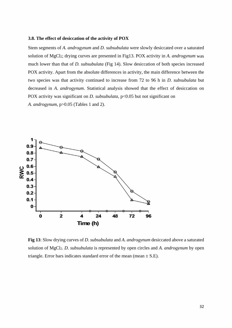
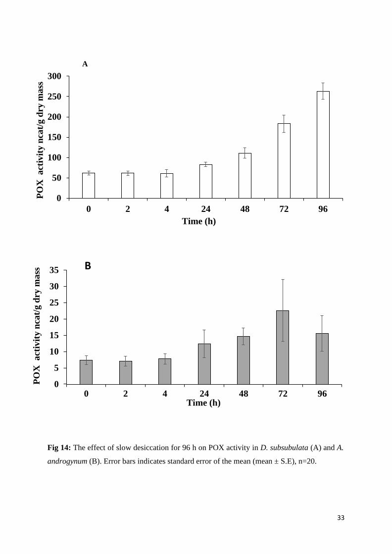
3.8 The effect of desiccation of the activity of POX ......................................................................... 32
3.9 Analysis of Peroxidase enzyme using PAGE electrophoresis. ................................................... 35
Chapter 4: Discussion ......................................................................................................................... 36
4.1. Response of photosynthesis to light intensity ............................................................................ 36
4.2. Photoinhibition ........................................................................................................................... 37
4.3. Tolerance to dehydration using CaCl2 ....................................................................................... 37
4.4. Effect of slow and fast desiccation on RWC and chlorophyll fluorescence .............................. 38
4.5. The effect of ABA pretreatment on desiccation tolerance ......................................................... 39
4.6. ABA and high light treatment .................................................................................................... 41
4. 7. Enzyme activity changes in mosses .......................................................................................... 41
4.7.1. Peroxidase activity .............................................................................................................. 41
4.7.2. Gel electrophoresis analysis ............................................................................................... 42
Chapter 5: General Conclusion and Recommendations ................................................................. 43
5.1. General conclusion ..................................................................................................................... 43
5.2. Future recommendations ............................................................................................................ 44
References ............................................................................................................................................ 45
vii
List of tables
Tables Pages
Table 1: The effect of time on POX activity of A. androgynum dried over a saturated solution of MgCl2
for 96 h. … ................................................................................................................................ 34
Table 2: The effect of time on POX activity in D. subsubulata dried over a saturated solution of MgCl2
for 96 h. … ................................................................................................................................ 34

viii
List of figures
Figures Pages
Figure 1: Evolutionary relationship between land plants and their algae relatives (Harris et al.,
2020) ...................................................................................................................................... 2
Figure 2: Diagrammatic representation of the sensing of stress by bryophytes, and signalling
during environmental stress (Bigot et al., 2018) .................................................................. 5
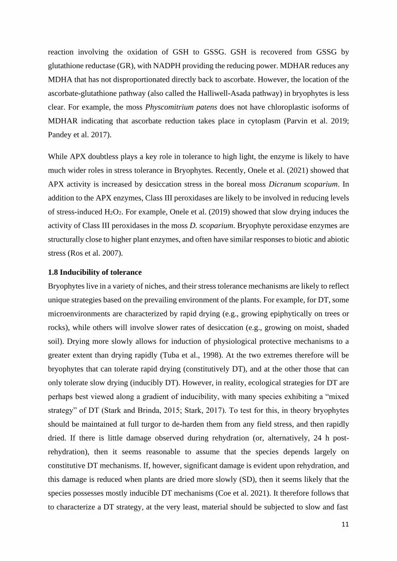
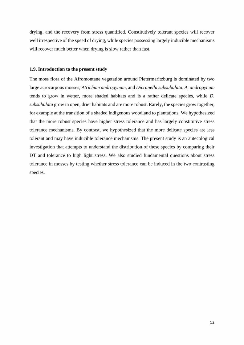
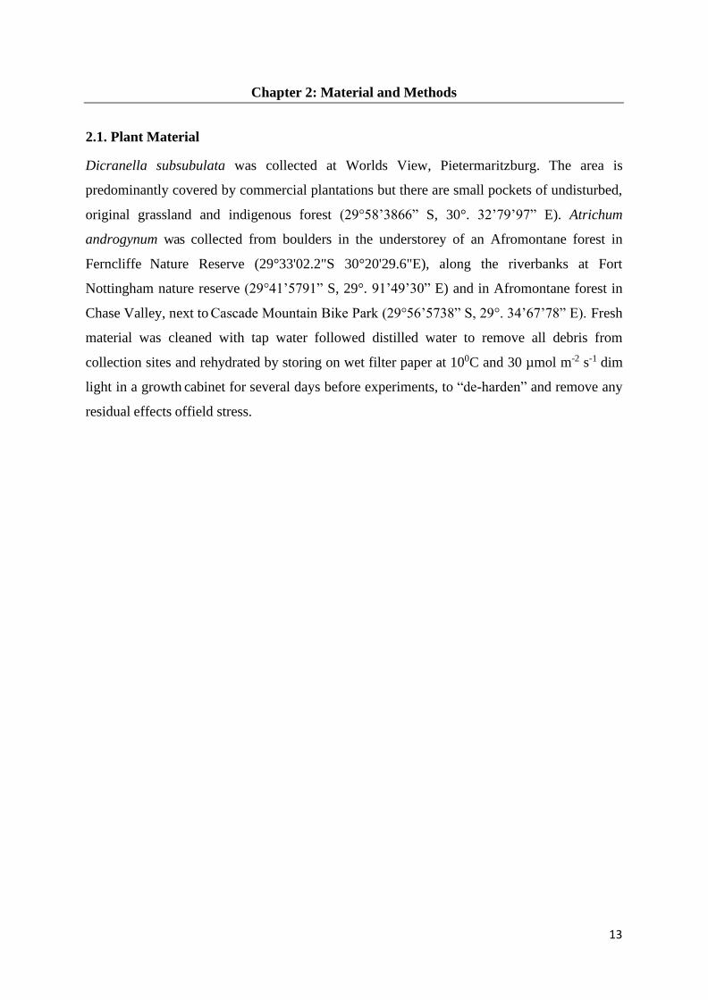
Figure 3: A. androgynum and D. subsubulata stem segment and the growth form of D.
subsubulata, together with reproduction structures .............................................................. 14
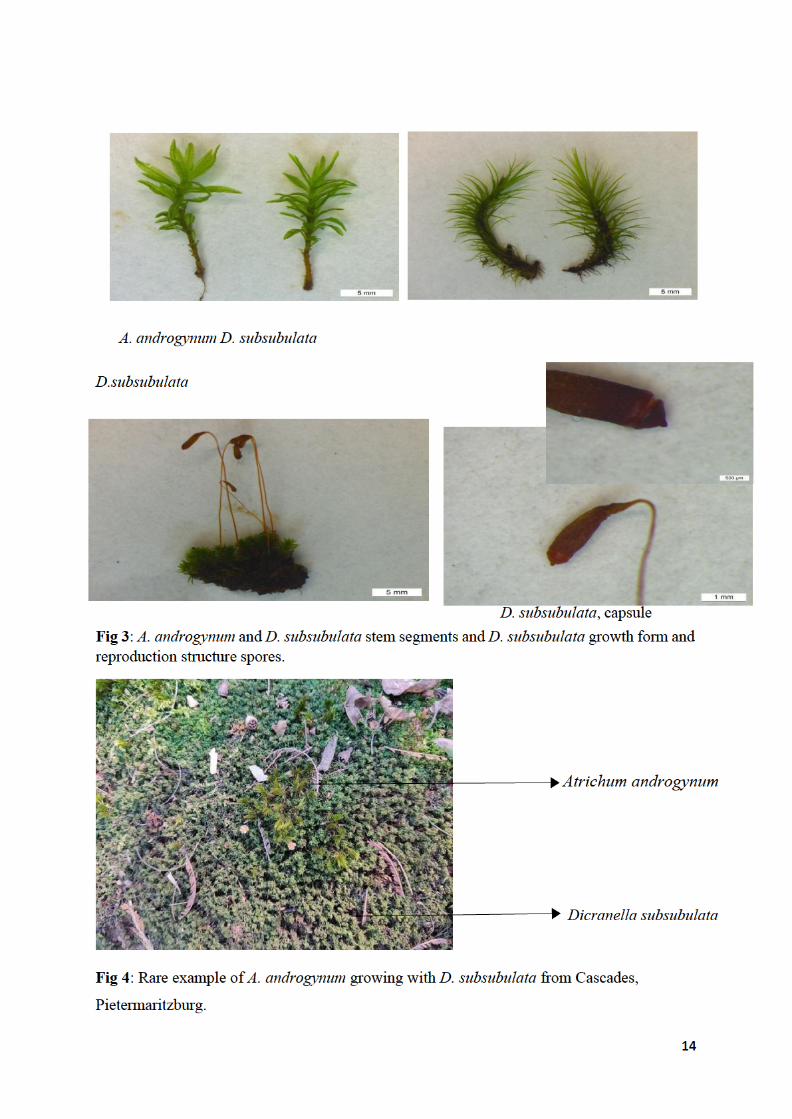
Figure 4: Rare example of A. androgynum growing with D. subsubulata from Cascades,
Pietermaritzburg… ................................................................................................................ 14
Figure 5: Light response curves of D. subsubulata (A, C) and A. androgynum (B, D) in
freshly collected material, and material stored at 10oC, 30 µmol m-2 s-1 for 7 d. Open circles
represent fresh and closed circles represent stored material. Error bars indicates standard error
of the mean, n=10 ................................................................................................................. 20
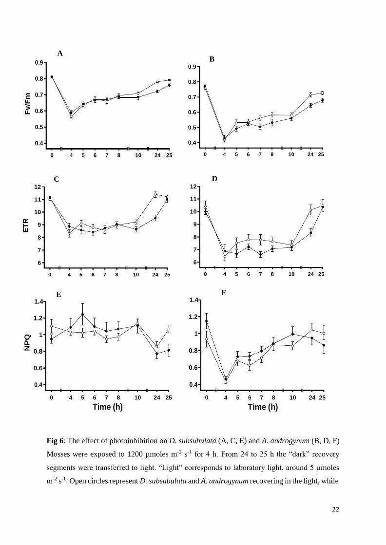
Figure 6: The effect of photoinhibition on D. subsubulata (A, C, E) and A. androgynum (B,
D, F) Mosses were exposed to 1200 µmoles m-2 s-1 for 4 h. From 24 to 25 h the “dark”
recovery segments were transferred to light. “Light” corresponds to laboratory light, around 5
µmoles m-2 s-1. Open circles represent D. subsubulata and A. androgynum recovering in the
light, while
ix
closed circles represent material recovering in the dark. Error bars indicates standard error of
the mean (mean ± S.E), n=10………………………………………………………………22
Figure 7: The effect of slow desiccation using calcium chloride (CaCl2) on D. subsubulata
(open circle) and A. androgynum (open triangles) for 1 week (A, C, E) and 48 h (B, D, F)
followed by rapid rehydration. Values at time zero correspond to initial values. Recovery
was measured every 10 min for 2 h and the next day, corresponding to 24 h after the start of
rehydration. Error bars indicate the standard error of the mean (mean ± S.E), n=10 ....... 24
Figure 8: The effect of desiccation on the RWC of D. subsubulata and A. androgynum. A.
The effect of slow desiccation using sodium chloride (NaCl) on the RWC of D. subsubulata
(open circle) and A. androgynum (closed circle). B. The effect on RWC in D. subsubulata
and A. androgynum desiccated initially over NaCl for 24 h followed by silica gel for 48 h
(total time 96 h). C. The effect of fast desiccation using silica gel on the RWC of A.
androgynum and D. subsubulata. Error bars indicate the standard error of the mean (mean ±
S.E), n=10 .......................................................................................................................... 26
x
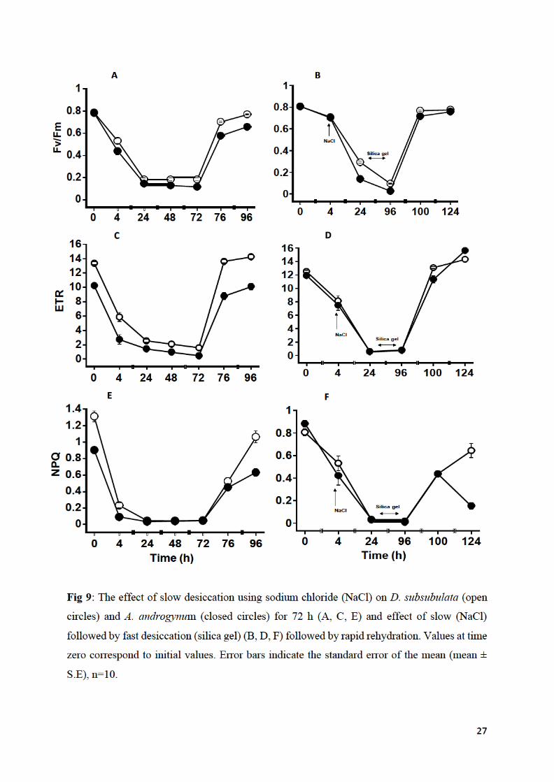
Figure 9: The effect of slow desiccation using sodium chloride (NaCl) on D. subsubulata
(open circles) and A. androgynum (closed circles) for 72 h (A, C, E) and effect of slow
(NaCl) followed by fast desiccation (silica gel) (B, D, F) followed by rapid rehydration.
Values at time zero correspond to initial values. Error bars indicate the standard error of the
mean (mean ± S.E), n=10 ............................................................................................... 27
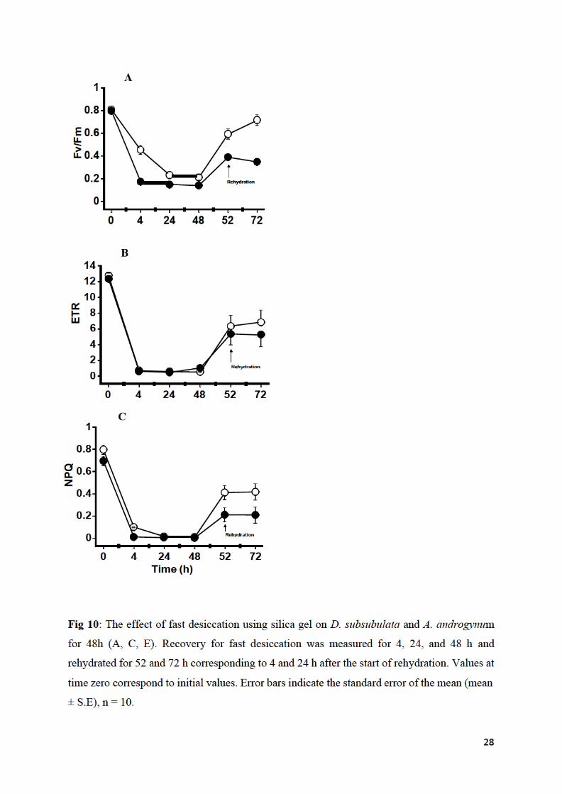
Figure 10: The effect of fast desiccation using silica gel on D. subsubulata and A.
androgynum for 48h (A, C, E). Recovery for fast desiccation was measured for 4, 24, and 48
h and rehydrated for 52 and 72 h corresponding to 4 and 24 h after the start of rehydration.
Values at time zero correspond to initial values. Error bars indicate the standard error of the
mean (mean ± S.E), n = 10 .............................................................................................. 28
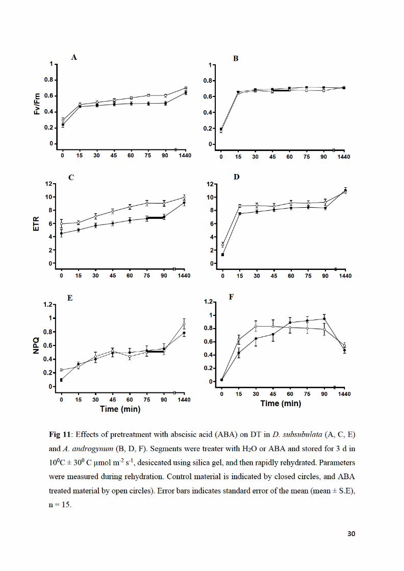
Figure 11: Effects of pretreatment with abscisic acid (ABA) on DT in D. subsubulata (A, C,
E) and A. androgynum (B, D, F). Segments were treated with H2O or ABA and stored for 3 d
in 100C and c. 30 µmol m-2 s-1, desiccated using silica gel, and then rapidly rehydrated.
Parameters were measured during rehydration. Control material is indicated by closed circles,
and ABA treated material by open circles. Error bars indicates standard error of the mean
(mean ± S.E), n = 15 ........................................................................................................ 30
Figure 12: The effect of pretreatment with a combination of light at 400 µmol m-2 s-1 and
ABA on the tolerance of chlorophyll fluorescence parameters to a high light stress of 1200
µmol m-2 s-1 in D. subsubulata (A, C) and A. androgynum (B, D). Open circles represent
mosses receiving ABA and light at 400 µmol m-2 s-1 as pre-treatments, and 1200 µmol m-2 s-1
treatment, open triangles represent mosses receiving only the treatment with 1200 µmol m-2 s-
1 light, squares represent control with neither ABA or light treatment. The light pretreatment
(400 µmol m-2 s- 1) was given from 0 to 4 h, and the light stress (1200 µmol m-2 s-1) from 24
to 28 h. Error bars indicate the standard error of the mean (mean ± S.E), n=10
……………………………………………………………….…………………..……….31
Figure 13: Slow drying curves of D. subsubulata and A. androgynum desiccated above a
saturated solution of MgCl2. D. subsubulata is represented by open circles and A.
androgynum by open triangles. Error bars indicates standard error of the mean (mean ± S.E)
……………………………………………………………………………………………. 32
Figure 14: The effect of slow desiccation for 96 h on POX activity in D. subsubulata (A) and
A. androgynum (B). Error bars indicates standard error of the mean (mean ± S.E), n=20.
…………………………………………………………………………………………….. 33

xi
Figure 15: Native PAGE electrophoresis gels of D. subsubulata stained for POX activity.
The electrophoresis was repeated four times, and this isoform was clearly visible in all
visualizations (B, C, D, E). Molecular weight markers are illustrated in lane A. .................. 35

xii
Abbreviations
RWC, Relative water content
WC, water content
min, minute
h, hour
d, day
ROS, reactive oxygen species
ABA, Abscisic acid
O2, Oxygen
H2O2, hydrogen peroxide
·OH, hydroxyl radical
H2O, water
MDA,malondialdehyde
POX, peroxidase
SOD, superoxide dismutase
GR, glutathione reductase
LEA, late embryogenesis abundant genes or proteins
A or Abs, absorbance
PYL, prybactin resistance
RoR, rate of rehydration
RoD, Rate of dehydration
Fo, minimum modulated chlorophyll fluorescence, QA in the RC of PSII oxidized
Fo´, minimum modulated fluorescence in the absence of water
Fm, maximum modulated fluorescence, QA reduced
NPQ, non-photochemical fluorescence quenching, usually calculated as (FM/FM´-1),
but (Fm/Fo´-1) after desiccation of lichens
PSI, photosystem I
PSII, photosystem II
PPFD, photosynthetically active photon flux density
FV/FM = (FM-Fo)/FM, quantum efficiency of (transiently stable) charge separation in PSII
mL, millilitre
xiii
mM, millimolar
µM,micromolar
PAR, photosynthetically active radiation
RH, relative humidity
Native PAGE, non-denaturing gel electrophoresis
RFO, raffinose
DT, desiccation tolerance
1
Chapter 1: Literature Review
1.1. Bryophytes
Bryophytes are desiccation tolerant organisms that can survive and grow in dry, high light
conditions. Although there is some debate, many consider that they were the “first land plants”,
probably because of their desiccation tolerance. Bryophytes are referred to as “ectohydric”
organisms and can receive not only water from the atmosphere but also inorganic nutrients
(Shaw and Renzaglia, 2004; Green et al., 2011). Today, there are 23000 species of bryophytes,
and they occur in a great diversity of forms, depending on the environment in which they grow
(Bahuguna et al., 2013). When studying the “autecology” of a specific species, it is important
to consider the response of the species to abiotic factors such as high light stress, desiccation,
and nutrient supply. It is also important to consider how these stresses vary throughout the
seasons. Understanding the extraordinary tolerance of mosses to some stresses could
potentially contribute to programs aimed at improving the tolerance of crop plants to abiotic
stress (Stark, 2017).
Bryophytes reach their greatest development as mats under cool and moist conditions, often on
the canopy floor. They can affect the nutrient cycling of an ecosystem by absorbing more
energy deposition and nutrients leached from dripping vegetation above the ground (Bahuguna
et al., 2013). Bryophytes are divided into three classes, namely hornworts, liverworts, and
mosses. They all play a significant role in the regulation of ecosystem nutrient cycling. Mosses
account for 25% of the annual accumulation of phosphorus from precipitation, and act as
competitors to other organisms reducing the number of minerals being shared (Bahuguna et
al., 2013). They are also desiccation tolerant (DT). Briefly, DT is the ability of an organism to
recover from being “air-dried” (Gaff and Oliver, 2013), a trait that was subsequently lost
during the evolution of angiosperms. Bryophytes have no control over water uptake and
release, and the great majority of species lack features such as a cuticle, stomata, and xylem
present in almost all vascular plants. Desiccation tolerance can be conceived of being true
“tolerance” to water stress; by comparison most vascular plants use a variety of “avoidance”
strategies to avoid drought, e.g., thick cuticles, sunken stomata, water storage, and rapid
stomatal closure (Gaff and Oliver, 2013).
Many bryophytes need to display tolerance to high light. Excess light can cause
photoinhibition, a reversible reduction in photosynthetic activities. Specific mechanisms are

2
used to avoid or repair damages caused by light stress. Some damage seems to occur to all
photosynthetic organisms even under moderate light intensities, and when the rate of repair
matches the rates of damages there is no net loss of photosynthetic activity (Keren and Krieger-
Liszkay, 2011). Photosynthetic organisms from cyanobacteria to vascular plants have various
mechanisms responsible for decreasing photoinhibitory damage (Keren and Krieger-Liszkay,
2011). These include the short-term acclimation that influences the structure and functions of
antennae complexes, for example, non-photochemical quenching (NPQ), state transitions,
reaction center quenching, alternative electron transport processes, and movement of
chloroplasts, leaves, or whole organisms away from intense light (Kehoe, 2010). Long-term
acclimatization also occurs and involves changes in the ratios of photosynthetic pigments, and
changes in the ratio of PSII to PSI (Kehoe, 2010). These mechanisms are found in all
photosynthetic organisms, including crop species that normally grow in less extreme
environments, but also cyanobacteria, lichens, and mosses that grow in extremely exposed soil
crusts (Keren and Krieger-Liszkay, 2011). If not prevented, photoinhibition will reduce
photosynthetic productivity. The main aim of the Introduction is to review tolerance to
desiccation and high light stress in plants, with an emphasis on bryophytes, as a way of
introducing the present study.
Fig 1: Evolutionary relationship between land plants and their algae relatives (Harris et al.,
2020).

3
1.2. Desiccation tolerance in bryophytes
As discussed above, almost all bryophytes are desiccation tolerant. A simple definition of DT
is an ability to survive at RWC below 10%, which they typically reach in an air-dried state
(Proctor et al., 2007). Although a bryophyte-like plant was probably the ancestor of present-
day angiosperms, later land plants developed reproductive structures such as pollen, spores,
and seeds that reduced the requirement for liquid water (Fig 1; Norwood et al., 2003; Gaff
and Oliver, 2013). Furthermore, Angiosperms have evolved cuticles, stomata, and conducting
tissues that buffer tissues relative to water contents against changes in the environment (Gaff
and Oliver, 2013). DT can be either constitutive or induced. Although one might think that
some species from for example those that form soil crusts would have exclusively
constitutive mechanisms, as they dry too quickly for mechanisms to be induced (and therefore
need high inherent tolerance) actually even in some of these species some DT can be induced
(Stark, 2017).
The severity of the effect of desiccation on bryophytes depends on the four factors according
to McLetchie and Stark (2019):
1. The duration a moss plant spends dry
2. The rate of rehydration (RoR)
3. The rate of dehydration (RoD)
4. The relative water content (RWC) in the dry state.
As discussed above, plants can be considered to be DT when an organism can return to normal
metabolism from the air-dry state. Typically, an air-dry state is achieved when the plant
material is in equilibrium with air of 50% RH or less and the plant material has reached a
relative water content c. 10%. The lack of a cuticle in most bryophytes allows their tissues to
equilibrate with the environment during dry conditions (McLetchie and Stark, 2019; Green et
al., 2011). As could be predicted, the highest DT is found within the species that are from dry
habitats (Green et al., 2011). One of the aims of studying DT bryophytes is to potentially
increase stress tolerance in higher plants. Liverworts and mosses are true green plants, and
studies on them can potentially be extrapolated to higher plants more easily than, for example,
lichens.
4
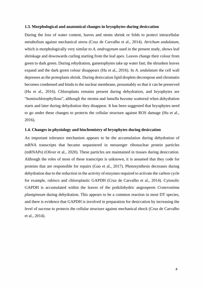
1.3. Morphological and anatomical changes in bryophytes during desiccation
During the loss of water content, leaves and stems shrink or folds to protect intracellular
metabolism against mechanical stress (Cruz de Carvalho et al., 2014). Atrichum undulatum,
which is morphologically very similar to A. androgynum used in the present study, shows leaf
shrinkage and downwards curling starting from the leaf apex. Leaves change their colour from
green to dark green. During rehydration, gametophytes take up water fast; the shrunken leaves
expand and the dark green colour disappears (Hu et al., 2016). In A. undulatum the cell wall
depresses as the protoplasts shrink. During desiccation lipid droplets decompose and chromatin
becomes condensed and binds to the nuclear membrane, presumably so that it can be preserved
(Hu et al., 2016). Chloroplasts remains present during dehydration, and bryophytes are
“homiochlorophyllous”, although the stroma and lamella become scattered when dehydration
starts and later during dehydration they disappear. It has been suggested that bryophytes need
to go under these changes to protects the cellular structure against ROS damage (Hu et al.,
2016).
1.4. Changes in physiology and biochemistry of bryophytes during desiccation
An important tolerance mechanism appears to be the accumulation during dehydration of
mRNA transcripts that became sequestered in messenger ribonuclear protein particles
(mRNAPs) (Oliver et al., 2020). These particles are maintained in tissues during desiccation.
Although the roles of most of these transcripts is unknown, it is assumed that they code for
proteins that are responsible for repairs (Gao et al., 2017). Photosynthesis decreases during
dehydration due to the reduction in the activity of enzymes required to activate the carbon cycle
for example, rubisco and chloroplastic GAPDH (Cruz de Carvalho et al., 2014). Cytosolic
GAPDH is accumulated within the leaves of the poikilohydric angiosperm Craterostima
plantgineum during dehydration. This appears to be a common reaction in most DT species,
and there is evidence that GAPDH is involved in preparation for desiccation by increasing the
level of sucrose to protects the cellular structure against mechanical shock (Cruz de Carvalho
et al., 2014).
5
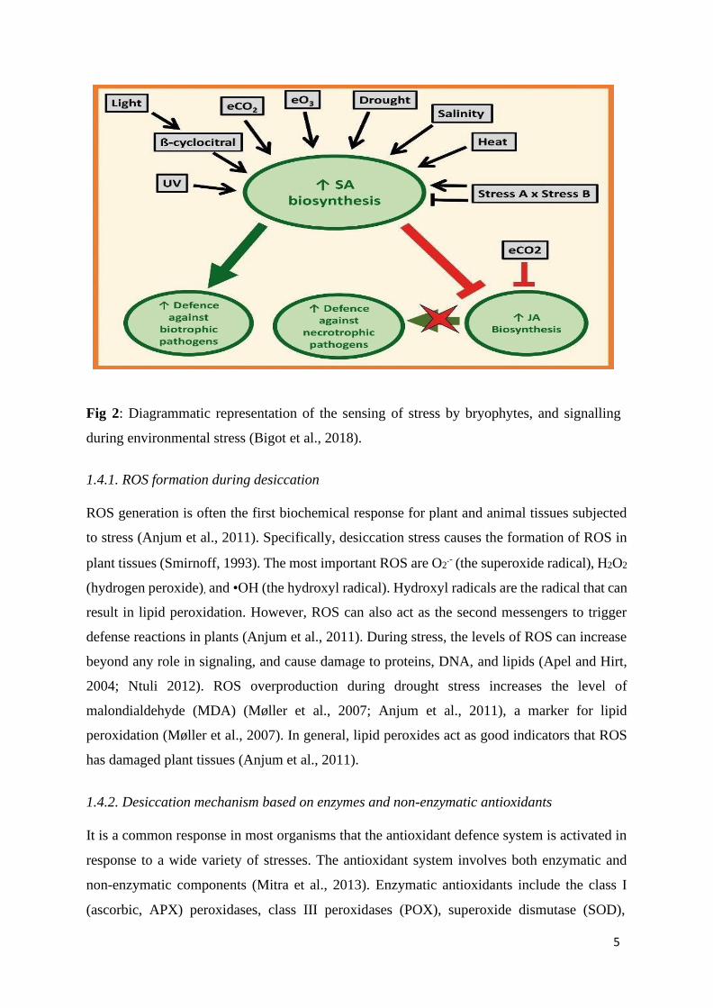
Fig 2: Diagrammatic representation of the sensing of stress by bryophytes, and signalling
during environmental stress (Bigot et al., 2018).
1.4.1. ROS formation during desiccation
ROS generation is often the first biochemical response for plant and animal tissues subjected
to stress (Anjum et al., 2011). Specifically, desiccation stress causes the formation of ROS in
plant tissues (Smirnoff, 1993). The most important ROS are O2.- (the superoxide radical), H2O2
(hydrogen peroxide), and •OH (the hydroxyl radical). Hydroxyl radicals are the radical that can
result in lipid peroxidation. However, ROS can also act as the second messengers to trigger
defense reactions in plants (Anjum et al., 2011). During stress, the levels of ROS can increase
beyond any role in signaling, and cause damage to proteins, DNA, and lipids (Apel and Hirt,
2004; Ntuli 2012). ROS overproduction during drought stress increases the level of
malondialdehyde (MDA) (Møller et al., 2007; Anjum et al., 2011), a marker for lipid
peroxidation (Møller et al., 2007). In general, lipid peroxides act as good indicators that ROS
has damaged plant tissues (Anjum et al., 2011).
1.4.2. Desiccation mechanism based on enzymes and non-enzymatic antioxidants
It is a common response in most organisms that the antioxidant defence system is activated in
response to a wide variety of stresses. The antioxidant system involves both enzymatic and
non-enzymatic components (Mitra et al., 2013). Enzymatic antioxidants include the class I
(ascorbic, APX) peroxidases, class III peroxidases (POX), superoxide dismutase (SOD),

6
catalase (CAT), and auxiliary enzymes, for example, mono- and dehydroascorbate reductases
and glutathione reductase (GR). Peroxidases are highly efficient scavengers of ROS,
specifically H2O2, but in addition peroxidases can also have a role in producing H2O2 which
may form part of the so-called “oxidative burst”, possibly to act in pathogen defence (Mayaba
et al. 2001). Catalases have lower affinity for H2O2 and are therefore less effective compared
to peroxidases in breaking down H2O2 with low concentration (Mayaba et al. 2001).
Peroxidases use different substrates such as ascorbate to breakdown H2O2 to H2O. It is well
known that an interplay exists between enzymatic and non-enzymatic antioxidants to scavenge
ROS in plants (Foyer and Halliwell, 1976).
Non-enzymatic antioxidants probably play a smaller role than enzymes but help in
controlling levels of ROS (Caverzan et al., 2016). Mitochondria synthesize ascorbate that is
transported across the cell membrane to other cell organelles (Ahmad et al., 2009). Ascorbate
is probably the most important antioxidant compound, and in addition to its role as a substrate
for peroxidase it can react directly with ROS. Another non-enzymatic ROS scavenger is
tocopherol. Tocopherol can scavenge peroxyl radicals that are found in membranes, including
those in the chloroplast (Caverzan et al., 2016). Perhaps the most important molecule
responsible for maintaining redox balance within the cellular compartments is glutathione
(GSH). GSH biosynthesis enzymes are upregulated during the abiotic stress (Apel and Hirt,
2004). Oxidized glutathione (GSSG) can be calculated as the percentage of total glutathione,
and the balance between GSSG and GSH is arguably the best indicator of redox status.
Damage caused by desiccation on protein synthesis are shown during rehydration.
Carotenoids produced by Marchantia polymorpha L. during desiccation can reduce the level
of ROS produced by desiccation stress (Kavitha and Murugan, 2016).
In humans, the imbalance of ROS and antioxidants is at least in part responsible for causing
many degenerative diseases (Ahmad et al., 2009; Gahtori and Chaturvedi, 2019). Interestingly,
some bryophytes are known as a good source of efficient natural antioxidants including phenols
and ferulic acid. It is therefore not surprising that bryophytes have been widely used as
medicinal plants. In China, bryophytes were traditionally used as medicines before they were
known as good source of antioxidants. Recently, a study was done using Oxytegus
tenuirostris, Eurhynchium striatum, and Rhynchostegium murale to investigates the activity of
antioxidants that showed that ecological factors (climate) plays a huge role in determining the
properties of antioxidant molecules in mosses (Gahtori and Chaturvedi, 2019).

7
1.4.3. Desiccation tolerance mechanisms based on sugar
An important component of DT in many organisms is the accumulation of sugars, specifically
sucrose and trehalose. Sucrose and trehalose stabilize the structure of macromolecules, protect
the biological membranes, stop diffusion, preserve the cytoskeleton, and possibly act as
antioxidants (Norwood et al., 2003). Trehalose-6-phosphate and trehalose are central
metabolite regulators impacting the status of carbohydrates energy and growth metabolism
(Gechev et al., 2012). During severe desiccation, carbon from carbohydrates such as starch and
stachyose can be metabolized to sucrose for extra osmoprotectant activity (Smeekens et al.,
2010; Gechev et al., 2012). Raffinose is made by raffinose synthase from sucrose and galactinol
(Smeekens et al., 2010).
1.4.4. Desiccation tolerance mechanisms based on LEA and other proteins
Late embryogenesis abundant genes or proteins (LEA) are proteins that are present both in
vegetative tissues and seeds. LEA proteins may supply a water hydration “shell” to specific
macromolecules and proteins to protect them against damage caused by dehydration (Ntuli,
2012). In addition to LEA proteins, small heat shock proteins (sHSPs) are expressed in a
desiccation-tolerant plant without stress and are also induced during drought stress (Gechev et
al., 2012). sHSPs can act as molecular chaperones for some proteins, protecting them from
denaturation and aggregation (Farrant et al., 2007). Rapid induction of heat shock genes often
occurs during dehydration in resurrection plants (Hundertmark and Hincha, 2008). Studies on
the “model moss” Physcomitrium patens have shown the importance of dehydrin genes such
as PpDHNA in mosses subjected to salt and osmotic stress (Saavedra et al., 2006). While the
precise function of LEA and sHSP proteins remain unclear, there is enough evidence to suggest
that they play an important role in the mechanism of DT in resurrection plants.
1.5. Abscisic acid
Abscisic acid (ABA) is an important plant phytohormone that plays a significant role in the
response of plants to abiotic stress, senescence, seed growth, and generally in plant
development (Kuromori et al., 2010). The most important role of ABA in vascular plants is
probably stomatal regulation, but the hormone is involved in many other stresses and normal
plant development. Land plants have multiple receptors for ABA, nucleocytoplasmic center is
the most important with two with specific receptors, PYR (pyrabactin resistance) or PYR-like
receptor. Although ABA is produced in plant roots, it can be translocated to the guard cells in
the xylem. The receptor AtABCG25 is responsible for intracellular ABA transport (Kuromori

8
et al., 2010). ABA regulates the synthesis of Late Embryogenesis Abundant protein genes
during the desiccation that accompanies seed maturation. LEA proteins are also responsible
for increasing dehydration tolerance in vascular plants (Koster et al., 2010).
Hartung (2010) reported that at that time, nine mosses species were known to possess
endogenous ABA (Hartung, 2010). The amount of ABA varies greatly; for example, Riccia
fluitans has high levels of ABA that may be related to its high DT (Hartung and Gimmler,
1994). There are only a few reports on the biosynthesis and metabolism of ABA in Bryophytes.
In lower plants, ABA is involved in signalling the upregulation of genes that are needed for
DT. For example, pre-treatment of the moss Atrichum androgynum increased the rate of
recovery the photosynthesis following desiccation, and also doubled NPQ, which may reduce
ROS formation (Mayaba et al. 2001). In the same work, ABA was shown to slightly increase
the accumulation of sugar, which as discussed above may be responsible for the protection of
the membrane during desiccation (Mayaba et al., 2001).
1.6. Tolerance to light stress by avoiding ROS formation.
1.6.1. Photoinhibition in mosses
High light can damage plants, and a major reason for the damage is the formation of ROS
(Pospíšil., 2016). When the light energy exceeds the maximum capacity of the photosystem to
utilize it, ROS formation in PSII can induce photoinhibition (Pospíšil et al., 2016).
Chlorophylls are found in PS antennae complex. When singlet state chlorophyll absorbs excess
energy, it is converted to the potentially harmful triplet state chlorophyll. Xanthophylls directly
quench excited chlorophyll preventing the formation of triplet chlorophyll (Pospíšil et al.,
2016). Indirect prevention of the formation of triplet chlorophyll involves the rearrangement
of Lhcb protein controlled by Psbs (Ruban et al., 2012). Quenching singlet chlorophyll can be
insufficient leading to conversion to triplet chlorophyll thus transferring energy from O2
forming 1O2 and other ROS, responsible for most photoinhibition (Pospíšil et al., 2016).
PSI is sensitive to inhibition induced by high light, particularly at low temperatures (Foyer,
2018). Light during the summer season is not regarded as the determining factor for low
productivity of bryophytes. Nutrient availability and water stress are often cited as having large
effects on productivity. However, photoinhibition can also limit the productivity of moss at
high latitudes where it is cold, possibly in part due to inhibition of PSI. In general, the optimal
light intensity of photosynthesis in Bryophytes is rather low in high latitudes (Barták et al.,

9
2012). Damage caused by high light in photosynthetic apparatus are limiting factors to the
productivity of Antarctic bryophytes (Adamson et al., 1988). Mosses experience stress in
polar habitats, where the combined effects of low temperature and bright light are extremely
stressful. (Adamson et., 1988). Interestingly, it has been observed that mosses growing in
shaded habitats can grow faster than mosses without a canopy above them (Murray et al.,
1993). High light and desiccation stress induces similar physiological effects on the
photosynthetic apparatus of bryophytes; both can promote ROS formation, particularly around
PSII (Barták et al., 2012). It seems likely that ROS formation is the major cause of damage to
the photosynthetic apparatus under the effects of dehydration and high light stress (Barták et
al., 2012).
1.6.2. Light screening by flavonoids and phenols in bryophytes
Flavonoid biosynthesis is an important response of plants to their environment. Many stresses
such as cold, UV-B light (UVB), desiccation, and salinity induce the production of flavonoids
(Davies et al., 2020). Flavonoid o-glycosides are the main important flavonoids acting to
provide UVB tolerance in both Marchantia and Arabidopsis (Yin and Ulm, 2017; Davies et
al., 2020). If for any reason (e.g. mutation) the production of flavonoids is reduced, the
susceptibility of Marchantia and Arabidopsis to UV increases. The production of flavonoids
can be reduced by excessive UV light. Interestingly, mutants with elevated levels of flavonoids
have high UVB tolerance (Soriano et al., 2018). However, in mosses and lycophytes the
flavonoid biosynthsis pathway that protects against UVB light appears to be absent. In mosses,
phenolics appear to be the most important compounds for UVB tolerance (Soriano et al., 2018).
Ultraviolet (UV) radiation is the noticeable environmental factors with an influence on
photosynthetic organism and water availability is the important environmental factor in
mosses. Two compounds p-coumaric and ferulic acid are induced by UV radiation in
Plagiochila asplenioides. In Bryophytes, unlike lichens, pigments that screen the
photosynthetic apparatus from high PAR are rare, rather these compounds and appear to be
produced more to stop UV light. Some bryophytes became pigmented during the exposure to
high light (Waterman et al., 2017), but more work is needed to assess whether these pigments
offer any photoprotection (Waterman et al., 2017; Soriano et al., 2018). Nevertheless, in the
context of the present study, neither D. subsubulata nor A. androgynum have ever been
observed to synthesize pigments.

10
1.6.3. Induction of Non-Photochemical quenching (NPQ) by light
Non-Photochemical quenching (NPQ) can protect plants from oxidative stress caused by high
light by dissipating excess energy radiating as heat (Gerotto et al., 2012). Photosynthetic
organisms harvest light from sunlight and convert it into chemical energy. During the
evolution of first land plants, plant needed to develop suitable mechanisms to protect plants
from high light and excessive UV radiation, first land plants needed to survive combinations
of desiccation and light stress. As the atmosphere became oxygenated by photosynthesis, it
seems likely that the risk of ROS production increased (Niyogi, 1999; Gerotto et al., 2012).
NPQ is a short-term method of protecting plants from excess light (Gerotto et al., 2012). NPQ
has two main components, energy (qE) and inhibitory quenching (qI). qE is generally believed
to be caused by the enzyme-catalyzed xanthophyll cycle, whereby the carotenoid violaxanthin
is enzymatically converted to zeaxanthin in a pH-regulated process that occurs during increases
in light intensity (Demmig-Adams et al. 2020). LHCSR 1, 2, and Psbs are involved in the
induction of NPQ, but the precise mechanism remains unclear (Gerotto et al., 2012). Using the
portable and lab chlorophyll fluorescence devices, NPQ can be measured in the field and in
more controlled environments. Studies showed that in general highly desiccation tolerant
bryophytes have higher levels of NPQ than more sensitive species (Deltoro et al., 1998).
Beckett et al. (2005) showed that in A. androgynum slow drying increases qE. However, NPQ
has not been studied in D. subsubulata.
1.7. Class I and Class III Peroxidases in mosses
As discussed above, antioxidant enzymes form an important part of the defence of organisms
against oxidative stress, irrespective of how such stress occurs. One of the most important
families of antioxidant enzymes are the peroxidases. Peroxidases are found in almost all
organisms from plants, animals to bacteria (Pandey et al., 2017). Peroxidases play the important
role of breaking down H2O2 to water (H2O) and oxygen (O2) (Pandey et al., 2017). Hydrogen
peroxide is a toxic product that is produced in multiple sites because of stress. In vascular
plants, the ascorbate-glutathione (or Halliwell-Asada) cycle removes harmful ROS,
particularly in the chloroplasts. Here, superoxide dismutase (SOD) dismutates O2.- to H2O2
which is broken down to H2O and O2 with reaction catalyzed by ascorbate peroxidase (APX)
or Class I peroxidases within the stroma. In the process, ascorbate is converted to
monodehydroascorbate (MDHA), some of which disproportionates to dehydroascorbate
(DHA). Ascorbate is regenerated from DHA by dehydroascorbate reductase (DHAR) in a
11
reaction involving the oxidation of GSH to GSSG. GSH is recovered from GSSG by
glutathione reductase (GR), with NADPH providing the reducing power. MDHAR reduces any
MDHA that has not disproportionated directly back to ascorbate. However, the location of the
ascorbate-glutathione pathway (also called the Halliwell-Asada pathway) in bryophytes is less
clear. For example, the moss Physcomitrium patens does not have chloroplastic isoforms of
MDHAR indicating that ascorbate reduction takes place in cytoplasm (Parvin et al. 2019;
Pandey et al. 2017).
While APX doubtless plays a key role in tolerance to high light, the enzyme is likely to have
much wider roles in stress tolerance in Bryophytes. Recently, Onele et al. (2021) showed that
APX activity is increased by desiccation stress in the boreal moss Dicranum scoparium. In
addition to the APX enzymes, Class III peroxidases are likely to be involved in reducing levels
of stress-induced H2O2. For example, Onele et al. (2019) showed that slow drying induces the
activity of Class III peroxidases in the moss D. scoparium. Bryophyte peroxidase enzymes are
structurally close to higher plant enzymes, and often have similar responses to biotic and abiotic
stress (Ros et al. 2007).
1.8 Inducibility of tolerance
Bryophytes live in a variety of niches, and their stress tolerance mechanisms are likely to reflect
unique strategies based on the prevailing environment of the plants. For example, for DT, some
microenvironments are characterized by rapid drying (e.g., growing epiphytically on trees or
rocks), while others will involve slower rates of desiccation (e.g., growing on moist, shaded
soil). Drying more slowly allows for induction of physiological protective mechanisms to a
greater extent than drying rapidly (Tuba et al., 1998). At the two extremes therefore will be
bryophytes that can tolerate rapid drying (constitutively DT), and at the other those that can
only tolerate slow drying (inducibly DT). However, in reality, ecological strategies for DT are
perhaps best viewed along a gradient of inducibility, with many species exhibiting a “mixed
strategy” of DT (Stark and Brinda, 2015; Stark, 2017). To test for this, in theory bryophytes
should be maintained at full turgor to de-harden them from any field stress, and then rapidly
dried. If there is little damage observed during rehydration (or, alternatively, 24 h post-
rehydration), then it seems reasonable to assume that the species depends largely on
constitutive DT mechanisms. If, however, significant damage is evident upon rehydration, and
this damage is reduced when plants are dried more slowly (SD), then it seems likely that the
species possesses mostly inducible DT mechanisms (Coe et al. 2021). It therefore follows that
to characterize a DT strategy, at the very least, material should be subjected to slow and fast
12
drying, and the recovery from stress quantified. Constitutively tolerant species will recover
well irrespective of the speed of drying, while species possessing largely inducible mechanisms
will recover much better when drying is slow rather than fast.
1.9. Introduction to the present study
The moss flora of the Afromontane vegetation around Pietermaritzburg is dominated by two
large acrocarpous mosses, Atrichum androgynum, and Dicranella subsubulata. A. androgynum
tends to grow in wetter, more shaded habitats and is a rather delicate species, while D.
subsubulata grow in open, drier habitats and are more robust. Rarely, the species grow together,
for example at the transition of a shaded indigenous woodland to plantations. We hypothesized
that the more robust species have higher stress tolerance and has largely constitutive stress
tolerance mechanisms. By contrast, we hypothesized that the more delicate species are less
tolerant and may have inducible tolerance mechanisms. The present study is an autecological
investigation that attempts to understand the distribution of these species by comparing their
DT and tolerance to high light stress. We also studied fundamental questions about stress
tolerance in mosses by testing whether stress tolerance can be induced in the two contrasting
species.
13
Chapter 2: Material and Methods
2.1. Plant Material
Dicranella subsubulata was collected at Worlds View, Pietermaritzburg. The area is
predominantly covered by commercial plantations but there are small pockets of undisturbed,
original grassland and indigenous forest (29°58’3866” S, 30°. 32’79’97” E). Atrichum
androgynum was collected from boulders in the understorey of an Afromontane forest in
Ferncliffe Nature Reserve (29°33'02.2"S 30°20'29.6"E), along the riverbanks at Fort
Nottingham nature reserve (29°41’5791” S, 29°. 91’49’30” E) and in Afromontane forest in
Chase Valley, next to Cascade Mountain Bike Park (29°56’5738” S, 29°. 34’67’78” E). Fresh
material was cleaned with tap water followed distilled water to remove all debris from
collection sites and rehydrated by storing on wet filter paper at 100C and 30 µmol m-2 s-1 dim
light in a growth cabinet for several days before experiments, to “de-harden” and remove any
residual effects of field stress.
15
2.2. Chlorophyll fluorescence on fresh and stored mosses
For both D. subsubulata and A. androgynum chlorophyll fluorescence was measured using a
Hansatech Instruments FMS 2 modulated fluorometer. Stem segments were clamped in
standard Hansatech leaf-clips. To take a measurement, each replicate was pretreated in
darkness for a minimum of 10 min, and then Fo and FM were recorded. The actinic light was
then switched on, and Ft and FM' measured after several minutes (when Ft had become stable).
To test the effect of storage, chlorophyll fluorescence measurements were taken for fresh
material before the material was stored for 7 d at 100C 30 µmol m-2 s-1. The maximal
efficiency of PSII (FV/FM) was calculated as:
FV/FM = (FM – Fo)/FM
where FM is the maximum fluorescence (photosynthetic reaction centres closed) and Fo is the
minimum fluorescence (photosynthetic reaction centres open).
The electron transfer rate (ETR) was estimated as:
ETR = ΦPSII x 0.5 x PFD
where ΦPSII (the operating efficiency of PSII) = (FM’ – Ft)/FM’
Ft is the stable fluorescence signal in the light, and FM’ is the maximum fluorescence with a
saturating light intensity pulse in the light.
Non-photochemical quenching (NPQ) was estimated using the Stern-Volmer quotient:
NPQ = (FM - FM')/FM'
2.3. The effect of light on recovery following photoinhibition
For both D. subsubulata and A. androgynum fresh 2 cm apical stem segments were used.
Mosses were dark-adapted for 10 min before fluorescence measurements. FV/FM, ETR, and
NPQ at 46 µ mol m-2s-1 were measured using a Hansatech Instruments (King's Lynn, UK) FMS
2 modulated fluorometer. Apical stem segments were placed in a petri dish with wet filter
paper, with each treatment comprising 10 replicates, and stored overnight at 100C 30 µmol m-
2 s-1, in growth chamber. Two open petri dishes with 10 stem segments were treated with 1200
µmol m-2s-1 bright lab light for 4 h. High-light stress was applied by a white LED panel (Model
SL-3500, Photon System Instruments, Brno, Czech Republic). During recovery, one petri dish
was kept in the dark, while the other was kept in the growth chamber. Measurements were

16
taken at 4, 5, 6, 7 8, and 10 h after the start of the experiment, and then again 24 and 25
h.
2.4. The effect of slow desiccation using CaCl2
Materials (10 segments from each species) were slowly dried above a saturated solution of
CaCl2 giving RH of c. 40% for 48 h and 1 week at 10°C, 30 µmol m-2 s-1. Segments were
rapidly rehydrated in the dark and chlorophyll fluorescence parameters were estimated every
10 min for 120 min and again after 1440 min.
2.5. Effect of slow and fast desiccation on RWC and chlorophyll fluorescence parameters
The turgid masses of the apical stem segments were determined by weighing blotted material
that had been soaked in 25 ml distilled H2O for 10 min, with gentle shaking on an orbital shaker.
The relative water content (RWC) during drying was estimated as (fresh mass after drying −
dry mass)/ (turgid mass − dry mass). Mosses were dried for 72 h at 70°C to obtain their dry
mass. RWC was therefore estimated as:
RWC= (FM-DM)/(TM-DM)
FM – Fresh mass (mass at any given time)
TM – Turgid mass
DM – Dried mass (oven dried at 70˚C).
For both D. subsubulata and A. androgynum, 2 cm apical stem segments were cut. Chlorophyll
fluorescence parameters were measured using a Hansatech Instruments FMS 2 modulated
fluorometer. Silica gel and sodium chloride (NaCl) were used as desiccants, providing fast and
slow drying respectively. For each treatment 10 stem segment for each species were used.
Treatments were as follows; First, both D. subsubulata and A. androgynum were desiccated
above a saturated solution of NaCl (giving a RH of c. 75%) for 72 h, with measurements being
made after 0, 4, 24, 48 and 72 h. Second, both D. subsubulata and A. androgynum were
desiccated using NaCl for 24 h followed by silica gel for 48 h, and measured 0, 4, 24 and 96 h
after the start of the experiment. In addition, A. androgynum was desiccated using silica gel for
48 h, and measured after 0, 4, 48 h. D. subsubulata was desiccated using NaCl for 24 h followed
by silica gel for 144 h, and measurements taken after 0, 4, 24, 48 and 144 h. In all cases, mosses
were rapidly rehydrated in liquid water, and measurements taken after 4 and 24 h.
17
2.6. The effect of ABA treatment on desiccation tolerance
D. subsubulata and A. androgynum pre-treated with 100 µM ABA (cis, trans; Sigma) which
was dissolved with 0.1% DMSO. The pHs of the ABA solution and the water control (also
containing 0.1% DMSO) were adjusted to 5.6 with HCl. Apical stem segments were cut, and
15 were gently shaken in ABA for 10 min, while 10 (the controls) were treated with distilled
H2O. Samples were first vacuum infiltrated in H2O or 100 µM ABA solution to promote ABA
uptake. Stem segments were transferred to Petri dishes with filter paper and left in the growth
cabinet (100C at 25 µ mol m-2 s-1) for 3 d without drying out. D. subsubulata was then dried
for 1 week over silica gel in a desiccation chamber. A. androgynum was dried for 48 h above
silica gel. Mosses were rehydrated using distilled H2O, and chlorophyll fluorescence
parameters (FV/FM, NPQ, and ETR) were estimated every 15 min for 3 h and 24 h as above
using the FMS 2 modulated fluorometer.
2.7. The effect of a combination of ABA and low light treatment on the susceptibility of
mosses to high light treatment
Apical stem segments (three replicates of 10) of D. subsubulata and A. androgynum were pre-
treated by gently shaking in 100 µM ABA (cis, trans; Sigma) (dissolved in 0.1% DMSO) or
distilled H2O for 1 h. The pHs of the ABA solution and the water control (also containing 0.1%
DMSO) were adjusted to 5.6 with HCl. Samples were first vacuum infiltrated in H2O or 100
µM ABA solution to promote ABA uptake. ABA treated stem segments were pretreated with
low light stress, 400 µmol m-2 s-1 for 4 h and left overnight at 12oC 30 µmol m-2 s-1 and then
treated with high light stress, 1200 µmol m-2 s-1 for 4 h the next day. Control 1 segments were
only treated with high light stress, 1200 µmol m-2 s-1 for 4 h on the second day, and Control 2
stem segments were placed on the laboratory light (c. 5 µmol m-2 s-1) for 3 d without high light
stress. FV/FM and NPQ were measured using a Hansatech FMS 2 modulated fluorometer at
specific time intervals throughout the experiment (0, 4, 24, 28, 48, and 52 h).
2.8. Determination of POX activity
Stem segments were homogenized in 0.05 M phosphate buffer, pH 7, and then centrifuged at
4oC, 10000 g for 5 min. The reaction mixture comprised of 1 mM o-dianisidine (3,3′-
dimethoxybenzidine), 1 mM H2O2, and 0.05 mM acetate buffer pH 5. The production of
oxidized dianisidine was measured spectrophotometrically (ε460 = 1.13 X 10⁴ M-1 cm-1).

18
2.9. Effect of slow desiccation on enzyme activity
For both D. subsubulata and A. androgynum changes in the activity of POX were studied
during a drying/wetting cycle. Each treatment (sampling time) contained five replicates each
comprising of five 2 cm apical stem segments (c. 0.04 g dry mass). The material was initially
fully hydrated by gentle shaking on an orbital shaker in distilled H2O for 1 h, then slow dried
by placing stems segment over a saturated solution of MgCl2 (resulting in a relative humidity
of 34%) in the growth cabinet for 96 h. At selected time intervals the relative water content
(RWC, see below) and POX activity was measured. After grinding as described above, extracts
were centrifuged at 4oC in Eppendorf tubes for 5 min at 1000 g and supernatants frozen at -
24oC until analysis.
2.10. Changes in RWC during slow and fast desiccation
For both D. subsubulata and A. androgynum 2 cm apical stem segments were cut and placed
and used to check for relative water content (RWC) during dehydration and rehydration.
Initially, the material was fully hydrated by gentle shaking on an orbital shaker in distilled
H2O for 1 h and weighed to get the turgid mass. Segments of both species were then placed in
small (65 mm) Petri dishes (5 replicates of 5 segments). To compare the effect of slow and
rapid dehydration, Petri dishes with stem segments were placed for 48 h above a saturated
solution of MgCl2 (giving a relative humidity of c. 40%) or silica gel (giving a relative humidity
of 0%) in the growth cabinet as described above. Segments were weighed after 4, 8, 24, 28, 32,
and 48 h and were then rapidly hydrated by placing in distilled water and weighed again after
4 and 8 h (corresponding to 52 and 56 h from the start of the experiment). Mosses were dried
for 48 h at 70°C to obtain their dry mass. RWC was estimated as above.
2.11. Gel electrophoresis
Polyacrylamide gel electrophoresis (PAGE) was used to test for the presence of peroxidase
isoforms in the two species. Fresh apical stem segments were homogenized using phosphate
buffer pH 7 and then the extracts were centrifuged at 3oC for 5 min at 10000 g. A. androgynum
extracts were concentrated by dialysis on solid sucrose overnight, and then reverse dialyzed
again with phosphate buffer pH 7 overnight, to remove sucrose from the samples. Extracts
were further concentrated by centrifugation in Millipore “microcons” with a molecular cut-off
of 10 000 Da according to manufactures recommendations. Extracts from D. subsubulata did
not need concentrating.

19
A modification of the method of Laemmli (1970) was used, using native 12% and 6% gels.
Peroxidase activity was visualized using acetate buffer (0.25 M, pH 5.0) containing 10%
glycerol and 1 mM o-dianisidine with 2 mM H2O2. To test for superoxide production, Na-
phosphate buffer (50 mM, pH 7.4) containing 10% glycerol, 0.1 mM MgCl2, and 1mM CaCl2
was used to wash the gel for 30 min. The gel was then incubated in the dark with the same
buffer containing 0.4 mM NADH and 0.5 mM NBT for 30 min. Serrano at al. (1994) method
of staining was followed to estimates the molecular sizes of DNA.

20
A B
C D
Chapter 3: Results
3.1 Light response curves
D. subsubulata and A. androgynum are adapted to relatively high light intensities, and both
species were affected by storage at 10oC, 30 µmol m-2 s-1 for one week (Fig 5). However,
ETR clearly saturated at higher light intensities in D. subsubulata than A. androgynum. Even
at 500 µmoles m-2s-1 ETR in D. subsubulata was not quite saturated, while in A. androgynum
ETR appeared to saturate at around 200 µmoles m-2 s-1. Similarly, NPQ kept increasing at
higher light intensities in D. subsubulata, while only increasing a little beyond 200 µmoles m-
2 s-1 in A. androgynum. In D. subsubulata storage in cool dim conditions reduced ETR and
NPQ. The effects were similar in A. androgynum, although at lower light intensities (below
200 µmoles m-2 s-1) storage increased NPQ.
Fig 5: Light response curves of D. subsubulata (A, C) and A. androgynum (B, D) in freshly
collected material and material stored at 10oC, 30 µmol m-2 s-1 for 7 d. Open circles represent
fresh and closed circles represent stored material. Error bars indicates standard error of the
mean (mean ± S.E), n=10.

21
3.2 Recovery from photoinhibition in dim light and the dark
The effect of light during the recovery from photoinhibition was assessed by measuring the
chlorophyll fluorescence parameters FV/FM, ETR and NPQ in D. subsubulata and A.
androgynum (Fig 6). Mosses were given a high light stress of 1200 µmol m-2 s-1 for 4 h. A.
androgynum was more sensitive than D. subsubulata. FV/FM was reduced to just below 0.6 in
D. subsubulata, but to c. 0.4 in A. androgynum, and ETR was reduced to a greater extent in D.
subsubulata than in A. androgynum. NPQ was reduced much more by high light stress in A.
androgynum than D. subsubulata. In both species, the recovery of FV/FM and ETR was similar
in the light and the dark for the first 6 h. However, for both species, recovery 20 h after exposure
was significantly better if material was allowed to recover in the light. For ETR, transferring
material that had been allowed to recover in the dark to the light resulted in rapid recovery to
values similar to those of material that the recovered in the light. In A. androgynum, while light
stress reduced NPQ, values gradually recovered, and were similar in the light and the dark.
22
ET
R
NP
Q
Fv/F
m
0.9
0.8
0.7
0.6
0.5
0.4
A
0 4 5 6 7 8 10 24 25
0.9
0.8
0.7
0.6
0.5
0.4
B
0 4 5 6 7 8 10 24 25
C 12
11
10
9
8
7
6
0 4 5 6 7 8 10 24 25
D 12
11
10
9
8
7
6
0 4 5 6 7 8 10 24 25
E 1.4
1.2
1
0.8
0.6
F 1.4
1.2
1
0.8
0.6
0.4
0 4 5 6 7 8 10 24 25
Time (h)
0.4
0 4 5 6 7 8 10 24 25
Time (h)
Fig 6: The effect of photoinhibition on D. subsubulata (A, C, E) and A. androgynum (B, D, F)
Mosses were exposed to 1200 µmoles m-2 s-1 for 4 h. From 24 to 25 h the “dark” recovery
segments were transferred to light. “Light” corresponds to laboratory light, around 5 µmoles
m-2 s-1. Open circles represent D. subsubulata and A. androgynum recovering in the light, while
23
closed circles represent material recovering in the dark. Error bars indicates standard error of
the mean (mean ± S.E), n=10.
3.3. Effect of slow desiccation over a saturated solution of CaCl2
The effect of slow desiccation on chlorophyll fluorescence parameters was assessed by storing
material above a saturated solution of CaCl2. A. androgynum was more sensitive to desiccation
than D. subsubulata (Fig 7). D. subsubulata recovered fast following desiccation for 48 h,
FV/FM, ETR and NPQ initially recovery quickly (within 10 min), ETR reached initial values
after 24 h. After desiccation for 1 week, recovery of fluorescence parameters was very slow,
and even after 24 h was only c. 30% of the initial values. By contrast, in D. subsubulata,
recovery was faster, and reached initial values even after desiccation for 1 week.


25
3.4. Effect of slow and fast desiccation on RWC
D. subsubulata and A. androgynum lost at least 60% and 80% respectively of their water
content after 72 h when desiccated above NaCl (Fig 8A). Desiccating D. subsubulata and
A. androgynum above NaCl for 24 h followed by 96 h above silica gel reduced water contents
to low values (Fig 8B). When desiccating the mosses above silica gel, A. androgynum quickly
lost 60% water after 4 h (Fig 8C). D. subsubulata only lost 30% of RWC in 4 h of slow drying
but lost 70% of its water content after 48 h of fast desiccation (Fig 8C).
3.5. Effect of slow and fast desiccation on photosynthesis
Slow desiccation for 72 h above NaCl, progressively reduced FV/FM, ETR and NPQ in both
D. subsubulata and A. androgynum (Fig 9A, C, and E). Chlorophyll fluorescence parameters
rapidly recovered to their original values following rehydration in both species. When
desiccated slowly above NaCl, and then placed above silica get, in both D. subsubulata and A.
androgynum FV/FM and ETR recovered rapidly and completely (Fig 9 B, D). While NPQ
fully recovered in D. subsubulata, in A. androgynum NPQ initially increased and then then
decreased to low values (Fig 9F). By contrast, following rapid drying for 48 h above silica gel,
none of the chlorophyll fluorescence parameters in A. androgynum fully recovered (Fig 10A,
B, C). In D. subsubulata FV/FM fully recovered (Fig 10A). Although the recovery of ETR
and NPQ was incomplete for both species, D. subsubulata recovered better than A.
androgynum (Fig 10B, C).


29
3.6. The effect of ABA pretreatment on desiccation tolerance
D. subsubulata and A. androgynum were both pre-treated with 100 µM ABA or distilled water
and desiccated using silica gel for 1 week. Chlorophyll fluorescence parameters of both species
of moss recovered well following desiccation (Fig 11). Pretreatment with ABA improved the
rate of recovery of ETR in both species, but only improved the rate of recovery of FV/FM in D.
subsubulata. Overall, D. subsubulata was more desiccation tolerant than A. androgynum (Fig
11). NPQ recovered more slowly than the other parameters, and ABA increased the rate of
recovery in A. androgynum but not D. subsubulata (Fig 11 E, F).
31
A
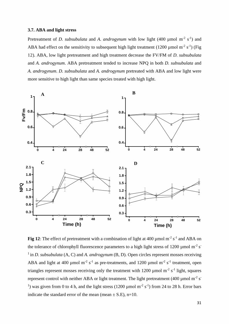
3.7. ABA and light stress
Pretreatment of D. subsubulata and A. androgynum with low light (400 µmol m-2 s-1) and
ABA had effect on the sensitivity to subsequent high light treatment (1200 µmol m-2 s-1) (Fig
12). ABA, low light pretreatment and high treatment decrease the FV/FM of D. subsubulata
and A. androgynum. ABA pretreatment tended to increase NPQ in both D. subsubulata and
A. androgynum. D. subsubulata and A. androgynum pretreated with ABA and low light were
more sensitive to high light than same species treated with high light.
B 1 1
0.8 0.8
0.6 0.6
0.4
2.1
1.8
1.5
1.2
0.9
0.6
0.3
0 4 24 28 48 52
C
0 4 24 28 48 52
Time (h)
0.4
2.1
1.8
1.5
1.2
0.9
0.6
0.3
0 4 24 28 48 52
D
0 4 24 28 48 52
Time (h)
Fig 12: The effect of pretreatment with a combination of light at 400 µmol m-2 s-1 and ABA on
the tolerance of chlorophyll fluorescence parameters to a high light stress of 1200 µmol m-2 s-
1 in D. subsubulata (A, C) and A. androgynum (B, D). Open circles represent mosses receiving
ABA and light at 400 µmol m-2 s-1 as pre-treatments, and 1200 µmol m-2 s-1 treatment, open
triangles represent mosses receiving only the treatment with 1200 µmol m-2 s-1 light, squares
represent control with neither ABA or light treatment. The light pretreatment (400 µmol m-2 s-
1) was given from 0 to 4 h, and the light stress (1200 µmol m-2 s-1) from 24 to 28 h. Error bars
indicate the standard error of the mean (mean ± S.E), n=10.
NP
Q
Fv/F
m
32
3.8. The effect of desiccation of the activity of POX
Stem segments of A. androgynum and D. subsubulata were slowly desiccated over a saturated
solution of MgCl2; drying curves are presented in Fig13. POX activity in A. androgynum was
much lower than that of D. subsubulata (Fig 14). Slow desiccation of both species increased
POX activity. Apart from the absolute differences in activity, the main difference between the
two species was that activity continued to increase from 72 to 96 h in D. subsubulata but
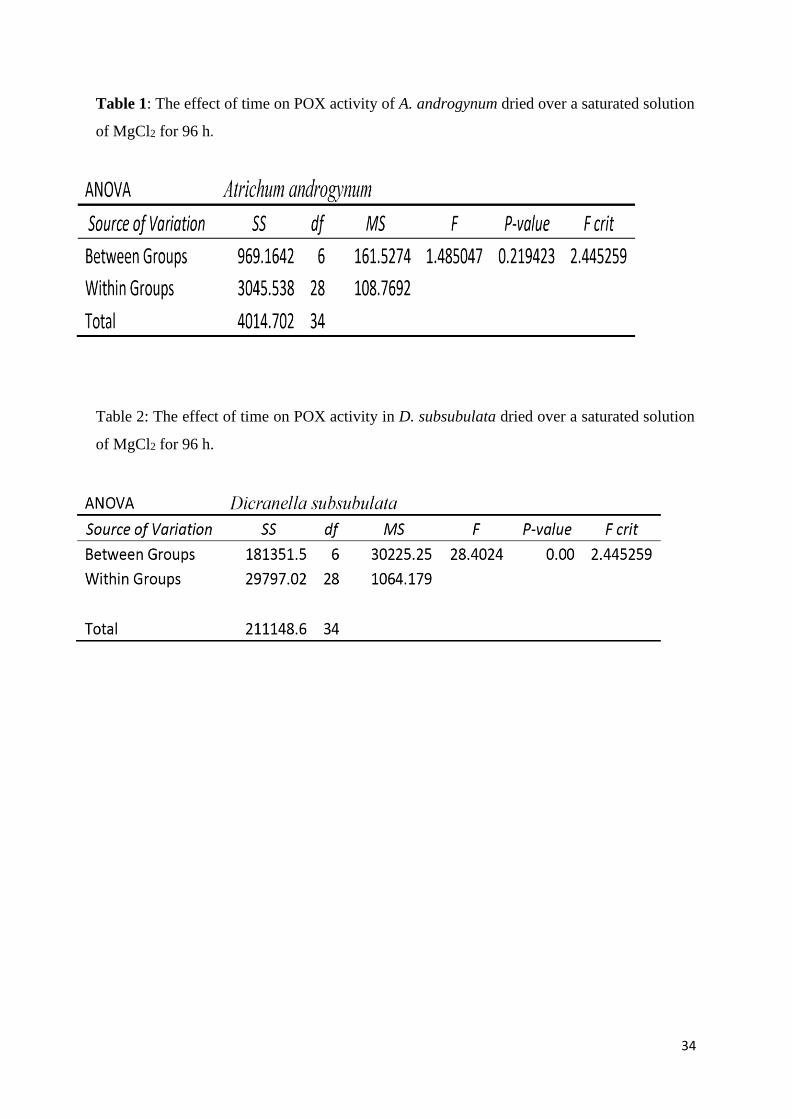
decreased in A. androgynum. Statistical analysis showed that the effect of desiccation on
POX activity was significant on D. subsubulata, p<0.05 but not significant on
A. androgynum, p>0.05 (Tables 1 and 2).
Fig 13: Slow drying curves of D. subsubulata and A. androgynum desiccated above a saturated
solution of MgCl2. D. subsubulata is represented by open circles and A. androgynum by open
triangle. Error bars indicates standard error of the mean (mean ± S.E).
33
A
300
250
200
150
100
50
0
0 2 4 24 48 72 96
Time (h)
35 B
30
25
20
15
10
5
0 0 2 4 24 48 72 96
Time (h)
Fig 14: The effect of slow desiccation for 96 h on POX activity in D. subsubulata (A) and A.
androgynum (B). Error bars indicates standard error of the mean (mean ± S.E), n=20.
PO
X a
ctiv
ity
nca
t/g d
ry m
ass
P
OX
a
ctiv
ity
nca
t/g d
ry m
ass
34
Table 1: The effect of time on POX activity of A. androgynum dried over a saturated solution
of MgCl2 for 96 h.
Table 2: The effect of time on POX activity in D. subsubulata dried over a saturated solution
of MgCl2 for 96 h.

35
3.8. Analysis of Peroxidase enzyme using PAGE electrophoresis.
Moss extracts were subjected to PAGE electrophoresis followed by visualizations of POX
activity using with o-dianisidine and H2O2. D. subsubulata had one POX isoform, with a
molecular mass of 57-55 kDa. The electrophoresis was repeated four times, and this
isoform was clearly visible in all 4 visualizations (Fig 15 B-E). A. androgynum had very low
enzyme activity and could not be visualized even after concentration of crude extracts using
dialysis on solid sucrose overnight, followed by reverse dialysis to remove sugar.
Staining gels of D. subsubulata for superoxide production indicated that extracts did not
contain measurable amounts of superoxide producing enzymes.
A B C D E
100 kDa
60 kDa
45 kDa
55 kDa
56 kDa 57 kDa
60 kD
12 kDa
Fig 15: Native PAGE electrophoresis gels of D. subsubulata stained for POX activity. The
electrophoresis was repeated four times, and this isoform was clearly visible in all
visualizations (B, C, D, E). Molecular weight markers are illustrated in lane A.
36
Chapter 4: Discussion
As discussed in the Introduction, the flora of larger mosses in the Afromontane vegetation
around Pietermaritzburg is dominated by two mosses, Atrichum androgynum, and Dicranella
subsubulata. A. androgynum tends to grow in wetter, more shaded habitats and is a rather
delicate species, while D. subsubulata grow in open, drier habitats and is more robust. Rarely,
the species grow together, for example at the transition of a shaded indigenous woodland to
plantations (Fig 3, 4). We hypothesized that the more robust species has higher stress
tolerance, and largely constitutive stress tolerance mechanisms. By contrast, we hypothesized
that the more delicate species is less tolerant and may rely more on inducible tolerance
mechanisms.
4.1. Response of photosynthesis to light intensity
Results showed that D. subsubulata and A. androgynum are adapted to shade environments.
However, D. subsubulata appears to be adapted to higher light levels than A. androgynum,
reaching AMAX at around 500 μmol m-2 s-1 (Fig 5A), while A. androgynum reached AMAX at
around 200 μmol m-2 s-1 (Fig 3B). Mosses survive in the shade by having specific adaptations
to low light intensity (Hájek et al., 2009; Marschall and Proctor, 2004). We expected that
species found in more exposure microsites in the exotic plantation forest would have higher
photosynthetic capacity than species growing in Afromontane forests. Results were consistent
with our expectations, with D. subsubulata having a higher maximum ETR than A.
androgynum. Hájek et al. (2009) reported that the moss genus Sphagnum, found in open
habitats had lower photosynthetic capacities than the species growing in more shaded
environments. As for almost all plants (Osmond et al. 1997), photosynthetic light responses are
plastic in the two species studied. Storage in cool dim light reduced maximum rates of ETR
(Fig 5). As would be expected from the reduced need for photoprotection, storage reduced
NPQ in D. subsubulata (Fig 5C) but counterintuitively increased NPQ in A. androgynum (Fig
5D). There are no obvious explanations for the result of light intensity in mosses. Results of
the present study confirm that D. subsubulata has a higher inherent tolerance to desiccation
and high light stress than A. androgynum.

37
4.2. Photoinhibition
Both species showed an excellent ability to recover from moderately severe photoinhibition,
caused by exposure to 1200 µmoles m-2 s-1 for 4 h (Fig 6). The recovery is likely due to re-
synthesis of the D1 protein which becomes preferentially damaged in moss (Rintamäki and
Riitta Salo, 1994). In general, FV/FM, ETR and NPQ were all more inhibited in A. androgynum
than D. subsubulata. Consistent with results obtained for lichen photobionts (Solhaug, 2018)
photosystems in the two species of mosses tested here need low light to recover from
photoinhibition. Although similar for the first 10 h, after 24 h recovery was much better if
mosses had been allowed to recover from photoinhibition in low light. Interestingly,
transferring mosses that had been kept in the dark to dim light (lab without light on) from 24
to 25 h after the start of the experiment enabled them to recover to similar FV/FM and ETR as
mosses allowed to recover from photoinhibition in dim light. Reasons for the better recovery of
mosses in dim light rather than darkness are unclear, but in higher plants it has been shown
that the D1 protein repair cycle is light dependent (Bergo et al., 2003).
4.3. Tolerance to dehydration using CaCl2
Mosses in general are highly desiccation tolerant (Oliver et al. 2020). In initial experiments we
tested the ability of D. subsubulata and A. androgynum to tolerate moderately fast drying for
48 h and 1 week over a saturated solution of CaCl2, corresponding to a relative humidity of c.
35% at 15oC. Both species rapidly recovered after 48 h of desiccation (Fig 7B, D, F),
although recovery was slightly faster and more complete in D. subsubulata than A.
androgynum. These mosses can therefore be classified as “fully desiccation-tolerant” (Wood,
2007). However, after desiccation for 1 week, recovery in D. subsubulata was much slower
than after 48 h, but complete after 24 h of rehydration (Fig 7A, C, E). By contrast, desiccation
of A. androgynum was probably lethal, with parameters not recovering after rehydration for
24 h. Results clearly showed that D. subsubulata was more tolerant to desiccation stress than
A. androgynum. It is well known that DT in Bryophytes varies greatly between species
(Wood, 2007). The differences in DT between D. subsubulata and A. androgynum here
reflect anecdotal observations that D. subsubulata grows in generally more exposed habitats
than A. androgynum, where it is likely to suffer more rapid and profound and desiccation.
38
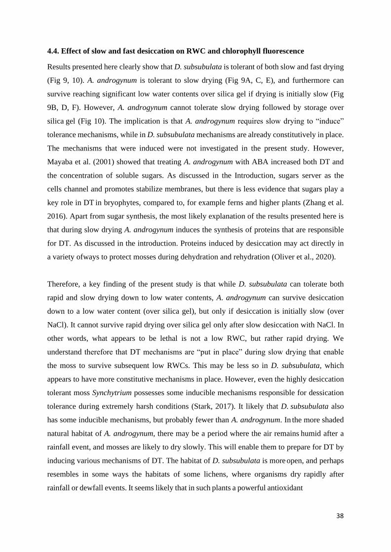
4.4. Effect of slow and fast desiccation on RWC and chlorophyll fluorescence
Results presented here clearly show that D. subsubulata is tolerant of both slow and fast drying
(Fig 9, 10). A. androgynum is tolerant to slow drying (Fig 9A, C, E), and furthermore can
survive reaching significant low water contents over silica gel if drying is initially slow (Fig
9B, D, F). However, A. androgynum cannot tolerate slow drying followed by storage over
silica gel (Fig 10). The implication is that A. androgynum requires slow drying to “induce”
tolerance mechanisms, while in D. subsubulata mechanisms are already constitutively in place.
The mechanisms that were induced were not investigated in the present study. However,
Mayaba et al. (2001) showed that treating A. androgynum with ABA increased both DT and
the concentration of soluble sugars. As discussed in the Introduction, sugars server as the
cells channel and promotes stabilize membranes, but there is less evidence that sugars play a
key role in DT in bryophytes, compared to, for example ferns and higher plants (Zhang et al.
2016). Apart from sugar synthesis, the most likely explanation of the results presented here is
that during slow drying A. androgynum induces the synthesis of proteins that are responsible
for DT. As discussed in the introduction. Proteins induced by desiccation may act directly in
a variety of ways to protect mosses during dehydration and rehydration (Oliver et al., 2020).
Therefore, a key finding of the present study is that while D. subsubulata can tolerate both
rapid and slow drying down to low water contents, A. androgynum can survive desiccation
down to a low water content (over silica gel), but only if desiccation is initially slow (over
NaCl). It cannot survive rapid drying over silica gel only after slow desiccation with NaCl. In
other words, what appears to be lethal is not a low RWC, but rather rapid drying. We
understand therefore that DT mechanisms are “put in place” during slow drying that enable
the moss to survive subsequent low RWCs. This may be less so in D. subsubulata, which
appears to have more constitutive mechanisms in place. However, even the highly desiccation
tolerant moss Synchytrium possesses some inducible mechanisms responsible for dessication
tolerance during extremely harsh conditions (Stark, 2017). It likely that D. subsubulata also
has some inducible mechanisms, but probably fewer than A. androgynum. In the more shaded
natural habitat of A. androgynum, there may be a period where the air remains humid after a
rainfall event, and mosses are likely to dry slowly. This will enable them to prepare for DT by
inducing various mechanisms of DT. The habitat of D. subsubulata is more open, and perhaps
resembles in some ways the habitats of some lichens, where organisms dry rapidly after
rainfall or dewfall events. It seems likely that in such plants a powerful antioxidant
39
system, and high concentrations of polyols and “DT proteins” such as HSP and LEA proteins
must be constitutively present (Gasulla et al., 2021).
4.5. The effect of ABA pretreatment on desiccation tolerance
Desiccation tolerance mechanisms in bryophytes are either inherent (“constitutive”) or can be
induced by hardening treatments such as partial desiccation or treatment with the stress
hormone ABA (Mayaba et al. 2001; Stark 2017). The moss, Physcomitrium patens had been
used as a model species to show that ABA plays a major role in inducing stress tolerance in
mosses (Oliver et al. 2020). Endogenous ABA in found moss species (Ergün et al. 2002;
Werner et al. 1991; Xiao et al. 2017). In the present study, mosses were treated with ABA, and
then rapidly dried, to reduce the induction of DT mechanisms during natural drying. ABA
treatment significantly improved the ability of the chlorophyll fluorescence parameters FV/FM,
and ETR of D. subsubulata to recover during rehydration (Fig 11A, C). By contrast, the
effect of ABA on the ability of A. androgynum to recover was much less (Fig 11B, D). ABA
had little effect on NPQ in D. subsubulata (Fig 11E), but slightly increased NPQ activity of
A. androgynum during rehydration (Fig 11F). Mayaba et al. (2001) also found that ABA
treatment tends to increase NPQ activity. However, increased NPQ did not appear to improve
the ability of the mosses to recover from desiccation stress. Taken together, results presented
here do not support the original hypothesis that D. subsubulata has more constitutive tolerance
mechanisms that A. androgynum.
Results suggest that D. subsubulata is more responsive to ABA than A. androgynum.
However, for desiccation tolerance, counter to the original hypothesis, results with
“hardening” pre-treatments and treatment with ABA showed that D subsubulata is as much if
not more dependent on inducible tolerance mechanisms as A. androgynum. While precise
tolerance mechanisms were not investigated here, for desiccation tolerance, induction of the
activity of antioxidative enzymes, specifically peroxidases seem likely to be an important
component of tolerance. It is worth noting that not all DT mechanisms are inducible by ABA
(Blomstedt et al. 2018), and possibly A. androgynum possesses more of such mechanisms than
D. subsubulata. Blomstedt et al. (2018) suggest that in higher plants further research is
needed to understand how desiccation tolerance is induced, specifically on the roles of
phytohormones such as, strigolactone and other potential xylem-messengers during drying
and rehydration. Within the bryophytes, Xiao et al. (2017) found that for Physcomitrium the
initial drying rate, and not the amount of endogenous ABA, may be critical in the acquisition
of desiccation tolerance, also suggesting the presence of non-ABA inducible DT mechanisms

40
in that species.

41
4.6. ABA and high light treatment
Attempts to modulate the effects of light stress with a combination of low light and ABA were
not successful. Short-term ABA treatment, in combination with a pretreatment with moderate
light (400 µmol m-2 s-1), did not increase the tolerance of either D. subsubulata or A.
androgynum to photoinhibition (Fig 12). Results were consistent with those of Jia and Lu
(2003) who reported that short term ABA treatment increases the sensitivity of maize (Zea
mays) to photoinhibition. Results presented here suggest that ABA treatment increases the
susceptibility of D. subsubulata and A. androgynum to light stress. An original aim of this
thesis was to attempt to modulate the tolerance of the mosses to photoinhibition by using
various ABA and moderate light intensities, with the intention to test the hypothesis that A.
androgynum displays greater plasticity than D. subsubulata. Unfortunately, attempts at
“hardening” were unsuccessful. While it seems likely that, as for lichens, tolerance to
photoinhibition is a plastic character in mosses, more work is needed to establish how tolerance
can be modulated to enable our original hypothesis to be tested.
4. 7. Enzyme activity changes in mosses
4.7.1. Peroxidase activity
In general, D. subsubulata possesses high peroxide activity and A. androgynum has low POX
activity (compare the y-axes in Fig 14). The best POX substate for both species was o-
dianisidine, a commonly used substrate for POX activity (Bania and Mahata 2012). Desiccation
induced the level of POX activity in both species (Fig 14). Peroxidase was induced by
desiccation even after 4 d (Fig 14). In this experiment, a saturated solution of MgCl2 was used
to induce a relatively slow drying of D. subsubulata and A. androgynum (Fig 13), which
would enable any upregulation of POX activity to occur. It seems logical to assume that in
both D. subsubulata and A. androgynum the level of antioxidant enzymes increases or is
maintained during dehydration to scavenge desiccation-induced ROS formation. Farrant et al.
(2007) reported this as a common feature of desiccant tolerance in higher plants. Liu et al.
(2019) studied the effects of various stress on SOD and POX activities in the moss Hypnum
plumaeforme. One‐time or repeated desiccation stress and cross‐stresses (low temperature
followed by desiccation stress) increased both SOD and POX activities. Dicranum scoparium,
closely related to Dicranella species, also had high peroxidase activity that increased
significantly during desiccation (Onele et al., 2018). Interesting, the effect of desiccation on
POX activity in A. androgynum was not statistically significant (Table 1), while in D.
subsubulata the increase in activity was significant (Table 2). Taken together, results on
42
peroxidase activity suggested that both species display an inducible tolerance mechanism, and
counter to our original hypothesis, the inducibility of the mechanism was greater not less in D.
subsubulata than A. androgynum. However, future work should focus on other antioxidant
enzymes not studied here e.g., SOD (Mayaba and Beckett, 2003).
4.7.2. Gel electrophoresis analysis
A. androgynum has too low peroxidase activity, and the enzyme could not be visualized using
gel electrophoresis, despite using sugar dialysis to concentrate samples, thus results could not
be presented. This was not the case in D. subsubulata peroxidase; perhaps surprisingly activity
occurred largely as a single isoform with a molecular mass of 55 to 60 kDa (Fig 15 B-D).
Most plants appear to possess multiple POX isoforms, and each isoform presumably has a
slightly different role in plants (Cosio and Dunand, 2009). Higher plant class III peroxidases
are involved in both ROS production and ROS scavenging, important components for stress
tolerance (Almagro et al., 2009). Dicranum scoparium displays multiple isoforms (Onele et
al., 2019). Possibly D. subsubulata possesses multiple isoforms with the same molecular mass.
This could be investigated in future using isoelectric focussing or more molecular biological
techniques. Interestingly, the D. subsubulata POX did not appear to produce superoxide
radicals (data not shown), unlike, for example, the POX of the liverwort Dumortiera hirsuta
which often grows in Afromontane forests in wetter microhabitats (Li et al. 2010). POX from
D. subsubulata therefore seem to be more involved in ROS scavenging than ROS production.
43
Chapter 5: General Conclusion and Recommendations
5.1. General conclusion
Desiccation and high light are probably the main abiotic stresses that limit the distribution of
the mosses D. subsubulata and A. androgynum in the KwaZulu Afromontane. Both species can
recovery from severe desiccation, and both are clearly DT. As would anecdotally be suggested
by looking at the plants in the field, results presented here suggest that D. subsubulata is
generally more stress-tolerant than A. androgynum. Intuitively, mosses and lichens grow in
particularly stressful habitats will be regularly subjected to sudden stressful events (e.g., rapid
desiccation). There is a general view that such mosses and lichens are therefore likely to depend
on constitutive rather than inducible tolerance mechanisms (Stark 2017; Gasulla et al. 2021).
Therefore, as outlined in the Introduction, the main hypothesis tested in this thesis was that D.
subsubulata depends more on constitutive stress tolerance mechanisms than A. androgynum.
While some of the results reported here support this hypothesis, evidence was also found that
significant stress tolerance can also be induced in D. subsubulata. In support of the hypothesis
that D. subsubulata depends more on constitutive tolerance mechanisms was the finding that
this species can readily survive rapid desiccation and storage over silica gel. By contrast, A.
androgynum can only survive storage over silica gel if desiccation in slow, presumably to
induce some tolerance mechanisms (Fig 9). However, at variance with this hypothesis was the
observation that while treatment with ABA clearly increased DT in D. subsubulata it had
very little effect on the tolerance of A. androgynum (Fig 11). Furthermore, while slow
desiccation significantly increases POX activity in D. subsubulata, in A. androgynum
desiccation tends to increase POX activity, but the increase is not significant (Fig 11). As
discussed in more detail above, the much lower effect of ABA in A. androgynum than D.
subsubulata may simply indicate that other hormones are involved in stress signalling in A.
androgynum. Nevertheless, it seems clear that even highly DT mosses such as D. subsubulata
do not rely exclusively on constitutive tolerance mechanisms, but in addition, possess some
inducible mechanisms.
Unfortunately, it could not be established from the experiments on high light stress whether D.
subsubulata possesses more constitutive tolerance mechanisms that A. androgynum. While
clearly showing that D. subsubulata is more tolerant to photoinhibition than A. androgynum
(Fig 6), our moderate light and ABA “hardening” treatment did not increase tolerance to
photoinhibition in either species. More work using a variety of different (possibly less stressful)
hardening treatments is needed to find out whether tolerance to high light stress in these two
44
species can be modulated, and whether modulation is greater in A. androgynum than D.
subsubulata.
5.2. Future recommendations
As outlined above, in future studies it would be desirable to test ways of modulating tolerance
to high light stress in these two species. Furthermore, desiccation tolerance of D. subsubulata
is poorly understood, but future work could focus on characterizing the peroxidase genes
present using the genome sequences available for closely related species D. scoparium. Other
tolerance mechanisms worth studying in more detail are the induction of potentially
protective sugars during desiccation. Furthermore, the induction of antioxidant enzymes
during desiccation in addition to POX such as SOD could be tested, and other proteins
associated with DT in other organisms such as HSP and LEA proteins.
A long-term gain would be to use knowledge on DT mechanisms in bryophytes to improve the
stress tolerance of crop plants. Bryophytes were the first land plants, and possess high DT, thus
genetic transformation using Bryophyte genes could be beneficial. To give examples from
other organisms, genetic engineering or modification was successful in tomatoes using an anti-
freeze gene from the Arctic fish to promote tolerance to frost damage (Le Gall et al., 2003).
Tobacco has been genetically modified to produce artemisinin drugs that have anti-malarial
properties (Farhi et al., 2011). In the Bryophytes, P. patens has been proposed to be a good
model species for genetic engineering, and indeed the entire genome has been sequenced
(Cove, 2005). However, simply to confine research to this species may “miss” tolerance
mechanisms found in other bryophytes. Therefore, it seems highly desirable that in addition to
P. patens, other bryophytes continued to be studied for stress tolerance mechanisms. The world
is predicted to face drastic climate change, and genetic engineering may be needed to ensure
security of food production in the future.

45
References
Adamson, H., Wilson, M., Selkirk, P., Seppelt, R., 1988. Photoinhibition in Antarctic
mosses. Polarforschung 58, 103-111.
Ahmad, P., Jaleel, C.A., Azooz, M.M., Nabi G., 2009. Generation of ROS and non-enzymatic
antioxidants during abiotic stress in plants. Botany Research International 2, 11-20.
Almagro, L., Gómez Ros, L.V., Belchi-Navarro, S., Bru, R., Ros Barceló, A., Pedreño, M.A.,
2009. Class III peroxidases in plant defence reactions. Journal of Experimental
Botany 60, 377-390.
Anjum, S.A., Xie, X.Y., Wang, L.C., Saleem, M.F., Man, C., Lei, W., 2011. Morphological
physiological and biochemical responses of plants to drought stress. African Journal of
Agricultural Research 6, 2026-2032.
Apel, K., Hirt, H., 2004. Reactive oxygen species: metabolism, oxidative stress, and signal
transduction. Annual Review of Plant Biology 55, 373-399.
Bahuguna, Y.M., Gairola, S., Semwal, D.P., Uniyal, P.L., Bhatt, A.B., 2013. Bryophytes and
ecosystem. Biodiversity of lower plants. IK International Publishing House Pvt. Ltd.,
New Delhi, 279-296.
Bania, I., Mahanta, R., 2012. Evaluation of peroxidases from various plant sources.
International Journal of Scientific and Research Publications 2, 1-5.
Barták, M., Hájek, J., Očenášová, P., 2012. Photoinhibition of photosynthesis in Antarctic
lichen Usnea antarctica. I. Light intensity-and light duration-dependent changes in
functioning of photosystem II. Czech Polar Reports 2, 42-51.
Beckett, R.P., Marschall, M., Laufer, Z., 2005. Hardening enhances photoprotection in the
moss Atrichum androgynum during rehydration by increasing fast rather than slow-
relaxing quenching. Journal of Bryology 27, 7-12.
Bergo, E., Segalla, A., Giacometti, G.M., Tarantino, D., Soave, C., Andreucci, F., Barbato, R.,
2003. Role of visible light in the recovery of photosystem II structure and function from
ultraviolet-B stress in higher plants. Journal of Experimental Botany 54, 1665-1673.
Bigot, S., Buges, J., Gilly, L., Jacques, C., Le Boulch, P., Berger, M., Delcros, P., Domergue,
J.B., Koehl, A., Ley‐Ngardigal, B., Tran Van Canh, L., 2018. Pivotal roles of
46
environmental sensing and signaling mechanisms in plant responses to climate
change. Global Change Biology 24, 5573-5589.
Blomstedt, C.K., Griffiths, C.A., Gaff, D.F., Hamill, J.D., Neale A.D., 2018. Plant desiccation
tolerance and its regulation in the foliage of resurrection “flowering-plant” species.
Agronomy 8, 146.
Caverzan, A., Casassola, A., Brammer, S.P., 2016. Reactive oxygen species and antioxidant
enzymes involved in plant tolerance to stress. SHANKER AK & SHANKER C. Abiotic
and biotic stress in plants-Recent advances and future perspectives. Publisher InTech,
463-480.
Coe, K.K., Greenwood, J.L., Slate, M.L., Clark, T.A., Brinda, J.C., Fisher, K.M., Mishler, B.D.,
Bowker, M.A., Oliver, M.J., Ebrahimi, S., Stark, L.R., 2021. Strategies of desiccation
tolerance vary across life phases in the moss Syntrichia caninervis. American Journal
of Botany 108, 249-262.
Cosio, C., Dunand, C., 2009. Specific functions of individual class III peroxidase
genes. Journal of Experimental Botany 60, 391–408.
Cove, D., 2005. The moss Physcomitrella patens. Annual Review of Genetics 39, 339-358.
Cruz De Carvalho, R., Bernardes Da Silva, A., Soares, R., Almeida, A.M., Coelho, A.V.,
Marques Da Silva, Jorge., Branquinho, C., 2014. Differential proteomics of
dehydration and rehydration in bryophytes: evidence towards a common desiccation
tolerance mechanism. Plant, Cell and Environment 37, 1499-1515.
Davies, K.M., Jibran, R., Zhou, Y., Albert, N.W., Brummell, D.A., Jordan, B.R., Bowman,
J.L., Schwinn, K.E., 2020. The evolution of flavonoid biosynthesis: A bryophyte
perspective. Frontiers in Plant Science 11, 7.
Deltoro, V., Calatayud, A., Gimeno, C., Barreno, E., 1998. Water relations, chlorophyll
fluorescence, and membrane permeability during desiccation in bryophytes from xeric,
mesic, and hydric environments. Canadian Journal of Botany 76, 1923–1929.
Demmig-Adams, B., López-Pozo, M., Stewart, J.J., Adams, W.W., 2020. Zeaxanthin and
lutein: Photoprotectors, anti-inflammatories, and brain food. Molecules 25,3607.
47
Ergün, N., Topcuoúlu, S, F., Yildiz, A., 2002. Auxin (indole-3-acetic acid), gibberellic acid
(GA3), abscisic acid (ABA) and cytokinin (zeatin) production by some species of
mosses and lichens. Turkish Journal of Botany 26, 13-18.
Farhi, M., Marhevka, E., Ben-Ari, J., Algamas-Dimantov, A., Liang, Z., Zeevi, V., Edelbaum,
O., Spitzer-Rimon, B., Abeliovich, H., Schwartz, B., Tzfira, T., 2011. Generation of
the potent anti-malarial drug artemisinin in tobacco. Nature Biotechnology 29, 1072-
1074.
Farrant, J., Brandt, W., Lidsey, G., 2007. An overview of mechanisms of desiccation tolerance
in selected angiosperm resurrection plants. Plant Stress 1, 72–84
Foyer, C.H., 2018. Reactive oxygen species, oxidative signaling and the regulation of
photosynthesis. Environmental and Experimental Botany 154, 134-142.
Foyer, C.H., Halliwell, B., 1976. The presence of glutathione and glutathione reductase in
chloroplasts: A proposed role in ascorbic acid metabolism. Planta 133, 21–25.
Gaff, D.F., Oliver, M., 2013. The evolution of desiccation tolerance in angiosperm plants: a
rare yet common phenomenon. Functional Plant Biology 40, 315-328.
Gahtori, D., Chaturvedi, P., 2019. Bryophytes: A Potential Source of Antioxidants.
In Bryophytes. IntechOpen 53-58
Gao, B., Li, X., Zhang, D., Liang, Y., Yang, H., Chen, M., Zhang, Y., Zhang, J., Wood, A.J.,
2017. Desiccation tolerance in bryophytes: The dehydration and rehydration
transcriptomes in the desiccation-tolerant bryophyte Bryum argenteum. Scientific
Reports 7, 1-14.
Gasulla, F., Del Campo, E.M., Casano, L.M., Guéra, A., 2021. Advances in Understanding of
Desiccation Tolerance of Lichens and Lichen-Forming Algae. Plants, 10, 807.
Gechev, T.S., Dinakar, C., Benina, M., Toneva, V., Bartels, D., 2012. Molecular mechanisms
of desiccation tolerance in resurrection plants. Cellular and Molecular Life Sciences 69,
3175–3186.
Gerotto, C., Alboresi, A., Giacometti, G.M., Bassi, R., Morosinotto, T., 2012. Coexistence of
plant and algal energy dissipation mechanisms in the moss Physcomitrella patens. New
Phytologist 196, 763-773.
Green, T.A., Sancho, L.G., Pintado, A., 2011. In Plant desiccation tolerance: Ecophysiology

48
of desiccation/rehydration cycles in mosses and lichens. Springer, Berlin, Heidelberg.
89-120.
Hájek, T., Tuittila, E.S., Ilomets, M., Laiho, R., 2009. Light responses of mire mosses–a key
to survival after water‐level drawdown? Oikos 118, 240-250.
Harris, B.J., Harrison, C.J., Hetherington, A.M., Williams, T.A., 2020. Phylogenomic evidence
for the monophyly of bryophytes and the reductive evolution of stomata. Current
Biology 30, 2001-2012.
Hartung, W., 2010. The evolution of abscisic acid (ABA) and ABA function in lower plants,
fungi, and lichens. Functional Plant Biology 37, 806-812.
Hartung, W., Gimmler, H., 1994. In Progress in Botany: A stress physiological role for abscisic
acid (ABA) in lower plants. Springer, Berlin, Heidelberg 55,157-173.
Hu, R., Xiao, L., Bao, F., Li, X., He, Y., 2016. Dehydration-responsive features of Atrichum
undulatum. Journal of Plant Research 129, 945-954.
Hundertmark, M., Hincha, D.K., 2008. LEA (late embryogenesis abundant) proteins and their
encoding genes in Arabidopsis thaliana. BMC genomics 9, 1-22.
Jia, H., Lu, C., 2003. Effects of abscisic acid on photoinhibition in maize plants. Plant
Science 165, 1403-1410.
Kavitha, C.H., Murugan, K., 2016. Desiccation-induced changes in lipid peroxidation
superoxide level and cell membrane H+ ATPase activity in forked fern – Dicranopteris
linearis (Burm. f.) Underw. International Journal of Advances in Research 3, 588-593.
Kehoe, D.M., 2010. Chromatic adaptation and the evolution of light color sensing in
cyanobacteria. Proceedings of the National Academy of Sciences 107, 9029-9030.
Keren, N., Krieger‐Liszkay, A., 2011. Photoinhibition: molecular mechanisms and
physiological significance. Physiologia Plantarum 142, 1-5.
Koster, K.L., Balsamo, R.A., Espinoza, C., Oliver, M.J., 2010. Desiccation sensitivity
and tolerance in the moss Physcomitrella patens: assessing limits and damage.
Plant Growth Regulation 62, 293–302.
Kuromori, T., Miyaji, T., Yabuuchi, H., Shimizu, H., Sugimoto, E., Kamiya, A., Moriyama,
Y., Shinozaki, K., 2010. ABC transporter AtABCG25 is involved in abscisic acid
transport and responses. Proceedings of the National Academy of Sciences 107, 2361-
49
2366.
Laemmli, U. K., 1970. Cleavage of structural proteins during the assembly of the head of
bacteriophage T4. Nature 227, 680-685.
Le Gall, G., DuPont, M.S., Mellon, F.A., Davis, A.L., Collins, G.J., Verhoeyen, M.E.,
Colquhoun, I.J., 2003. Characterization and content of flavonoid glycosides in
genetically modified tomato (Lycopersicon esculentum) fruits. Journal of Agricultural
and Food Chemistry 51, 2438-2446.
Li, J.L.Y., Sulaiman, M., Beckett, R.P., Minibayeva, F.V., 2010. Cell wall peroxidases in the
liverwort Dumortiera hirsuta are responsible for extracellular superoxide production
and can display tyrosinase activity. Physiologia Plantarum 138, 474–484.
Liu, W., Xu, J., Fu, W., Wang, X., Lei, C., Chen, Y., 2019. Evidence of stress imprinting with
population‐level differences in two moss species. Ecology and Evolution 9, 6329-6341.
Marschall, M., Proctor, M.C., 2004. Are bryophytes shade plants? Photosynthetic light
responses and proportions of chlorophyll a, chlorophyll b and total carotenoids. Annals
of Botany 94, 593-603.
Mayaba, N., Beckett, R.P., 2003. Increased activities of superoxide dismutase and catalase are
not the mechanism of desiccation tolerance induced by hardening in the moss Atrichum
androgynum. Journal of Bryology 25, 281-286.
Mayaba, N., Beckett, R.P., Csintalan, Z., Tuba, Z., 2001. ABA increases the desiccation
tolerance of photosynthesis in the Afromontane understorey moss Atrichum
androgynum. Annals of Botany 88, 1093-1100.
McLetchie, D.N., Stark, L.R., 2019. Rate of drying influences tolerance of low water contents
in the moss Funaria hygrometrica (Funariaceae). The Bryologist 122, 271-280.
Mitra, J., Xu, G., Wang, B., Li, Meijing., Deng, X., 2013. Understanding desiccation tolerance
using the resurrection plant Boea hygrometrica as a model system. Frontiers in Plant
Science 4, 1-10.
Møller, I.M., Jensen, P.E., Hansson, A., 2007. Oxidative modifications to cellular components
in plants. Annual Revolution Plant Biology 58, 459-481.
Murray, K.J., Tenhunen, J.D., Nowak, R.S., 1993. Photoinhibition as a control on
photosynthesis and production of Sphagnum mosses. Oecologia 96, 200-207.
50
Niyogi, K, K., 1999. Photoprotection revisited: genetic and molecular approaches. Annual
Review of Plant Physiology and Plant Molecular Biology 50: 333–359.
Norwood, M., Toldi, O., Richter, A., Scott, P., 2003. Investigation into the ability of roots of
the poikilohydric plant Craterostigma plantagineum to survive dehydration stress.
Journal of Experimental Botany 54, 2313–2321.
Ntuli, T.M., 2012. Botany: Drought and desiccation-tolerance and sensitivity in plants. Mworia JK
(ed.) Botany. In Tech, Johannesburg, 29-60.
Oliver, M.J., Farrant, J.M., Hilhorst, H.W., Mundree, S., Williams, B., Bewley, J.D., 2020.
Desiccation tolerance: Avoiding cellular damage during drying and
rehydration. Annual Review of Plant Biology 71, 435-460.
Onele, A.O., Chasov, A., Viktorova, L., Beckett, R.P., Trifonova, T., Minibayeva, F., 2018.
Biochemical characterization of peroxidases from the moss Dicranum
scoparium. South African Journal of Botany 119, 132-141.
Onele, A.O., Chasov, A.V., Trifonova, T.V., Minibayeva, F.V., 2019. Ascorbate peroxidase of
the moss Dicranum scoparium: Gene identification and enzyme activity. Doklady
Biochemistry and Biophysics 489, 380-384.
Onele, A.O., Chasov, A.V., Viktorova, L.V., Minibayeva, F.V., Beckett, R.P., 2021.
Characterization and expression analysis of ascorbate peroxidase from the moss
Dicranum scoparium during abiotic stresses. The Bryologist 124, 68-84.
Osmond, B., Badger, M., Maxwell, K., Bjorkman, O., Leegood, R., 1997. Too many photons:
photorespiration, photoinhibition and photooxidation. Trends in Plant Science 2, 119–
121.
Pandey, V.P., Awasthi, M., Singh, S., Tiwari, S., Dwivedi, U.N., 2017. A comprehensive
review on function and application of plant peroxidases. Biochemistry and Analytical
Biochemistry 6, 1-16.
Parvin, K., Hasanuzzaman, M., Bhuyan, M.H.M., Mohsin, S.M.,Fujita, M., 2019. Quercetin
mediated salt tolerance in tomato through the enhancement of plant antioxidant defense
and glyoxalase systems. Plants 8, 247.
Pospíšil, P., 2016. Production of reactive oxygen species by photosystem II as a response to
light and temperature stress. Frontiers in Plant Science 7, 1950.
51
Proctor, M.C., Oliver, M.J., Wood, A.J., Alpert, P., Stark, L.R., Cleavitt, N.L., Mishler, B.D.,
2007. Desiccation-tolerance in bryophytes: a review. The Bryologist 110, 595-621.
Rintamäki, E., Riitta Salo, E, M, A., 1994. Rapid turnover of the D1 reaction-center protein of
photosystem II as a protection mechanism against photoinhibition in a moss, Ceratodon
purpureus (Hedw.) Brid. Planta 193, 520-529.
Ros, L.G., Gabaldón, C., Pomar, F., Merino, F., Pedreño, M.A., Barceló, A.R., 2007. Structural
motifs of syringyl peroxidases predate not only the gymnosperm–angiosperm
divergence but also the radiation of tracheophytes. New Phytologist 173, 63-78.
Ruban, A. V., Johnson, M. P., Duffy, C. D. P., 2012. The photoprotective molecular switch in
the photosystem II antenna. Biochimica Biophysica Acta 1817, 167–181.
Saavedra, L., Svensson, J., Carballo, V., Izmendi, D., Welin B., Vidal, S., 2006. A dehydrin
gene in Physcomitrella patens is required for salt and osmotic stress tolerance. Plant
Journal 45, 237–249.
Serrano, A., Córdoba, F., González-Reyes, J.A., Navas, P., Villalba, J.M., 1994. Purification
and characterization of two distinct NAD (P) H dehydrogenases from onion (Allium
cepa L.) root plasma membrane. Plant Physiology 106, 87-96.
Shaw, J., Renzaglia, K., 2004. Phylogeny and diversification of bryophytes. American Journal
of Botany 91, 1557-1581.
Smeekens, S., Ma, J., Hanson, J., Rolland, F., 2010. Sugar signals and molecular networks
controlling plant growth. Current Opinion in Plant Biology 13, 274–279.
Smirnoff, N., 1993. The role of active oxygen in the response of plants to water deficit and
desiccation. New phytologist, Tansley. 52, 27-58
Solhaug, K.A., 2018. Low-light recovery effects on assessment of photoinhibition with
chlorophyll fluorescence in lichens. The Lichenologist 50, 139-145.
Soriano, G., Del-Castillo-Alonso, M. A., Monforte, L., Nunez-Olivera, E., Martinez-Abaigar,
J., 2018. Phenolic compounds from different bryophyte species and cell compartments
respond specifically to ultraviolet radiation, but not particularly quickly. Plant
Physiology and Biochemistry 134, 137-144.
Stark, L.R., Brinda, J.C., 2015. Developing sporophytes transition from an inducible to a
constitutive ecological strategy of desiccation tolerance in the moss Aloina ambigua:

52
effects of desiccation on fitness. Annals of Botany 115,593-603.
Stark, L.R., 2017. Ecology of desiccation tolerance in bryophytes: A conceptual framework
and methodology. The Bryologist 120, 130-166.
Tuba, Z., Protor, C.F., Csintalan, Z., 1998. Ecophysiological responses of
homoiochlorophyllous and poikilochlorophyllous desiccation tolerant plants: a
comparison and an ecological perspective. Plant Growth Regulation 24, 211-217.
Waterman, M. J., Nugraha, A. S., Hendra, R., Ball, G. E., Robinson, S. A., Keller, P. A., 2017.
Antarctic moss biflavonoids show high antioxidant and ultraviolet-screening activity.
Journal of Natural Products, 80, 2224–2231.
Werner, O., Espin R, M, R., Bopp, M., Atzorn, R., 1991. Abscisic acid induced drought
tolerance in Funaria hygrometrica Hedw. Planta 186, 99–103.
Wood, A, J., 2007. The nature and distribution of vegetative desiccation-tolerance in
hornworts, liverworts and mosses. The Bryologist 110, 163–177.
Xiao, L.H., Yobi, A., Koster, K.L., He, Y.K., Oliver, M.J., 2017. Desiccation tolerance in
Physcomitrella patens: Rate of dehydration and the involvement of endogenous
abscisic acid (ABA). Plant Cell and Environment 41, 275-284.
Yin, R., Ulm, R., 2017. How plants cope with UV-B: from perception to response. Current
Opinion in Plant Biology 37, 42-48.
Zhang, Q., Song, X., Bartels, D., 2016. Enzymes and metabolites in carbohydrate metabolism
of desiccation tolerant plants. Proteomes, 4, 40.