EFFECTS OF GALACTOSE AND FRUCTOSE ON THE EXPRESSION OF ISOCITRATE...
41
EFFECTS OF GALACTOSE AND FRUCTOSE ON THE EXPRESSION OF ISOCITRATE LYASE (ICL1) ENZYME IN Candida albicans TING SENG YEAT UNIVERSITI SAINS MALAYSIA 2016
Transcript of EFFECTS OF GALACTOSE AND FRUCTOSE ON THE EXPRESSION OF ISOCITRATE...

EFFECTS OF GALACTOSE AND FRUCTOSE ON
THE EXPRESSION OF ISOCITRATE LYASE
(ICL1) ENZYME IN Candida albicans
TING SENG YEAT
UNIVERSITI SAINS MALAYSIA
2016

EFFECTS OF GALACTOSE AND FRUCTOSE ON
THE EXPRESSION OF ISOCITRATE LYASE
(ICL1) ENZYME IN Candida albicans
by
TING SENG YEAT
Thesis submitted in fulfillment of the requirements
for the degree of Master of Science
FEBRUARY 2016

ii
ACKNOWLEDGEMENTS
I would like to express my gratitude to my supervisors, Dr. Doblin Anak Sandai and Dr.
Hasni Arsad for their undivided support and advice. For without their encouragement,
guidance and advice, this dissertation would not be completed. I would also like to thank
my laboratory members: Puan Nur Hikmah, Laina Zarisa, Ishola Oluwaseun, Siti
Fadhilah, Cheah Hong Leong, Khirun, Nalini and Muwafaq Adam for their support and
company. Their company throughout this project keep me cheerful and motivated. I
would also like to thank Dr. Kumitaa Theva Das, Dr. Amir Yunus and Dr. Citartan
Marimuthu for their generous support and advice.
A special thank to MyBrain15 (Ministry of Higher Education, Malaysia) for the funding
of MyMaster scholarship scheme. Thank also extended to the Advanced Medical and
Dental Institute (AMDI, USM) for the funding of my project of study.
Last but not least, I would like to express my sincere gratitude to my family for their
unconditional love and understanding. Their encouragement and continuous support is
the main pillar that keeps me motivated.

3
LIST OF CONTENTS
Page
ACKNOWLEDGEMENTS....................................................................................... ii
LIST OF CONTENTS .............................................................................................. iii
LIST OF TABLES ..................................................................................................... vi
LIST OF FIGURES ................................................................................................. vii
LIST OF SYMBOLS AND ABBREVIATIONS ..................................................... ix
ABSTRAK ............................................................................................................... xiii
ABSTRACT .............................................................................................................. xv
CHAPTER 1: INTRODUCTION ............................................................................. 1
1.1 Candida spp. and Candidiasis ............................................................................................... 1
1.1.1 Asexual and Parasexual Reproduction in C. albicans .................................................... 2
1.1.2 Clinical Manifestation .................................................................................................... 2
1.2 Antifungal Agents.................................................................................................................. 3
1.3 Antifungal Resistance............................................................................................................ 6
1.4 Virulence and Pathogenicity in C. albicans .......................................................................... 7
1.4.1 Adhesion......................................................................................................................... 9
1.4.2 Secreted Degradative Enzymes .................................................................................... 10
1.4.3 Morphological Transition and Phenotypic Switching .................................................. 11
1.4.4 Biofilms ........................................................................................................................ 12
1.5 Transmigration .................................................................................................................... 14
1.6 Fitness Attribute .................................................................................................................. 15
1.6.1 Heat-shocks .................................................................................................................. 15
1.6.2 Osmotic and Cationic Stress ........................................................................................ 16
1.6.3 Cell Wall Stress ............................................................................................................ 18
1.6.4 Oxidative Stress ........................................................................................................... 19
1.7 Central Metabolism Pathways............................................................................................. 20
1.7.1 Glycolysis and Gluconeogenesis.................................................................................. 22
1.7.2 The Tricarboxylate Acid (TCA) and Glyoxylate Cycle................................................ 23
1.8 Glucose Sensing .................................................................................................................. 24
1.9 Catabolite Inactivation ........................................................................................................ 26
1.10 Ubiquitination ................................................................................................................... 27

4
1.11 Project Aims ...................................................................................................................... 31
CHAPTER 2: MATERIALS AND METHODS .................................................... 33
2.1 Chemicals and Suppliers ..................................................................................................... 33
2.2 Growth Media and Storage Conditions ............................................................................... 35
2.2.1 C. albicans and S. cerevisiae Growth and Storage Conditions .................................... 35
2.2.2 Growth Curves ............................................................................................................. 36
2.3 Strains.................................................................................................................................. 37
2.3.1 C. albicans Strains........................................................................................................ 37
2.3.2 S. cerevisiae Strains...................................................................................................... 37
2.4 DNA Analysis...................................................................................................................... 38
2.4.1 Small Scale Preparation of Yeasts Genomic DNA ....................................................... 38
2.4.2 DNA Quantification ..................................................................................................... 39
2.4.3 Agarose Gel Electrophoresis ........................................................................................ 40
2.4.4 Polymerase Chain Reaction (PCR) .............................................................................. 40
2.5 RNA Analysis ...................................................................................................................... 42
2.5.1 Preparation of RNase-free Materials ............................................................................ 42
2.5.2 RNA purification from S. cerevisiae and C. albicans .................................................. 42
2.5.3 RNA Quality Control ................................................................................................... 43
2.5.4 Quantitative Real Time Polymerase Chain Reaction ................................................... 45
2.6 Protein Analysis .................................................................................................................. 46
2.6.1 C. albicans Protein Extraction ..................................................................................... 46
2.6.2 Protein Quantification .................................................................................................. 47
2.6.3 Polyacrylamide Gel Electrophoresis ............................................................................ 47
2.6.4 Immunodetection of Myc-tagged Proteins ................................................................... 48
CHAPTER 3: RESULTS ......................................................................................... 51
3.1 Growth on Galactose or Fructose and Alternative Carbon Sources .................................... 51
3.2 The C. albicans ICL1 mRNAs are Repressed by Either Galactose or Fructose.................. 53
3.2.1 Analysis of mRNA Levels Upon Galactose or Fructose Addition to Lactate Growing
S. cerevisiae Cells ................................................................................................................. 53
3.2.2 Analysis of mRNA Levels Upon Galactose or Fructose Addition to Lactate Growing
C. albicans Cells ................................................................................................................... 55
3.3 Role of Ubiquitination in Sugar Phosphate-accelerated Protein Degradation in
C. albicans ................................................................................................................................ 57
3.4 The C. albicans Icl1 Protein is destabilized by Fructose but Not Galactose....................... 60

5
3.4.1 Impact of Galactose or Fructose Addition Upon ScIcl1 in S. cerevisiae During Growth on
Lactate ................................................................................................................................... 60
3.4.2 Impact of Galactose or Fructose Addition Upon CaIcl1 in C. albicans During Growth on
Lactate ................................................................................................................................... 63
3.4.3 C. albicans has Retained the Molecular Apparatus to Destabilize Proteins in Response to
Galactose or Fructose ............................................................................................................ 66
3.4.4 C. albicans Isocitrate Lyase has Lost Signal That Triggers Its Destabilization in Response
to Galactose but Not in Response to Fructose....................................................................... 69
3.4.5 Addition of Ubiquitination Site to C. albicans Icl1 Destabilizes the Protein Upon Addition
of Galactose or Fructose to Lactate Grown Cells.................................................................. 72
CHAPTER 4: DISCUSSIONS ................................................................................ 75
CHAPTER 5. CONCLUSION AND SUGGESTION TO FUTURE STUDY .... 82
REFERENCES ......................................................................................................... 83
APPENDICES ........................................................................................................ 102
6
LIST OF TABLES
Page
Table 2.1
Table 2.2
Table 2.3
Table 2.4
Table 2.5
Materials used in this study 33
C. albicans strains 37
S. cerevisiae strains 37
PCR reaction mixture 41
Primers for real-time qPCR analysis 46

vii
LIST OF FIGURES
Page
Figure 1.1
Figure 1.2
Figure 1.3
Figure 3.1
Figure 3.2
Figure 3.3
Figure 3.4
Figure 3.5
Figure 3.6
Figure 3.7
Figure 3.8
Figure 3.9
Figure 3.10
Virulence factors of C. albicans 8
The pathways of ß- oxidation, the glyoxylate cycle, 21
gluconeogenesis and glycolysis are shown with the
C. albicans gene names
The ubiquitin-proteosome pathways 29
Growth of S. cerevisiae and C. albicans on galactose or 52
fructose versus lactate
Impact of galactose or fructose addition upon ICL1 mRNAs 54
levels in S. cerevisiae during growth on lactate
Impact of galactose or fructose addition upon ICL1 mRNAs 56
levels in C. albicans during growth on lactate
Comparison of an amino acid pile-up between S. cerevisiae 58
and C. albicans
Western blotting analysis of ScIcl-Myc3 DS3-Y30 protein 61
expression after galactose or fructose addition to
S. cerevisiae cells growing on lactate
Graphs showing the degradation of ScIcl-Myc3 proteins 62
after addition of 0 or 2% galactose or fructose to lactate
grown cultures and was normalized to house keeping
protein beta Actin.
Western blotting analysis of CaIcl-Myc3 CA1395 protein 64
expression after galactose or fructose addition to
C. albicans cells growing on lactate.
Graphs showing the degradation of CaIcl-Myc3 proteins 65
after addition of 0 or 2% galactose or fructose to lactate
grown cultures and was normalized to house keeping
protein beta Actin
Western blotting analysis of Ca(ScIcl-Myc3) DSCO1 67
protein expression after galactose or fructose addition to
C. albicans DSCO1 cells growing on lactate
Graphs showing the degradation of ScIcl-Myc3 proteins in 68
C. albicans after addition of 0 or 2% galactose or fructose to lactate grown cultures and was normalized to house keeping
protein beta Actin

8
Figure 3.11
Figure 3.12
Figure 3.13
Figure 3.14
Western blotting analysis of Sc(CaIcl-Myc3) DS4-Y40 70
protein expression after galactose or fructose addition to
S. cerevisiae DS4-Y40 cells growing on lactate
Graphs showing the degradation of CaIcl-Myc3 proteins in 71
S. cerevisiae after addition of 0 or 2% galactose or fructose to lactate grown cultures and was normalized to house
keeping protein beta Actin
Western blotting analysis of CaIcl-Ubi-Myc3 DSCO4 73
protein expression after galactose or fructose addition to C.
albicans UBI4/UBI4 cells growing on lactate
Graphs showing the degradation of CaIcl-Ubi-Myc3 74
DSCO4 proteins in C. albicans UBI4/UBI4 after addition of
0 or 2% galactose or fructose to lactate grown cultures and
was normalized to house keeping protein beta Actin

9
LIST OF SYMBOLS AND ABBREVIATIONS
ADP
ALS
AMP
ASM
ATP
Bp
cAMP
cDNA
CGD
CLSI
dATP
dCTP
dGTP
DNA
DNase
dNTP
ECL
ECM
EDTA
ESR
EtBr
EUCAST
Adenosine 5’-diphosphate
Agglutinin like sequence
Adenosine monophosphate
American Society of Microbiology
Adenosine 5’-triphosphate
Base pair
Adenosine 3’,5’- cyclic monophosphate
Complementary DNA
Candida genome database
Clinical and Laboratory Standards Institute
2’-deoxyadenosine 5’-triphosphate
2’-deoxycytodine 5’ triphospahte
2’-deoxycytosine triphosphate
Deoxyribonucleic acid
Deoxyribonuclease
Deoxynucleic triphosphate
Enhanced chemiluminescence
Extracellular matrix material
Ethyenediaminetetraacetic acid
Environmental stress response
Ethidium bromide
European Committee on Antimicrobial Susceptibility Testing

10
GFP
GPI
HBEC
HIV
HOG
HRP
HSE
HSF
HSP
IDSA
kDa
mRNA
MAP
MAPK
MAPKK
MgCl2
MIC
MTL
NaCl
NAD
NADPH
OD
Oligo
ORF
PBS
Green fluorescence protein
glycosylphosphatidylinositol
human buccal epithelial cells
Human immunodeficiency virus
High osmolarity glycerol response
Horseradish peroxidase
Heat shock element
Heat shock factor
Heat shock protein
Infectious Diseases Society of America
kiloDaltons
messenger RNA
mitogen activated protein mitogen
activated protein kinase mitogen
activated protein kinase kinase
magnesium chloride
minimum inhibitory concentration
mating-type-like
sodium chloride
nicotinamide adenine dinucleotide
nicotinamide adenine dinucleotide phosphate
optical density
oligonucleotide open
reading frame
phosphate buffer saline

11
PCR
PL
PVDF
REC
RNA
RNase
ROS
Rpm
RT
SAP
SAPK
SDS
SGD
TAE
TCA
TE
Tris
U
%
oC
w/v
v/v
mL
µL
mm
polymerase chain reaction
Phospholipase
polyvinykidene difluoride
Reconstitute human epithelium
ribonucleic acid
ribonuclease
reactive oxygen species
revolutions per minute
reverse transcription
secreted aspartyl proteinase
stress activated protein kinase
sodium dodecyl sulphate
Saccharomyces Genome database
Tris-acetate/EDTA
Tricarboxylic acid
Tris-EDTA
Tris (hydromethyl)aminomethane
Unit (enzyme activity)
Percentage
Degree Celsius
Weight per volume
Volume per volume
Milliliter
Microliter
Millimeter

xii
Nm
cfu/mL
mg/L
ng/µL
rpm
x g
mM
nM
EDTA
≥
MIC
Ct
α
β
Nanometer
Colony forming unit per milliliter
Milligram per liter
Nanogram per microliter
Rotations per minute
Relative centrifugal force
Millimolar
Nanomolar
Ethylenediaminetetraacetate
Greater or equal to
Minimum inhibitory concentration
Cycle threshold
Alpha
Beta

13
KESAN GALAKTOSA DAN FRUKTOSA KE ATAS EKSPRESI ENZIM
ISOSITRAT LIASE (ICL1) DALAM Candida albicans
ABSTRAK
Sifat kepatogenan dalam C. albicans bergantung kepada atribut kecergasan dan
juga faktor-faktor virulen yang lain. Ini termasuk sifat kental terhadap tindak balas
tekanan dan penyesuaian metabolik. C. albicans boleh menduduki kebanyakkan
persekitaran dalam ruang badan manusia yang mengandungi kepelbagaian sumber
karbon yang berbeza. Asimilasi sumber karbon adalah penting untuk pertumbuhan dan
juga untuk meneruskan jangkitan. Tesis ini mengkaji kesan galaktosa atau fruktosa
terhadap asimilasi sumber karbon sekunder seperti laktat oleh C. albicans. Gen
C. albicans Isositrat liase (CaICL1) ditindas oleh penambahan 2% galaktosa atau
fruktosa kepada sel-sel yang sedang membiak pada laktat. Gen CaICL1 yang mengekod
kitaran glioksilat enzim Icl1 diperlukan untuk pertumbuhan dalam mengunakan sumber
karbon sekunder seperti laktat. Enzim CaIcl1 didapati stabil dalam galaktosa tetapi tidak
stabil dalam fruktosa. Sebaliknya, kedua-dua galaktosa dan fruktosa mendegradasikan
protein S. cerevisiae Icl1 (ScIcl1). Saringan laman pengubikuitinan oleh
http://www.ubpred.org/ menunjukkan C. albicans tidak mempunyai tapak
pengubikuitinan dalam enzim glukoneogenik dan kitaran glioksilat berbanding dengan
S. cerevisiae. Penambahan tapak pengubikuitinan daripada ScIcl1 kepada CaIcl1
memberi kesan kepada degradasi protein Icl1 sebagai tindakbalas kepada galaktosa
dalam sel C. albicans melalui satu proses yang bergantung pada ubikuitin. CaIcl selepas

14
penambahan tapak pengubikuitinan menunjukan peningkatan kepada kelajuan degradasi
protein apabila terdedah kepada fruktosa. Dengan itu C. albicans telah kehilangan radas
molekul yang mencetuskan ketidakstabilan protein sasaran sebagai tindak balas kepada
galaktosa dan kesannya boleh mengasimilasikan sumber karbon alternatif dan galaktosa
dalam masa yang serentak. Ini merupakan faktor dominan yang menyumbang kepada
fleksibiliti metabolik seterusnya kevirulenan dalam C. albicans.

15
EFFECTS OF GALACTOSE AND FRUCTOSE ON THE EXPRESSION OF
ISOCITRATE LYASE (ICL1) ENZYME IN Candida albicans
ABSTRACT
The virulence of C. albicans is dependent upon fitness attributes as well as
virulence factors. These attributes include robust stress responses and metabolic
flexibility. C. albicans can occupy a variety niches in human, many of which contain a
range of different carbon sources. The assimilation of these carbon sources is important
for growth and essential for establishment of infections by C. albicans. This thesis
examines the impact of galactose or fructose upon the assimilation of secondary carbon
sources such as lactate by C. albicans Isocitrate lyase gene (CaICL1) is repressed upon
addition of 2% galactose or fructose to lactate- grown cells. The CaICL1 gene, which
encode the glyoxylate cycles enzymes isocitrate lyase are required for growth on non-
fermentable carbon sources such as lactate. However the enzyme CaIcl1 was not
destabilized by galactose, but was degraded in response to fructose. In contrast,
S. cerevisiae Icl1 (ScIcl1) was rapidly degraded in response to either galactose or
fructose. Screening of ubiquitination site by http://www.ubpred.org/ showed that
C. albicans lacks ubiquitination site in gluconeogenic and glyoxylate cycles enzymes as
compare to S. cerevisiae, Addition of a putative S. cerevisiae ubiquitination site at
carboxy terminus of CaIcl1 led to galactose- accelerated degradation of this protein in
C. albicans cell via a ubiquitin-dependent process. In the other hand, CaIcl prior to
addition of ubiquitination site was degraded upon exposure to fructose; addition of
16
S. cerevisiae ubiquitination site to CaIcl1 further increased the rate of protein
degradation. Thereby in conclusion, C. albicans has lost the molecular apparatus that
triggers the destabilization of target proteins in response to galactose and can
simultaneously assimilate alternative carbon sources and galactose, a dominant factor
that contribute to metabolic flexibility subsequently virulency of C. albicans.

1
CHAPTER 1: INTRODUCTION
1.1 Candida spp. and Candidiasis
Approximately 200 species of fungi were recognized as human/animal pathogen
(Kwon-Chung & Bennett, 1992; Rippon, 1988). Fungal diseases (mycosis) are often
caused by around 50 of these species. Yeast such as Candida species, Cryptococcus (Cr.)
neoformans, and Cr. gattii are the well known for causing diseases (Kurtzman et al.,
2011). Candida spp. are normally harmless and exist in a symbiotic mutualistic
relationship with humans and inhabited skin, mucocutaneous tissues, and
gastrointestinal tract (Kurtzman et al., 2011). However given opportunity, they can take
advantage of local and systemic weakness in host immune system, such as patients
undergoing cancer chemotherapy treatment, or infected with HIV or in neonates or when
environmental niche become available (for example, after antibiotic treatment), to cause
superficial infections, such as vaginitis and oral thrush, and blood stream infection
(Pappas, Silveira, et al., 2009; Berman & Sudbery, 2002; Kao et al., 1999). Such
infections, termed candidiasis, are the fourth most common hospital-acquired infection
in USA (8 out of 100,000 per annum), with 40% mortality rate and approximately
10,000 death per year (Edmond et al., 1999; Kao et al., 1999). Out of all the disease
causing Candida spp., C. albicans is accounted for 40-60% of the cases (Bassetti et al.,
2006).

2
1.1.1 Asexual and Parasexual Reproduction in C. albicans
Candida albicans are eukaryotic diploid (2N) sexual yeasts (Kurtzman & Fell, 1998)
and can divide asexually or can undergo parasexual reproduction (Ene & Bennett, 2014).
C. albicans MTL (mating-type-like) loci is homologous to the Saccharomyces cerevisiae
MAT (mating type) loci (Hull & Johnson, 1999). Members of the genus Candida are
very incongruous and can grow in at least 3 different structures; yeast, pseudohyphae
and hyphae, such as C. albicans and C. dubliniensis (Sudbery et al., 2004; R.A.
Calderone, 2002). Other morphology occur during colony switching, white domed
colonies switch reversibly to opaque flat colonies (white-opaque switching system)
(Slutsky et al., 1987). To enable Candida cells to mate, mating type-like locus a (MTLa)
and MTLα cells must switch from white to opaque (Butler et al., 2009). Opaque cells
secreted pheromones to form conjugation tubes, and subsequently, tetraploid (4N) cells
through nuclear fusion. In order to return to the diploid state, mating products can be
induced to undergo concerted chromosome loss (Forche et al., 2008; Bennett &
Johnson, 2003).
1.1.2 Clinical Manifestation
Mycoses are generally categorized into three groups: systemic mycose, dermotophytose
and superficial mycoses. Although these categories are useful, some mycoses overlap
and fall into all three categories, e.g. candidiasis (Bulmer, 1995).
Systemic candidiasis or invasive candidiasis occurs when Candida invade and spread
via the bloodstream to multiple organs (Parker et al., 1976). Candida contamination of

3
indwelling intravascular catheters may result in Candidemia (Karlowsky et al., 1997).
Candidiasis can also occurs after surgical procedures, or injuries to the skin, respiratory
tract, or gastrointestinal (Wenzel, 1995) and lengthy therapy with wide range of
antibiotics or corticosteroids (Pfaller, 1996). Initially, the patients have irregular, lengthy,
or continuous fever and are unresponsive to antimicrobial chemotherapy. Many organ
ailment may rise from haematological dissemination, some of these ailment may resolve
impromptu under antimycotic therapy; others may result in permanent defects such as
loss of vision after endophthalmitis. Substantial Candida colonization of the
gastrointestinal, respiratory, or urinary tract of a post-surgical or impaired immune
system patients, may cause invasion of mucosa or serosa subsequently producing
infections such as, cystitis, esophagitis, peritonitis or pyelonephritis via penetration into
adjacent deeper tissue. In small children, blockage or unsuccessful discharge in the
gastrointestinal or urinary tract be capable of leading to gathering of Candida biomass
and subsequently forming fungus balls in the the stomach, renal pelvis, or other natural
body cavities. There is normally no invasion of the deeper tissue or mucosa. Removal of
the fungal mass through surgical drainage should resolves the problem (Jucker, 2003).
1.2 Antifungal Agents
Candida spp. produce a broad spectrum of infections, from non-lethal mucocutaneous
disease to invasive process that involve wide range of organs. In order to counter such a
wide range of infections, a variety of diagnostic and treatments were required.
According to the latest guidelines of the Infectious Diseases Society of America (IDSA),
the choice of anti-fungal treatment should be based on the clinical condition of the
patient, information on the species and antifungal susceptibility of the fungus isolated

4
from the patient, drug toxicity level, signs of organ failure and secondary affected drug
clearance, and the condition of patient before exposure to anti-fungal drugs (De Rosa et
al., 2009; Pappas, Kauffman, et al., 2009). The major groups of antifungals are
polyenes, azoles, echinocandins, allylamine and morpholine, and antimetabolite, such as
5-fluorocytosine. These groups are distinguished primarily by the chemical structure and
mechanism of action.
Polyene antifungal agents, such as Amphotericin B, pimaricin and nystatin interact with
cell membrane components, such as ergosterol in fungi and cholesterol in humans, to
form channels, thereby perturbing membrane function causing small leakage of cellular
contents from the inside of the fungal cell to the outside (F. C. Odds et al., 2003).
Amphotericin B has a relatively broad spectrum of action against dimorphic fungi,
molds (e.g. Aspergillus spp.), and yeasts (e.g. Candida spp., Cryptococcus neoformans).
Amphotericin B is the only fungal polyene that can be administered systemically to treat
visceral infection. The only disadvantages from the use of Amphotericin B are its poor
aqueous solubility and toxicity to mammalian cells (F. C. Odds et al., 2003).
Azole antifungal agents inhibit cytochrome P-450-dependent enzyme lanosterol
demethylase (also known as 14α-sterol demethylase), which involved in the biosynthesis
of ergosterol. Ergosterol is required for construction of fungal cell membrane structure
and function (Neely & Ghannoum, 2000; Georgopapadakou & Walsh, 1996). As a
result, ergosterol in the cell membrane is depleted; membrane functions and structure are
altered, thus inhibiting the fungal growth (Pfaller, 2012).
Echinocandins (anidulafungin, caspofungin and micafungin) are fungal secondary

5
metabolites, they have a cyclic hexapeptide core with a lipid side-chain responsible for
anti-fungal activity by targeting and inhibits the synthesis of β-1,3-D glucan (F. C. Odds
et al., 2003). β-1,3-D glucan is essential to the function and structure of the fungal cell
wall. Echinocandins causes the formation of a faulty cell wall and leads to instability
subsequently cell rupture in yeasts, and abnormal growth of hyphae in molds.
Echinocandins have no effect against, Fusarium, Cryptococcus, Scedosporium and
Trichosporon species or zygomycetes but are are highly effective against Candida and
Aspergillus species (Espinel-Ingroff, 2003).
Allylamine, such as naftifine and terbinafine inhibit squalene epoxidase in ergosterol
biosynthesis pathway. Similarly amorolfine, the morpholine drug inhibits downstream of
the same pathway (Ryder & Mieth, 1992). The allylamine antimycotic terbinafine is
efficient in vitro against a variety of disease causing fungi. It can be administered orally
and can be applied directly to body surface in the therapy of fungal infections of the hair,
skin and nails (Balfour & Faulds, 1992). Clinical study have shown topical and oral
terbinafine to be effective in Candida nail infections and skin candidiasis (Ryder et al.,
1998).
Antimetabolite fungal agents, 5-Fluorocytosine (5-FC) functions by preventing fungi’s
DNA and RNA synthesis, 5-FC can be used in combination with Amphotericin B to treat
serious systemic infection, such as candidosis, cryptococcosis, aspergillosis and
chromoblastomycosis. 5-FC is rarely used alone (monotherapy) because of frequent
development of resistance. However usage of 5-FC has severe side effects such as
hepatoxicity and bone-marrow depression (Vermes et al., 2000).
6
1.3 Antifungal Resistance
Clinical and Laboratory Standards Institute (CLSI) in the United States has issued
standardized methods to test in vitro susceptibility of antifungal resistance. Similar
testing protocol was issued by the European Committee on Antimicrobial Susceptibility
Testing (EUCAST) in Europe (Cuenca-Estrella et al., 2010). A fungus strain is classified
as resistance when the fungus are no longer susceptible to an antifungal agent or when
the Minimal Inhibitory Concentration (MIC) of the drug exceeds the susceptibility
breakpoint for the organism (Kanafani & Perfect, 2008). Antifungal resistance can be
categorized into microbial or clinical resistance, or mixed of both category (Turnidge &
Paterson, 2007). Clinical resistance occurs when the administration of an antifungal
drugs fails to treat a fungal infection (Kanafani & Perfect, 2008). Resistance can be
intrinsic (primary) or acquired (secondary). Intrinsic resistance belongs naturally to the
fungi. Fungi with intrinsic resistance are resistance to the antifungal agent even before
coming in contact with the drug. Example includes resistance of C. krusei to
fluconazole. Fluconazole resistance among C. albicans is an acquired resistance due to
the exposure of susceptible strain to the antifungal agent (Marichal et al., 1999).
Resistance also occurs when environmental factors causes the replacement of the
susceptible species with a resistant one (Pfaller, 2012). MIC values do not always
correspond to antifungal therapy and MIC levels are not always reliable (Rex & Pfaller,
2002). The difference between in vivo and in vitro data followed a pattern of “90-60”
rule, infection due to non-resistant isolate react to therapy ~90% of the time, whereas
infections due to resistant isolate react ~60% of the time (Rex & Pfaller, 2002).
Better understanding for mechanisms of action of different antifungal agents is

7
necessary to understand the mechanism of resistance. Mechanisms of resistance in fungi,
are caused by mutation in gene encoding target proteins, development of active efflux
pumps to reduce concentration of the drug, development of detour pathways and up-
regulation of target enzyme. For example, depletion of ergosterol from the fungal
membrane is consequence of exposure to azole anti-fungal agent and leads to
accumulation of toxic product 14α-methyl-3,6-diol, therefore resulted in redundant
growth. erg3/erg3 mutant inhibits formation of 14α-methyl-3,6-diol (Spampinato &
Leonardi, 2013; Pfaller, 2012; Peman et al., 2009; Kanafani & Perfect, 2008).
1.4 Virulence and Pathogenicity in C. albicans
Pathogen is microorganism that has gained ability to cause infection of a host and yield
disease. It depends on the expression of virulence factors on both sides of host and
pathogen. A fungus pathogen can acclimate to the tissue environment and confront the
lytic activity of the host’s defense. Virulence is the ability of a pathogen to multiply,
cause harm to its host and produce disease in patient (Casadevall, 2007). Virulence is
attributed to several factors including adherence, secreted degradative enzymes,
morphology switching, biofilms and quorum sensing (Fig. 1.1) (Lim et al., 2012).
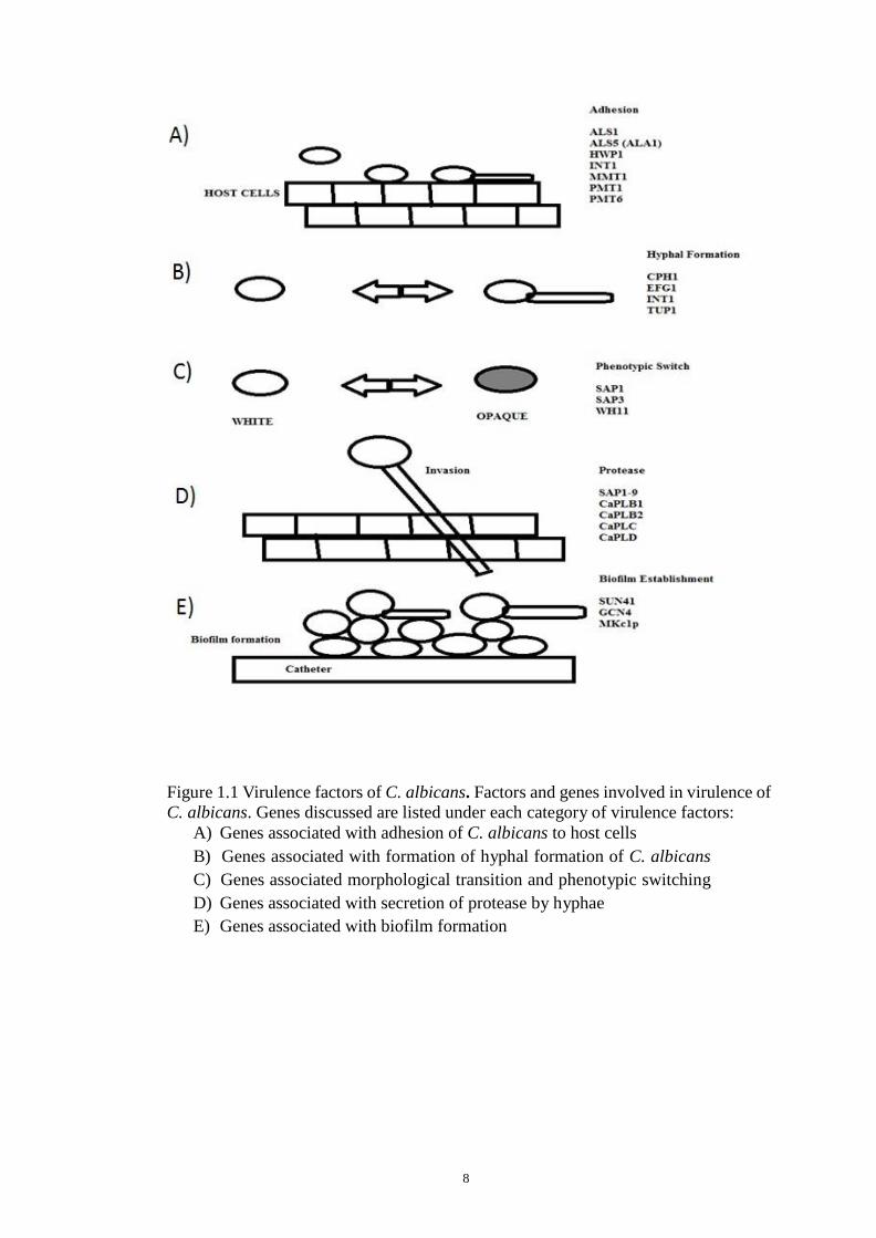
8
Figure 1.1 Virulence factors of C. albicans. Factors and genes involved in virulence of
C. albicans. Genes discussed are listed under each category of virulence factors:
A) Genes associated with adhesion of C. albicans to host cells
B) Genes associated with formation of hyphal formation of C. albicans
C) Genes associated morphological transition and phenotypic switching
D) Genes associated with secretion of protease by hyphae
E) Genes associated with biofilm formation

9
1.4.1 Adhesion
Adherence of C. albicans to host cells is important for commencing and maintaining a
commensal relationship (Gaur et al., 1999), as well as for establishment of disease
through the settlement of host niches (Chaffin et al., 1998). It is a complex mechanism
utilizing several types of adhesins, including agglutinin- like sequence (ALS) and hyphal
wall protein (Hwp1) (Mayer et al., 2013; Yang, 2003). Glycosylphosphatidylinositol
(GPI)- linked cell surface glycoproteins (Als1-7 and Als9) was encoded by ALS gene ,
these proteins are homologous to α-agglutinin, required for cell-cell recognition during
mating in S. cerevisiae (R. A. Calderone & Fonzi, 2001). Als1, Als3 and Als5 (Ala1)
have an attachment function to human buccal epithelial cells (HBEC) and fibronectin,
collagen, laminin and endothelial cells (Hoyer, 2001; Hawser & Douglas, 1994). Als4p
binds to endothelial cells, Als5 is for cell aggregation, Als6 bind to collagen and Als9
binds to laminin. Als1 is vital for the attachment of the microorganism to the oral
mucosa during the initial phase of the infection (Kamai et al., 2002). Hypha-associated
adhesion Als3 is vital for adhesion (Murciano et al., 2012; Phan et al., 2007). ALS3 gene
was highly expressed during infection of in vivo vaginitis and during infection of oral
epithelial cells in vitro (Naglik et al., 2011; Wachtler et al., 2011; Cheng et al., 2005).
HWP1 encodes an outer surface mannoprotein on the hyphal wall (transglutaminase
substrate, TGase) and form covalent binding with HBEC (Staab et al., 1996). An
hwp1/hwp1 mutant strain was greatly defective in the ability to adhere to HBEC and
display attenuated virulence in a murine model of systemic candidiasis (Staab et al.,
1999; Chaffin et al., 1998).

10
1.4.2 Secreted Degradative Enzymes
Following attachment to host cell surfaces and hyphae growth, proteinases was secreted
by pathogen’s hyphae in order to degrade the tissue lining and acquire nutrition at the
infection site (Wachtler et al., 2012; Naglik et al., 2003). Secreted aspartyl proteinases
(SAP) from Candida dissolved many proteins at laceration sites, such as collagen,
haemoglobin, keratin, albumin, Immunoglobulin A, fibronectin, cystatin A, salivary
lactoferin, interleukin1β, mucin, and laminin (Hube et al., 1998). SAP comprises of ten
members, Sap1-10. Sap9 and Sap10 remain constrained to the cell-surface and Sap1-8
are released to the surrounding medium (Taylor et al., 2005; Naglik et al., 2003).
Expression of Saps 1, 2 and 3 by the yeast phase are required for virulence in a murine
model of systemic infection, and for invasion of reconstituted human epithelium (RHE)
in vitro (Schaller et al., 1999; Hube et al., 1997). Saps 4, 5 and 6 are expressed in the
passage from yeast to hyphae phase at neutral pH. Sap7 was never detected in vitro
whereas, Saps 9 and 10 manifest in both forms (yeast and hyphae) (Albrecht et al.,
2006).
Enzymes such as phospholipases (PL) digest ester linkages of glycophospholipids and
allow Candida cells to invade tissue. There are four different classes of phospholipases
(PLA, PLB, PLC and PLD); out of these four classes only the five member of PLB
(PLB1-5) are situated outside a cell and has both fatty acid release (hydrolase) and
lysophospholipase- transacylase activities (Mavor et al., 2005; Niewerth & Korting,
2001). There is higher level of phospholipases in C. albicans strains isolated from blood
than commensal strains (Ibrahim et al., 1995); cells producing less phospholipase are
less likely to cause disease than strains producing high phospholipase in mouse model

11
(Theiss et al., 2006; Ghannoum, 2000). Therefore high level of phospholipase activity is
discovered at where the hyphae were in direct contact with the object (Pugh & Cawson,
1977).
1.4.3 Morphological Transition and Phenotypic Switching
Morphological transition in C. albicans is the ability to switch between the unicellular
yeast form, such as blastospores, and the filamentous form, such as pseudohyphae or
hyphae. Out of all the Candida spp., only C. dubliniensis and C. albicans can undergo
morphogenesis. Morphological conversion from yeast cells (round/oxoid in shape and
readily separate) to pseudohyphae (extended ellipsoidal cells with constraints at the
septa) or parallel-walled true hyphae is facilitated by ~pH 7, nutrients, temperature of
37-40ºC, approximately 5.5% of CO2 concentration and presence of biotin, amino acid,
serum and N-acetyl-D-glucosamine. Reverse conversion from hyphae to yeast form
occurs when cells are grown in higher concentration of glucose, absence of serum,
acidic pH, and lower temperature (Eckert et al., 2007; Corner & Magee, 1997). In
C. albicans, this conversion is controlled by 2 regulatory proteins, Cph1 and Efg1,
which are homolog to S. cerevisiae, Ste12 and Phd1 respectively (Lewis, Lo, et al.,
2002). C. albicans mutant strains (cph1 and efg1) have morphogenesis defects, such as
defective in filamentous growth and are unable to form filaments when it is exposed to
many stimuli including macrophage and serum. Candida mutant strains, cph1/cph1 and
efg1/efg1 are avirulent in mouse model (Lewis, Lo, et al., 2002). This transition (yeast to
psedohyphae or hyphae and vice versa) is termed dimorphism and it is required for
pathogenesis, as yeast forms are suited for dissemination in tissue and hyphae forms are
required for invasion and tissue damage. The yeast cell that develops into hyphae is able

12
to kill macrophages (upon phagocytozed by macrophages) by secreting hyphae
associated proteinases; these factors also help hyphae cells in resist to neutrophils.
Hyphae cells also induce transmigration through endothelial cells from bloodstream
(Hube, 2004; Gow et al., 2002; Molero et al., 1998).
The colonies of C. albicans show distinct morphological form, including white round-
oxoid cells, gray colonies with elongated shaped cells (opaque), hat, irregular wrinkle,
strippled, fuzzy, star, and rough at high frequency (10-4
to 10-1
) (Sudbery et al., 2004). It
is not known what causes phenotypic switching and how switching affect virulence of
C. albicans. Switching happen more frequently in cells isolated directly from vaginitis
or systemically infected patients (Jones et al., 1994; Soll, 1988). White-phase cells have
lower frequency for mating and lower capability to colonize skin in a cutaneous model
than opaque-phase cells. White-phase cells are more virulent in systemic infection while
opaque-phase cells in cutaneous infection of murine model (Kvaal et al., 1999).
Expression of gene SAP1 and SAP3 only occur in opaque cells, while expression of
SAP2, WHI1, and EFG1 occur specifically in white-phase cells (Miller & Johnson,
2002). Morphology transition is important for C. albicans in its adaptation or infection
of the fungus to specific organs, such as skin, kidney and endothelial lining in blood
vessels.
1.4.4 Biofilms
A vital virulence factor of C. albicans is its ability to forms biofilms. Formation of
biofilms starts with adherence of C. albicans cells to non biotic material, such as
catheters and dentures, or to biotic surfaces, such as mucosal cell surfaces. Biofilms
13
results in an increase in candidemia and antifungal resistance (Chandra, Kuhn, et al.,
2001; Hawser & Douglas, 1994). Devices like catheters and dentures provide a platform
for Candida cells to form biofilms. Formations of biofilms proceeds in three
development phases: early (attachment of Candida cells to abiotic or biotic platform and
expansion of these cells), intermediate (accumulation of extracellular matrix material
(ECM) and development of hyphae cells on upper part of the biofilm), and mature
(biofilm complex distribute Candida cells to the surrounding). Mature biofilms are more
resistant to the exposure of antifungal agents and host immune factors in comparison to
planktonic cells (Finkel & Mitchell, 2011). C. albicans biofilm is resistant to variety of
azoles, including voriconazole (Lewis, Kontoyiannis, et al., 2002), miconazole (Lamfon
et al., 2004), ketoconazole, itraconazole and fluconazole, (Hawser & Douglas, 1995),
polyenes nystatin and Amphotericin B, (Chandra, Kuhn, et al., 2001; Chandra,
Mukherjee, et al., 2001) and flucytosine (Hawser & Douglas, 1995). Azole resistant in
C. albicans biofilms may be attributed to the high levels expression of the drug efflux
determinants MDR1, major facilitator superfamily transporter, and the ATP-binding
cassette transporters CDR1 and CDR2 (Lupetti et al., 2002).
One important component of C. albicans biofilms is ECM, which is proposed to provide
a structural scaffold and protection for biofilm cells (Taff et al., 2012). ECM is mainly
made from β-1,3 glucan (encoded by SKN1 and KRE1). Higher Amphotericin B
resistance corresponded to upregulation of KRE1 transcript in yeast (Breinig et al.,
2004). β-1,3 glucan in ECM also protect C. albicans from neutrophils attacks and do not
trigger production of reactive oxygen species (ROS) (Z. Xie et al., 2012).

14
1.5 Transmigration
Adhesion of C. albicans to the endothelium of the blood vessels triggers the invasion or
transmigration of C. albicans across the endothelium. Several mechanisms were
proposed for Candida migrates across the endothelium, Two of the mechanisms are
induced endocytosis and active penetration (Naglik et al., 2011; Zhu & Filler, 2010). For
induced endocytosis, adherent organism was endocytosed by endothelium allowing
adherent cell to cross abluminal surface of the endothelium. On the cell surface of
endothelium, the fungus synthesized specialized proteins that binds to ligand E-cadherin
on host epithelium cells and N-cadherin on endothelium cells via the Candida protein
Als3, thereby triggering endocytosis of the C. albicans across the endothelial barrier
(Phan et al., 2007; Phan et al., 2005). So far two genes associated to invasion have been
identified, SSA1 and ALS3 which functions as both adhesin and invasin (Sun et al.,
2010; Phan et al., 2007). SSA1 induced the expression of heat shock protein 70 (HSP70)
family on the cell surface and deletion of both SSA1 and ALS3 genes resulted in reduced
epithelial adherence and invasion. Similarly mutant strains show less frequency in
causing oropharyngeal candidiasis in a mouse model (Naglik et al., 2011; Sun et al.,
2010). Als1 and Ssa1 bind to host E-cadherin subsequently induces tyrosine
phosphorylation of unknown intracellular endothelial cell proteins (Belanger et al.,
2002). These binding produce pseudopods through microfilament rearrangement
subsequently trigger endocytosis of adherent C. albicans hyphae cells (Phan et al., 2007;
Filler et al., 1995). Active penetration of C. albicans into endothelial cells requires the
elongation of hyphae to penetrate through the endothelial cells and likely kill the cells
along the process (Wachtler et al., 2011; Dalle et al., 2010). It is not well understood
what exactly facilitates active penetration of host cells. However, it is presumed that

15
factors such as physical forces and fungal adhesion are crucial (Wachtler et al., 2011). It
is not only the hyphal form of C. albicans that can undergoes endocytosis; unchanged
strains can also undergo endocytosis to a certain degree causing minimal damage to the
endothelial cells (Klotz et al., 1983). Induced endocytosis is a passive process and does
not required living fungal cells as even dead hyphae cells are endocytosed (Dalle et al.,
2010; Park et al., 2005).
1.6 Fitness Attribute
Adaptation to the environment is vital to pathogens such as C. albicans to allow them to
grow and expand in wide range environments, such as within their mammalian host and
cause infection. C. albicans can grows in variety niches in human and metabolize
different carbon sources that are present in the host. Environment adaptation involves
both metabolic and stress adaptation. Stress responses can be initiated in response to a
wide-range of environmental stimulus including heat-shocks, osmotic and cationic,
oxidation, nitrosative and cell wall stresses (A. J. Brown et al., 2014). Regulation of
C. albicans metabolism and activation of specific stress responses are initiated upon
contact with host. Metabolic flexibility and stress response represent crucial fitness
attributes that have evolves alongside with virulence attributes in C. albicans and is
important for cells viability (Barelle et al., 2006; Lorenz & Fink, 2001).
1.6.1 Heat-shocks
Numerous studies have shown that the pathogenicity of C. albicans is influenced by heat
shock response. Stress response by heat shock proteins (Hsp70 and Hsp90) mark

16
damaged or aggregated proteins for degradation, or encourage the folding of target
proteins (Feder & Hofmann, 1999; Parsell et al., 1993). The response in C. albicans is
regulated by the heat shock transcription factor Hsf1 (Nicholls et al., 2009). Hsf1 is
essential for cells viability in yeast. C. albicans exposure to acute-heat shock causes
Hsf1 phosphorylation and through the canonical heat shock elements (HSEs) in their
promoters promote the expression of target heat shock protein (HSP) genes (Nicholls et
al., 2009). Inductions of cellular adaptation to the heat shock are by refolding or
degradation of damaged proteins through activation of HSP gene. Following thermal
adaptation, heat shock protein 90 (HSP90) gene is activated and interacts with Hsf1 to
repressed the stress response in C. albicans (Leach et al., 2012). Hsp90 is required for
C. albicans to establish systemic infection. Autoantibodies against Hsp90 gives mild
protection against the infection (Matthews et al., 1991).
Heat shock proteins are found on C. albicans cell surface and are immunogenic during
infections (Lopez-Ribot et al., 1996; Matthews et al., 1987). Induction of heat shock
proteins by mild thermal insult is associated with yeast-hyphae morphogenesis, which is
considered to be virulence attribute (Swoboda et al., 1996). A mutant strain with
mutation that inhibits promotion of the heat shock response in C. albicans has attenuated
virulence.
1.6.2 Osmotic and Cationic Stress
Prolonged exposure to salts, such as sodium chloride, NaCl and potassium chloride, KCl
resulted in dehydration of cells, causing loss of turgor pressure and a reduction in cell
size due to cationic and osmotic stress (Kuhn & Klipp, 2012). These events will

17
subsequently triggers the expression and phosphorylation of the stress-activated protein
kinases (SAPK) Hog1 that results in reprogram of the gene expression pattern required
for cell survival upon osmostress. Examples include regulation of gene encoding
glycerol biosynthetic enzymes (Enjalbert et al., 2006; Smith et al., 2004). This will
results in the build-up of glycerol in the cells which will then allow them to balance their
osmotic pressure with that of surrounding and allowing the resumption of growth.
Adaptation of C. albicans cells to osmotic/cationic stresses relies on HOG1. It is a major
regulator of the osmostress-regulated transcription, including activation of glycerol
biosynthetic gene which will then cause aggregation of glycerol (Smith et al., 2004; San
Jose et al., 1996).
Common signaling modules found in both higher and lower eukaryotic cells, such as
mitogen-activated protein kinase (MAPK) cascades are composed of three succesively
activated tiers of kinases (MAP kinase kinase kinase, MAP kinase kinase and MAP
kinase). Hog1 is a component of a highly conserved mitogen-activated protein (MAP)
kinase pathway. In C. albicans, this MAPK is activated by the MAPKK, Pbs2, which in
turn is activated by a single MAPKKK, Ssk2 (Arana et al., 2005). The regulators
network upstream of gene expressions that activate this MAPK module due to exposure
to osmotic stress in C. albicans is open to interpretation.
Hog1 (SAPK) regulates both morphogenesis and stress response in C. albicans and is a
dominant factor for virulence of C. albicans (Cheetham et al., 2011). Adaptation to
other stresses, such as regulation of cellular morphogenesis and metabolism, oxidative
stress response, and influence cell wall performance required Hog1 (Alonso-Monge et
al., 2009; Eisman et al., 2006; Alonso-Monge et al., 2003). Based on several studies,

18
adaptation to osmotic and cationic stress are important in host niches, such as in the
kidney, where the concentrations of NaCl in urine can approach 600 mmol l−1
(Z. Zhang
et al., 2004; Ohno et al., 1997).
1.6.3 Cell Wall Stress
The fungal cell wall is important for survival and interaction with the surrounding
environment; it is the point of contact between fungus and target surfaces, and processes
such as adhesion, dimorphism, and biofilms formation take place. These factors are
responsible for the pathogenicity of C. albicans.
Congo Red and Caspofungin are antifungal drug that inhibit and disturb synthesis and
assembly of β-glucan, whereas Calcofluor White disrupt formation of chitin. These
antifungal drugs are used to pressure the C. albicans cell wall in vitro (Eisman et al.,
2006; Wiederhold et al., 2005). The Hog1 pathway regulates cell wall stress responses
and enables the synthesis of chitin (Munro et al., 2007; Eisman et al., 2006).
Two additional highly conserved MAPK pathways are responsible for regulations of
stress response in C. albicans cell wall. The first pathway, the Mkc1 facilitated MAPK
or the cell integrity pathway, and a second pathway, involves in Cek1-mediated MAPK
or yeast-hypha morphogenesis (Roman et al., 2007).
Bck1-mediated MAPKKK, Mkk1-mediated MAPKK and Mkc1-mediated MAPK are
MAPKK module that involves in cell integrity pathway (Navarro-Garcia et al., 1998).
Cascade of phosphorylation reactions, Mkc1 governed by protein kinase (Pkc1) plays an

19
essential role in the generation of a stable cell wall in yeast (Paravicini et al., 1996). The
inactivation of Mkc1 increases the cell wall sensitivity to thermal insult and other
stresses (Navarro-Garcia et al., 1998). Inactivation of Mkc1 does reduce and weaken the
virulence of C. albicans (Diez-Orejas et al., 1997). However inactivation of this protein
does not contribute and increase the probability of cell death in response to neutrophils
or macrophages (Arana et al., 2007).
MAPKKK Ste11, MAPKK Hst7 and MAPK Cek1 are components of morphogenetic
MAPK (Cek1) pathway. This pathway also regulate C. albicans mating response and are
required for efficiency of mating (Chen et al., 2002). Cell surface sensor Msb2 triggers
Cek1 pathway upon exposure to antifungal agents that target cell wall integrity or
mutation of gene required for integrity of cell wall (Cantero & Ernst, 2011; Roman et
al., 2009). Inactivation of Cek1 in C. albicans growing under certain conditions resulted
in retarded growth of filament and is more prone to cell wall stresses (Eisman et al.,
2006; Csank et al., 1998; Leberer et al., 1996). C. albicans cek1 mutants display
attenuated virulence but does not affect sensitivity of the cells in response to
macrophage or neutrophil (Arana et al., 2007; Csank et al., 1998).
1.6.4 Oxidative Stress
Reactive oxygen species (ROS) is an acting compound causing damage and
inflammation to the tissue. Resistant to this compound by C. albicans require Cap1, an
orthologue of AP-1-like transcription factor Yap1 in S. cerevisiae (Alarco & Raymond,
1999). Following oxidative stress, cysteine residues near the carboxyl terminus Cap1
which are redox-sensitive was oxidized and subsequently leads to the nuclear

20
accumulation of Cap1 and Yap1-responsive elements (YRE) in their promoters that will
activates the target genes. These processes are Hog1 independent (Znaidi et al., 2009; X.
Zhang et al., 2000). Upon accumulation of Cap1 in C. albicans, genes involved in the
removal of oxidative stress, such as oxidative damage repair, superoxide dismutase and
catalase, and redox homeostasis glutathione synthesis was expressed and functions by
detoxifying ROS and regulates stress adaptation in cells. Deletion of CAP1 will increase
C. albicans sensitivity to oxidative stress due to decrease promotion of genes and
enzymes mentioned above (Enjalbert et al., 2006; Alarco & Raymond, 1999). Moreover
C. albicans mutant strains (cap1/cap1 and hog1/hog/1) display attenuated virulence and
increased response to phagocytes (Arana et al., 2007; Fradin et al., 2005).
1.7 Central Metabolism Pathways
In order to proliferate in a broad range of environmental niches, metabolic flexibility
and virulence factors are important so that they can metabolize different carbon sources
that are scarce or the only available carbon source at a specific environmental niche.
Carbohydrates are necessary to produce biomolecules and for generating energy for
basic functions of cells. Before entering the glycolytic pathway, sugars undergo
transition to fructose 6-phosphate or glucose 6-phosphate. ATP and NADH are produce
from the conversion of sugar phosphates into pyruvate in glycolysis pathway. From
there, fermentation and respiration are carried out by cells. Although NAD+ is
regenerated by both processes, respiration produces more ATP than fermentation
through the oxidative phosphorylation and tricarboxylic acid (TCA) cycle. Glycolysis
pathway is important for carbon metabolism and it is frequently used for both
fermentation and respiration. This pathway is important to the virulence in pathogenic

21
bacteria, parasites, and fungi, and was up-regulated during infections (Costa et al., 2007;
Barelle et al., 2006; Rodaki et al., 2006). Glycolysis, gluconeogenesis, and the
glyoxylate cycle are part of C. albicans central metabolism (Fig. 1.2).
Figure 1.2: The pathways of ß-oxidation, the glyoxylate cycle, gluconeogenesis, and
glycolysis are shown, with the C. albicans gene names (Taken from Lorenz et al., 2004).
22
1.7.1 Glycolysis and Gluconeogenesis
Transcript of central metabolic pathway such as glycolysis is strictly regulated in
response to environmental conditions, such as availability of carbon source, oxygen
levels, energy needs and metabolite concentrations. Most of the regulators of gene
expression in glycolytic of other species have not been identified. Understanding of
transcriptional control of glycolysis in eukaryotes is mainly based on the experimental
paradigm S. cerevisiae; nonfermentable yeast (Chambers et al., 1995). The transcription
regulators (Gcr1 and Gcr2) are responsible for inducing the expression of the glycolytic
genes in S. cerevisiae (Uemura & Fraenkel, 1990; Clifton et al., 1978). Inactivation of
either gene (GCR1 and GCR2) result in growth defects in response to glucose due to
unexpressed glycolytic genes (Uemura & Fraenkel, 1990).
The Crabtree-positive Saccharomyces yeasts is a facultative anaerobes and has glucose
repression circuit. This is in contrast to that of most other obligate aerobes and
facultative eukaryotes, which lack the glucose repression circuit. Pyruvate is oxidized to
carbon dioxide through the TCA cycle in Crabtree-negative organism under aerobic
conditions. Most aerotolerant organisms are able to metabolize energy anaerobically
through fermentation pathways to a certain extent to subsequently regenerate NAD+,
such as Neurospora crassa, Aspergillus oryzae, and Trichoderma reesei. These fungi
metabolize energy aerobically and the TCA cycle is not affected during growth in
glucose rich which is different in S. cerevisiae that rely heavily on fermentation pathway
(Maeda et al., 2004; X. Xie et al., 2004; Chambergo et al., 2002).

23
C. albicans is an opportunistic fungus that is capable of metabolizing carbon sources
through respiration and fermentation pathway. C. albicans is a Crabtree-negative
organism that does not have GCR1/2 homologs and it regulates transcription of
glycolytic genes in different way compared to S. cerevisiae. Tye7 and Gal4 activates the
glycolytic pathway in C. albicans and severe growth defects was observed in the mutant
strains (tye7 and gal4) cultured on a certain condition. C. albicans mutant strains (tye7
and gal4) showed attenuated virulence in Galleria mellonella infection model. Therefore
TYE7 and GAL4 genes are required for pathogenicity and virulence of C. albicans
(Askew et al., 2009).
Gluconeogenesis is required for yeast cells to generate sugar phosphates for the
synthesis of essential cellular components, during growth on non-fermentable carbon
sources. During growth on unbalanced carbon sources, S. cerevisiae and C. albicans
shunt glycolysis pathway, this is achieved by enzymes of gluconeogenesis, such as
phosphoenolpyruvate carboxykinase, and fructose-1,6-bisphosphatase. The enzymes of
the glyoxylate cycle are necessary for gluconeogenesis, such as malate synthase (MLS)
and isocitrate lyase (ICL).
1.7.2 The Tricarboxylate Acid (TCA) and Glyoxylate Cycle
The glyoxylate cycle is also known as a “modified tricarboxylic acid (TCA) cycle”.
Isocitrate lyase, ICL1 and malate synthase, MLS1 are enzymes in glyoxylate cycle that
functions by converting isocitrate and acetyl-CoA into succinate and malate respectively
(Fig 1.2). In glyoxylate cycle, isocitrate was metabolized into succinate and glyoxylate
which are subsequently condensed by malate synthase, MLS1 and accompanied by

24
acetyl-CoA to produce free CoA-SH and malate. Consequently malate is further
processed by malate dehydrogenase and produce succinate as a final product. Succinate
can be reused in the TCA cycle or to operate as carbohydrate biosynthesis indicator or
biosynthesis of amino acid. Fatty acids or C2-units such as ethanol or acetate are utilized
by glyoxylate cycle to produce succinate, a C4-units carbon sources through various
catabolic processes. Succinate is then metabolize by glyoxylate cycle to produce energy
(Kornberg & Madsen, 1958). In contrast, carbon sources such as ethanol, acetate or oleic
acid cannot be metabolized by S. cerevisiae because it has deficiency in Icl1 or Mls1.
Moreover growth on these carbon sources greatly repress the expression of gene
involved in metabolism of nitrogen, malate synthase (DAL7) (Kunze et al., 2002;
Fernandez et al., 1992; Hartig et al., 1992). In recent year, key enzyme such as isocitrate
lyase in glyoxylate cycle is greatly studied. Mutant strain (icl/icl) has attenuated
virulence in mouse model (Lorenz & Fink, 2001). This provide a platform for anti-
fungal testing to reduce the virulency of C. albicans
1.8 Glucose Sensing
In the presence of glucose, C. albicans undergoes yeast-to-hyphal transition which is
crucial for virulence and invasion of host cells (Hudson et al., 2004), as explained
previously (Section 1.4.3) morphological plasticity is important factor for C. albicans
virulency. The hyphal form can diffused through the tissues or form mycelial biofilms,
and the yeast form is presume to be able to propogate easily via body fluids (Bendel et
al., 2003).