EFFECTS OF FARADIC STIMULATION OF THE ... OF FARADIC STIMULATION OF THE CEREBRAL CORTEX ON LIMB AND...
18
EFFECTS OF FARADIC STIMULATION OF THE CEREBRAL CORTEX ON LIMB AND RENAL VOLUMES IN THE CAT AND MONKEY1 HAROLD D. GREEN AND EBBE C. HOFF From the Laboratory of Physiology, Yale University School of Medicine, New Haven Received for publication June 1, 1936 Dusser de Barenne and Kleinknecht (1) observed falls of blood pressure following electrical stimulation of the motor areas of dogs, cats, and rab- bits anesthetized with ether, urethane or morphine. Later Hoff and Green (2) found that rises of blood pressure could be obtained from simi- lar cortical areas of cats and monkeys when anesthetized with ether. Since mean blood pressure as recorded in these experiments represents only the resultant of simultaneous vascular changes in different parts of the body, the problem of whether a rise or a fall in pressure occurs may depend upon various factors such as anesthesia and the character of cere- bral representation of the vasomotor system. The problem has there- fore been investigated afresh by combining studies of blood pressure with measurement of organ volumes. METHODS. Bilateral limb volumes, renal volume, and blood pressure were optically recorded while stimulating the cortex of 14 cats and 14 monkeys anes- thetized with ether, or with Dial-Ciba (containing urethane) supplemented with ether. While curare was usually given in addition to paralyze muscular movements, a few experiments were performed without this drug to demonstrate that volume changes were obtainable in the absence of normal muscular movement with stimuli below the threshold for muscular response. In all cases in which curare was ad- ministered, artificial respiration was maintained throughout the experiment. The limb plethysmographs were closed at the upper end by rubber diaphragms made of dental dam pierced with a hole large enough to admit the limb, without obstructing the venous return, and were adjusted so as to include the fore-arms and as much of the upper arms as possible. An air-tight seal was obtained by shav- ing or closely clipping the hair and cementing the rubber dam to the arm with gum- rubber cement. The plethysmographs were filled with water at 35-40°C., except in those instances in which the limbs were skinned when Ringer-Locke’s solution was used. The changes in volume were recorded with Frank segment capsules (3) covered with condom rubber, using air transmission. The use of a combined air- fluid transmission system introduces certain artefacts, namely, extraneous fine vibra- tions which may be noticed in some records, but these do not interfere with the 1 A preliminary report of these experiments was communicated to the American Physiological Society, March 26, 1936. These investigations were aided by a grant from the Research Funds, Yale Uni- versity School of Medicine. 641 by 10.220.33.4 on April 17, 2017 http://ajplegacy.physiology.org/ Downloaded from
Transcript of EFFECTS OF FARADIC STIMULATION OF THE ... OF FARADIC STIMULATION OF THE CEREBRAL CORTEX ON LIMB AND...

EFFECTS OF FARADIC STIMULATION OF THE CEREBRAL CORTEX ON LIMB AND RENAL VOLUMES IN THE
CAT AND MONKEY1
HAROLD D. GREEN AND EBBE C. HOFF
From the Laboratory of Physiology, Yale University School of Medicine, New Haven
Received for publication June 1, 1936
Dusser de Barenne and Kleinknecht (1) observed falls of blood pressure following electrical stimulation of the motor areas of dogs, cats, and rab- bits anesthetized with ether, urethane or morphine. Later Hoff and Green (2) found that rises of blood pressure could be obtained from simi- lar cortical areas of cats and monkeys when anesthetized with ether. Since mean blood pressure as recorded in these experiments represents only the resultant of simultaneous vascular changes in different parts of the body, the problem of whether a rise or a fall in pressure occurs may depend upon various factors such as anesthesia and the character of cere- bral representation of the vasomotor system. The problem has there- fore been investigated afresh by combining studies of blood pressure with measurement of organ volumes.
METHODS. Bilateral limb volumes, renal volume, and blood pressure were optically recorded while stimulating the cortex of 14 cats and 14 monkeys anes- thetized with ether, or with Dial-Ciba (containing urethane) supplemented with ether. While curare was usually given in addition to paralyze muscular movements, a few experiments were performed without this drug to demonstrate that volume changes were obtainable in the absence of normal muscular movement with stimuli below the threshold for muscular response. In all cases in which curare was ad- ministered, artificial respiration was maintained throughout the experiment.
The limb plethysmographs were closed at the upper end by rubber diaphragms made of dental dam pierced with a hole large enough to admit the limb, without obstructing the venous return, and were adjusted so as to include the fore-arms and as much of the upper arms as possible. An air-tight seal was obtained by shav- ing or closely clipping the hair and cementing the rubber dam to the arm with gum- rubber cement. The plethysmographs were filled with water at 35-40°C., except in those instances in which the limbs were skinned when Ringer-Locke’s solution was used. The changes in volume were recorded with Frank segment capsules (3) covered with condom rubber, using air transmission. The use of a combined air- fluid transmission system introduces certain artefacts, namely, extraneous fine vibra- tions which may be noticed in some records, but these do not interfere with the
1 A preliminary report of these experiments was communicated to the American Physiological Society, March 26, 1936.
These investigations were aided by a grant from the Research Funds, Yale Uni- versity School of Medicine.
641
by 10.220.33.4 on April 17, 2017
http://ajplegacy.physiology.org/D
ownloaded from

642 HAROLD D. GREEN AND EBBE C. HOFF
interpretation since gross changes only, and not the contour, are being studied. This air-fluid system was used because it greatly diminished shifting of the base level of the volume curves due to changing temperature of the air contained within the plethysmographs. Renal volume was recorded with an oncometer composed of two shallow cups hinged together covered with light rubber dam and arranged to surround the kidney without constricting the pedicle. The oncometer halves were filled with either water or air and connected by air-filled tubes to a Frank segment capsule.
In nearly all instances, blood pressure was recorded from a femoral artery with a Wiggers optical manometer (4), using paraffin-coated glass cannulae and boiled 20 per cent sodium sulphate as the anticoagulant. The manometer and cannula had a natural frequency of 75 to 100 per second. A constant slow intravenous in- fusion of mammalian Ringer-Locke’s solution was maintained throughout all experi- ments. Although in a few instances a thyratron stimulator was used, the stimulating current was usually obtained from a 1100 ohm potentiometer shunted across the standard 60 cycle 110 volt alternating current. The potential across the stimulating electrodes could be readily adjusted within 0.1 volt over the range of 0 to 110 volts. Bipolar electrodes separated about 1.0 mm. were used, the cortex being covered with warm, moist pledgets between stimuli which were applied not oftener than once in 2 minutes and lasted 5 to 15 seconds.
RESULTS. I. Either a rise or a fall of mean blood pressure may result from the stimulation of the motor area of the cortex. In a previous paper (2) it was demonstrated that under ether anesthesia, plus curare to pre- vent muscular activity, only a rise of blood pressure could consistently be obtained from the motor area of the cat’s cortex and from the motor and premotor areas of that of the monkey. This observation has been confirmed in the course of these experiments but, in addition, it has been observed that in animals anesthetized by Dial supplementled by ether, no change, or a fall of mean blood pressure more often resulted from excitation of the same areas of the cortex (see fig. 2, A-D). This latter observation therefore confirms Dusser de Barenne and Kleinknecht’s findings that decline of pressure may be elicited by stimulation of the motor cortex of animals.
2. The blood JEow in di$erent regions of the body as measured by alteration of the volume of these regions, is a$ected by stimulation of the cortex. a. Renal volume is usually decreased. A diminution of renal volume con- sistently occurs in curarized animals under ether anesthesia during the rise of blood pressure evoked by stimulation of the motor cortex. A typical record of such a response is reproduced in figure 1D. In this and all subsequent records, the kidney volume is labeled RV and downward movement of the line indicates a decrease in volume. The diminution of renal volume slightly precedes the rise of blood pressure. In a series of experiments this amounted to 0.5 to 1.0 second, the latency of the renal volume changes varying from 1.5 to 3.5 seconds and that of the simul- taneously recorded blood pressure from 2.0 to 4.5 seconds. An equiva- lent change in volume of a given kidney occurred following stimulation
by 10.220.33.4 on April 17, 2017
http://ajplegacy.physiology.org/D
ownloaded from

CORTICAL REPRESENTATION OF VASOMOTOR SYSTEM 643
of either cerebral hemisphere. After sectioning the nerves to the kidney on one side, that kidney now dilated during the rise of mean blood prcs- sure. That the rise of blood pressure is not dependent solely on vascular const’riction in the viscera was shown in two experiments by a consider- able rise of pressure even after complete abdominal evisceration.
b. Limb volume is usually increased. Stimulation of the sigmoid gyrus of the cat’s cortex, and of areas 4 and 6 in the monkey produced in almost every instance an alteration of limb volume. The most frequent response in both cat and monkey was a dilatation of the limbs occurring after a latency of 1.5 to 13 seconds with an average latency of 4.1 seconds. Very weak stimuli (as low as 0.8 volt A.C. 60 cycle) were often more effective than stronger stimuli. This dilatation was greatest about 9 seconds after onset of stimulation and occurred regardless of any alteration of the mean blood pressure. Thus in figure In, a record obtained under ether ancs- thesia, the limbs dilated during a rise in blood pressure while, as illustrated in figure lB, dilatation occurred in the absence of change of blood pressure and in figure 2, A-D, during a decline of blood pressure. In the experi- ments from which the last two figures were taken, Dial anesthesia was used. In these as in all subsequent records, the limb volume curves arc labeled LL or LA for the left extremity and RL or RA for the right. Approximation of the limb volume curves, i.e., downward movement of the line for the left extremity and upward movement of that for the right, indicates increased volume of the extremities. Usually three waves may be seen in each record : a small one, the pulse wave; a sligMy larger and slower one, thct respiratory wave, and the large slow wave indicating the change of volume or of pressure as a result of the stimulat,ion.
c. Limb volume is occasionally diminished. While stimulation of the motor areas both of monkeys and cats anesthetized with ether usually produced a dilatation of the extremities, in 5 monkeys and 1 cat, anesthe- tized with Dial, stimulation of similar areas frequently resulted in a con- striction of the limbs with a latency of 1.5 to 11 seconds (average 6.0 sec.). This constriction was usually accompanied by a rise of blood pres- sure, although occasionally there was no change or even a slight fall of blood pressure. Records showing constriction of limbs during stimulation of the cortex are reproduced in figures 1E; 3A-B, and 4A.
Either constriction or dilatation of the limbs was followed by return to normal approximately 8 to 13 seconds after the end of stimulation. usu- ally no after-response was encountered although in some instances excita- tion of the cortex produced an initial constriction of the limbs followed by a delayed dilatation which became more marked just after cessation of the current. In all experiments the alteration of limb volumes during stimulation occurred in the same direction on both sides of the body. The latencies of the response were equal as were the durations of the
by 10.220.33.4 on April 17, 2017
http://ajplegacy.physiology.org/D
ownloaded from

644 HAROLD D. GREEN AND EBBE C. HOFF
by 10.220.33.4 on April 17, 2017
http://ajplegacy.physiology.org/D
ownloaded from
CORTICAL REPRESENTATION OF VASOMOTOR SYSTEM 645
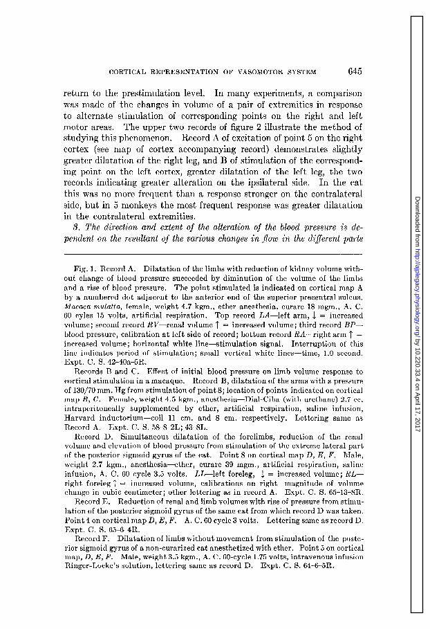
return to the prestimulation level. In many experiments, a comparison was made of the changes in volume of a pair of extremities in response to alternate stimulation of corresponding points on the right and left motor areas. The upper two records of figure 2 illustrate the method of studying this phenomenon. Record A of excitation of point 5 on the right cortex (see map of cortex accompanying record) demonstrates slightly greater dilatation of the right leg, and B of stimulation of the correspond- ing point on the left cortex, greater dilatation of the left leg, the two records indicating greater alteration on the ipsilateral side. In the cat this was no more frequent than a response stronger on the contralateral side, but in 5 monkeys the most frequent response was greater dilatation in the contralateral extremities.
3. The direction and extent of the alteration of the blood pressure is de- pendent on the resultant of the various changes in flow in the different parts
Fig. 1. Record A. Dilatation of the limbs with reduction of kidney volume with- out change of blood pressure succeeded by diminution of the volume of the limbs and a rise of blood pressure. The point stimulated is indicated on cortical map A by a numbered dot adjacent to the anterior end of the superior precentral sulcus. 2Macaca mulaita, female, weight 4.7 kgm., ether anesthesia, curare 18 mgm., A. C. 60 cyles 15 volts, artificial respiration. Top record LA-left arm, I = increased volume; second record R&-renal volume l’ = increased volume; third record BP- blood pressure, calibration at left side of record; bottom record R&--right arm T = increased volume; horizontal white line-stimulation signal. Interruption of this line indicates period of stimulation; small vertical white lines-time, 1.0 second. Expt. C. S. 42-40a-5R.
Records B and C. Effect of initial blood pressure on limb volume response to cortical stimulation in a macaque. Record B, dilatation of the arms with a pressure of 130/70 mm. Hg from stimulation of point 8; location of points indicated on cortical map B, C. Female, weight 4.5 kgm., anesthesia- Dial-Ciba (with urethane) 2.7 cc. intraperitoneally supplemented by ether, artificial respiration, saline infusion, Harvard inductorium-coil 11 cm. and 8 cm. respectively. Lettering same as Record A. Expt. C. S. 58-8-21;; 43-8L.
Record D. Simultaneous dilatation of the forelimbs, reduction of the renal volume and elevation of blood pressure from stimulation of the extreme lateral part of the posterior sigmoid gyrus of the cat. Point 8 on cortical map D, E, F. Male, weight 2.7 kgm., anesthesia-ether, curare 39 mgm., artificial respiration, saline infusion, A. C. 60 cycle 3.5 volts. U-left foreleg, 1 = increased volume; RL- right foreleg 1 = increased volume, calibrations on right-magnitude of volume change in cubic centimeter; other lettering as in record A. Expt. C. S. 65-13-8R.
Record E. Reduction of renal and limb volumes with rise of pressure from stimu- lation of the posterior sigmoid gyrus of the same cat from which record D was taken. Point 4 on cortical map D, E, F. A. C. 60 cycle 3 volts. Lettering same as record D. Expt. C. S. 65-64R.
Record F. Dilatation of limbs without movement from stimulation of the poste- rior sigmoid gyrus of a non-curarized cat anesthetized with ether. Point 5 on cortical map, D, E, F. Male, weight 3.5 kgm., A. C. 60-cycle 1.75 volts, intravenous infusion Ringer-Locke’s solution, lettering same as record D. Expt. C. S. 64-6-5R.
by 10.220.33.4 on April 17, 2017
http://ajplegacy.physiology.org/D
ownloaded from

646 HAROLD D. GREEN AND EBBE C. HOFF
of the body. In those conditions in which cortical stimulation produced no change in systemic blood pressure, the limb and kidney volumes varied reciprocally and the duration of the changes was approximately the same in each organ. This same relationship was also often present when the pressure fell or when it rose. Figure ID is an example of the latter condi- tion. In many instances both the limbs and kidney were constricted, and in these the blood pressure always rose. Figure 1E is an example of these phenomena. Dilatation of both the kidney and the limbs was never observed. In one experiment a fortuitous occurrence (recorded in fig. 1A) demonstrates clearly that the mean blood pressure is the resultant of somatic and visceral vascular reactions. The initial effect of cortical stimulation was dilatation of the extremities with constriction of the kidneys, the net effect being no change in blood pressure. Approxi- mately 11 seconds after the onset of stimulation the limbs began to diminish in volume while the kidney remained constricted and the blood pressure began to rise.
4. Evidence that the vascular changes are active and not passive phenomena. Weber (5), observing the effect of cortical stimulation on blood pressure, limb, and gut volume recorded simultaneously, found that the rise in blood pressure was followed a few beats later by a slight increase in the volume of both limbs while a great reduction of gut volume occurred with the elevation of pressure. He concluded at that time that this rise in blood pressure resulting from cortical stimulation was due mainly to constriction of vessels in those viscera innervated by the splanchnic nerves and that the increase in limb volume was a passive enlargement associated with increased systemic blood pressure. Later, however, Weber (6) found that the increase in limb volume after cortical stimula- tion was greater following evisceration than before, despite the fact that the rise in pressure was less after this procedure. He was therefore led to revise his previous opinion and to suggest an active dilatation in the vessels of the limbs. That the somatic vascular areas do participate ac- tively in the change of systemic blood pressure and the redistribution of blood in the body induced by cortical stimulation and that the volume changes are mediated through nervous channels is substantiated in our own experiments by the following lines of evidence:
a. Variations of limb volume are independent of blood pressure changes. In the earlier experiments, using ether anesthesia, increase in limb volume was consistently obtained in both cats and monkeys, but was always associated with a rise of pressure. However, under Dial anesthesia the blood pressure either rose negligibly or not at all, or fell during cortical stimulation, while the limb volume showed as great a dilatation as with ether. Although it is quite possible that the dilatation of limbs with increase of pressure recorded in the ether experiments may have been
by 10.220.33.4 on April 17, 2017
http://ajplegacy.physiology.org/D
ownloaded from

CORTICAL REPRESENTATION OF VASOMOTOR SYSTEM 647
passively produced, the degree of dilatation was usually greater than that occurring with a simple rise of pressure; furthermore it is difficult to con- ceive of an increase of limb volume during a declining pressure except as a result of an active relaxation of the vascular bed of the limbs. More- over, under certain conditions the limbs have definitely constricted during a rise of pressure, and again in these circumstances such an occurrence is impossible except as a result of active constriction of the vessels of the limbs. Finally the diminution of the volume of the extremities is not a result of shunting of blood to other parts of the body such as the abdom- inal viscera, since renal volume records usually indicated a simultaneous constriction of the renal vascular bed.
b. Limb volume changes persist after adrenalectomy. In one cat and one monkey in which the adrenal arteries and veins had been ligated and the adrenal glands removed, active changes of limb volume continued to occur, so that it does not seem likely that they are due to the liberation of adrenalin as a result of stimulation of the cerebral cortex. Further- more, as shown in figure 4C, injection of adrenalin produced a marked dilatation of an extremity of a monkey in which cortical stimulation had just previously yielded a reduction of limb volume.
c. Cortically induced changes in limb volume are abolished by denervating an extremity. In one monkey showing constriction of the forelimbs with rise of pressure on cortical stimulation, the right extremity was denervated by section of the nerves in the axilla. After the section, stimulation of the cortex produced a slight passive dilatation in the denervated arm with the rise of pressure, while the volume of the normal arm was diminished. The denervated arm responded to intravenous injection of adrenalin as did the normal by dilatation associated with a greatly delayed rise of pressure but an immediate fall of kidney volume (see fig. 4A-C). A similar procedure was carried out in another monkey in which cortical stimulation produced a dilatation of the forelimbs with fall of blood pres- sure. Upon denervation of the left forelimb by section of the nerve trunks in the axilla, this extremity decreased in volume as the blood pressure fell while the right extremity continued to dilate during stimu- lation of the cortex. These experiments indicate that the variations in pc’ripheral vascular resistance produced by cortical st,imulation arc not, mcdiat,td by any humoral agent liberated in other part/s of the body and are not, passive eff&s due! t,o alteration in general blood prcssurq bu1, are caused by nerve impulses passing to the vascular artla from whit+ t,hc responses arc obtained.
5. The muscles of the extremities participate in the limb volume dilatation. In order to examine directly the changes in volume which occur in the muscle as a result of cortical stimulation, limb volumes have been recorded before and after skinning and compared with records obtained simul-
by 10.220.33.4 on April 17, 2017
http://ajplegacy.physiology.org/D
ownloaded from

648 HAROLD D. GREEN AND EBBE C. HOFF
by 10.220.33.4 on April 17, 2017
http://ajplegacy.physiology.org/D
ownloaded from

CORTICAL REPRESENTATION OF VASOMOTOR SYSTEM 649
taneously from the opposite normal extremity. In one cat in which stimulation of the posterior sigmoid gyrus yielded a decline in pressure and simultaneous dilatation of the forelimbs, the response after complete removal of all the skin from that part of a forelimb included in the plethysmograph still took place with the same latency and the same duration in comparison with the opposite normal extremity. This is illustrated in the records given in figure 2A-D. Stimulation of the motor area on either side in 2 cats still produced bilateral forelimb dilatation after removing the skin from both limbs (fig. 3C and D). Similarly vaso- constriction associated with no change or a rise in pressure was obtained in cats both before and after skinning the limbs (fig. 3A and B). Inas- much as these alterations in volume recorded from skinned limbs are active changes, and since in every case the blood pressure varied in the opposite direct’ion to the volume change, it is concluded that the varia- tions in limb volume are at least partly if not chiefly attributable to an active vasodilatation or vasoconstriction in the muscles.
6. Limb volume responses occur without epileptic muscular contractions. It was previously shown (2) that alterations of blood pressure resulting from cortical stimulation can occur in non-curarized animals in the ab- sence of simple muscular response or epileptic seizures. A similar con- clusion may be reached with regard to limb volume changes. They have been produced without the slightest accompanying muscular movements in curarized animals. They occur with short latency and the limb vol- umes return to the prestimulation level shortly after the end of stimu- lation with no suggestion of an aft’er-response. Furthermore in lightly etherized non-curarized animals, well-marked volume changes were evoked with cortical stimuli so weak as to be below the threshold for somatic muscular responses (see fig. IF). In the same animals, slightly more intense stimuli evoked discrete muscular movements of the opposite side.
7. Factors affecting the vascular response. In a given animal under constant experimental conditions successive stimulation of various points in the motor and premotor cortex of monkeys and of the sigmoid gyri
Fig. 2. Limb volume changes elicited from stimulation of corresponding areas on the right and left posterior sigmoid gyri of the cat before and after removing the skin from one of the extremities. Fall of pressure from stimulation of these areas under dial anesthesia. Records A and H, stimulation of right and left cortex respec- tively before skinning; and records C and D, the same areas after skinning the left forelimb. Points of stimulation indicated by numbered dots on cortical map. Male, weight 3.0 kgm., anesthesia-Dial-Ciba (with urethane) 1.5 cc. intraperitoneally supplemented by ether, curare 30 mgm., artificial respiration, intravenous infusion Ringer-Locke’s solution, A. C. BO-cycle 1.5 volts for A and B, and 1.25 volts for C and D. Lettering same as in figure 1, record D. Expt. C. S. 67-5-5R; 6-51,; 19-5R; 20-51,.
by 10.220.33.4 on April 17, 2017
http://ajplegacy.physiology.org/D
ownloaded from

650 HAROLD D. GREEN AND EBBE C. HOFF
by 10.220.33.4 on April 17, 2017
http://ajplegacy.physiology.org/D
ownloaded from

COKTICAL l~EPKliSENTATION OF VASOMOTOH SYSTEM 651
and gyrus proreus of cats has yielded a similar result from each point. This result may be any of the combinations of vascular change previ- ously referred to, namely, dilat,ation or constriction of the limbs with fall or rise or no change of pressure. However there are a number of fact,ors which appear to affect the direction and extent of these changes: 1. In three instances weak stimuli, 0.8 to 2.0 volts A. C. 60-cycle, evoked a dila- tation of the limbs but with stronger stimuli there was constriction of the limbs. 2. In six experiments in which cortical stimulation produced constriction of the limbs, the diminution of volume occurred most fre- quently when the systolic blood pressure ranged from 60 to 100 mm. Hg (see fig. 1C I and fig. 4A), but rarely with a pressure above 140 (fig. 1 E). Dilatation of the limbs on the other hand occurred at all ranges of pres- sure. 3. In 2 experimentas in which cortical st,imulation was consistentlv producing dilatation of the limbs associated with a considerable fall of pressure, diminution of the depth of the artificial respiration converted the response to a reduction of limb volume with a slight fall of pressure. After restoration of the pulmonary ventilation to its original depth, cortical stimulation again gave rise to dilatation of the limbs and a con- siderable fall in the blood pressure. Records from one of these experi- ments are reproduced in figure 3, A-D. 4. The difference in the effects of Dial and ether anesthesias on the vascular responses has already been described (fig. 2A and B; fig. 3C; fig. 1D). 5. Toward the end of pro- longed experiments, stimulation of the cortex with weak stimuli (1.5 t)o 2.5 volts, A. C.) frequently resulted in a dilatation of the exbremities wllic$
was followed through on0
by alternate constriction and dilatation of the limbs, Usually
passing thf? cx- or more cycles before becoming stabilized.
tent of the blood pressure shift was considerable only during the init,ial
Fig. 3. Limb volume responses from stimulation of a point on the posterior sigmoid gyrus of a cat anesthetized with Dial. Records taken in the following order -C, A, B, D, E.
Record C. Dilatation of limbs with fall of pressure. Record A. Diminution of limb volumes elicited from cortical stimulation during
a period of reduced pulmonary ventilation. Record B. Illustrates reduction of limb volumes, taken after removal of the
skin from the extremities, evoked by cortical stimulation during a period of reduced pulmonary ventilation, compare with record A.
Record D. Taken after removal of the skin from the extremities and restoration of the normal volume of pulmonary ventilation. Dilatation of the limbs followed by a short interval of periodic oscillation of limb volumes occurring late in the experiment after removing the skin from the two extremities. Point of stimulation indicated by the numbered dot on the cortical map. Female, weight 2.7 kgm., anesthesia-Dial-Ciba (with urethane) 1.4 cc. intraperitoneally supplemented by ether, curare 34 mgm., artificial respiration, intravenous infusion Ringer-Locke’s solution, A. C. 60-cycle 2.5 volts. Lettering same as in figure 1, record D. Expt. C. S. 7O-35-5R; 37-5R; 33a-5R; 39c-5R.
by 10.220.33.4 on April 17, 2017
http://ajplegacy.physiology.org/D
ownloaded from

652 HAROLD D. GREEN AND EBBE C. HOFF
by 10.220.33.4 on April 17, 2017
http://ajplegacy.physiology.org/D
ownloaded from

CORTICAL REPRESENTATION OF VASOMOTOR SYSTEM 653
dilatation of the limbs (see fig. 3D). This tendency toward instability became very pronounced in experiments in which the skin had been re- moved from a pair of cxt’remities and in one experiment stimulation of the cortex had to be discontinued shortly after removal of the skin because of the onset of a constant rhythmic dilat,ation and constriction of the limbs. A record showing a series of these oscillations in the volumes of skinned limbs is given in figure 3E.
DIWWSION. In clinical literature2 one finds numerous reports of al- teration of skin temperature and the presence in hemiplegic limbs of edema, In early experimental literature Eulenburg and Landois (12, 13) observed cooling of the opposite side of the body on stimulation of the dog’s cort,ex. Raudnitz (14) obtained the same result but found it nec- essary to use stimuli of an intensity which evoked an epileptic seizure. Kennard (8) has recorded, during warming and cooling of the body, differ- ences in the skin temperature of opposite extremities of monkeys and chimpanzees which had had unilateral ablation of motor and premotor areas of the cortex.
While these observations appear to implicate the cortex in the integra- tion of vasomotor reactions in the skin, doubt has been thrown on them by certain recent clinical studies. Thus Zollinger in 1935 (15) studied the temperature of the skin in a patient with a tumor of the left frontal lobe, and failed to find any significant differences in opposite sides of the body, even after ablation of one cerebral hemisphere (death from meningitis seventeen days after operation). Uprus, Gaylor, Williams and Car- michael (16, see also 20) simultaneously recorded the temperature of the skin of three limbs during warming and cooling of the fourth ex- tremity. In their records on long standing hemiplegic patients, the shift of temperature was of the order of 11 to 14OC. in the finger and 0.9 to 1.1” in the rectum, Since there was no difference in the rate of warming and cooling of opposite extremities in these patients, they concluded
2 For references see 2, 7, 8, 9, 10 and II.
Fig. 4. Effect of dcnervation on limb volume responses to stimulation of a point adjacent to the superior precentral sulcus in a macaque.
Record A. Constriction of arms with rise of blood pressure. Record B. Taken after complete denervation of the left arm. Constriction of
normal right urm, slight passive dilatation of left arm with small rise of blood pressure.
Record C. Marked dilatation of both the normal right and the denervated left arms produced by intravenous injection of 2 cc. of 1: 10,000 solution of epinephrine. Female, weight 3.4 kgm., anesthesia-Dial-Ciba (with urethane), 0.68 cc. intra- peritoneally supplemented by ether, curare 15 mgm., A. C. 60-cycle 3 volts for A and 8 volts for I.!!, artificial respiration, saline infusion. Lettering same as in figure I, record A. Expt. C. S. 61-7-1R; 33-IR; 42.
by 10.220.33.4 on April 17, 2017
http://ajplegacy.physiology.org/D
ownloaded from
654 HAROLD D. GREEN AND EBBE C. HOFF
that lesions of the cerebral hemipheres do not alter the functional capac- ity of the vessels of the hemiplegic limb to constrict or dilate in response to sensory stimuli or to changes in temperature of the blood.
During the course of the experiments presented in this paper, skin temperatures were recorded from seven monkeys with unilateral lesions of the motor or premotor cortex. In four of these, the temperature of the paralyzed extremity lagged behind the normal when warming the animal after a period of cooling which had been sufficient to lower the rectal temperature 34OC. and the temperature of the skin lo-15”C., an observa- tion in harmony with those of Kennard (8).
While such data indicate that the cortex is concerned in the finer adjustment of the body to changes in environmental temperature, they do not permit analysis of the physiological role that the cerebral cortex plays in the control of the circulation of the body as a whole. Goldstein (17) has studied this problem by measurement of blood pressure in the extremities of patients with various cerebral lesions. In his subjects, the blood pressure in the paralyzed side was higher than in the normal ex- tremity. Kahler (18) also found in 55 patients with cerebral tumors (16 of which came to autopsy) that the blood pressure in the affected extrem- ities was 10 to 20 mm. higher than in the normal. These last observa- tions suggest a difference in the blood flow in the two sides of the body in the presence of cerebral lesions.
More pertinent experimental and clinical studies have utilized registra- tion of limb and organ volumes. The earliest studies of this type were made by Weber (5, 6). On the basis of cortical stimulation experiments in animals and suggestion of muscular movement to hypnotized subjects in whom no movement was actually made, he arrived at the conception that the cerebral cortex exerts an influence on the distribution of blood in the body. He found that the limbs dilated bilaterally as a result of cortical stimulation and therefore concluded that the observations of a difference in the temperature response on the two sides to cortical stimu- lation must be incorrect. Danielopolu, Radovici, Carnial and Aslan (19) studying the limb volume changes in patients with capsular hemi- plegia or cortical lesions, observed that the affected extremity at some time showed conspicuous volume changes not present in the normal limb; at a subsequent examination very slight pulsations synchronous with respirations were recorded. They postulated the existence of cortical zones which normally influence the lower vegetative centers, and concluded from their experiments that release of this control by cortical lesions results in a state of exaggerated automatism which permits the limbs to pass from time to time from a condition of extreme vasodilata- tion to one of extreme vasoconstriction. Sturup, Bolton, Williams and Carmichael (20) in a study of finger volumes found less conspicuous
by 10.220.33.4 on April 17, 2017
http://ajplegacy.physiology.org/D
ownloaded from

CORTICAL REPRESENTATION OF VASOMOTOR SYSTEM 655
differences in responses to cold, pressure, pain, noise, deep breathing, altered blood temperature, visceral, mental activity, etc.
Since it has been shown that changes in external temperature result in a difference in the skin temperature on the two sides of the body of animals deprived of a part of one cerebral hemisphere, and since stimula- tion of the cerebral cortex appears to produce an alteration of skin tem- perature greater on one side than on the other, it might be expected that the limb volume response to cortical stimulation would also be greater on one side than on the other. However since the changes of limb vol- ume as a result of cortical stimulation were only slightly different on the two sides, since the responses were but little diminished by skinning and since the alterations in volume remained bilateral, and the duration, extent and direction of the responses were the same in the skinned and normal extremities, it is concluded that the greater part of the volume change is due to alterations in the blood supply of the muscles and that therefore any asymmetry in the response in the skin vessels is masked by the variations in the volume of the muscles. Studies of finger volume are thus less significant than measurements of forearm volumes (cf. 20).
In our experiments, alteration of cardiac output has not been measured but evidence has been presented that a change in peripheral vascular resistance occurs in various parts of the body which may account for the modifications of blood pressure. Thus a decrease in renal volume slightly preceded the general rise in blood pressure and persisted approximately as long as the pressure rise from cortical stimulation. The increased peripheral resistance necessary for such rise of blood pressure was not limited to the abdominal viscera, for a considerable rise still occurred during cortical stimulation after removal of the adrenals and complete abdominal evisceration. In most instances of stimulation of the motor cortex, there was a decrease of kidney volume and an increase of limb volume irrespective of changes in aortic pressure, the variable factor being the type of anesthetic. While a quantitative comparison of the results of various experiments cannot be made, it is possible that under ether the splanchnic constriction was relatively greater than the somatic dilatation so that the net result was an increase in the mean peripheral resistance and a rise in blood pressure; whereas in those carried out under Dial and ether, the somatic vasodilatation was relatively greater than the splanchnic constriction resulting in a reduction of the mean peripheral resistance and a decline of blood pressure. Such effect of different anes- thetics may explain the observations of Howell and Austin (21) that a rise of pressure was obtained from the sigmoid area of the dog when morphia and curare were used and a fall when morphia and ether were administered.
On the basis of these stimulation experiments, it may be concluded
by 10.220.33.4 on April 17, 2017
http://ajplegacy.physiology.org/D
ownloaded from

656 HAROLD D. GREEN AND EBBE C. HOFF
that there are certain nervous pathways, originating in the motor cortex, excitation of which results in a redistribution of blood, decreasing the supply to the abdominal viscera and increasing that to those parts which would be made active by the simultaneous excitation of the motor efferent pathways from the same areas of the cortex. Such excitation may presumably occur under conditions of normal voluntary activity. Since an apparently adequate circulation may be maintained in decerebrate animals (cats) in which vascular reflexes can be elicited, the question arises what essential relationship exists between the cerebral cortex and vasomotor activity in normally functioning individuals. In an attempt to clarify this problem, certain deductions from stimulation experiments are presented: 1, most of the responses to cortical stimulation are “type” responses, i.e., essen- tially the same irrespective of the part of the motor cortex stimulated; 2, this “type” response is usually such that it causes a shift of blood from the splanchnic to the muscular regions; but 3, this response is subject to profound alteration by factors extraneous to the site of stimulation and the character of the stimulus, i.e., the general blood pressure, the type and depth of anesthesia, the depth of the pulmonary ventilation and the stlate of the animal as influenced by the duration of the experiment.
Two theories of the relationship of the cortex to the vascular system may be advanced to account for these observations. The first implies that the ordinary distribution of blood is maintained primarily by re- flexes operating through medullary or hypothalamic centers. According to this conception, corticofugal impulses, which may be considered affer- ent with regard to these subcortical centers, act to upset temporarily this balance in favor of a different blood distribution, while the lower centers reestablish the original state after the cessation of impulses from the cortex. Whether the effect of stimulation of the cortex is a rise or fall of pressure or a constriction or dilatation of the limbs, in any case the response is mediated through the same cortifugal pathways passing to those subcortical centers, the difference in the end result being due to the various factors considered above which operate in such a manner as to modify the state of reactivity of the subcortical vascular reflex centers.
The second theory presupposes that vasomotor responses are integrated at the level of the cerebral cortex as well as at subcortical levels. The cortex may be more particularly concerned with the finer integration of somatic muscular and vasomotor activities and the adjustment of these responses to exteroceptive aff erent impulses. On the basis of this theory, the various anesthetics, the depth of the respiration, etc., may alter the response to cortical stimulation not only by affecting subcortical centers but also by direct action on the cortex itself. This theory is in harmony with the view expressed by Pinkston, Bard and Rioch (22) that “several levels of the nervous system including the cortical, are necessary for nor- mal temperature control.”
by 10.220.33.4 on April 17, 2017
http://ajplegacy.physiology.org/D
ownloaded from

CORTICAL REPRESENTATION OF VASOMOTOR SYSTEM 657
Since conclusive evidence of the exact part played by the cortex in the integration of cardiovascular responses is not yet forthcoming, it is impossible to decide between the two theories presented above. Future investigation will probably reveal that the two views are not mutually exclusive, but that the correct explanation of the higher control of auto- nomic functions will involve a combination of them both. However, the present experiments do establish the experimental fact that excitation of efferent nervous pathways from the motor and premotor areas of the cerebral cortex results in alterations, both in direction and in extent, of the vasoconstrictor state of widely separated areas and that a mechanism thus exists by which the cortex may influence the distribution of blood in the body.
SUMMARY
1. Stimulation of the sigmoid gyrus of cats and of the cortex adjacent to the superior precentral sulcus in monkeys anesthetized with Dial- Ciba plus curare usually evokes no change or a fall of pressure, whereas with ether anesthesia the pressure usually rises.
2. When limb and renal volumes are recorded simultaneously, the limb volumes usually increase while the kidney volume diminishes during exci- tation of the motor and premotor areas of both cats and monkeys.
3. The alterations of volume of the limbs in response to stimulation of the motor areas of the cortex involve the muscle predominantly, since the changes persist essentially unaltered after removing the skin. They occur in the same direction in opposite extremities but in the monkey are often, though not consistently, somewhat greater in the contralateral extremities.
4. Since dilatation of the limbs occurs with a fall of blood pressure and constriction with a rise; since these alterations of limb volume are abol- ished by denervation of the limbs, and since they can be produced in non- curarized animals with stimuli too weak to cause muscular activity, it is concluded that alterations of blood flow in the extremities occurring as a result of excitation of the cortex are active phenomena mediated through nervous channels and not the product of epileptic seizure nor passive changes due to alteration in blood pressure.
5. In spite of the fact that the blood pressure usually rises with excita- tion of the motor cortex of animals under ether anesthesia and falls in animals anesthetized with Dial-Ciba, the direction of the simultaneous changes of volume of the kidney and limbs is usually the same under both anesthetics. It is suggested therefore that the rise of pressure in the former may be due to a predominance of splanchnic vasoconstriction while the fall of pressure in the latter may be due to predominance of somatic vasodilatation. However, occasionally in instances of low initial systemic blood pressure or diminution of pulmonary ventilation, reduc-
by 10.220.33.4 on April 17, 2017
http://ajplegacy.physiology.org/D
ownloaded from

658 HAROLD D. GREEN AND EBBE C. HOFF
tion of limb volume instead of dilatation is induced by the stimulation. It is concluded therefore that a synergic or .reciprocal relationship exists between the degree of constriction and dilatation of the arterioles in vari- ous parts of the body and that this relationship may be disturbed by excitation of the cortex. While such excitation usually alters this rela- tionship in such manner as to favor the circulation in active muscles of the body, the distribution, extent and direction of the alteration may be affected by many factors. Therefore any conceivable variation of mean blood pressure can occur.
6. On the basis of these experiments it is concluded that a mechanism exists by which the motor and premotor areas of the cerebral cortex may influence the distribution of blood to various regions of the body and that through this mechanism the requirements for increased blood supply in muscles brought into action by the motor cortex may possibly be anticipated.
The authors wish to express their grateful thanks to Prof. John F. Fulton to whom they are indebted for his generous aid and advice during this investigation.
REFERENCES
(1) DUSSER DE BARENNE, J. G. AND F. KLEINKNECHT. Ztschr. f. Biol. 82: 13, 1924.
(2) HOFF, E. C. AND H. D. GREEN. This Journal 117: 411, 1936. (3) WIGGERS, C. J. Modern aspects of the circulation in health and disease.
Philadelphia, Lea and Febiger. viii + 662 pp., 1923. (4) WIGGERS, C. J. AND W. R. BAKER. J. Lab. Clin. Med. 10: 54, 1924. (5) WEBER, E. Arch f. Anat. u. Physiol. (Physiol. Abth.) Suppl. 495, 1906. (6) WEBER, E. Der Einfluss psychischer Vorgange auf den Korper, insbesondere
auf die Blutverteilung. Berlin, Julius Springer. viii + 426 pp., 1910. (7) FULTON, J. F. J. Mich. State Med. Sot. 33: 175, 235, 1934. (8) KENNARD, M. A. Arch. Neurol. Psychiat. 33: 537, 1935. (9) KENNARD, M. A., H. R. VIETS AND J. F. FULTON. Brain 67: 69, 1934.
(10) FULTON, J. F. Proc. Institute Med. Chicago 11: 21, 1936. (11) FULTON, J. F. Medicine 16: 247, 1936. (12) EULENBURG, A. AND L. LANDOIS. Centralb. f. d. med. Wissensch. 14: 260,1876. (13) EULENBURG, A. AND L. LANDOIS. Arch. f. path. Anat. u. Physiol. 68: 245,1876. (14) RAUDNITZ, R. W. Arch. f. path. Anat. u. Physiol. 101: 276, 1885. (15) ZOLLINGER, R. Arch. Neurol. Psychiat. 34: 1055, 1935. (16) UPRUS, V., J. B. GAYLOR, D. J. WILLIAMS AND E. A. CARMICHAEL. Brain 68:
448, 1935. (17) GOLDSTEIN, K. Munch. med. Wchnschr. 66: 65, 104, 1918. (18) KAHLER, H. Wiener klin. Wchnschr. 36I 219, 1922. (19 DANI~LOPOLU, D., A. RADOVICI, A. CARNIOL AND A. ASLAN. J. de Physiol. et
de Path. g&r. 24: 541, 1926. (20) STORUP, G., B. BOLTON, D. G. WILLIAMS AND E. A. CARMICHAEL. Brain 68:
456, 1935. (21) HOWELL, W. H. AND M. F. AUSTIN. This Journal 3: 22, 1899. (22) PINKSTON, J. O., P. BARD AND D. McK. RIOCH. This Journal 109: 515, 1934.
by 10.220.33.4 on April 17, 2017
http://ajplegacy.physiology.org/D
ownloaded from


![ETA-USA for 1 minute without defect, faradic current=10[mA] Shock 196m/s^2 Cooling Convection cooling O pen-fr ame ty e / rox. 430[g] AC2000V for 1 minute without defect, faradic current=10[mA]](https://static.fdocuments.net/doc/165x107/5ac365de7f8b9a57528c1429/eta-for-1-minute-without-defect-faradic-current10ma-shock-196ms2-cooling.jpg)