Effects of dietary fibers, micronutrients, and ...
18
Beane et al. Appl Biol Chem (2021) 64:36 https://doi.org/10.1186/s13765-021-00605-6 REVIEW Effects of dietary fibers, micronutrients, and phytonutrients on gut microbiome: a review Kaleigh E. Beane 1† , Mersady C. Redding 2† , Xiaofan Wang 3† , Jeong Hoon Pan 2 , Brandy Le 2 , Cara Cicalo 2 , Suwon Jeon 2 , Young Jun Kim 4 , Jin Hyup Lee 4 , Eui‑Cheol Shin 5 , Ying Li 6 , Jiangchao Zhao 3* and Jae Kyeom Kim 1,2* Abstract The human gastrointestinal tract harbors a magnitude of bacteria, which are collectively known as the gut microbi‑ ome. Research has demonstrated that the gut microbiome significantly impacts the health of its host and alters the host’s risk for various chronic diseases. Many factors, such as diet, could potentially be manipulated to alter the host gut microbiome and induce subsequent preventative and/or therapeutic effects. It has been established that diet partakes in the regulation and maintenance of the gut microbiome; however, specific crosstalk between the micro‑ biome, gut, and host has not been clearly elucidated in relation to diet. In this review of the scientific literature, we outline current knowledge of the differential effects of major plant‑derived dietary constituents (fiber, phytochemi‑ cals, vitamins, and minerals) on the diversity and composition of the gut microbiome. Keywords: Gut microbiome, Dietary fiber, Micronutrients, Phytonutrients © The Author(s) 2021. This article is licensed under a Creative Commons Attribution 4.0 International License, which permits use, sharing, adaptation, distribution and reproduction in any medium or format, as long as you give appropriate credit to the original author(s) and the source, provide a link to the Creative Commons licence, and indicate if changes were made. The images or other third party material in this article are included in the article’s Creative Commons licence, unless indicated otherwise in a credit line to the material. If material is not included in the article’s Creative Commons licence and your intended use is not permitted by statutory regulation or exceeds the permitted use, you will need to obtain permission directly from the copyright holder. To view a copy of this licence, visit http://creativeco mmons.org/licenses/by/4.0/. Introduction e human gut harbors more than 100 trillion bacteria, along with fungi and viruses that form the gut micro- biome. is astonishingly diverse population includes significant amounts of genetic information, which is estimated to be over 150 times more than the human genome [1], thus playing a key role in host health and diseases. Recent advances in technology have made stud- ies on the human microbiome more feasible; the major- ity of studies have categorized the gut microbiome per operational taxonomic units based on bacterial 16S ribo- somal DNAs. Now, vast studies are generating a map of the entire metagenome to better understand functional signatures of the gut microbiome and their potential roles in one’s health status (i.e., Crohn’s disease [2]). Of the many known factors influencing the dynamics of the gut microbiome and its functional signatures, diet has been implicated as one of the most relevant factors. For instance, in a 2.5-year case study of the human infant gut microbiome, major taxonomic groups showed dramatic shifts corresponding to changes in the diet while age only resulted in a smooth temporal gradient [3]. Further, other studies showed that the gut microbiome (and their clus- ters) was strongly associated with long-term diet, par- ticularly macro-nutrients in healthy young subjects [4] and elderly subjects [5], reinforcing the notion that diet is a critical determinant of the gut microbiome. Given that (1) the gut microbiome is critical in host’s health and disease etiology, and (2) diet plays a signifi- cant role in regulating and maintaining signatures of gut microbiome, it is important and timely to comprehen- sively review and update evidence regarding the effects of Open Access *Correspondence: [email protected]; [email protected] † Kaleigh E. Beane, Mersady C. Redding and Xiaofan Wang contributed equally to this work 2 Department of Behavioral Health and Nutrition, University of Delaware, Newark, DE 19716, USA 3 Department of Animal Science, Division of Agriculture, University of Arkansas, Fayetteville, AR 72701, USA Full list of author information is available at the end of the article
Transcript of Effects of dietary fibers, micronutrients, and ...
Beane et al. Appl Biol Chem (2021) 64:36 https://doi.org/10.1186/s13765-021-00605-6
REVIEW
Effects of dietary fibers, micronutrients, and phytonutrients on gut microbiome: a reviewKaleigh E. Beane1†, Mersady C. Redding2†, Xiaofan Wang3†, Jeong Hoon Pan2, Brandy Le2, Cara Cicalo2, Suwon Jeon2, Young Jun Kim4, Jin Hyup Lee4, Eui‑Cheol Shin5, Ying Li6, Jiangchao Zhao3* and Jae Kyeom Kim1,2*
Abstract
The human gastrointestinal tract harbors a magnitude of bacteria, which are collectively known as the gut microbi‑ome. Research has demonstrated that the gut microbiome significantly impacts the health of its host and alters the host’s risk for various chronic diseases. Many factors, such as diet, could potentially be manipulated to alter the host gut microbiome and induce subsequent preventative and/or therapeutic effects. It has been established that diet partakes in the regulation and maintenance of the gut microbiome; however, specific crosstalk between the micro‑biome, gut, and host has not been clearly elucidated in relation to diet. In this review of the scientific literature, we outline current knowledge of the differential effects of major plant‑derived dietary constituents (fiber, phytochemi‑cals, vitamins, and minerals) on the diversity and composition of the gut microbiome.
Keywords: Gut microbiome, Dietary fiber, Micronutrients, Phytonutrients
© The Author(s) 2021. This article is licensed under a Creative Commons Attribution 4.0 International License, which permits use, sharing, adaptation, distribution and reproduction in any medium or format, as long as you give appropriate credit to the original author(s) and the source, provide a link to the Creative Commons licence, and indicate if changes were made. The images or other third party material in this article are included in the article’s Creative Commons licence, unless indicated otherwise in a credit line to the material. If material is not included in the article’s Creative Commons licence and your intended use is not permitted by statutory regulation or exceeds the permitted use, you will need to obtain permission directly from the copyright holder. To view a copy of this licence, visit http:// creat iveco mmons. org/ licen ses/ by/4. 0/.
IntroductionThe human gut harbors more than 100 trillion bacteria, along with fungi and viruses that form the gut micro-biome. This astonishingly diverse population includes significant amounts of genetic information, which is estimated to be over 150 times more than the human genome [1], thus playing a key role in host health and diseases. Recent advances in technology have made stud-ies on the human microbiome more feasible; the major-ity of studies have categorized the gut microbiome per operational taxonomic units based on bacterial 16S ribo-somal DNAs. Now, vast studies are generating a map of
the entire metagenome to better understand functional signatures of the gut microbiome and their potential roles in one’s health status (i.e., Crohn’s disease [2]). Of the many known factors influencing the dynamics of the gut microbiome and its functional signatures, diet has been implicated as one of the most relevant factors. For instance, in a 2.5-year case study of the human infant gut microbiome, major taxonomic groups showed dramatic shifts corresponding to changes in the diet while age only resulted in a smooth temporal gradient [3]. Further, other studies showed that the gut microbiome (and their clus-ters) was strongly associated with long-term diet, par-ticularly macro-nutrients in healthy young subjects [4] and elderly subjects [5], reinforcing the notion that diet is a critical determinant of the gut microbiome.
Given that (1) the gut microbiome is critical in host’s health and disease etiology, and (2) diet plays a signifi-cant role in regulating and maintaining signatures of gut microbiome, it is important and timely to comprehen-sively review and update evidence regarding the effects of
Open Access
*Correspondence: [email protected]; [email protected]†Kaleigh E. Beane, Mersady C. Redding and Xiaofan Wang contributed equally to this work2 Department of Behavioral Health and Nutrition, University of Delaware, Newark, DE 19716, USA3 Department of Animal Science, Division of Agriculture, University of Arkansas, Fayetteville, AR 72701, USAFull list of author information is available at the end of the article
Page 2 of 18Beane et al. Appl Biol Chem (2021) 64:36
nutrients on gut microbiome dynamics and their poten-tial mechanisms. Therefore, in this review, we aimed to review the recent evidence regarding associations between the human gut microbiome and vegetables, spe-cifically major plant-derived dietary constituents: fiber, vitamins, minerals, and nutritive phytonutrients.
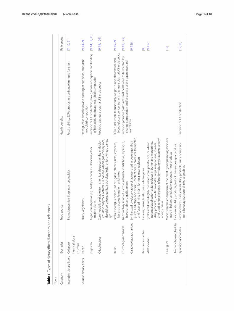
Health promoting effects of dietary fibers and suggested mechanismsTypes of fibers and dietary sourcesDietary fibers have proven to have beneficial physiologi-cal effects on humans (e.g., body weight management [6]). Dietary fiber can be subdivided into non-fermenta-ble/insoluble and fermentable/soluble forms, which can vary in their potential impact on health (Table 1). Physic-ochemical characteristics of fiber include origin, solubil-ity and viscosity, fermentability, and chemical structure [7].
Insoluble dietary fibers (e.g., celluloses, hemicellu-lose, and fructans [8]) are present in foods such as whole wheat flour, brown rice, nuts, beans, and vegetables (e.g., cauliflower, broccoli, celery) [9, 10]. These fibers are characterized by their bulking effect and high fermenta-tion by the gut microbiota, resulting in short-chain fatty acid (SCFAs) production [7]. A recent study showed that butyrate, an SCFA, causes an immunologic alteration in macrophages, and increased expression in antibacterial and host defense genes [11]. Along with the beneficial increase in SCFAs, insoluble fiber can lead to a healthy microbial composition [7]. In contrast, Desai et al., dem-onstrated that a fiber-deprived microbiota can increase disease susceptibility by a reduction in protective mucus; the decrease in mucus membrane, which aids in blocking pathogens entering the system, was due to an increase in mucin-degrading bacteria resulting from a deficiency in fiber [12].
Soluble fibers (e.g., pectin, guar gum, and some inulin) are present in whole grains (e.g., oats, wheat), legumes (e.g., lentils, split peas, various types of beans), seeds and nuts (e.g., flax seed), and some fruits and vegetables (e.g., carrots, apples) [13]. In contrast to insoluble fibers, soluble fibers are characterized as viscous, creating a gel-like form in the intestine which may slow absorption of nutrients (e.g., glucose and lipids [7]). In a recent study, Bang et al. found that consumption of pectin, a soluble fiber, results in the use of galacturonic acid, as an energy source for microbes, hence increasing reducing sugar levels in human stool [14]. This study shows the protec-tive potential of pectin (a soluble fiber) against metabolic diseases, such as type 2 diabetes via controlling blood glucose levels [14].
Prebiotics are another class of insoluble fibers, and were originally defined as “non-digestible compounds that,
when consumed, induce changes in composition and/or activity of the gastrointestinal bacteria, thus causing benefit(s) upon host health” [15]. Classification of prebi-otics is based on three criteria; (a) resistance to gastric acidity, hydrolysis by mammalian enzymes, and gastroin-testinal absorption, (b) fermentation by intestinal micro-biota, and (c) selective stimulation of the growth and/or activity of intestinal bacteria associated with health and well-being [16, 17]. Positive functional characteristics of prebiotics have been reported including, but not limited to: (a) selective fermentation, (b) modulation of gut pH, (c) fecal bulking, (d) the prevention of gut colonization by pathogens, and (e) the control of putrefactive bacte-ria, thus reducing the host’s exposure to toxic metabolites [18]. Types (and sources) of prebiotics, include β-glucan (in mushrooms, and cereal grains) [9], fructooligosaccha-ride [14, 19], oligofructose [19], inulin [10, 12, 14], galac-tooligosaccharides (glycosylation of primary lactose) [19], guar gum (in cereal grains) [19], resistance starches and maltodextrin (e.g., starches) [10, 20], xylooligosac-charides and arabinooligosaccharides (in cereals, bars, and dairy products) [19]. Consumption of prebiotics is known to change intestinal microbiota diversity and to increase the production of SCFAs (i.e., propionate, and butyrate) [9, 19, 21, 22]. For instance, Carlson et al. found that prebiotics (e.g., β-glucan, xylooligosaccharides, and pure inulin) were effective to promote the formation of beneficial SCFAs in human subjects [19].
Production of SCFAsSCFAs are produced by the intestinal microbiota via fermentation of carbohydrates [9, 14] and other non-absorbable nutrients [19, 21]. The most abundant SCFAs include acetate (C2), propionate (C3), and butyrate (C4), which exist in a 3:1:1 ratio [23, 24], and represent 90–95% of all SCFAs produced in the colon [14, 19, 22]. Produc-tion of these SCFAs can either be used by downstream bacterial species (cross feeding) and/or by the host as nutrient sources. Specifically, acetate and propionate can be absorbed by the lumen and enter peripheral circula-tion to be involved in overall metabolic homeostasis [25], while butyrate (C4) serves over 70% of the energy supply for colonocytes [26]. Among the three SCFAs, butyrate was frequently reported to be involved in other functions such as immune regulation [27], cell growth [28, 29], intestinal barrier function [30], and ion transport [31].
Supplementation of insoluble fibers may result in increased butyrate production which is highly associ-ated with an individual’s gut microbiome composition [32]. Two main steps are involved in the butyrate produc-tion: butyrate synthesis and polysaccharide degradation [32]. Primary degraders attack specific polymer bonds to generate mono and di-oligosaccharides, allowing them
Page 3 of 18Beane et al. Appl Biol Chem (2021) 64:36
Tabl
e 1
Type
s of
die
tary
fibe
rs, f
unct
ions
, and
refe
renc
es
Fibe
rs
Cate
gory
Exam
ples
Food
sou
rce
Hea
lth b
enefi
tsRe
fere
nces
Inso
lubl
e di
etar
y fib
ers
Cellu
lose
Bean
s, br
own
rice,
flou
r, nu
ts, v
eget
able
sFe
cal b
ulki
ng, S
CFA
pro
duct
ion,
enh
ance
imm
une
func
tion
[7–1
2, 2
1]
Hem
icel
lulo
se
Fruc
tans
Solu
ble
diet
ary
fiber
sPe
ctin
Frui
ts, v
eget
able
sSl
ow g
luco
se a
bsor
ptio
n an
d bi
ndin
g of
bile
aci
ds, m
odul
ate
mic
robi
al c
ompo
sitio
n[9
, 14,
21]
β‑gl
ucan
Alg
ae, c
erea
l gra
ins
(e.g
., ba
rley
or o
ats)
, mus
hroo
ms,
othe
r m
arin
e pl
ants
Preb
iotic
, SC
FA p
rodu
ctio
n, s
low
glu
cose
abs
orpt
ion
and
bind
ing
of b
ile a
cids
, mod
ulat
e m
icro
bial
com
posi
tion
[9, 1
4, 1
9, 2
1]
Olig
ofru
ctos
eCo
mm
erci
ally
ava
ilabl
e fro
m c
hem
ical
deg
rada
tion
by e
ndog
ly‑
cosi
dase
enz
ymes
; nat
ural
ly in
ban
ana,
asp
arag
us, c
hico
ry ro
ot,
dand
elio
n gr
eens
, gar
lic, a
rtic
hoke
s, le
eks,
onio
n, w
heat
, bar
ley,
ry
e
Preb
iotic
, dec
reas
e pl
asm
a LP
S in
dia
betic
s[9
, 19,
124
]
Inul
inLe
eks,
aspa
ragu
s, on
ions
, whe
at, g
arlic
, chi
cory
, oat
s, so
ybea
ns,
bana
nas,
agav
e, J
erus
alem
art
icho
kes
SCFA
pro
duct
ion,
redu
ce b
ody
wei
ght,
bloo
d ch
oles
tero
l, an
d bl
ood
gluc
ose
conc
entr
atio
ns, d
ecre
ase
plas
ma
LPS
in d
iabe
tics
[9, 1
9, 2
1]
Fruc
tool
igos
acch
arid
eTr
ansf
ruct
osyl
atio
n of
suc
rose
; nat
ural
ly in
art
icho
ke, a
spar
agus
, ba
nana
, chi
cory
, gar
lic, o
nion
Preb
iotic
, pro
mot
e ga
stro
inte
stin
al h
ealth
due
to fe
rmen
tabi
lity,
ch
ange
com
posi
tion
and/
or a
ctiv
ity o
f the
gas
troi
ntes
tinal
m
icro
biot
a
[9, 1
9, 1
25]
Gal
acto
olig
osac
char
ides
Synt
hesi
zed
by g
lyco
syla
tion
of la
ctos
e; u
sed
in b
ever
ages
(fru
it ju
ices
and
oth
er a
cid
drin
ks),
conf
ectio
nery
pro
duct
s, fe
rmen
ted
and
flavo
red
milk
s, in
fant
form
ula,
mea
l rep
lace
rs
[9, 1
26]
Resi
stan
ce s
tarc
hes
Bana
nas,
bean
s, le
ntils
, pea
s, w
hole
gra
ins
[9]
Mal
tode
xtrin
Synt
hesi
zed
from
hig
hly
proc
esse
d co
rn, p
otat
o, ri
ce, o
r whe
at;
pote
ntia
l app
licat
ion
as fa
t rep
lace
r in
butt
er a
nd m
arga
rines
, da
iry p
rodu
cts;
low
‑fat s
alad
dre
ssin
gs, m
ayon
nais
e, s
prea
ds,
and
used
in n
onal
lerg
enic
infa
nt fo
rmul
a, re
hydr
atio
n/re
cove
ry/
ener
gy d
rinks
[9, 1
27]
Gua
r gum
Mad
e fro
m th
e en
dosp
erm
of t
he p
lant
Cya
mop
sis te
trag
onol
obus
; us
ed in
bak
ery,
cer
eal,
dairy
pro
duct
s, m
eat p
rodu
cts
[19]
Ara
bino
olig
osac
char
ides
Bars
, cer
eals
, dai
ry p
rodu
cts,
isot
onic
bev
erag
es, s
port
s dr
inks
Xylo
olig
osac
char
ides
Bam
boo
shoo
ts, b
ars,
cere
als,
dairy
pro
duct
s, fru
its, h
oney
, iso
‑to
nic
beve
rage
s, sp
orts
drin
ks, v
eget
able
s,Pr
ebio
tic, S
CFA
pro
duct
ion
[19,
21]

Page 4 of 18Beane et al. Appl Biol Chem (2021) 64:36
to be further fermented into butyrate by secondary pro-ducers. These two steps can vary among different strains, and the metabolites synthesized during these two steps could cross-feed each other through a complex metabolic process. However, both reaction efficiencies need to be balanced or the polysaccharide degrader will consume the majority of the available carbon and energy [32]. Eubacterium rectale and Faecalibacterium prausnitzii are two main species involved in butyrate production in the human gut [33], while other species are resistant to starch degradation such as Ruminococcus and Bifido-bacterium [34]. The efficiencies of butyrate production differ in people with various dietary, genetic, health and geographic backgrounds [35, 36], warranting a greater understanding of individual supplementation plans.
Effects of dietary fibers on epithelial barrier functionsThe intestine is a multi-functional tubular organ con-taining developed vasculature, lymphatic drainage, and extensive innervation. The outside layer of the intesti-nal tubulin is muscular tissue, and the innermost layer is structured as a single layer of polarized epithelial cells interfacing between the external environment and inner host tissues. The single layered enterocytes are concat-enated through tight junctions (TJs) to form the first layer of immune defensive translocation of food-borne pathogens or other ingested toxic compounds [37]. An intact intestinal cell wall is a prerequisite for regular gastrointestinal homeostasis but still, pathogens such as Salmonella typhimurium, Clostridium perfringens, and enteropathogenic Escherichia coli can invade the host by disturbing the TJ complex [38]. The construction of TJs is dynamic and regulated by enterocytes’ histological development and certain signaling pathways (i.e., tumor necrosis factor-α) that are initiated by pathogens. TJs also serve as the point of attack for receptors of pathogenic bacteria to invade the host and cause increased perme-ability, which further result in destructive intravenous electrolyte exchange, microbial dysbiosis, and diarrhea [33].
Dietary fibers and SCFAs are implicated with epithelial barrier functions. Specifically, butyrate has a bi-direc-tional effect on epithelial barrier function. For instance, when experiencing diarrhea and antibiotic induced dys-biosis, commensal bacteria responsible for butyrate production are significantly decreased [39]. Decreased commensal bacteria causes a shortage of available butyrate as an energy source for colonocytes, thereby dis-rupting the enterocyte refreshment. In humans, supple-mentation of certain fibers can enrich butyrate producing bacteria in the gut; as the butyrate production increases, a corresponding alteration and reduction in the diarrhea condition occurs [40]. Butyrate is also known to improve
the barrier function through nourishing TJs such as beta defensin, cingulin, ZO-1 and ZO-2 proteins in chicken HD11 macrophage cells, primary monocytes, bone marrow cells, and jejunal and cecal explants [41]. Simi-lar findings were found in another study using in vitro Caco-2 human epithelial colorectal adenocarcinoma cell line, in which butyrate was able to offset barrier impair-ment of Campylobeacter jejuni. In addition, intracellular signaling pathways initiated by butyrate related to barrier function have been studied. For example, it is reported that butyrate enhances the TJs through activating Akt/mTOR pathways and ATP replenishment [42]. Another study indicated that dietary butyrate promotes barrier function by repressing interleukin 10 (IL-10) receptor-dependent claudin-2 [43]. IL-10 receptor contains two ligand-binding subunits: ligand-binding alpha subunit (IL-10RA) and beta subunit (IL-10B) [44]. When IL-10 binds to IL10R, it activates JAK-STAT signaling path-way, in which STAT3 activation is critical to anti-inflam-matory activity [45, 46]. Butyrate was found to promote epithelial barrier function through IL-10RA repression of Claudin-2, which regulates paracellular channels for small cations and water, stimulating diarrhea via the leak-flux mechanism [43].
Anti‑inflammatory effects of dietary fibersThe gut possesses one of the largest immune systems in humans, exerting both physical (i.e., barrier function) and biochemical defenses against invading foodborne pathogens and other hazards [47]. The pro-inflammatory response acts by first being triggered in the primary reac-tion to invaders, and then proceeding recruitment or ini-tiation of the amplification [48]. Subsequently, cells are differentiated into multiple sub-types of immune cells to the impaired locations as a natural response to invad-ing antigens [49]. This whole process gradually accu-mulates and strengthens within the first couple of days at the expense of great amounts of energy and nutrient consumption. The gastrointestinal epithelial cells, cov-ered with an array of ligand receptors, are constantly encountering various toxic compounds or pathogens that could easily and frequently activate the immune response. Therefore, certain immunologic derangement diseases such as inflammatory bowel disease (IBD) and autoimmune disease could occur [50, 51]. Anti-inflam-mation, which serves as an inhibitory effect on excessive inflammation, is needed to maintain a balanced immune homeostasis.
Butyrate has been widely studied as an anti-inflam-mation regulator through modulating cytokine produc-tion, kinase activity, and immune-associated signaling pathways. Previously reported mechanisms are related to the up-regulation of immunosuppressive IL-10 [52],
Page 5 of 18Beane et al. Appl Biol Chem (2021) 64:36
G-protein coupled receptors (GPRs), specifically GPR41 [53], and inhibition of multiple cellular immune media-tors such as toll-like receptor engaged IL-12/23p40 [54], nuclear factor (NF)-κB [55], and histone deacetylase [53, 56]. IBD including both ulcerative colitis and Crohn’s disease are a typical chronic relapsing inflammatory disease caused by genetically related anti-inflammation deficiency [57]. It is believed that the sensing capacity of anti-inflammatory mediators such as butyrate is impaired in IBD patients. However, supplementing higher levels of butyrate dense dietary fibers could counteract the sever-ity of these syndromes to some degree [55, 58, 59].
Phytochemicals and gut microbiomeOnly 5–10% of phytochemicals are absorbed in the small intestine, while the remaining 90–95% of phytochemi-cals are transformed by the resident colonic microbiota [60]. The microbiota yields highly bioavailable metabo-lites from these phytochemicals. The metabolism and absorption of phytochemicals may have potential sys-temic health effects on the host (i.e., cardioprotective [61, 62], and protection against glucose toxicity [63]). These systemic health effects are partly due to an increase of polyphenols in the diet, which has been linked with prevention of diverse chronic diseases [i.e., metabolic syndrome [64, 65]; will be further discussed below]. Last, unabsorbed dietary phytochemicals can directly
modulate the microbiota. For instance, phenolic com-pounds from tea leaves inhibited growth and adhesion of Clostridium spp., E. coli, and S. typhimurium [66].
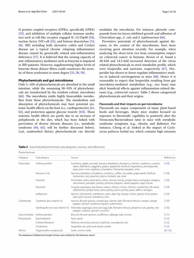
Preventive potentials of phytochemicals against dis-eases, in the context of the microbiome, have been receiving great attention recently. For example, when analyzing the short-term rice bran consumption impact on colorectal cancer in humans, Brown et al. found a 28-fold and 14.5-fold increased detection of the citrus related phytochemicals in stool metabolite profile, which were hesperidin and narirutin, respectively [67]. Hes-peridin has shown to down-regulate inflammatory mark-ers in induced carcinogenesis in mice [68]. Hence it is reasonable to expect that hesperidin resulting from gut microbiota-mediated metabolism (e.g., rice bran) may elicit beneficial effects against inflammation-related dis-eases (e.g., colorectal cancer). Table 2 shows categorized phytochemicals and their origins.
Flavonoids and their impacts on gut microbiomeFlavonoids are major components of many plant-based foods and beverages. Many mice studies focused on exposure to flavonoids’ capability to positively alter the Firmicutes/Bacteroideses ratio in mice with metabolic syndrome symptoms (e.g., obesity, and diabetes). For instance, Cheng et al. looked at the impact of Cyclo-carya paliurus herbal tea, which contains high amounts
Table 2 Searched phytochemicals subcategories, sources, and references
The databases PubMed and Google Scholar were utilized for the literature search
Phytochemicals
Category Subcategory Source References
Flavonoids Anthocyanidins Acai berry, apple, avocado, banana, blackberry, blueberry, cherries, cranberry, currants, dates, elderberry, eggplant, grapes, grapefruit, kiwifruit, lingonberry, pomegranate, pear, plum, nuts, raspberry, strawberry, Tasmanian peppers, wine
[128]
Flavone‑3‑ols Apricots, blackberry, blueberry, cranberry, coffee, chocolate, grape seeds, kiwifruit, nectarines, nuts, peaches, plums, rhubarb, tea, wine
[128]
Flavones Artichokes, celery seed spice, celery, chicory, honey, juniper berry, kumquats, oregano, olive leaves, pumpkin, parsley, pimento peppers, sweet peppers, sage, thyme
[128]
Flavonols Arugula, asparagus, bay leaves, capers, cilantro, chives, cherries, cranberries, dill weed, elderberries, juniper berry, okra, parsley, plum, prickly, pears, saffron, tarragon
[128]
Isoflavones Apricot, clementine, cranberries, raisin, date, fig, mango, melon, passion fruit, peach, pear, plum, prune, strawberry, nuts
[129]
Carotenoids Carotenes (pro‑vitamin A) Apricot, Brussel sprouts, cantaloupe, cilantro, kale, Romaine lettuce, mango, orange pepper, spinach, butternut squash, watermelon
[130]
Xanthopylls (non‑pro vitamin A) Artichoke, asparagus, broccoli, egg, kale, Romaine lettuce, pistachio nuts, parsley, red pepper, scallions, spinach, zucchini
[130]
Glucosinolates Isothiocyanates Broccoli, Brussel sprouts, cauliflower, cabbage, kale, turnips [131]
Phytosterols Epicholesterol Nuts, seeds [132]
6‑Ketochilestanol Walnuts, almonds, peanuts, hazelnuts, macadamia nuts [132]
Cholesteryl Vegetable oils, spelt and wheat cereals [132]
Allicins Organosulfur compounds Garlic, onions, leeks [81, 82]
Page 6 of 18Beane et al. Appl Biol Chem (2021) 64:36
of flavonoids, on obesity-related metabolic disorders [69]. Cyclocarya paliurus flavonoids (CPF) decreased the Firmicutes/Bacteroideses ratio and Proteobacteria at the phylum level while fecal microbial diversity was improved, indicating CPF influence on the microbial community and between host and microbe provide ben-eficial effects. Additionally, an abundance of Prevotella was detected, which is beneficially involved in glucose metabolism [70] and fermentation of amino acids [71], suggesting dietary flavonoids could produce protective and therapeutic effects on high-fat induced obesity via modulation of the microbiome [69].
Similarly, two short-term mice studies focused on fla-vonoid modification of the gut microbiome in non-dis-ease specific states. Wankhade et al. analyzed blueberry consumption, finding significant modifications in both α-diversity and β-diversity [65]. Specifically, the Firmi-cutes/Bacteroideses ratio, Tenericutes, and Deferibacte-res were decreased at the phylum level. Interestingly, the sex was a significant factor at the genus level; metabolic pathways (i.e., fatty acid metabolism, lipid metabolism) were significantly different in blueberry-fed male mice, and not in blueberry-fed female mice. While this study does suggest the influence of flavonoids from blueberry on gut microbial α-diversity and β-diversity in male mice, it is less clear why there was no effect on the female mice [65].
The other short-term mice study looked at black rasp-berry effect on the colonic microbiome. Black raspber-ries improved the Firmicutes/Bacteroideses ratio, and high amounts of polyphenols ellagitannins (i.e., urolith-ins) and anthocyanins were identified in colon tissue and plasma. Luminal Clostridium was significantly decreased after intervention, which is possibly due to pathogenic Clostridium (i.e., Clostidium perfingens, Clostridium dif-ficile). Dietary black raspberries ultimately increased mucosal microbial composition, via reduction of luminal Clostridium, on the luminal microbiota [64].
Supporting the potential therapeutic role of flavonoids, Petersen et al. utilized long-term strawberry supplemen-tation for microbiome modification in diabetic mice [72]. Microbial composition was significantly altered at the phylum and genus levels in both α-diversity and β-diversity, by decreasing Verrucomicrobia and Bifi-dobacterium in diabetic mice. Additionally, there were multiple significantly predicted functional metagenomic profiles identified through the PICRUSt (i.e., lipid biosyn-thesis proteins, insulin signaling pathway, and phosphati-dylinositol signaling pathway), indicating a correlation between these pathways and bacterial abundance [72].
Another study looked at the effect of long-term sup-plementation of dietary flavonoid isoquercetin and solu-ble fiber inulin effects on mice that were fed a high-fat
diet (to induce metabolic disorders). Tan et al. found that mice receiving both isoquercetin and inulin sup-plementation had slower weight gain, improved glucose tolerance and insulin sensitivity, reduced hepatic lipid accumulation, adipocyte hypertrophy, circulating leptin and adipose fibroblast growth factor 21 levels. Interest-ingly, the compared groups receiving isoquercetin or inu-lin independently had no significant results. This suggests inulin changes the microbiome composition through its prebiotic effects, enhancing the metabolism and absorp-tion of the flavonoid isoquercetin to have beneficial effects on the prevention of metabolic syndrome devel-opment with high-fat diets [73].
An abundance of future studies is needed to fully understand broad impacts of flavonoids on the gut micro-biome. While many of these studies identified microbial composition and disease states, they found were mainly focused on the Firmicutes/Bacteroideses ratio, and the metabolic disease state. More studies are needed to identify the alterations of other microbial strain due to flavonoid intervention, and how they potentially impact disease states. Additionally, more research is needed to identify the potential impact of flavonoids on other dis-eases, such as cancers and inflammation. Long-term studies would further help solidify sex-specific changes on the microbiome in flavonoid interventions, as there are currently no long-term studies published in this area. Lastly, many of these studies failed to isolate the effects of flavonoids from whole fruit and vegetable consumption. Perhaps a single flavonoid may have difficulty making a significant effect, making it difficult to determine if the results are due to the phytochemical metabolite or due to the beneficial effects of fiber. As noted above, fiber has a significant impact on the modulation of the microbi-ome, and without this separation it could prove difficult to understand the significant effect of the phytochemical.
Carotenoids and their impacts on gut microbiomeTwo primary research articles reported implications of carotenoids on the microbiome, where both analyzed the effect of increased serum carotenoid concentrations on the gut microbiome. High serum carotenoids are asso-ciated with decreased risk of chronic diseases [74], and contribute to gut microbiome health. While carotenoids are found in multiple plant-based dietary sources (e.g., carrots), Ramos et al. analyzed the carotenoid source of Tucumã oil and its effect on microbial diversity and SCFA production in cows. Carotenoids from Tucumã oil resulted in a lower abundance in Fibrobacter and Rikenellacea RC9 gut group, and enriched Pyramidobac-ter, Megasphaera, Anaerovibrio, and Selenomonas. These results suggest the use of Tucumã oil as a catenoid source and shows favorable shifts in gut microbiome health [75].
Page 7 of 18Beane et al. Appl Biol Chem (2021) 64:36
Comparatively, a long-term randomized control study in humans analyzed the relationship between colonic mucosal bacteria and serum carotenoid concentrations. Djuric et al. found that colonic mucosal bacteria were associated with serum carotenoid concentrations at base-line, and long-term exposure had no significant effects. Indicating the need for increased research on long-term exposure to better understand the effect of dietary change maintaining a beneficial microbial change. It was identified, however, that 11 operational taxonomic units were associated with higher serum carotenoid levels. Fac-tors affecting the level of carotenoid levels included body mass index, smoking, and dietary intakes (represent-ing 12% of the total variance in carotenoid levels). These results further suggest the impact of multifaceted factors (i.e., behavioral and metabolic factors) on the gut micro-biota’s capability for carotenoid absorption [74]. Further research is needed on carotenoids and effects on micro-biota diversity and impacting disease states in hosts, as only these two articles were identified in the search.
Glucosinolates and their impacts on gut microbiomeGlucosinolates are sulfur-containing compounds pro-foundly present in cruciferous vegetables. Thus far only one study examined effects of glucosinolates on the gas-trointestinal microbiota which is somewhat unexpected given their widely known benefits against chronic dis-eases (i.e., cancers [76–78]). Specifically, broccoli and cabbage are good sources of glucosinolates. Kaczmarek et al. analyzed the effects of broccoli on the gut micro-biota via a short-term randomized controlled feeding study in healthy adult subjects. β-diversity alterations indicated that bacterial communities were impacted by broccoli feeding; Firmicutes abundance was decreased by 9% while Bacteroides abundance was increased by 8%. The strongest associations between bacterial rela-tive abundance and glucosinolate metabolites were seen in participants with a body mass index < 26 kg/m2. Addi-tionally, broccoli consumption significantly altered a few key metabolic pathways: endocrine system, transport and catabolism, and energy metabolism [79]. Clearly, further studies are warranted in the context of the disease pre-ventive potential of cruciferous vegetables through gut microbiome modulation, particularly considering that gastrointestinal microbiota can metabolize glucosinolates into isothinocyanates (ITCs) [80]. Also, additional mech-anistic studies are needed to explain different responses to glucosinolates in participants with higher body mass index.
Allicins and their impacts on gut microbiomeAllicins are organic sulfur phytochemicals that are par-ticularly found in garlic [81]. One short-term randomized
control study examined the effects of garlic extract on gut microbiota, inflammation, and cardiovascular mark-ers (e.g., blood pressure, pulse wave velocity, and arterial stiffness). Reid et al. found that aged garlic, converted to the vaso-active component S-allylcysteine, inccreased Lactobacillus and Clostridia. Additionally, blood pres-sure was significantly reduced by 10 ± 3.6 mmHg systolic, and 5.4 ± 2.3 mmHg diastolic. Central blood pressure and arterial stiffness were significantly reduced, show-ing improvements in overall hypertension. These findings may provide hints between gut microbiome modulation and cardiovascular health although a causal relation-ship is still not clear [82]. As shown in this study, allicins may have beneficial cardiovascular effects; however, this short-term feeding study is the only evidence dem-onstrating impacts of allicins on the gut microbiome. Similar to other groups of phytochemicals, long-term feeding in addition to mechanistic/functional validation are needed; in addition, in order to uncouple with effects of fibers, it will be informative to explore if the garlic-induced gut microbiome modulation can be recapitu-lated with isolated allicin exposure.
Micronutrients and microbiomeA multidirectional relationship lies between the host and its microbiome. Although, as aforementioned, diet sig-nificantly impacts the gut microbiome, gut microbiota per se also play a role in human metabolic functions [83]. Furthermore, the microbiome may alter the host’s absorption of various dietary nutrients and, thereby, indirectly impact micronutrient physiology [84]. Spe-cifically, some microbial strains synthesize vitamins and cofactors, and evidence suggests that microbial metabo-lites can affect metabolic and physiological pathways of micronutrients in the human body [85, 86]. In addition, production of vitamins and cofactors can provide essen-tial nutrients to colonocytes, promote competition with pathogenic organisms, and modulate immune responses [87]. Because bacteria alter the efficiency of bile acids to emulsify dietary lipids and form micelles, the microbi-ome potentially influences the absorption of lipid-solu-ble vitamins as well [88]. With consideration for luminal nutrient effects on the microbiome and microbial effects on host nutrition status, it is reasonable to believe that identification and manipulation of microbial interactions with hosts could be pivotal to human health.
Numerous factors, including physiochemical proper-ties of foods, nutrient availability, colonic transit time, and age of host may modulate dietary effects on the colonic microbiota [89]. Substrates that are not defined as “prebiotics”, such as phytochemicals, potentially alter microbial composition and function; vitamins and min-erals are also examples of these substrates, yet they are
Page 8 of 18Beane et al. Appl Biol Chem (2021) 64:36
not well-recognized or well-understood regarding their effects on the gut microbiome [90]. This may be partially due to the fact that most vitamins are absorbed in the upper small intestine, which results in low concentra-tions of vitamins or minerals reaching the distal parts of the gastrointestinal tract [90]. Nevertheless, studies have found micronutrient-induced changes in the mammalian gut microbiome, and modulation by vitamins and min-erals warrants further investigation [85]. In fact, it was reported that several micronutrients have potential to alter gastrointestinal function, immune response, and, as a result, microbial populations [85, 91].
It is estimated that more than three billion people worldwide suffer from micronutrient deficiencies, pre-dominantly vitamin A, iron, and zinc [91]. Disease states (e.g., malnutrition) may alter microbiome-mediated transport, metabolism, and synthesis of cofactors and vitamins [91]. Therefore, interference of microbiota func-tion by micronutrient deficiency could explain the rela-tionship between dysbiosis and malnutrition [92]. Few existing studies have examined the transportation of vita-mins between the microbiota and intestine or the impact of luminal vitamins on the microbiota (host-microbe-metabolic axis) [92, 93]. Understanding the influence of low micronutrient supply on microbiota development, composition, and metabolism is an integral part of imple-menting new strategies to overcome deleterious effects of malnutrition [92].
Mapping of metabolic relationshipA microbiome has approximately 150-fold more genes than the human genome [94]. Using high-throughput technologies such as next-generation sequencing and mass spectrometry–based metabolomics, researchers are able to sequence the metagenome of the microbiome and associate this information with the genomics, epig-enomics, transcriptomics and metabolomics of the host [94]. Mapping of human and microbe-derived enzymes to gut metabolic pathways has revealed that gut microbe-derived enzymes are essential components of various human pathways [83]. A total of 518 enzymatic reactions were shared between host and gut-bacterial species [83], suggesting significant cross-talk between the host and microbiota. Furthermore, mapping of known and pre-dicted enzymes to canonical human pathways resulted in identification of 48 pathways that have at least one bacteria-encoded enzyme [83]. Bacterial communities contribute to human gut metabolism by complement-ing enzymes that are not encoded by the human genome but are essential for digestion and metabolism [83]. Most of these pathways are involved in metabolizing dietary nutrients, including cofactors and micronutrients [83]. Thirty exclusively microbe-derived enzymes complement
vitamin/cofactor metabolic pathways, which underscores the dependency of human pathways on microbe-derived enzymes [83]. Microbes not only complement, but also supplement some of the enzyme functions that are com-mon to both human and gut microbiome [83]. Further support is provided by the findings of a meta-genome analysis of the human gut microflora, which revealed the presence of Clustered Orthologous Groups of pro-teins involved in production of essential vitamins [95, 96]. Most of the production and absorption of microbial vitamins takes place in the colon; however, the micro-biome may contribute to maintaining systemic levels and minimizing deficiency effects [95, 96]. Microbiome multi-omics analyses coinciding with host omics data-sets will be important for revealing the microbiome’s role in human health [94]. Unbiased and untargeted omics approaches could unveil the involvement of vitamins and minerals within the interrelationship of the host and microbiome [94].
VitaminsOf the essential micronutrients, microbial synthesis of vitamin K and B vitamins had the greatest body of research regarding effects on host systemic status. There is also evidence of microbial modulation of colonocyte nutrient absorption that potentially influences host nutri-tion status [84, 88]. In contrast, little is known regarding effects of vitamin supplementation on the gut microbi-ome, and the relationship between micronutrient defi-ciencies and dysbiosis is yet to be elucidated.
Vitamin BIntestinal microbiome may be a primary source of B vita-mins [97]. It is known that these vitamins modulate host’s epigenome [98]. Because the vitamins produced by the microbiota can result in epigenetic changes in host cells, they may play a significant role in modulating host gene expression [98]. Of the 8 B vitamins, 7 have colonic bac-terial sources, and a human population with abnormal intestinal microbiota may have unexpected B vitamin deficiencies that are unrelated to food consumption [97], reinforcing the importance of gut microbiome in B vita-mins metabolism and thus host health.
Vitamin B1 (thiamine) is an essential cofactor for organisms. Humans acquire most vitamin B1 through their diet [99]; however, evidence indicates that bacte-rial-derived vitamin B1 can be absorbed into human colonocytes, which may contribute to colonic vitamin B1 status and affect systemic status. A study published in 2017 investigated vitamin B1′s role in the gut utiliz-ing Bacteroides thetaiotaomicron as a model gut microbe [99]. RNA sequencing revealed global downregulation of vitamin B1 and amino acid biosynthesis, glycolysis, and
Page 9 of 18Beane et al. Appl Biol Chem (2021) 64:36
purine metabolism in the presence of vitamin B1, and expression of the major biosynthetic operon was upregu-lated under vitamin B1-deficient conditions [99]. Further investigation using genetic mutants suggested thiamine biosynthesis and transport is critical for growth when vitamin B1 is deficient [99]. The ability of microbes to transport, synthesize, and compete for vitamin B1 may impact the structure and function of the microbiome during shifts in luminal availability of vitamin B1. Gut microbes that are wholly dependent on vitamin B1 trans-port, such as members of the genus Alistipes and many members of the Bacilli, are predicted to be adversely affected by deficient conditions [99]. In a study of Crohn’s disease (CD) patients, microbial genes for pathways involved in biosynthesis of vitamins B1, B2, B9, and B12 were decreased during exacerbation; [100]. The study suggested that intestinal abundances of vitamin B1, B2, B9, and B12 could be involved in CD exacerbation and inflammation associated with dysbiosis [100]. Therefore, manipulating the presence or concentration of vitamin B1, and possibly of other water-soluble vitamins, could be an effective method for treating or preventing dysbio-sis [99].
Free absorbable and protein-bound forms of vita-min B2 are synthesized by the human microbiota, but the uptake of vitamin B2 into colonocytes is concentra-tion dependent; the higher the luminal concentration, the lower the uptake, and vice versa [91]. As a result, the systemic effects of vitamin B2 production in the lumen may be mediated. Furthermore, genomic and functional analysis of one gut microbe, Romboutsia ilealis CRIBT, revealed genes for de novo purine and pyrimidine syn-thesis, as well as for production of coenzymes NAD and FAD via salvage pathways from vitamin B2 and B3 [101]. R. ilealis CRIBT relies on these pathways or exogenous sources for the supply of precursors, mainly in the form of B vitamins, suggesting that luminal B vitamin avail-ability may alter microbiome function and composition [101]. Vitamin B2 affects the growth of Faecalibacterium prausnitzii, which has a specialized use of vitamin B2 as an extracellular electron transporter, allowing it to toler-ate limited levels of oxygen [90, 101]. The stimulation of F. prausnitzii growth by exposure to vitamin B2 may be a function of this vitamin in microbiome-modulation that could be of clinical interest [90]. A first pilot open-label study with vitamin B2 (100 mg vitamin B2/day) showed an increase in Faecalibacterium and a reduction in E. coli in most participants [101]. In addition, multiple articles suggest that a double-blind, parallel-group, placebo-controlled “Ribogut” trial was approved in 2018. The trial was to examine the effects of vitamin B2 on the gut microbiota composition of healthy volunteers adminis-tered 50 or 100 mg vitamin B2/day for 14 days; however,
no results have been published [101]. Further vitamin B2-microbiome studies may present new possibilities for vitamin combinations as supplements for prevention of human diseases through microbiome modulation [90].
It was suggested that vitamin B5 (pantothenic acid) is potentially supplied by normal intestinal bacteria more so than by natural food sources [97]. However, the rela-tionship between the microbiome and vitamin B5, as well as vitamin B3 (niacin) and B6 (pyridoxine), is oth-erwise unclear. However, the human microbiota contains enzymes that depend on vitamin B6 [91]. Certain bacte-ria or the host may contribute the vitamin B6 needed for aminotransferase metabolism in bacteria [91]. Virulence and motility of Helicobacter pylori depend on the pres-ence of functional enzymes important for bacterial de novo vitamin B6 synthesis [91].
Humans cannot produce vitamin B7 (biotin) and, therefore, evidently depend on dietary intake or the intes-tinal microbiota to maintain healthy levels [53]. However, no pathways for how the microbiome affects systemic status have been elucidated.
Bacteria commonly found in the colon can produce vitamin B9 (folate) [93]. Although vitamin B9 is primar-ily absorbed in the small intestine, absorption does occur to some extent in the colon as well [93]. Vitamin B9 pro-duction by intestinal microbiota can modify the effects of vitamin B9 ingested in the diet and has already been considered in studies of diet and colon cancer risk [93]. However, it is still questionable whether high vitamin B9 production will result in higher absorption because administration of high-producing strains results in higher fecal vitamin B9 concentrations [91]. Neverthe-less, radiolabeled vitamin B9 precursor (p-aminobenzoic acid) originating in the colons of rats resulted in radiola-beled vitamin B9 within various tissues. More research is needed to elucidate whether microbiota-derived vitamin B9 affects systemic folate status [91].
Vitamin B12 (cobalamin) is synthesized exclusively by bacteria and archaea. Although it is frequently argued that microbiome-derived vitamin B12 is a source for humans, experimental data shows this is not the case because colonic vitamin B12 is not bioavailable due to the lack of required enzymes and receptors in the colon [91]. Despite the lack of absorption, vitamin B9 and B12 regu-late microbiota gene expression and may control genomic interactions between the microbiome and host [91]. The gut microbiome has vitamin and mineral require-ments for growth and proliferation, and studies have found several pairs of organisms with vitamin synthesis pathway patterns that complement each other, implying a vitamin-dependent cross-feeding between microbes for growth [88]. B12 production by Eubacterium hal-lii and the interdependent production of propionate by

Page 10 of 18Beane et al. Appl Biol Chem (2021) 64:36
Akkermansia muciniphila demonstrates this mutualistic symbiosis [102]. In a study surveying over 300 sequenced microbiota-derived bacterial strains, 83% of the strains were shown to have enzymes dependent on vitamin B12 as a cofactor [91]. Most of these species lack genes required to synthesize vitamin B12 de novo and, there-fore, rely on transport to meet their requirements [91]. Gut microbial species consequently compete with other species and the host for dietary vitamin B12, indicating that luminal vitamin B12 is an important nutrient for the gut microbiome [91, 93]. Furthermore, B vitamin pro-duction by intestinal bacteria mediates or modifies the effects of other ingested nutrients, which partly explains the heterogeneity in the results of the studies investi-gating these nutrients to date [93]. There is now strong evidence that water-soluble vitamins synthesized by the microbiota contribute to interactions with the host [91], but specific mechanisms for cross-talk require further investigation.
Vitamin C and vitamin EAntioxidants, including vitamin C, are being explored as new targets for the treatment of dysbiosis, but few reports are available regarding vitamin C in relation to the micro-biome [103]. One study explored vitamins C and E, as part of an antioxidant blend with tea polyphenols, lipoic acid, and microbial antioxidants in early-weaned pig-lets [104]. Early weaning caused oxidative stress (rep-resented by malondialdehyde, hydrogen peroxide, and hydroxyl radicals) and changes in intestinal microbiota, including significantly decreased total bacteria, Lacto-bacillus, and Bifidobacterium counts, and increased E. coli counts [104]. In contrast, antioxidant status (repre-sented by antioxidant enzymes) demonstrated a positive correlation with Lactobacillus and Bifidobacterium, and a negative correlation with E. coli [104]. Therefore, oxida-tive stress was directly related to changes of gut micro-biota, and vitamin C and E, as part of an antioxidant blend, affected microbiome composition in weaned pig-lets [104]. Dietary overnutrition and metabolic syndrome may cause overgrowth of Gram-negative bacteria in the gut that cause inflammation, impaired gut function, and endotoxemia [105]. A review paper published in 2019 concluded that, whereas endotoxemia depletes vitamin C and impairs vitamin E trafficking, high vitamin C intake has the ability to restore gut-liver functions and antioxi-dant status [105].
Vitamin A and vitamin DIn contrast to the B vitamins, gut microbiota do not synthesize carotenes, but, as dietary carotene may be adsorbed to fibers, bacterial digestion of fibers potentially liberates carotene for absorption into colonocytes [91].
Therefore, the microbiome potentially affects host vita-min A status.
It is well known that vitamin A or vitamin D deficiency significantly affects the immune response [91]. As is vita-min D, vitamin A is involved in the induction of anti-microbial gene expression; consequently, retinoic acid is likely responsible for adequate immune response and barrier function of the intestinal mucosa against patho-genic bacteria [91]. Impaired response and decreased mucin and defensin expression may allow penetration of the intestinal barrier [91]. The total amount of bacteria (including E. coli) in the rat intestinal tract was increased by vitamin A deficiency, and the prevalence of Lacto-bacillus spp. was decreased in the intestinal segments of the jejunum, ileum, and colon [106]. Similar reduc-tions of these species were found in the small intestines of vitamin A deficient mice, along with a reduction in segmented filamentous bacteria, but, in contrast to rats, total bacteria were reduced in deficient mice [106]. Over-all, mechanistic support for how vitamin A deficiency leads to changes in intestinal bacterial populations is lacking [106].
Vitamin D and its receptor [vitamin D receptor (VDR)], are known as regulators of microbiome and health [107]. Specifically, conditions such as low levels of vitamin D or inactivating polymorphisms in VDR, have been impli-cated in the development of inflammatory and metabolic disorders [108, 109]. Vitamin D deficiency is associated with dysbiosis, and supplementation has the potential to modulate the gut microbiome [108]. Studies in 1-month old infants and in ages 3–6 months have found influ-ences of vitamin D on key bacterial taxa [110, 111]. In 1 month old infants, Bifidobacterium spp. and C. diffi-cile appeared to have negative correlations with vitamin D consumption, whereas B. fragilis presented a positive linear relationship [110]. Lower proportions of Clostridi-ales were seen in 3 to 6-month-old breastfed infants, and cord blood vitamin D was linked to increased Lach-nobacterium, but decreased Lactococcus [111]. In addi-tion, Vitamin D supplementation has a positive effect on inflammatory bowel disease and cystic fibrosis patients through the modulation of the microbiome [108]. When it comes to mechanisms, vitamin A and D play roles in the induction of antimicrobial gene expression and affect intestinal immune and barrier function that plays a role in suppression of microbial invasion into the epithe-lium [90, 112, 113]. More specifically, vitamin D induces expression of antimicrobial peptides (defensin and cathelicidin) and maintains adequate TJ formation [113]. Additionally, VDR knockout mice and mice fed a vitamin D-deficient diet exhibited altered microbiota composi-tion [114], supporting a notion that vitamin D may pro-mote health via gut microbiome.
Page 11 of 18Beane et al. Appl Biol Chem (2021) 64:36
VDR is absent in prokaryotic cells, so any effects of vitamin D on microbiota must be through indirect effects of the host that alter the microbiome [91]. Vari-ations in the human VDR gene shapes the gut microbi-ome at the genetic level [107]. Conditional knockout of intestinal epithelial VDR leads to dysbiosis, particularly altered bacterial abundance; VDR knockout mice exhibit a decrease in Lactobacillus and an increase in Clostrid-ium and Bacteroides [107]. Although VDR expression modulates the microbiome, some gut microbiota have the ability to enhance VDR gene expression in intestinal epithelial cells [115]. Genome-wide association analysis identified variations in the human VDR gene and other host factors that influence gut microbiota [116]. Specific associations were identified between overall microbial and individual taxa at multiple genetic loci, including the VDR gene [107]. In summary, studies have demonstrated that vitamin D and variants of VDR have a relationship with the gut microbiome.
With evidence that vitamin D deficiency results in dys-biosis, vitamin D supplementation may be an effective modulator of the microbiome [86, 97, 116]. With oral intake of vitamin D3, Bacteriodetes and Firmicutes were the dominating phylum and less Proteobacteria were recorded in the mucus of the upper gastrointestinal tract than in the lower [86]. Vitamin D3 also decreased the cell numbers of Helicobacter spp. in the pylori-positive sub-group [86]. Vitamin D3, however, did not have an effect on the microbial population of the lower gastrointesti-nal tract [86]. Because dysbiosis has become epidemic in parallel with vitamin D deficiency, another study exam-ined the possible link between vitamin D deficiency and dysbiosis in irritable bowel syndrome (IBS) patients [97]. It was expected that vitamin D supplementation would improve IBS and induce weight loss, but with two years of careful vitamin D supplementation, there was no improvement in patient-reported IBS symptoms and no weight loss [97]. To test the hypothesis that second-ary vitamin B5 deficiency impeded healthy microbiome composition, B100 (100 mg of all B vitamins, except 100 μg of B12 and vitamin B7 and 400 μg of vitamin B9) was added to vitamin D supplementation [97]. When B100 was added to vitamin D supplementation, differen-tial results were more prominent [97]. Three months of vitamin D and B100 resulted in improved sleep, reduced pain, and resolution of bowel symptoms that suggested a return of the four phyla Actinobacteria, Bacteriodetes, Firmicutes, and Proteobacteria that make up the nor-mal human microbiome [97]. Based on patient-related IBS symptoms, the “healthy four” bacterial phyla did not return with just vitamin D but did appear with both large doses of vitamin D and B vitamins in a full complement of 8 [97]. Therefore, the study suggests that vitamin D
supplementation alone does not positively impact the gut microbiome.
Vitamin KResearch has demonstrated that gut microbiome syn-thesizes vitamin K2, but clear understanding as to its bioavailability is lacking [91]. Absorption of vita-min K generally takes place in the small intestine and requires bile salts and pancreatic enzymes, which are both absent in the colon [91]. However, humans on low vitamin K diets for 3–4 weeks did not develop vita-min deficiency, whereas subjects treated with broad-spectrum antibiotics showed a significant decrease in plasma prothrombin levels [88], indicating potential role(s) of gut microbiome in vitamin K metabolism. In line with this, uremia in chronic kidney disease patients can affect absorption of nutrients due to changes in the patient’s gut microbiome, as evidenced by altered SCFA concentrations and reduced serum vitamin K [117]. It is possible that gut microbiome alterations due to ure-mia result in lower production and absorption of vita-min K and, thus, higher prevalence of deficiency [117]. In summary, vitamin K is consumed through dietary intake, but gut bacteria may also affect systemic vita-min K status [88]. However, exact mechanism(s) under-lying these interactions and factors influencing vitamin K bioavailability are to be elucidated.
Overall, despite assumptions regarding microbiome and vitamins, little is known about the influence of the human microbiome on systemic micronutrient status. Note that literature has, all together, not discussed many of the B vitamins, vitamin C, or vitamin E in rela-tion to the microbiome. Clearly, the lack of primary research experiments underscores the need for further research.
MineralsAt the intestinal level, microbiota affect absorption of calcium, magnesium, and iron [84]. Iron transporters are present in the cecum and right colon, and, in the pres-ence of prebiotics that promote growth of bacteria to pro-duce propionate, iron absorption increases [91]. Luminal minerals, including calcium, iron, and other micronu-trients, may also have microbiome-modulating effects as well [90]. As discussed, bacteria present different growth requirements, and thus have selective advantages and disadvantages according to environment. Specifi-cally, a study examined nutrient transport reactions (i.e., exchange reactions) in silico to understand effects of gut nutrients on bacterial growth using four bacteria spe-cies [118]. In the study, environmental exchange of car-bon dioxide, copper, potassium, magnesium, manganese,
Page 12 of 18Beane et al. Appl Biol Chem (2021) 64:36
sulfate, zinc, and ferrous (Fe2+) iron were found to be essential reactions for cell growth shared by the four bac-terial species.
IronIron (Fe) is an essential nutrient for many microbes, and it has been recently demonstrated that the gut microbi-ome has the potential to influence iron uptake and stor-age by modulating iron transport proteins [119]. Iron supports some microbial growth, which may include both normal and pathogenic strains [112]. Bacteria use different forms of iron as cofactors [101]. As multiple transporters involved in the transport of iron compounds are predicted, it is possible that uptake of iron provides a competitive advantage to microbes that are dependent on iron for respiration and other metabolic processes [101]. In particular, Lactobacilli seem to depend on iron, and it has been recently shown that iron-deficiency ane-mia is related to a depletion of Lactobacilli [91]. This may explain the reduction of Lactobacilli in iron-deficient microbiomes [91]. Recent studies using animal mod-els of dietary iron deficiency have shown decreased lev-els of Bacteriodes spp. and Roseburia spp./Eubacterium rectale, and increased levels of Lactobacilli and Entero-bacteriaceae [119]. In rodent models, iron deficiency lead to a significant reorganization of the microbiota composition with a decrease in the microbial diversity [106]. Interestingly, repletion of iron-deficient rats with iron sulfate (FeSO4) caused a partial restoration of the microbiota but decreased numbers of Enterobacteriaceae and Lactobacillus/Pediococcus, Leuconostoc spp. [106]. A similar study in mice also found that iron deficiency decreases Xylanibacter, Ruminococcaceae, and Prevotella and increases Prevotellaceae,, but iron repletion only par-tially restored microbiota diversity [106]. Feeding iron in the form of heme to mice increased Bacteriodetes and decreased Firmicutes [106].
It is clear that dietary components, such as iron, influ-ence host gut microbiota composition and metabolism [119]. A large body of evidence suggests that poorly bio-available iron can stimulate the growth and virulence of pathogenic microbes [119]. Studies in both humans and animals have reported iron supplement-induced changes in microbiome composition, including increases in Bacteroides spp. and Enterobacteriaceae, decreases in Bifidobacteria and Lactobacilli, and an expansion of opportunistic pathogens such as Salmonella, E. coli, and C. difficile [119]. When Gallus gallus (broiler chickens) were fed an iron biofortified diet versus a diet with poorly bioavailable iron, no significant changes in phylogenetic diversity was observed [119]. However, there were signifi-cant differences in microbiota composition, with the iron biofortified group harboring fewer taxa that participate
in iron uptake, greater abundance of bacteria involved in phenolic catabolism, and greater abundance of beneficial butyrate-producers [119]. Additionally, improvements of iron bioavailability led to decreased cecal iron avail-ability for bacterial utilization [119]. Significant remod-eling of the gut microbiota occurred in animals receiving clinically-relevant iron-biofortified diet [119]. Not only was the biofortified diet not associated with an increase in dysbiotic or pathogenic microbial load, but this group harbored more SCFA-producers and other beneficial bacteria and fewer bacterial genes encoding infectious diseases compared to the mildly iron-deficient group [119].
ZincIn addition to iron, the interrelations of zinc status and the microbiome have been studied using animal models. A study using chicken as a model found that zinc defi-ciency resulted in remarkable changes in the microbiota, including metabolic changes, reduced output of SCFAs, and a subsequent reduction in zinc bioavailability [91]. Furthermore, mice fed a high zinc diet (1000 mg/kg diet) showed decreased microbial diversity with shifts in bac-terial species, while an adequate zinc (29 mg/kg) or low zinc (0 mg/kg) diet did not [106]. Zinc-supplemented mice were also more susceptible to C. difficile infection [106]. However, other studies have found that high rates of trace mineral supplementation in swine and poul-try altered microbial colonization of the gut, resulting in improved gut health [91]. More specifically, supple-mentation of zinc increased gram-negative facultative anaerobic bacterial groups, the colonic concentration of SCFAs, as well as overall species richness and diver-sity [91]. For example, supplementation with high levels of zinc increased Lactobacillus in the gut microbiome of weaned pigs and resulted in favorable effects on develop-ment and metabolic activity of the intestinal microbiota [91, 120]. In contrast, trace mineral treatment in cattle had no effect on phylum and family fecal microbial rela-tive abundance, with the exception of bacteria in phylum Spirochaetes and family Spirochaetaceae; however, fecal abundance may not be indicative of gut microbiota com-position [120].
SeleniumSelenium (Se) status correlates with higher diversity of gut microbiome in mice, and specific phylotypes are altered in the order Bacteriodales and Firmicutes [106]. A study in 2011 assessed the effect of dietary Se on the composition of mouse microbiota by subjecting mice to a Se-deficient diet and diets supplemented with 0.1, 0.4, or 2.25 ppm of Se in the form of sodium selenite [121]. After 8 weeks, Se status was examined by analyzing the
Page 13 of 18Beane et al. Appl Biol Chem (2021) 64:36
levels of SelP, the main selenoprotein in plasma of mam-mals [121]. SelP decreased dramatically in mice fed the Se-deficient diet, whereas the differences between mice fed the other diets were minimal [121]. Effects were inde-pendent of germ-free, conventionalized, and colonization conditions. Se supplementation (at all doses) increased microbiome diversity [121]. Relative proportions of major taxonomic groups were not different; however, specific phylotypes were affected [121]. For example, some phylotypes of Bacteriodales increased in response to Se, whereas others decreased [121].
The intestinal microbiota may be sensitive to changes in trace element levels; therefore, gut colonization may influence trace element status, either through competi-tion for certain elements or modification of food absorp-tion or digestion [121]. Experimentation revealed Se levels in the liver, kidney, and spleen of mice were sen-sitive to dietary Se, whereas concentration in brain and testes did not differ with Se intake [121]. Despite the pos-sibility of Se-induced changes in other nutrient statuses, analysis of trace elements, including Mn, Fe, Zn, Mo, Cu, and As, did not reveal statistically significant differences between different Se diets [121].
Additionally, in 2018, four experiments using mice models investigated the effects of Se content in diet on the gut microbiome [122]. In mice with only differen-tial Se supplementation, the overall richness of the gut microbiome was not altered, but different Se intakes induced changes in the compositions of the gut micro-biome [122]. Dorea levels were increased when diet was Se-deficient (< 0.01 mg/kg diet), and overgrowth of Dorea has been proposed to have adverse health effects [122]. In contrast, increases in Turicibacter and Akker-mansia and decreases in Mucispirillum were detected
in mice supplied with supranutritional Se (0.40 mg/kg diet) when compared to Se-sufficient (0.15 mg/kg diet) [122]. Research suggests these alterations to the micro-biome and, therefore, Se supplementation may have health benefits [122]. To bypass the effects of Se itself and determine the effects of Se-induced microbiota modula-tion, transplantation of fecal microbiota from different Se-supplied donors was investigated in mice and showed varied effects on intestinal barrier status and immune response [122]. Additionally, gut microbiota from Se-supplemented fecal donors showed resistance against DSS-induced colitis and an increased survival rate of 100% compared to 60% in a Se-deficient fecal transplant group [122]. Mice given fecal transplants from sufficient or supranutritional Se-supplied donors were also more resistant to Salmonella typhimurium compared to Se-deficient donor fecal transplanted mice, supported by decreased S. typhimurium load in the tissues, greater barrier function, and less intestinal inflammation [122].
Se and selenoproteins may impact inflammatory signal-ing pathways implicated in the pathogenesis of IBD [123]. In particular, Se status impacts two transcription factors, NF-κB and peroxisome proliferator activated receptor (PPAR)-γ, which are involved in activation of immune cells and are implicated in various stages of inflamma-tion and resolution [123]. The relationship of Se, NF-κB, and PPAR-γ in relation to the gut microbiome and gut inflammation may provide alternative therapy methods for IBD [123]. The literature indicates that mice have been utilized in multiple studies to elucidate the effects of Se on the gut microbiome [123]. However, no clinical tri-als have been conducted to understand the implications of selenium-induced changes to the human microbiome profile.
Page 14 of 18Beane et al. Appl Biol Chem (2021) 64:36
The above findings give promising evidence that phyto-chemicals, vitamins, and minerals alter the microbiome. This review presents well-established beneficial effects of fiber and SCFAs on the host microbiome and suggests a symbiotic relationship between the microbiome and host that is mediated by diet (Fig. 1). Furthermore, adequate amounts of phytochemicals, vitamins, and minerals may pose therapeutic or preventative effects against diseases. There are obvious limitations in the amount of literature investigating direct influences of phytochemicals, vita-mins, and minerals on the microbiome. Each individual phytochemical, vitamin, and mineral lacks a large body of evidence to support controlled modification of the gut microbiome for health benefits by oral consumption/sup-plementation. A thorough understanding of other fac-tors that regulate effects (e.g., sex differences, synergistic effects, mechanisms) is also lacking. Moreover, there are limitations in translating animal models to human significance, and the available results are conflicting as to whether supplements disrupt or improve microbial diversity and richness.
The impact of the microbiome on host micronutrient status is also still controversial due to uncertainty regard-ing the absorption and metabolism of synthesized micro-nutrients in the colon. Despite the lack of quality support for specific pathways, it is evident that vitamins and minerals play a role in the multidirectional relationship between the microbiome and host. In the future, phyto-chemicals, vitamins, and minerals may be components of prevention and treatment measures taken to combat dis-eases linked to the microbiome. However, the interplay of the microbiome and each micronutrient is far from clear. The gap in the literature presents significant opportu-nity to explore micronutrient pathways within the host-microbe-metabolic axis.
AcknowledgementsNot applicable
Authors’ contributionsConceptualization, JZ and JKK; methodology, JKK, KEB and MCR; investigation, KEB, MCR, XW, JHP, BL, CC, SJ, YJK, JHL, E‑CS, and YL; visualization, MCR, JHP and SJ; writing‑original draft, KEB, MCR and XW; writing‑review and editing, JHP, BL, CC, SJ, YJK, JHL, E‑CS, YL, JZ and JKK; supervision, JZ and JKK. All authors have read and agreed to the published version of the manuscript.
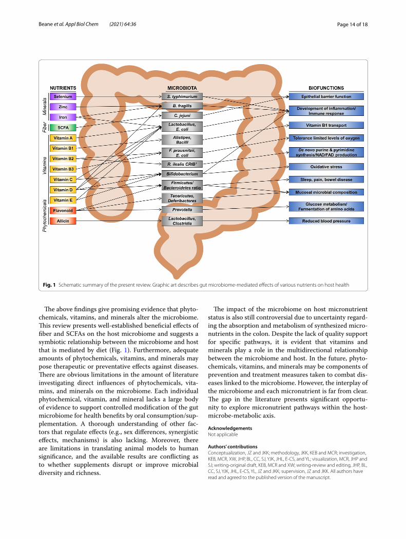
Fig. 1 Schematic summary of the present review. Graphic art describes gut microbiome‑mediated effects of various nutrients on host health
Page 15 of 18Beane et al. Appl Biol Chem (2021) 64:36
FundingThis work was supported by the University of Delaware Start‑Up fund (BHAN175183 to JKK), by an Institutional Development Award (IDeA), Center of Biomedical Research Excellence, from the National Institute of General Medical Sciences of the National Institutes of Health [P20GM113125‑03 to JKK], and by the National Research Foundation of Korea (NRF) grants funded by the Korea Government (MSIP) (NRF‑2019R1A2C1090007).
Availability of data and materialsNot applicable.
Declarations
Competing interestsThe authors declare that they have no competing interests.
Author details1 School of Human Environmental Sciences, University of Arkansas, Fayette‑ville, AR 72701, USA. 2 Department of Behavioral Health and Nutrition, Uni‑versity of Delaware, Newark, DE 19716, USA. 3 Department of Animal Science, Division of Agriculture, University of Arkansas, Fayetteville, AR 72701, USA. 4 Department of Food and Biotechnology, Korea University, Sejong 30019, Republic of Korea. 5 Department of Food Science, Gyeongsang National Uni‑versity, Jinju 52828, Republic of Korea. 6 Key Laboratory of Molecular Design and Precision Breeding of Animal in Guangdong Province, Foshan University, Foshan 528000, Guangdong, China.
Received: 15 January 2021 Accepted: 17 March 2021
References 1. Qin J, Li R, Raes J, Arumugam M, Burgdorf KS, Manichanh C, Nielsen
T, Pons N, Levenez F, Yamada T, Mende DR, Li J, Xu J, Li S, Li D, Cao J, Wang B, Liang H, Zheng H, Xie Y, Tap J, Lepage P, Bertalan M, Batto JM, Hansen T, Le Paslier D, Linneberg A, Nielsen HB, Pelletier E, Renault P, Sicheritz‑Ponten T, Turner K, Zhu H, Yu C, Li S, Jian M, Zhou Y, Li Y, Zhang X, Li S, Qin N, Yang H, Wang J, Brunak S, Dore J, Guarner F, Kristiansen K, Pedersen O, Parkhill J, Weissenbach J, Meta HITC, Bork P, Ehrlich SD, Wang J (2010) A human gut microbial gene catalogue established by metagenomic sequencing. Nature 464:59–65
2. Mills RH, Vazquez‑Baeza Y, Zhu Q, Jiang L, Gaffney J, Humphrey G, Smarr L, Knight R, Gonzalez DJ (2019) Evaluating metagenomic prediction of the metaproteome in a 4.5‑year study of a patient with Crohn’s disease. mSystems 4:e00337‑e1318
3. Koenig JE, Spor A, Scalfone N, Fricker AD, Stombaugh J, Knight R, Angenent LT, Ley RE (2011) Succession of microbial consortia in the developing infant gut microbiome. Proc Natl Acad Sci USA 108(Suppl 1):4578–4585
4. Wu GD, Chen J, Hoffmann C, Bittinger K, Chen YY, Keilbaugh SA, Bewtra M, Knights D, Walters WA, Knight R, Sinha R, Gilroy E, Gupta K, Baldas‑sano R, Nessel L, Li H, Bushman FD, Lewis JD (2011) Linking long‑term dietary patterns with gut microbial enterotypes. Science 334:105–108
5. Claesson MJ, Jeffery IB, Conde S, Power SE, O’Connor EM, Cusack S, Harris HM, Coakley M, Lakshminarayanan B, O’Sullivan O, Fitzgerald GF, Deane J, O’Connor M, Harnedy N, O’Connor K, O’Mahony D, van Sinderen D, Wallace M, Brennan L, Stanton C, Marchesi JR, Fitzgerald AP, Shanahan F, Hill C, Ross RP, O’Toole PW (2012) Gut microbiota composi‑tion correlates with diet and health in the elderly. Nature 488:178–184
6. Slavin JL (2005) Dietary fiber and body weight. Nutrition 21:411–418 7. Deehan EC, Duar RM, Armet AM, Perez‑Munoz ME, Jin M, Walter J (2017)
Modulation of the gastrointestinal microbiome with nondigestible fermentable carbohydrates to improve human health. Microbiol Spectr 5:1–24
8. El Kaoutari A, Armougom F, Gordon JI, Raoult D, Henrissat B (2013) The abundance and variety of carbohydrate‑active enzymes in the human gut microbiota. Nat Rev Microbiol 11:497–504
9. Holscher HD (2017) Dietary fiber and prebiotics and the gastrointestinal microbiota. Gut Microbes 8:172–184
10. Van Rymenant E, Abranko L, Tumova S, Grootaert C, Van Camp J, Wil‑liamson G, Kerimi A (2017) Chronic exposure to short‑chain fatty acids modulates transport and metabolism of microbiome‑derived phenolics in human intestinal cells. J Nutr Biochem 39:156–168
11. Schulthess J, Pandey S, Capitani M, Rue‑Albrecht KC, Arnold I, Franchini F, Chomka A, Ilott NE, Johnston DGW, Pires E, McCullagh J, Sansom SN, Arancibia‑Carcamo CV, Uhlig HH, Powrie F (2019) The short chain fatty acid butyrate imprints an antimicrobial program in macrophages. Immunity 50(432–445):e437
12. Desai MS, Seekatz AM, Koropatkin NM, Kamada N, Hickey CA, Wolter M, Pudlo NA, Kitamoto S, Terrapon N, Muller A, Young VB, Henrissat B, Wilmes P, Stappenbeck TS, Nunez G, Martens EC (2016) A dietary fiber‑deprived gut microbiota degrades the colonic mucus barrier and enhances pathogen susceptibility. Cell 167(1339–1353):e1321
13. Delcour JA, Aman P, Courtin CM, Hamaker BR, Verbeke K (2016) Prebiot‑ics, fermentable dietary fiber, and health claims. Adv Nutr 7:1–4
14. Bang SJ, Kim G, Lim MY, Song EJ, Jung DH, Kum JS, Nam YD, Park CS, Seo DH (2018) The influence of in vitro pectin fermentation on the human fecal microbiome. AMB Express 8:98
15. Gibson GR, Roberfroid MB (1995) Dietary modulation of the human colonic microbiota: introducing the concept of prebiotics. J Nutr 125:1401–1412
16. Roberfroid M (2007) Prebiotics: the concept revisited. J Nutr 137:830S‑837S
17. Gibson GR, Probert HM, Loo JV, Rastall RA, Roberfroid MB (2004) Dietary modulation of the human colonic microbiota: updating the concept of prebiotics. Nutr Res Rev 17:259–275
18. de Jesus Raposo MF, de Morais AM, de Morais RM (2016) Emergent sources of prebiotics: seaweeds and microalgae. Mar Drugs 14:27
19. Carlson JL, Erickson JM, Lloyd BB, Slavin JL (2018) Health effects and sources of prebiotic dietary fiber. Curr Dev Nutr 2:nyz005
20. Bindels LB, Segura Munoz RR, Gomes‑Neto JC, Mutemberezi V, Martinez I, Salazar N, Cody EA, Quintero‑Villegas MI, Kittana H, de Los Reyes‑Gavilan CG, Schmaltz RJ, Muccioli GG, Walter J, Ramer‑Tait AE (2017) Resistant starch can improve insulin sensitivity independently of the gut microbiota. Microbiome 5:12
21. Carlson JL, Erickson JM, Hess JM, Gould TJ, Slavin JL (2017) Prebiotic dietary fiber and gut health: comparing the in vitro fermentations of beta‑glucan, inulin and xylooligosaccharide. Nutrients 9:1361
22. Day L, Gomez J, Oiseth SK, Gidley MJ, Williams BA (2012) Faster fermen‑tation of cooked carrot cell clusters compared to cell wall fragments in vitro by porcine feces. J Agric Food Chem 60:3282–3290
23. Cook SI, Sellin JH (1998) Review article: short chain fatty acids in health and disease. Aliment Pharmacol Ther 12:499–507
24. Cummings JH, Pomare EW, Branch WJ, Naylor CP, Macfarlane GT (1987) Short chain fatty acids in human large intestine, portal, hepatic and venous blood. Gut 28:1221–1227
25. Wong JM, de Souza R, Kendall CW, Emam A, Jenkins DJ (2006) Colonic health: fermentation and short chain fatty acids. J Clin Gastroenterol 40:235–243
26. Hamer HM, Jonkers D, Venema K, Vanhoutvin S, Troost FJ, Brummer RJ (2008) Review article: the role of butyrate on colonic function. Aliment Pharmacol Ther 27:104–119
27. Magnusson MK, Isaksson S, Ohman L (2020) The anti‑inflammatory immune regulation induced by butyrate is impaired in inflamed intestinal mucosa from patients with ulcerative colitis. Inflammation 43:507–517
28. Mathew OP, Ranganna K, Mathew J, Zhu M, Yousefipour Z, Selvam C, Milton SG (2019) Cellular effects of butyrate on vascular smooth muscle cells are mediated through disparate actions on dual targets, histone deacetylase (hdac) activity and pi3k/akt signaling network. Int J Mol Sci 20:2902
29. Morais JAV, Rodrigues MC, Ferreira FF, Ranjan K, Azevedo RB, Pocas‑Fonseca MJ, Muehlmann LA (2020) Photodynamic therapy inhibits cell growth and enhances the histone deacetylase‑mediated viability impairment in Cryptococcus spp. in vitro. Photodiagnosis Photodyn Ther 29:101583
30. Uerlings J, Schroyen M, Bautil A, Courtin C, Richel A, Sureda EA, Brugge‑man G, Tanghe S, Willems E, Bindelle J, Everaert N (2020) In vitro prebi‑otic potential of agricultural by‑products on intestinal fermentation, gut barrier and inflammatory status of piglets. Br J Nutr 123:293–307
Page 16 of 18Beane et al. Appl Biol Chem (2021) 64:36
31. Canani RB, Costanzo MD, Leone L, Pedata M, Meli R, Calignano A (2011) Potential beneficial effects of butyrate in intestinal and extraintestinal diseases. World J Gastroenterol 17:1519–1528
32. Baxter NT, Schmidt AW, Venkataraman A, Kim KS, Waldron C, Schmidt TM (2019) Dynamics of human gut microbiota and short‑chain fatty acids in response to dietary interventions with three fermentable fibers. MBio 10:e02566‑02518
33. Guttman JA, Finlay BB (2009) Tight junctions as targets of infectious agents. Biochim Biophys Acta 1788:832–841
34. Louis P, Flint HJ (2017) Formation of propionate and butyrate by the human colonic microbiota. Environ Microbiol 19:29–41
35. Kaczmarek JL, Musaad SM, Holscher HD (2017) Time of day and eating behaviors are associated with the composition and function of the human gastrointestinal microbiota. Am J Clin Nutr 106:1220–1231
36. Karl JP, Hatch AM, Arcidiacono SM, Pearce SC, Pantoja‑Feliciano IG, Doherty LA, Soares JW (2018) Effects of psychological, environmental and physical stressors on the gut microbiota. Front Microbiol 9:2013
37. Guzman JR, Conlin VS, Jobin C (2013) Diet, microbiome, and the intesti‑nal epithelium: an essential triumvirate? Biomed Res Int 2013:425146
38. Carlson JL, Erickson JM, Hess JM, Gould TJ, Slavin JL (2018) Potential of butyrate to influence food intake in mice and men. Gut 67:1203–1204
39. Whelan K, Schneider SM (2011) Mechanisms, prevention, and man‑agement of diarrhea in enteral nutrition. Curr Opin Gastroenterol 27:152–159
40. O’Keefe SJ, Ou J, Delany JP, Curry S, Zoetendal E, Gaskins HR, Gunn S (2011) Effect of fiber supplementation on the microbiota in critically ill patients. World J Gastrointest Pathophysiol 2:138–145
41. Sunkara LT, Achanta M, Schreiber NB, Bommineni YR, Dai G, Jiang W, Lamont S, Lillehoj HS, Beker A, Teeter RG (2011) Butyrate enhances disease resistance of chickens by inducing antimicrobial host defense peptide gene expression. PLoS ONE 6:e27225
42. Yan H, Ajuwon KM (2017) Butyrate modifies intestinal barrier function in ipec‑j2 cells through a selective upregulation of tight junction proteins and activation of the akt signaling pathway. PLoS ONE 12:e0179586
43. Zheng L, Kelly CJ, Battista KD, Schaefer R, Lanis JM, Alexeev EE, Wang RX, Onyiah JC, Kominsky DJ, Colgan SP (2017) Microbial‑derived butyrate promotes epithelial barrier function through il‑10 receptor‑dependent repression of claudin‑2. J Immunol 199:2976–2984
44. Commins S, Steinke JW, Borish L (2008) The extended il‑10 superfamily: Il‑10, il‑19, il‑20, il‑22, il‑24, il‑26, il‑28, and il‑29. J Allergy Clin Immunol 121:1108–1111
45. El Kasmi KC, Holst J, Coffre M, Mielke L, de Pauw A, Lhocine N, Smith AM, Rutschman R, Kaushal D, Shen Y, Suda T, Donnelly RP, Myers MG Jr, Alexander W, Vignali DA, Watowich SS, Ernst M, Hilton DJ, Murray PJ (2006) General nature of the stat3‑activated anti‑inflammatory response. J Immunol 177:7880–7888
46. Riley JK, Takeda K, Akira S, Schreiber RD (1999) Interleukin‑10 receptor signaling through the jak‑stat pathway. Requirement for two distinct receptor‑derived signals for anti‑inflammatory action. J Biol Chem 274:16513–16521
47. Hammami R, Fernandez B, Lacroix C, Fliss I (2013) Anti‑infective proper‑ties of bacteriocins: an update. Cell Mol Life Sci 70:2947–2967
48. Mogensen TH (2009) Pathogen recognition and inflammatory signaling in innate immune defenses. Clin Microbiol Rev 22:240–273
49. Mazzurana L, Rao A, Van Acker A, Mjösberg J (2018) The roles for innate lymphoid cells in the human immune system. Semin Immunopathol 40:407–419
50. Blander JM (2016) Death in the intestinal epithelium‑basic biology and implications for inflammatory bowel disease. FEBS J 283:2720–2730
51. van der Beek CM, Dejong CHC, Troost FJ, Masclee AAM, Lenaerts K (2017) Role of short‑chain fatty acids in colonic inflammation, carcino‑genesis, and mucosal protection and healing. Nutr Rev 75:286–305
52. Sun M, Wu W, Chen L, Yang W, Huang X, Ma C, Chen F, Xiao Y, Zhao Y, Ma C, Yao S, Carpio VH, Dann SM, Zhao Q, Liu Z, Cong Y (2018) Microbiota‑derived short‑chain fatty acids promote th1 cell il‑10 production to maintain intestinal homeostasis. Nat Commun 9:3555
53. Liss MA, White JR, Goros M, Gelfond J, Leach R, Johnson‑Pais T, Lai Z, Rourke E, Basler J, Ankerst D, Shah DP (2018) Metabolic biosynthesis pathways identified from fecal microbiome associated with prostate cancer. Eur Urol 74:575–582
54. Segain JP, Raingeard D, de la Bletiere A, Bourreille VL, Gervois N, Rosales C, Ferrier L, Bonnet C, Blottiere HM, Galmiche JP (2000) Butyrate inhibits inflammatory responses through nfkappab inhibition: implications for Crohn’s disease. Gut 47:397–403
55. Kovarik JJ, Tillinger W, Hofer J, Holzl MA, Heinzl H, Saemann MD, Zlabinger GJ (2011) Impaired anti‑inflammatory efficacy of n‑butyrate in patients with ibd. Eur J Clin Investig 41:291–298
56. Vinolo MA, Rodrigues HG, Nachbar RT, Curi R (2011) Regulation of inflammation by short chain fatty acids. Nutrients 3:858–876
57. Fellermann K, Stange DE, Schaeffeler E, Schmalzl H, Wehkamp J, Bevins CL, Reinisch W, Teml A, Schwab M, Lichter P, Radlwimmer B, Stange EF (2006) A chromosome 8 gene‑cluster polymorphism with low human beta‑defensin 2 gene copy number predisposes to Crohn disease of the colon. Am J Hum Genet 79:439–448
58. Hamer HM, Jonkers DM, Vanhoutvin SA, Troost FJ, Rijkers G, de Bruine A, Bast A, Venema K, Brummer RJ (2010) Effect of butyrate enemas on inflammation and antioxidant status in the colonic mucosa of patients with ulcerative colitis in remission. Clin Nutr 29:738–744
59. Komiyama Y, Andoh A, Fujiwara D, Ohmae H, Araki Y, Fujiyama Y, Mit‑suyama K, Kanauchi O (2011) New prebiotics from rice bran ameliorate inflammation in murine colitis models through the modulation of intestinal homeostasis and the mucosal immune system. Scand J Gastroenterol 46:40–52
60. Saura‑Calixto F, Serrano J, Goñi I (2007) Intake and bioaccessibility of total polyphenols in a whole diet. Food Chem 101:492–501
61. Hung LM, Chen JK, Huang SS, Lee RS, Su MJ (2000) Cardioprotective effect of resveratrol, a natural antioxidant derived from grapes. Cardio‑vasc Res 47:549–555
62. Cassidy A (2018) Berry anthocyanin intake and cardiovascular health. Mol Aspects Med 61:76–82
63. Kazuhiko Uchiyama YN, Hasegawa G, Nakamura N, Takahashi J, Yoshi‑kawa T (2013) Astaxanthin protects β‑cells against glucose toxicity in diabetic db/db mice. Redox Rep 7:290–293
64. Gu J, Thomas‑Ahner JM, Riedl KM, Bailey MT, Vodovotz Y, Schwartz SJ, Clinton SK (2019) Dietary black raspberries impact the colonic micro‑biome and phytochemical metabolites in mice. Mol Nutr Food Res 63:e1800636
65. Wankhade UD, Zhong Y, Lazarenko OP, Chintapalli SV, Piccolo BD, Chen JR, Shankar K (2019) Sex‑specific changes in gut microbiome composition following blueberry consumption in c57bl/6j mice. Nutrients 11:313
66. Gyawali R, Ibrahim SA (2012) Impact of plant derivatives on the growth of foodborne pathogens and the functionality of probiotics. Appl Microbiol Biotechnol 95:29–45
67. Brown DG, Borresen EC, Brown RJ, Ryan EP (2017) Heat‑stabilised rice bran consumption by colorectal cancer survivors modulates stool metabolite profiles and metabolic networks: a randomised controlled trial. Br J Nutr 117:1244–1256
68. Saiprasad G, Chitra P, Manikandan R, Sudhandiran G (2013) Hesperi‑din alleviates oxidative stress and downregulates the expressions of proliferative and inflammatory markers in azoxymethane‑induced experimental colon carcinogenesis in mice. Inflamm Res 62:425–440
69. Cheng L, Chen Y, Zhang X, Zheng X, Cao J, Wu Z, Qin W, Cheng K (2019) A metagenomic analysis of the modulatory effect of Cyclo-carya paliurus flavonoids on the intestinal microbiome in a high‑fat diet‑induced obesity mouse model. J Sci Food Agric 99:3967–3975
70. Kovatcheva‑Datchary P, Nilsson A, Akrami R, Lee YS, De Vadder F, Arora T, Hallen A, Martens E, Bjorck I, Backhed F (2015) Dietary fiber‑induced improvement in glucose metabolism is associated with increased abundance of prevotella. Cell Metab 22:971–982
71. Vargas‑Bello‑Perez E, Cancino‑Padilla N, Romero J, Garnsworthy PC (2016) Quantitative analysis of ruminal bacterial populations involved in lipid metabolism in dairy cows fed different vegetable oils. Animal 10:1821–1828
72. Petersen C, Wankhade UD, Bharat D, Wong K, Mueller JE, Chintapalli SV, Piccolo BD, Jalili T, Jia Z, Symons JD, Shankar K, Anandh Babu PV (2019) Dietary supplementation with strawberry induces marked changes in the composition and functional potential of the gut microbiome in diabetic mice. J Nutr Biochem 66:63–69
73. Si Tan JAC‑M, Matthews VB, Koch H, O’Gara F, Croft KD, Ward NC (2018) Isoquercetin and inulin synergistically modulate the gut
Page 17 of 18Beane et al. Appl Biol Chem (2021) 64:36
microbiome to prevent development of the metabolic syndrome in mice fed a high fat diet. Sci Rep 8:10100
74. Djuric Z, Bassis CM, Plegue MA, Ren J, Chan R, Sidahmed E, Turgeon DK, Ruffin MT IV, Kato I, Sen A (2018) Colonic mucosal bacteria are associated with inter‑individual variability in serum carotenoid con‑centrations. J Acad Nutr Diet 118:606‑616.e603
75. Ramos AFO, Terry SA, Holman DB, Breves G, Pereira LGR, Silva AGM, Chaves AV (2018) Tucuma oil shifted ruminal fermentation, reducing methane production and altering the microbiome but decreased substrate digestibility within a rusitec fed a mixed hay—concentrate diet. Front Microbiol 9:1647
76. Lin T, Zirpoli GR, McCann SE, Moysich KB, Ambrosone CB, Tang L (2017) Trends in cruciferous vegetable consumption and associations with breast cancer risk: a case‑control study. Curr Dev Nutr 1:e000448
77. Kim JK, Strapazzon N, Gallaher CM, Stoll DR, Thomas W, Gallaher DD, Trudo SP (2017) Comparison of short‑ and long‑term exposure effects of cruciferous and apiaceous vegetables on carcinogen metabolizing enzymes in Wistar rats. Food Chem Toxicol 108:194–202
78. Veeranki OL, Bhattacharya A, Tang L, Marshall JR, Zhang Y (2015) Cruciferous vegetables, isothiocyanates, and prevention of bladder cancer. Curr Pharmacol Rep 1:272–282
79. Kaczmarek JL, Liu X, Charron CS, Novotny JA, Jeffery EH, Seifried HE, Ross SA, Miller MJ, Swanson KS, Holscher HD (2019) Broccoli consumption affects the human gastrointestinal microbiota. J Nutr Biochem 63:27–34
80. Angelino D, Dosz EB, Sun J, Hoeflinger JL, Van Tassell ML, Chen P, Harnly JM, Miller MJ, Jeffery EH (2015) Myrosinase‑dependent and ‑independent formation and control of isothiocyanate products of glucosinolate hydrolysis. Front Plant Sci 6:831
81. Bianchini F, Vainio H (2001) Allium vegetables and organosulfur compounds: do they help prevent cancer? Environ Health Perspect 109:893–902
82. Ried K, Travica N, Sali A (2018) The effect of kyolic aged garlic extract on gut microbiota, inflammation, and cardiovascular markers in hypertensives: the gargic trial. Front Nutr 5:122
83. Mohammed A, Guda C (2015) Application of a hierarchical enzyme classification method reveals the role of gut microbiome in human metabolism. BMC Genomics 16(Suppl 7):S16
84. Pascale A, Marchesi N, Marelli C, Coppola A, Luzi L, Govoni S, Giustina A, Gazzaruso C (2018) Microbiota and metabolic diseases. Endocrine 61:357–371
85. Nagy‑Szakal D, Ross MC, Dowd SE, Mir SA, Schaible TD, Petrosino JF, Kel‑lermayer R (2012) Maternal micronutrients can modify colonic mucosal microbiota maturation in murine offspring. Gut Microbes 3:426–433
86. Dicks LMT, Geldenhuys J, Mikkelsen LS, Brandsborg E, Marcotte H (2018) Our gut microbiota: a long walk to homeostasis. Benef Microbes 9:3–20
87. Ryan PM, Ross RP, Fitzgerald GF, Caplice NM, Stanton C (2015) Sugar‑coated: exopolysaccharide producing lactic acid bacteria for food and human health applications. Food Funct 6:679–693
88. Rowland I, Gibson G, Heinken A, Scott K, Swann J, Thiele I, Tuohy K (2018) Gut microbiota functions: metabolism of nutrients and other food components. Eur J Nutr 57:1–24
89. Holen T, Norheim F, Gundersen TE, Mitry P, Linseisen J, Iversen PO, Drevon CA (2016) Biomarkers for nutrient intake with focus on alterna‑tive sampling techniques. Genes Nutr 11:12
90. La Fata G, Rastall RA, Lacroix C, Harmsen HJM, Mohajeri MH, Weber P, Steinert RE (2017) Recent development of prebiotic research‑statement from an expert workshop. Nutrients 9:1376
91. Biesalski HK (2016) Nutrition meets the microbiome: micronutrients and the microbiota. Ann N Y Acad Sci 1372:53–64
92. Mach N, Clark A (2017) Micronutrient deficiencies and the human gut microbiota. Trends Microbiol 25:607–610
93. Sharma S, Litonjua A (2014) Asthma, allergy, and responses to methyl donor supplements and nutrients. J Allergy Clin Immunol 133:1246–1254
94. Wang Q, Wang K, Wu W, Giannoulatou E, Ho JWK, Li L (2019) Host and microbiome multi‑omics integration: applications and methodologies. Biophys Rev 11:55–65
95. Gill SR, Pop M, Deboy RT, Eckburg PB, Turnbaugh PJ, Samuel BS, Gordon JI, Relman DA, Fraser‑Liggett CM, Nelson KE (2006) Metagenomic analy‑sis of the human distal gut microbiome. Science 312:1355–1359
96. Mamo G (2016) Anaerobes as sources of bioactive compounds and health promoting tools. Adv Biochem Eng Biotechnol 156:433–464
97. Gominak SC (2016) Vitamin d deficiency changes the intestinal micro‑biome reducing b vitamin production in the gut. The resulting lack of pantothenic acid adversely affects the immune system, producing a “pro‑inflammatory” state associated with atherosclerosis and autoim‑munity. Med Hypotheses 94:103–107
98. Bhat MI, Kapila R (2017) Dietary metabolites derived from gut micro‑biota: critical modulators of epigenetic changes in mammals. Nutr Rev 75:374–389
99. Costliow ZA, Degnan PH (2017) Thiamine acquisition strategies impact metabolism and competition in the gut microbe bacteroides thetaio‑taomicron. mSystems 2:e00116‑00117
100. Klaassen MAY, Imhann F, Collij V, Fu J, Wijmenga C, Zhernakova A, Dijkstra G, Festen EAM, Gacesa R, Vich Vila A, Weersma RK (2019) Anti‑inflammatory gut microbial pathways are decreased during crohn’s disease exacerbations. J Crohns Colitis 13:1439–1449
101. Gerritsen J, Hornung B, Renckens B, van Hijum SAFT, Martins VAP, dos Santos GT, Rijkers PJS, de Vos WM, Smidt H (2017) Genomic and func‑tional analysis of romboutsia ilealis cribt reveals adaptation to the small intestine. PeerJ 5:e3698
102. Belzer C, Chia LW, Aalvink S, Chamlagain B, Piironen V, Knol J, de Vos WM (2017) Microbial metabolic networks at the mucus layer lead to diet‑independent butyrate and vitamin b(12) production by intestinal symbionts. MBio 8:e00770‑00717
103. Mohajeri MH, Brummer RJM, Rastall RA, Weersma RK, Harmsen HJM, Faas M, Eggersdorfer M (2018) The role of the microbiome for human health: from basic science to clinical applications. Eur J Nutr 57:1–14
104. Xu J, Xu C, Chen X, Cai X, Yang S, Sheng Y, Wang T (2014) Regulation of an antioxidant blend on intestinal redox status and major microbiota in early weaned piglets. Nutrition 30:584–589
105. Traber MG, Buettner GR, Bruno RS (2019) The relationship between vitamin csstatus, the gut‑liver axis, and metabolic syndrome. Redox Biol 21:101091
106. Smith AD, Panickar KS, Urban JF Jr, Dawson HD (2018) Impact of micro‑nutrients on the immune response of animals. Annu Rev Anim Biosci 6:227–254
107. Wang J, Thingholm LB, Skieceviciene J, Rausch P, Kummen M, Hov JR, Degenhardt F, Heinsen FA, Ruhlemann MC, Szymczak S, Holm K, Esko T, Sun J, Pricop‑Jeckstadt M, Al‑Dury S, Bohov P, Bethune J, Sommer F, Ellinghaus D, Berge RK, Hubenthal M, Koch M, Schwarz K, Rimbach G, Hubbe P, Pan WH, Sheibani‑Tezerji R, Hasler R, Rosenstiel P, D’Amato M, Cloppenborg‑Schmidt K, Kunzel S, Laudes M, Marschall HU, Lieb W, Nothlings U, Karlsen TH, Baines JF, Franke A (2016) Genome‑wide association analysis identifies variation in vitamin d receptor and other host factors influencing the gut microbiota. Nat Genet 48:1396–1406
108. Kanhere M, He J, Chassaing B, Ziegler TR, Alvarez JA, Ivie EA, Hao L, Hanfelt J, Gewirtz AT, Tangpricha V (2018) Bolus weekly vitamin d3 sup‑plementation impacts gut and airway microbiota in adults with cystic fibrosis: a double‑blind, randomized, placebo‑controlled clinical trial. J Clin Endocrinol Metab 103:564–574
109. Chen Y, Liu W, Sun T, Huang Y, Wang Y, Deb DK, Yoon D, Kong J, Thadhani R, Li YC (2013) 1,25‑dihydroxyvitamin d promotes negative feedback regulation of tlr signaling via targeting microrna‑155‑socs1 in macrophages. J Immunol 190:3687–3695
110. Talsness CE, Penders J, Jansen E, Damoiseaux J, Thijs C, Mommers M (2017) Influence of vitamin d on key bacterial taxa in infant microbiota in the koala birth cohort study. PLoS ONE 12:e0188011
111. Sordillo JE, Zhou Y, McGeachie MJ, Ziniti J, Lange N, Laranjo N, Savage JR, Carey V, O’Connor G, Sandel M, Strunk R, Bacharier L, Zeiger R, Weiss ST, Weinstock G, Gold DR, Litonjua AA (2017) Factors influencing the infant gut microbiome at age 3–6 months: findings from the ethnically diverse vitamin d antenatal asthma reduction trial (vdaart). J Allergy Clin Immunol 139(482–491):e414
112. Aleksandrova K, Romero‑Mosquera B, Hernandez V (2017) Diet, gut microbiome and epigenetics: emerging links with inflammatory bowel diseases and prospects for management and prevention. Nutrients 9:962
113. Gombart AF (2009) The vitamin d‑antimicrobial peptide pathway and its role in protection against infection. Future Microbiol 4:1151–1165
Page 18 of 18Beane et al. Appl Biol Chem (2021) 64:36
114. Wu S, Zhang YG, Lu R, Xia Y, Zhou D, Petrof EO, Claud EC, Chen D, Chang EB, Carmeliet G, Sun J (2015) Intestinal epithelial vitamin d receptor deletion leads to defective autophagy in colitis. Gut 64:1082–1094
115. Resta SC (2009) Effects of probiotics and commensals on intestinal epithelial physiology: Implications for nutrient handling. J Physiol 587:4169–4174
116. Sun J (2018) Dietary vitamin D, vitamin D receptor, and microbiome. Curr Opin Clin Nutr Metab Care 21:471–474
117. Steiber AL (2014) Chronic kidney disease: considerations for nutrition interventions. JPEN J Parenter Enteral Nutr 38:418–426
118. Rosario D, Benfeitas R, Bidkhori G, Zhang C, Uhlen M, Shoaie S, Mardino‑glu A (2018) Understanding the representative gut microbiota dysbiosis in metformin‑treated type 2 diabetes patients using genome‑scale metabolic modeling. Front Physiol 9:775
119. Reed S, Neuman H, Glahn RP, Koren O, Tako E (2017) Characterizing the gut (Gallus gallus) microbiota following the consumption of an iron biofortified rwandan cream seeded carioca (Phaseolus vulgaris L.) bean‑based diet. PLoS ONE 12:e0182431
120. Faulkner MJ, Wenner BA, Solden LM, Weiss WP (2017) Source of sup‑plemental dietary copper, zinc, and manganese affects fecal microbial relative abundance in lactating dairy cows. J Dairy Sci 100:1037–1044
121. Kasaikina MV, Kravtsova MA, Lee BC, Seravalli J, Peterson DA, Walter J, Legge R, Benson AK, Hatfield DL, Gladyshev VN (2011) Dietary selenium affects host selenoproteome expression by influencing the gut micro‑biota. FASEB J 25:2492–2499
122. Zhai Q, Cen S, Li P, Tian F, Zhao J, Zhang H, Chen W (2018) Effects of dietary selenium supplementation on intestinal barrier and immune responses associated with its modulation of gut microbiota. Environ Sci Technol Lett 5:724–730
123. Nettleford SK, Prabhu KS (2018) Selenium and selenoproteins in gut inflammation—a review. Antioxidants 7:36
124. Moshfegh AJ, Friday JE, Goldman JP, Ahuja JK (1999) Presence of inulin and oligofructose in the diets of Americans. J Nutr 129:1407S‑1411S
125. Sabater‑Molina M, Larque E, Torrella F, Zamora S (2009) Dietary fruc‑tooligosaccharides and potential benefits on health. J Physiol Biochem 65:315–328
126. Torres DPM, Goncalves M, Teixeira JA, Rodrigues LR (2010) Galacto‑oligosaccharides: production, properties, applications, and significance as prebiotics. Compr Rev Food Sci Food Saf 9:438–454
127. Hofman DL, van Buul VJ, Brouns FJ (2016) Nutrition, health, and regulatory aspects of digestible maltodextrins. Crit Rev Food Sci Nutr 56:2091–2100
128. Harnly JM, Doherty RF, Beecher GR, Holden JM, Haytowitz DB, Bhagwat S, Gebhardt S (2006) Flavonoid content of US fruits, vegetables, and nuts. J Agric Food Chem 54:9966–9977
129. Liggins J, Bluck LJ, Runswick S, Atkinson C, Coward WA, Bingham SA (2000) Daidzein and genistein content of fruits and nuts. J Nutr Bio‑chem 11:326–331
130. Perry A, Rasmussen H, Johnson EJ (2009) Xanthophyll (lutein, zeaxan‑thin) content in fruits, vegetables and corn and egg products. J Food Compos Anal 22:9–15
131. McNaughton SA, Marks GC (2003) Development of a food composi‑tion database for the estimation of dietary intakes of glucosinolates, the biologically active constituents of cruciferous vegetables. Br J Nutr 90:687–697
132. Lagarda MJ, Garcia‑Llatas G, Farre R (2006) Analysis of phytosterols in foods. J Pharm Biomed Anal 41:1486–1496
Publisher’s NoteSpringer Nature remains neutral with regard to jurisdictional claims in pub‑lished maps and institutional affiliations.