Effects of Corticotropin-Releasing Factor 1 Receptor...
9
Effects of Corticotropin-Releasing Factor 1 Receptor Antagonism on the Hypothalamic-Pituitary-Adrenal Axis of Rodents □ S Donald R. Gehlert, Jeffrey Cramer, and S. Michelle Morin Lilly Research Laboratories, a Division of Eli Lilly and Company, Indianapolis, Indiana Received November 8, 2011; accepted March 7, 2012 ABSTRACT Corticotropin-releasing factor (CRF) is the major hypothalamic neuropeptide responsible for stimulation of the hypothalamic- pituitary-adrenal axis (HPAA), resulting in the synthesis and release of glucocorticoids from the adrenal cortex. In a recent study, we reported the discovery of the CRF1 receptor antag- onist, 3-(4-chloro-2-morpholin-4-yl-thiazol-5-yl)-8-(1-ethylpro- pyl)-2,6-dimethyl-imidazo[1,2-b]pyridazine (MTIP), which has efficacy in preclinical models of stress-induced alcohol con- sumption. Because CRF1 is important in HPAA activation, we evaluated the effects of MTIP administration on rodent HPAA function. Initial studies established the MTIP doses required for brain and pituitary CRF1 occupancy and those associated with the inhibition of intracerebroventricular CRF on the HPAA in mice. Then, rat basal plasma corticosterone (CORT) concen- trations were measured hourly by radioimmunoassay for 24 h after three daily doses of MTIP or vehicle. In these studies, the early phase of the nocturnal CORT surge was reduced; how- ever, the area under the CORT curve was identical for the 24-h period. In subsequent studies, increases in plasma CORT due to direct pharmacological manipulation of the HPAA axis or by stressors were evaluated after MTIP treatment in mice. MTIP attenuated CORT responses generated by immediate bolus administration of insulin or ethanol; however, MTIP did not affect activation of the HPAA by other stressors and pharma- cological agents. Therefore, MTIP can modulate basal HPAA activity during the CORT surge and reduced activation after a select number of stressors but does not produce a lasting suppression of basal CORT. The ability of MTIP to modulate plasma CORT after hyperinsulinemia may provide a surrogate strategy for a target occupancy biomarker. Introduction Corticotropin-releasing factor (CRF) is an important regu- lator of the endocrine, immune, behavioral, and autonomic responses to stress (Vale et al., 1981). This peptide produces its biological effects by binding to two pharmacologically distinct G protein-coupled receptors, CRF1 and CRF2 (Bale and Vale, 2004). CRF exhibits high affinity for CRF1 but substantially lower affinity for CRF2. Although CRF1 is abundant in the pituitary, CRF2 predominates in tissues such as the heart, skeletal muscle, and gastrointestinal tract. In the brain, CRF1 is found in the cerebral cortex, amygdala, cerebellum, and brainstem, whereas CRF2 is only abundant in the lateral septum and hypothalamus. Hypothalamically derived CRF is a key regulator of the hypothalamo-pituitary- adrenal axis (HPAA) through stimulation of pituitary release of adrenocorticotropin stimulating the release of corticoste- rone (CORT) by the rat adrenal (Vale et al., 1981). Whereas the role of CRF at the level of the pituitary is well established (Timpl et al., 1998; Preil et al., 2001), CRF also has distinct actions via the central nervous system (Mu ¨ ller et al., 2003). Central administration of CRF produces activation of the HPAA, and little is known about the ability of CRF1 antag- onists to modulate this response (Song et al., 1995). Distur- bances in the central CRF system have been proposed to play an important role in the etiology of major depression (Hols- boer, 2000) and alcoholism (Sommer et al., 2008). In addition, there is a correlation between remission of depressive symp- toms and a normalization of HPAA function (Holsboer, 2000). The discovery of the related urocortin family of peptides Article, publication date, and citation information can be found at http://jpet.aspetjournals.org. http://dx.doi.org/10.1124/jpet.111.189753. □ S The online version of this article (available at http://jpet.aspetjournals.org) contains supplemental material. ABBREVIATIONS: CRF, corticotropin releasing factor; CRF1, corticotropin-releasing factor receptor type 1; CRF2, corticotropin-releasing factor receptor type 2; CORT, corticosterone; HPAA, hypothalamic-pituitary-adrenal axis; MTIP, 3-(4-chloro-2-morpholin-4-yl-thiazol-5-yl)-8-(1-ethylpropyl)- 2,6-dimethyl-imidazo[1,2-b]pyridazine; R121919, 2,5-dimethyl-3-(6-dimethylamino-4-methylpyridin-3-yl)-7-dipropylaminopyrazolo[1,5-a]pyrimidine; SSR125543, 4-(2-chloro-4-methoxy-5-methylphenyl)-N-[(1S)-2-cyclopropyl-1-(3-fluoro-4-methylphenyl)ethyl]-5-methyl-N-prop-2-ynyl-1,3- thiazol-2-amine; RIA, radioimmunoassay; ANOVA, analysis of variance; AST, astressin; NBI-34041, 2-(2,4-dichlorophenyl)-4-methyl-6-(1-propylbutyl)- 7,8-dihydro-6H-1,3,6,8a-tetraaza-acenaphthylene. 1521-0103/12/3413-672–680$25.00 THE JOURNAL OF PHARMACOLOGY AND EXPERIMENTAL THERAPEUTICS Vol. 341, No. 3 Copyright © 2012 by The American Society for Pharmacology and Experimental Therapeutics 189753/3770103 JPET 341:672–680, 2012 672 http://jpet.aspetjournals.org/content/suppl/2012/03/08/jpet.111.189753.DC1 Supplemental material to this article can be found at: at ASPET Journals on July 10, 2018 jpet.aspetjournals.org Downloaded from
Transcript of Effects of Corticotropin-Releasing Factor 1 Receptor...
Effects of Corticotropin-Releasing Factor 1 ReceptorAntagonism on the Hypothalamic-Pituitary-Adrenal Axisof Rodents□S
Donald R. Gehlert, Jeffrey Cramer, and S. Michelle MorinLilly Research Laboratories, a Division of Eli Lilly and Company, Indianapolis, Indiana
Received November 8, 2011; accepted March 7, 2012
ABSTRACTCorticotropin-releasing factor (CRF) is the major hypothalamicneuropeptide responsible for stimulation of the hypothalamic-pituitary-adrenal axis (HPAA), resulting in the synthesis andrelease of glucocorticoids from the adrenal cortex. In a recentstudy, we reported the discovery of the CRF1 receptor antag-onist, 3-(4-chloro-2-morpholin-4-yl-thiazol-5-yl)-8-(1-ethylpro-pyl)-2,6-dimethyl-imidazo[1,2-b]pyridazine (MTIP), which hasefficacy in preclinical models of stress-induced alcohol con-sumption. Because CRF1 is important in HPAA activation, weevaluated the effects of MTIP administration on rodent HPAAfunction. Initial studies established the MTIP doses required forbrain and pituitary CRF1 occupancy and those associated withthe inhibition of intracerebroventricular CRF on the HPAA inmice. Then, rat basal plasma corticosterone (CORT) concen-trations were measured hourly by radioimmunoassay for 24 h
after three daily doses of MTIP or vehicle. In these studies, theearly phase of the nocturnal CORT surge was reduced; how-ever, the area under the CORT curve was identical for the 24-hperiod. In subsequent studies, increases in plasma CORT dueto direct pharmacological manipulation of the HPAA axis or bystressors were evaluated after MTIP treatment in mice. MTIPattenuated CORT responses generated by immediate bolusadministration of insulin or ethanol; however, MTIP did notaffect activation of the HPAA by other stressors and pharma-cological agents. Therefore, MTIP can modulate basal HPAAactivity during the CORT surge and reduced activation after aselect number of stressors but does not produce a lastingsuppression of basal CORT. The ability of MTIP to modulateplasma CORT after hyperinsulinemia may provide a surrogatestrategy for a target occupancy biomarker.
IntroductionCorticotropin-releasing factor (CRF) is an important regu-
lator of the endocrine, immune, behavioral, and autonomicresponses to stress (Vale et al., 1981). This peptide producesits biological effects by binding to two pharmacologicallydistinct G protein-coupled receptors, CRF1 and CRF2 (Baleand Vale, 2004). CRF exhibits high affinity for CRF1 butsubstantially lower affinity for CRF2. Although CRF1 isabundant in the pituitary, CRF2 predominates in tissuessuch as the heart, skeletal muscle, and gastrointestinal tract.In the brain, CRF1 is found in the cerebral cortex, amygdala,cerebellum, and brainstem, whereas CRF2 is only abundant
in the lateral septum and hypothalamus. Hypothalamicallyderived CRF is a key regulator of the hypothalamo-pituitary-adrenal axis (HPAA) through stimulation of pituitary releaseof adrenocorticotropin stimulating the release of corticoste-rone (CORT) by the rat adrenal (Vale et al., 1981). Whereasthe role of CRF at the level of the pituitary is well established(Timpl et al., 1998; Preil et al., 2001), CRF also has distinctactions via the central nervous system (Muller et al., 2003).Central administration of CRF produces activation of theHPAA, and little is known about the ability of CRF1 antag-onists to modulate this response (Song et al., 1995). Distur-bances in the central CRF system have been proposed to playan important role in the etiology of major depression (Hols-boer, 2000) and alcoholism (Sommer et al., 2008). In addition,there is a correlation between remission of depressive symp-toms and a normalization of HPAA function (Holsboer, 2000).The discovery of the related urocortin family of peptides
Article, publication date, and citation information can be found athttp://jpet.aspetjournals.org.
http://dx.doi.org/10.1124/jpet.111.189753.□S The online version of this article (available at http://jpet.aspetjournals.org)
contains supplemental material.
ABBREVIATIONS: CRF, corticotropin releasing factor; CRF1, corticotropin-releasing factor receptor type 1; CRF2, corticotropin-releasing factorreceptor type 2; CORT, corticosterone; HPAA, hypothalamic-pituitary-adrenal axis; MTIP, 3-(4-chloro-2-morpholin-4-yl-thiazol-5-yl)-8-(1-ethylpropyl)-2,6-dimethyl-imidazo[1,2-b]pyridazine; R121919, 2,5-dimethyl-3-(6-dimethylamino-4-methylpyridin-3-yl)-7-dipropylaminopyrazolo[1,5-a]pyrimidine;SSR125543, 4-(2-chloro-4-methoxy-5-methylphenyl)-N-[(1S)-2-cyclopropyl-1-(3-fluoro-4-methylphenyl)ethyl]-5-methyl-N-prop-2-ynyl-1,3-thiazol-2-amine; RIA, radioimmunoassay; ANOVA, analysis of variance; AST, astressin; NBI-34041, 2-(2,4-dichlorophenyl)-4-methyl-6-(1-propylbutyl)-7,8-dihydro-6H-1,3,6,8a-tetraaza-acenaphthylene.
1521-0103/12/3413-672–680$25.00THE JOURNAL OF PHARMACOLOGY AND EXPERIMENTAL THERAPEUTICS Vol. 341, No. 3Copyright © 2012 by The American Society for Pharmacology and Experimental Therapeutics 189753/3770103JPET 341:672–680, 2012
672
http://jpet.aspetjournals.org/content/suppl/2012/03/08/jpet.111.189753.DC1Supplemental material to this article can be found at:
at ASPE
T Journals on July 10, 2018
jpet.aspetjournals.orgD
ownloaded from
revealed additional peptides that interact with CRF recep-tors (Bale and Vale, 2004). Urocortin I has high affinity forboth CRF1 and CRF2, whereas urocortin II and urocortin IIIexhibit high affinity and selectivity for CRF2. CRF2-selectivepeptides suppress feeding while having a more modulatoryeffect on stress-like responses (Hashimoto et al., 2004; Ja-mieson et al., 2006).
Additional insight into the roles of CRF receptors in theneuroendocrine response can be found in observations usingCRF1- and CRF2-deficient mice. CRF1-deficient mice havedecreased adrenocorticotropin and CORT responses after re-straint stress compared with wild-type controls (Smith et al.,1998; Timpl et al., 1998); however, they exhibit very littledifference in basal plasma CORT concentrations. It should benoted that the CRF1-KO exhibited adrenal agenesis attrib-uted to insufficient adrenocorticotropin during development(Smith et al., 1998), and, therefore, it is not surprising thatthese animals cannot mount an appropriate endocrine stressresponse. CRF2-deficient mice (Bale et al., 2000; Coste et al.,2000) exhibit normal basal plasma adrenocorticotropin andCORT and a normal circadian hormone rhythm. In contrastto the CRF1-deficient mice, CRF2-deficient mice exhibit anincreased endocrine responsiveness to restraint stress and,in some studies, have exhibited a prolongation of the CORTresponse to stress (Coste et al., 2000). Finally, CRF1- andCFR2-deficient mice (Preil et al., 2001; Bale et al., 2002)exhibit minor changes in plasma adrenocorticotropin andCORT in response to restraint stress. Whereas there was nodifference in basal CORT, these animals exhibited a reduc-tion in morning CORT. Based on these results, it is thoughtthat both CRF1 and CRF2 participate in the endocrine stressresponse although CRF1 has the major stimulatory role sub-sequent to physiological stressors.
The behavioral effects of centrally administered CRF alongwith the behavioral phenotype of the deficient mice havemade CRF1 an attractive target for drug discovery and de-velopment (Nielsen, 2006). Preclinical antagonist studies in-dicate the potential for antidepressant- and anxiolytic-likeactivities (Kehne and De Lombaert, 2002) as well as thepotential for the treatment of alcoholism (Gehlert et al.,2007) and substance abuse (Koob and Kreek, 2007). An earlyopen-label clinical study indicated the potential for antide-pressant activity (Zobel et al., 2000) and improved sleep(Held et al., 2004); however, recent clinical studies did notdemonstrate anxiolytic (Coric et al., 2010) or antidepressant(Binneman et al., 2008) activities. At present, there is noclinical occupancy biomarker to establish central CRF1 re-ceptor occupancy in the human studies, and it is not clearwhether there was adequate brain or pituitary CRF1 occupancyin these clinical trials. In a previous article, we disclosed a novelCRF1 antagonist 3-(4-chloro-2-morpholin-4-yl-thiazol-5-yl)-8-(1-ethylpropyl)-2,6-dimethyl-imidazo[1,2-b]pyridazine (MTIP)that reduced ethanol consumption in preclinical models(Gehlert et al., 2007). Brain receptor occupancy was estimatedusing ex vivo autoradiography and would not be suitable forclinical studies (Heinrichs et al., 2002; Gehlert et al., 2007). Inthe present study, we first established that MTIP could reducethe activation of the HPAA after central administration of CRF.Then, we evaluated the effects of MTIP on basal and stress-induced CORT secretion to better understand the influence ofbrain and pituitary CRF1 occupancy on measures of HPAAfunctionality. To activate the HPAA, we used several strategies
that were shown to increase plasma CORT by central and/orperipheral mechanisms including caffeine (Spindel et al., 1983),yohimbine (Marinelli et al., 2007), ethanol (Zgombick and Er-win, 1988), predator odor stress (Anisman et al., 2001), andinsulin (Muret et al., 1992).
Materials and MethodsAll studies adhered to the National Institutes of Health Guide for
the Care and Use of Laboratory Animals (Institute of LaboratoryAnimal Resources, 1996) and were performed with approval from theLilly Animal Care and Use Committee.
Effect of CRF1 Antagonists on Plasma CORT afterIntracerebroventricular Administration of CRF
Male C57BL6 mice (Harlan, Indianapolis, IN) were administeredvehicle (2 ml/kg, 3% dimethyl sulfoxide and 20% Emulphor in water) orMTIP, 2,5-dimethyl-3-(6-dimethylamino-4-methylpyridin-3-yl)-7-dipropylaminopyrazolo[1,5-a]pyrimidine (R121919), or 3, 4-(2-chloro-4-methoxy-5-methylphenyl)-N-[(1S)-2-cyclopropyl-1-(3-fluoro-4-methyl-phenyl)ethyl]-5-methyl-N-prop-2-ynyl-1,3-thiazol-2-amine (SSR125543)(30 mg/kg p.o. in vehicle) for 3 days [all three compounds were synthesizedat Eli Lilly and Company (Gehlert et al., 2007)]. The in vitro potency, invivo potency, and single-dose pharmacokinetics of all these compoundshave been reported previously (Gehlert et al., 2007) (SupplementalTable 1). The 30 mg/kg dose was selected because this was the maximaldose that provided a suspension suitable for a single bolus oral dosewith all three compounds, and all three compounds had similar in vitroaffinity for the receptor. One hour after the final dose, animals wereadministered 3 �g of CRF via direct, vertical, free-hand intracerebro-ventricular injection to a depth of 3.5 mm below bregma with a cuffed27-gauge needle attached to a 25-�l Hamilton syringe. Mice were sac-rificed 15 min later, and trunk blood was collected. In some experi-ments, randomly assigned mice were gently restrained by hand andinjected with artificial cerebrospinal fluid or the CRF1/2 antagonistastressin (various doses in a total volume of 3 �l). After sacrifice, trunkblood was collected, and the plasma was separated by centrifugation (5min at 7000 rpm in an Eppendorf tabletop centrifuge). Plasma wasfrozen at �80°C until assayed for CORT by radioimmunoassay (RIA)(MP Biomedicals, Salon, OH). In some experiments, plasma adreno-corticotropin was also assessed using RIA (MP Biomedicals). Datawere analyzed and plotted using Microsoft Excel (Microsoft, Red-mond, WA) and GraphPad Prism (GraphPad Software Inc., SanDiego, CA).
Measurement of Plasma CORT and Adrenocorticotropinby RIA
Plasma corticosterone and adrenocorticotropin were measuredwith the corresponding ImmunoChem Double Antibody 125I radio-immunoassay kit from MP Biomedicals. In all experiments in whichplasma CORT or adrenocorticotropin was measured, blood was col-lected in EDTA-containing tubes and kept on ice until centrifuged forplasma collection. The plasma was stored at �80°C until assayedfollowing the standard protocol provided with the kit.
The range of detection of the corticosterone kit is approximately7.7 to 1000 ng/ml with inter- and intra-assay coefficients of variationof less than 5% in our laboratory. The reported specificity of theassay by the manufacturer indicates less than 0.1% cross-reactivitywith any other steroids commonly found in plasma.
For the adrenocorticotropin kit, the range of detection is ap-proximately 5.7 to 1000 pg/ml with inter- and intra-assay coeffi-cients of variation of less than 8%. The manufacturer reportsthat the assay is specific for adrenocorticotropin1–39 as well asadrenocorticotropin1–24.
CRF1 Receptor Antagonism and the HPAA 673
at ASPE
T Journals on July 10, 2018
jpet.aspetjournals.orgD
ownloaded from
Ex Vivo Binding of 125I-Tyr0-Sauvagine
The method for CRF-1 receptor binding ex vivo using rat cerebel-lum has been described previously (Gehlert et al., 2005). Rat cere-bellum was selected because it had been previously shown to containpredominantly CRF-1 receptors at a density suitable to performbinding assays. In addition, this tissue is within the blood-brainbarrier and allowed us to estimate central occupancy by the com-pound. Rats were gavaged with vehicle (2 ml/kg, 3% dimethyl sul-foxide and 20% Emulphor in water) or compound in vehicle for 3consecutive days. One hour after the last dose, rats were decapitatedand pituitary glands and cerebella were removed, frozen on dry ice,and stored at �70°C. On the day of the assay, tissue was thawed,homogenized in buffer (50 mM Tris-HCl, 2 mM EGTA, and 10 mMMgCl2) and incubated at 37°C for 1 h. For the binding assay, approx-imately 200 �g of prepared homogenate was combined with a finalconcentration of 0.175 nM 125I-Tyr0-Sauvagine (PerkinElmer Lifeand Analytical Sciences, Waltham, MA) in assay buffer with 0.1%bovine serum albumin, 0.1% bacitracin, and 100 kU/ml aprotinin.Nonspecific binding was determined by addition of 1 �M ovine CRF(American Peptide Company, Sunnyvale, CA). After incubation atroom temperature for 120 min, the assay was terminated by centrif-ugation, and binding was assessed using a gamma counter. ED50
values were derived from specific binding calculated in GraphPadPrism using the four-parameter sigmoidal dose-response model.
Effect of MTIP on Basal CORT in the Rat
To determine this, we used the BASi Culex API system that allowsfor administration of the compound via gastric cannulae and bloodsampling via implanted jugular cannulae. This method eliminatesthe need to restrain the animal to accomplish these tasks and theresulting perturbations in stress hormones. For the following stud-ies, all animals were surgically modified and dosed at BASi facilities(West Lafayette, IN) using the Culex API system as described pre-viously (Bohs et al., 2000; Peters et al., 2000).
BASi Study LLY-4767. Twenty-two male Sprague-Dawley rats(300–350 g; Harlan) were used. All rats were surgically implantedwith gastric catheters, allowed 7 days of recovery, and then im-planted with femoral vein catheters. The rats were divided into twogroups of 11 for the experiment and placed in the Culex system atBASi. Group 1 received bolus infusions of vehicle (0.5% Tween 80 in0.1 N HCl, pH 4.0) via the intragastric catheter, and group 2 receivedMTIP at 30 mg/kg in a volume of 7.5 ml/kg. All animals were doseddaily at noon for 3 days. After the last dose of compound, 75-�lblood samples were taken hourly for 24 h via the Culex system andstored at 4°C until the end of the study. The blood samples werecentrifuged and plasma was collected and stored at �80°C untilassayed for CORT. Because of technical issues with the blood collec-tion, 7 of the original 22 animals did not complete the study, and datapoints from those animals were excluded from the study. Fifteenanimals completed the study (six vehicle-treated and nine MTIP-treated). Plasma CORT was measured using RIA. All samples wererun in duplicate, and the assay was conducted according to kitinstructions. The resulting CORT levels were averaged by group(mean � S.E.M.) for each time point and statistically evaluated by arepeated-measures, two-way analysis of variance (ANOVA) usingLilly internal Discovery Statistics software.
BASi Study LLY-4783. Thirty-six male Sprague-Dawley rats(350–375 g; Harlan) were used for the study. All animals had intra-gastric and femoral artery catheters implanted before the initiationof the study. Arterial catheters were used for blood collection in thisexperiment because BASi historically has had fewer catheter prob-lems with arterial than with venous lines, particularly in longerduration studies. The rats were divided into two groups of 18 andplaced in the Culex chambers for the duration of the study. In aninitial study, one group of rats was administered three daily doses ofvehicle, and a second group of rats were given three daily doses ofMTIP. Blood samples were taken hourly thereafter for 24 h. In a
second study, the dose-response relationship was explored. For thisexperiment, the study design consisted of three consecutive cycles ofdosing for 3 days followed by blood sample collection at eight timepoints during the following 24 h. For each cycle, group 1 receivedvehicle (0.5% Tween 80 in 0.1 N HCl, pH 4.0) via the intragastriccatheter, and group 2 received MTIP in an ascending dose per cycleof 0.1, 1.0, or 10 mg/kg in a volume of 7.5 ml/kg. At the end of eachblood collection, the samples were centrifuged, and plasma was col-lected and stored at �80°C until the end of the study.
Measurement of Plasma MTIP Concentrations
The plasma concentrations of MTIP were determined by positiveTurboIonSpray liquid chromatography-mass spectrometry using aSciex 4000 system (AB Sciex, Framingham, MA). Samples wereprepared by methanol precipitation and centrifuged to remove par-ticulate matter. An aliquot of the supernatant was transferred anddiluted with 80% water-20% methanol. The lower limit of quantita-tion was 1 ng/ml. Pharmacokinetic parameters were calculated bynoncompartmental analysis, using the trapezoidal rule for area un-der the curve calculation using a validated pharmacokinetic calcu-lation program (WinPTK; Eli Lilly and Co., Indianapolis, IN). Todetermine the plasma concentration of MTIP associated with themaximal concentration (Cmax), a single oral 10 mg/kg dose was givento rats, and the Cmax was determined to be 2400 ng/ml. Dose linearitywas assumed, and the plasma concentration associated with 1.5mg/kg (the single-dose ED50) was determined to be 360 ng/ml.
Effect of MTIP on Plasma CORT after Predator Odor orTreatment with Pharmacological Agents
Hyperinsulinemia. C57BL/6 mice (male, 22–26 g; Harlan) werehandled for 3 days before the experiment. The mice were dosed orallywith vehicle (1% carboxymethyl cellulose) or MTIP (3, 10, or 30mg/kg, 10 ml/kg) for 3 days (n � 8/group). On the 3rd day, insulin(Humalin R, 1 U/kg, 10 ml/kg i.p.; Eli Lilly and Company) or vehicle(saline intraperitoneally) was injected 1 h after the compound dose.Trunk blood was collected 1 h after insulin administration and kepton ice until the plasma was separated by centrifugation (10 min at8000 rpm in an Eppendorf tabletop centrifuge). Plasma was frozen at�80°C until assayed for CORT. Blood glucose was measured at timeof sacrifice with an AccuCheck Advantage monitor and ComfortCurve strips (Roche, Indianapolis, IN).
Predator Odor Stress. Animals were handled for 3 days beforethe experiment day (n � 10/group). The mice were dosed intracere-broventricularly 45 min before predator odor stress. Mice were ex-posed to predator odor (soiled rat bedding) for 10 min and then werereturned to their home cage for 20 min. The home cage control groupwas briefly picked up and then returned to their home cage for 30min. Trunk blood was collected in EDTA tubes at 30 min after oralexposure and kept on ice until centrifuged to collect plasma (10 minat 8000 rpm in an Eppendorf table top centrifuge) Plasma was frozenat �80°C until assayed for CORT.
Metyrapone. Animals were handled for 3 days before the exper-iment day and were dosed orally with 30 mg/kg MTIP (n � 8/group).On the experiment day animals were administered metyrapone (2-methyl-1,2-di-3-pyridyl-1-propanone, 75 mg/kg i.p.; Sigma-Aldrich,St. Louis, MO) 1 h after MTIP (orally) and 60 min before sacrifice. Acontrol group was dosed intracerebroventricularly with astressin(1 �g/3 �l) 45 min before metyrapone. Trunk blood was collectedand kept on ice until the plasma was separated by centrifugation(10 min at 8000 rpm in an Eppendorf tabletop centrifuge). Plasmawas collected and frozen at �80°C until assayed for CORT andadrenocorticotropin.
Caffeine. Animals were handled for 3 days before the experiment.The mice were administered MTIP (30 mg/kg p.o.) or vehicle for 3days. On the final experiment day, MTIP or vehicle were dosed 2 hbefore caffeine. Astressin was administered immediately (1 �g/3 �li.c.v.) 30 min before caffeine. Caffeine was injected (30 mg/kg, i.p.) 30
674 Gehlert et al.
at ASPE
T Journals on July 10, 2018
jpet.aspetjournals.orgD
ownloaded from

min before sacrifice and blood collection. Plasma was collected andfrozen at �80°C until assayed for CORT.
Yohimbine. Mice were dosed for 3 days with vehicle (1% carboxy-methyl cellulose) or MTIP (30 mg/kg p.o.). Two hours after the lastMTIP dose, the mice were administered vehicle (saline) or yohimbine(2.0 mg/kg i.p.; Sigma-Aldrich) and sacrificed 30 min later. Twoadditional groups were administered astressin (1 �g/3 �l i.c.v.) 30min before intraperitoneal vehicle or yohimbine (n � 6–9/group).Trunk blood was collected in tubes containing EDTA and kept on iceuntil the plasma was separated by centrifugation (5 min at 7000 rpmin an Eppendorf tabletop centrifuge). Plasma was frozen at �80°Cuntil assayed for CORT.
Ethanol. Animals were dosed for 3 days with MTIP (30 mg/kgp.o.) On the 3rd day, the animals were dosed with MTIP 1 h beforeethanol (Decon Laboratories, King of Prussia, PA; orally, 32% inH2O) or vehicle (H2O) (n � 8/group). The animals were administeredethanol or water 30 min before sacrifice. Trunk blood was collectedand kept on ice until the plasma was separated by centrifugation (10min at 8000 rpm in an Eppendorf tabletop centrifuge). Plasma wasfrozen at �80°C until assayed for CORT. Blood alcohol content wasmeasured with an ethanol L3K assay kit (Diagnostic Chemicals,Ltd., Charlottetown, PE, Canada).
ResultsThe pharmacological and pharmacokinetic properties of
the CRF1 antagonists used in the present study were pub-lished previously (Gehlert et al., 2007) and are summarizedin Supplemental Table 1. We first examined the effects ofR121919, SSR125543, and MTIP on plasma CORT concen-trations after central CRF administration. Because thesecompounds have similar in vitro and in vivo potencies (Sup-plemental Table 1), we used a maximal dose of 30 mg/kg forboth mouse and rat studies. We were limited to this as amaximal dose because higher concentrations in this vehiclewere too viscous to deliver through a syringe and needle. In
addition, this dose provided near maximal receptor occu-pancy in the ex vivo binding assays (Supplemental Table 1).In these studies, the nonpeptide CRF1 antagonists were ad-ministered orally for 3 days and then CRF was administeredinto the lateral cerebral ventricle using a freehand technique.Thirty minutes later, the animals were sacrificed, andplasma CORT was measured by RIA. The peptide CRF1/CRF2 antagonist, astressin was administered intracere-broventricularly before CRF in some animals to determinethe relative contributions of CRF1 and CRF2 receptor stim-ulation. The results from these experiments are summarizedin Fig. 1. At a dose of 30 mg/kg, MTIP was the only CRF1antagonist that could produce a statistically significant re-duction in plasma CORT. Central administration of 3 �g ofastressin produced complete inhibition of the CRF-inducedincrease in CORT. It was interesting to note that the CRF1antagonists appeared to produce a reduction in basal concen-trations of CORT, but these were not statistically significantin this study. Because MTIP produced the greatest reductionin CRF-stimulated CORT, subsequent studies were con-ducted using MTIP. First, we evaluated the ability of MTIPto occupy the pituitary CRF1 receptor. Using this method, wefound that three days of orally administered MTIP inhibited125I-sauvagine binding to rat pituitary glands ex vivo with anED50 of 7.5 mg/kg compared with 7.8 mg/kg observed whenusing the cerebella from the same animals (Fig. 2). Thesevalues were somewhat greater than that seen in single-dosestudies (Gehlert et al., 2007). Therefore, a dose of 30 mg/kg
Fig. 1. Effect of CRF1 antagonists (30 mg/kg p.o., 3-day dosing) onintracerebroventricular CRF-induced plasma CORT concentrations. Inthese studies, mice were treated with maximal doses of three nonpeptideCRF1 receptor antagonists for 3 days. One hour after the final dose,animals were administered 3 �g of CRF intracerebroventricularly, andblood was collected 15 min later. Plasma CORT concentrations weremeasured by radioimmunoassay. Astressin was administered in the op-posite ventricle 30 min before intracerebroventricular CRF. In thesestudies, Astressin completely antagonized the increase in plasma concen-trations of CORT produced by intracerebroventricular CRF. MTIP wasthe only nonpeptide antagonist that produced a statistically significantreduction in CORT, and this antagonist was selected for further study. Inaddition to the reduction in CRF-stimulated CORT, there was also areduction in basal CORT noted that was studied in the rat (Fig. 3). �, p �0.05 versus vehicle (veh)/CRF group, ANOVA with Bonferroni post hoctesting (n � 8).
Fig. 2. Inhibition of 125I-Tyr0-sauvagine binding to rat pituitary ex vivo.Rats were administered various doses of MTIP by gavage, and the specificbinding of 125I-Tyr0-sauvagine was assessed as described under Materialsand Methods. In these studies, MTIP produced a dose-dependent de-crease in the binding of 125I-Tyr0-sauvagine to the pituitary (A) ex vivowith an estimated ED50 of 7.5 mg/kg compared with an ED50 of 7.8 mg/kgfor the cerebellum (B) (n � 5).
CRF1 Receptor Antagonism and the HPAA 675
at ASPE
T Journals on July 10, 2018
jpet.aspetjournals.orgD
ownloaded from
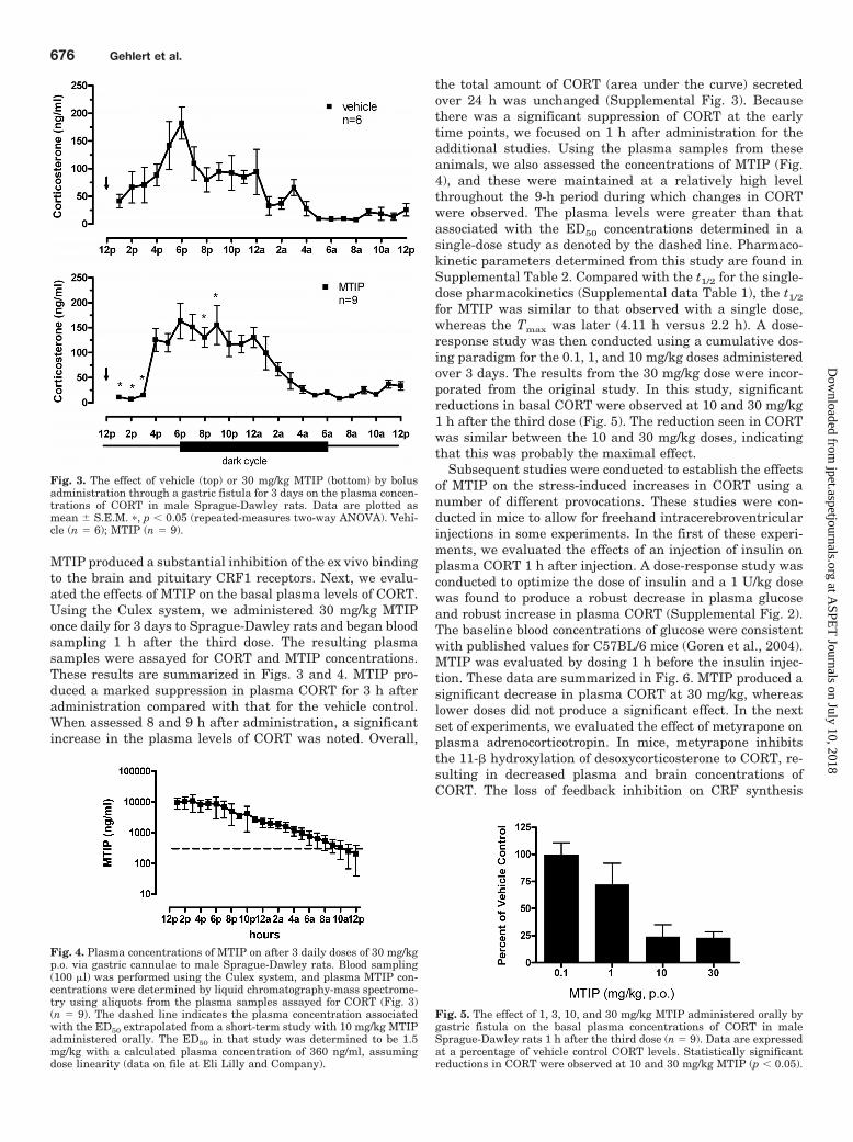
MTIP produced a substantial inhibition of the ex vivo bindingto the brain and pituitary CRF1 receptors. Next, we evalu-ated the effects of MTIP on the basal plasma levels of CORT.Using the Culex system, we administered 30 mg/kg MTIPonce daily for 3 days to Sprague-Dawley rats and began bloodsampling 1 h after the third dose. The resulting plasmasamples were assayed for CORT and MTIP concentrations.These results are summarized in Figs. 3 and 4. MTIP pro-duced a marked suppression in plasma CORT for 3 h afteradministration compared with that for the vehicle control.When assessed 8 and 9 h after administration, a significantincrease in the plasma levels of CORT was noted. Overall,
the total amount of CORT (area under the curve) secretedover 24 h was unchanged (Supplemental Fig. 3). Becausethere was a significant suppression of CORT at the earlytime points, we focused on 1 h after administration for theadditional studies. Using the plasma samples from theseanimals, we also assessed the concentrations of MTIP (Fig.4), and these were maintained at a relatively high levelthroughout the 9-h period during which changes in CORTwere observed. The plasma levels were greater than thatassociated with the ED50 concentrations determined in asingle-dose study as denoted by the dashed line. Pharmaco-kinetic parameters determined from this study are found inSupplemental Table 2. Compared with the t1/2 for the single-dose pharmacokinetics (Supplemental data Table 1), the t1/2
for MTIP was similar to that observed with a single dose,whereas the Tmax was later (4.11 h versus 2.2 h). A dose-response study was then conducted using a cumulative dos-ing paradigm for the 0.1, 1, and 10 mg/kg doses administeredover 3 days. The results from the 30 mg/kg dose were incor-porated from the original study. In this study, significantreductions in basal CORT were observed at 10 and 30 mg/kg1 h after the third dose (Fig. 5). The reduction seen in CORTwas similar between the 10 and 30 mg/kg doses, indicatingthat this was probably the maximal effect.
Subsequent studies were conducted to establish the effectsof MTIP on the stress-induced increases in CORT using anumber of different provocations. These studies were con-ducted in mice to allow for freehand intracerebroventricularinjections in some experiments. In the first of these experi-ments, we evaluated the effects of an injection of insulin onplasma CORT 1 h after injection. A dose-response study wasconducted to optimize the dose of insulin and a 1 U/kg dosewas found to produce a robust decrease in plasma glucoseand robust increase in plasma CORT (Supplemental Fig. 2).The baseline blood concentrations of glucose were consistentwith published values for C57BL/6 mice (Goren et al., 2004).MTIP was evaluated by dosing 1 h before the insulin injec-tion. These data are summarized in Fig. 6. MTIP produced asignificant decrease in plasma CORT at 30 mg/kg, whereaslower doses did not produce a significant effect. In the nextset of experiments, we evaluated the effect of metyrapone onplasma adrenocorticotropin. In mice, metyrapone inhibitsthe 11-� hydroxylation of desoxycorticosterone to CORT, re-sulting in decreased plasma and brain concentrations ofCORT. The loss of feedback inhibition on CRF synthesis
Fig. 3. The effect of vehicle (top) or 30 mg/kg MTIP (bottom) by bolusadministration through a gastric fistula for 3 days on the plasma concen-trations of CORT in male Sprague-Dawley rats. Data are plotted asmean � S.E.M. �, p � 0.05 (repeated-measures two-way ANOVA). Vehi-cle (n � 6); MTIP (n � 9).
Fig. 4. Plasma concentrations of MTIP on after 3 daily doses of 30 mg/kgp.o. via gastric cannulae to male Sprague-Dawley rats. Blood sampling(100 �l) was performed using the Culex system, and plasma MTIP con-centrations were determined by liquid chromatography-mass spectrome-try using aliquots from the plasma samples assayed for CORT (Fig. 3)(n � 9). The dashed line indicates the plasma concentration associatedwith the ED50 extrapolated from a short-term study with 10 mg/kg MTIPadministered orally. The ED50 in that study was determined to be 1.5mg/kg with a calculated plasma concentration of 360 ng/ml, assumingdose linearity (data on file at Eli Lilly and Company).
Fig. 5. The effect of 1, 3, 10, and 30 mg/kg MTIP administered orally bygastric fistula on the basal plasma concentrations of CORT in maleSprague-Dawley rats 1 h after the third dose (n � 9). Data are expressedat a percentage of vehicle control CORT levels. Statistically significantreductions in CORT were observed at 10 and 30 mg/kg MTIP (p � 0.05).
676 Gehlert et al.
at ASPE
T Journals on July 10, 2018
jpet.aspetjournals.orgD
ownloaded from
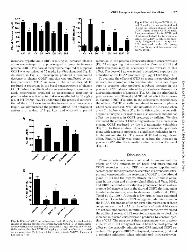
increases hypothalamic CRF, resulting in increased plasmaadrenocorticotropin in a physiological attempt to increaseplasma CORT. The dose of metyrapone required to suppressCORT was optimized at 75 mg/kg i.p. (Supplemental Fig. 1).As shown in Fig. 7B, metyrapone produced a pronounceddecrease in plasma CORT, and this was unaffected by pre-treatment with MTIP. As seen in the rat studies, MTIPproduced a reduction in the basal concentrations of plasmaCORT. When the effects of adrenocorticotropin were evalu-ated, metyrapone produced an approximate doubling ofplasma adrenocorticotropin that was unaffected by 30 mg/kgp.o. of MTIP (Fig. 7A). To understand the potential contribu-tion of the CRF2 receptor to this increase in adrenocortico-tropin, we administered the peptidic CRF1/CRF2 antagonistastressin at a dose of 1 �g i.c.v. and observed a partial
reduction in the plasma adrenocorticotropin concentrations(Fig. 7A) suggesting that a combination of central CRF1 andCRF2 receptors may be necessary to see reversal of thiseffect. The dose of 1 �g was found to completely suppress theactivation of the HPAA produced by 3 �g of CRF (Fig. 1).
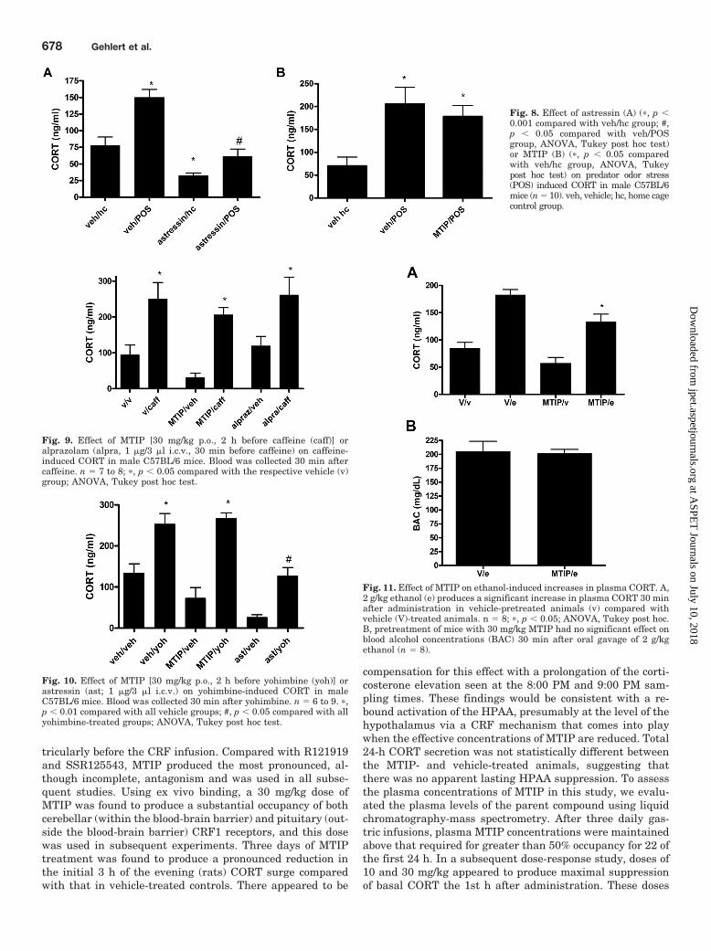
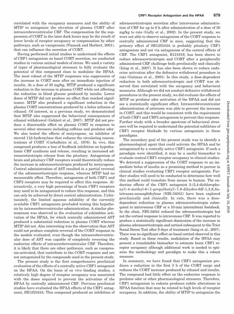
To evaluate the effects of MTIP on a putative psychologicalstressor, we exposed mice to predator odor (rat urine). Expo-sure to predator odor produced a robust increase in theplasma CORT that was reduced by prior intracerebroventric-ular administration of astressin (Fig. 8A). On the other hand,pretreatment with 30 mg/kg MTIP did not affect the increasein plasma CORT (Fig. 8B). In the subsequent experiments,the effects of MTIP on caffeine-induced increases in plasmaCORT were assessed. MTIP did not affect the increase whengiven 2 h before caffeine (Fig. 9). In this study, the benzodi-azepine anxiolytic alprazolam was also assessed and did notaffect the increases in CORT produced by caffeine. We alsoevaluated the effects of CRF antagonists on the increases inplasma CORT produced by the �-2 antagonist yohimbine(Fig. 10). In these studies, intracerebroventricular pretreat-ment with astressin produced a significant reduction in yo-himbine-stimulated CORT whereas MTIP had no significanteffect. Finally, MTIP was found to reduce the increase inplasma CORT after the immediate administration of ethanol(Fig. 11).
DiscussionThese experiments were conducted to understand the
effects of CRF1 antagonism on basal and stress-inducedCORT secretion in vivo. CRF is the major hypothalamicsecretagogue that regulates the secretion of adrenocorticotro-pin and consequently, the secretion of CORT in the adrenalgland. CRF1 has the highest affinity for CRF and is abun-dant in the brain and pituitary gland (Bale and Vale, 2004),and CRF1-deficient mice exhibit a pronounced basal cortico-sterone deficiency, a loss in the diurnal CORT rhythm, and ablunted endocrine response to stressors (Smith et al., 1998;Timpl et al., 1998). Although a few studies have examinedthe effect of short-term CRF1 antagonist administration onthe HPAA, the impact of longer term administration of thesecompounds on the HPAA or the effects of central blockadeare not well understood. In initial experiments, we evaluatedthe ability of several CRF1 receptor antagonists to block theincrease in plasma corticosterone produced by central injec-tion of CRF. The purpose of these experiments was to deter-mine which CRF1 antagonist would have the most profoundeffect on the centrally administered CRF-induced CORT se-cretion. The peptide CRF1/2 antagonist, astressin, produceda complete inhibition when administered intracerebroven-
Fig. 6. Effect of 3 days of MTIP (3, 10,and 30 mg/kg p.o.) on insulin-induced(1 U/kg i.p.) plasma glucose (left) andCORT (right) in male C57BL/6 mice.Insulin was dosed 1 h after MTIP, andblood was collected 1 h after insulin. v,vehicle for MTIP; V, vehicle for insu-lin; I, 1 U/kg insulin. n � 7 to 8; �, p �0.05 compared with v/V group;ANOVA (Tukey post hoc test) or ver-sus v/I group.
Fig. 7. Effect of MTIP on metyrapone (met; 75 mg/kg i.p.)-induced in-creases in plasma adrenocorticotropin (A) and CORT (B) in mice. Intrac-erebroventricularly administered astressin (1 �g/3 �l) was able to par-tially reduce this, but MTIP (30 mg/kg p.o.) had no effect. �, p � 0.05versus vehicle (veh)/veh; #, p � 0.05 versus veh/met. ANOVA, Tukey posthoc test (n � 8).
CRF1 Receptor Antagonism and the HPAA 677
at ASPE
T Journals on July 10, 2018
jpet.aspetjournals.orgD
ownloaded from
tricularly before the CRF infusion. Compared with R121919and SSR125543, MTIP produced the most pronounced, al-though incomplete, antagonism and was used in all subse-quent studies. Using ex vivo binding, a 30 mg/kg dose ofMTIP was found to produce a substantial occupancy of bothcerebellar (within the blood-brain barrier) and pituitary (out-side the blood-brain barrier) CRF1 receptors, and this dosewas used in subsequent experiments. Three days of MTIPtreatment was found to produce a pronounced reduction inthe initial 3 h of the evening (rats) CORT surge comparedwith that in vehicle-treated controls. There appeared to be
compensation for this effect with a prolongation of the corti-costerone elevation seen at the 8:00 PM and 9:00 PM sam-pling times. These findings would be consistent with a re-bound activation of the HPAA, presumably at the level of thehypothalamus via a CRF mechanism that comes into playwhen the effective concentrations of MTIP are reduced. Total24-h CORT secretion was not statistically different betweenthe MTIP- and vehicle-treated animals, suggesting thatthere was no apparent lasting HPAA suppression. To assessthe plasma concentrations of MTIP in this study, we evalu-ated the plasma levels of the parent compound using liquidchromatography-mass spectrometry. After three daily gas-tric infusions, plasma MTIP concentrations were maintainedabove that required for greater than 50% occupancy for 22 ofthe first 24 h. In a subsequent dose-response study, doses of10 and 30 mg/kg appeared to produce maximal suppressionof basal CORT the 1st h after administration. These doses
Fig. 8. Effect of astressin (A) (�, p �0.001 compared with veh/hc group; #,p � 0.05 compared with veh/POSgroup, ANOVA, Tukey post hoc test)or MTIP (B) (�, p � 0.05 comparedwith veh/hc group, ANOVA, Tukeypost hoc test) on predator odor stress(POS) induced CORT in male C57BL/6mice (n � 10). veh, vehicle; hc, home cagecontrol group.
Fig. 9. Effect of MTIP [30 mg/kg p.o., 2 h before caffeine (caff)] oralprazolam (alpra, 1 �g/3 �l i.c.v., 30 min before caffeine) on caffeine-induced CORT in male C57BL/6 mice. Blood was collected 30 min aftercaffeine. n � 7 to 8; �, p � 0.05 compared with the respective vehicle (v)group; ANOVA, Tukey post hoc test.
Fig. 10. Effect of MTIP [30 mg/kg p.o., 2 h before yohimbine (yoh)] orastressin (ast; 1 �g/3 �l i.c.v.) on yohimbine-induced CORT in maleC57BL/6 mice. Blood was collected 30 min after yohimbine. n � 6 to 9. �,p � 0.01 compared with all vehicle groups; #, p � 0.05 compared with allyohimbine-treated groups; ANOVA, Tukey post hoc test.
Fig. 11. Effect of MTIP on ethanol-induced increases in plasma CORT. A,2 g/kg ethanol (e) produces a significant increase in plasma CORT 30 minafter administration in vehicle-pretreated animals (v) compared withvehicle (V)-treated animals. n � 8; �, p � 0.05; ANOVA, Tukey post hoc.B, pretreatment of mice with 30 mg/kg MTIP had no significant effect onblood alcohol concentrations (BAC) 30 min after oral gavage of 2 g/kgethanol (n � 8).
678 Gehlert et al.
at ASPE
T Journals on July 10, 2018
jpet.aspetjournals.orgD
ownloaded from
correlated with the occupancy measures and the ability ofMTIP to antagonize the elevation of plasma CORT afterintracerebroventricular CRF. The compensation for the sup-pression of CORT in the later dark hours may be the result oflower levels of receptor occupancy or compensation by otherpathways, such as vasopressin (Pinnock and Herbert, 2001),that can influence the secretion of CORT.
Having performed initial studies to understand the effectsof CRF1 antagonism on basal CORT secretion, we conductedstudies in various animal models of stress. We used a varietyof types of pharmacological stressors to fully evaluate thepotential of this compound class to modulate the HPAA.The most robust of the MTIP responses was suppression ofthe increase in CORT seen after an immediate injection ofinsulin. At a dose of 30 mg/kg, MTIP produced a significantreduction in the increase in plasma CORT while not affectingthe reduction in blood glucose produced by insulin. Lowerdoses of MTIP did not produce an effect that reached signif-icance. MTIP also produced a significant reduction in theplasma CORT concentrations produced by a bolus infusion ofethanol. Of interest, in a previous study, we demonstratedthat MTIP also suppressed the behavioral consequences ofethanol withdrawal (Gehlert et al., 2007). MTIP did not pro-duce a discernable effect on plasma CORT in response toseveral other stressors including caffeine and predator odor.We also tested the effects of metyrapone, an inhibitor ofsteroid 11�-hydroxylase that reduces the circulating concen-trations of CORT (Carballeira et al., 1976). In vivo, thiscompound produces a loss of feedback inhibition on hypotha-lamic CRF synthesis and release, resulting in increased ad-renocorticotropin release from the pituitary. Antagonism ofbrain and pituitary CRF receptors would theoretically reducethe increase in adrenocorticotropin produced by metyrapone.Central administration of AST resulted in a partial reversalof the adrenocorticotropin response, whereas MTIP had nomeasurable effect. Therefore, antagonism of both CRF1 andCRF2 receptors may be required to affect this response. Al-ternatively, a very high percentage of brain CRF1 receptorsmay need to be antagonized to reduce this response, and thiscan only be achieved by direct central administration. Unfor-tunately, the limited aqueous solubility of the currentlyavailable CRF1 antagonists precluded testing this hypothe-sis by intracerebroventricular administration. A similar phe-nomenon was observed in the evaluation of yohimbine acti-vation of the HPAA, for which centrally administered ASTproduced a substantial reduction in plasma CORT whereasMTIP did not. Also interesting was the observation that ASTcould not produce complete reversal of the CORT response inthe models evaluated, even though the intracerebroventric-ular dose of AST was capable of completely reversing theendocrine effects of intracerebroventricular CRF. Therefore,it is likely that there are other pathways, such as vasopres-sin-activated, that contribute to the CORT response and arenot antagonized by the compounds used in the present study.
The present study is the first comprehensive preclinicalevaluation of the effects of a centrally active CRF1 antagoniston the HPAA. On the basis of ex vivo binding studies, arelatively high degree of receptor occupancy was associatedwith the doses required to prevent the activation of theHPAA by centrally administered CRF. Previous preclinicalstudies have evaluated the HPAA effects of the CRF1 antag-onist, SR125543A. This compound produced an inhibition of
adrenocorticotropin secretion after intravenous administra-tion of CRF for up to 6 h after administration of a dose of 30mg/kg to rats (Gully et al., 2002). In the present study, wewere not able to observe antagonism of the CORT response tocentrally administered CRF in mice, suggesting that theprimary effect of SR125543A is probably pituitary CRF1antagonism and not via antagonism of the central effects ofCRF. The CRF1 antagonist, R121919, has been shown toreduce adrenocorticotropin and CORT after a peripherallyadministered CRF challenge both preclinically and clinically(Ising et al., 2007). It has also been shown to reduce endo-crine activation after the defensive withdrawal procedure inrats (Gutman et al., 2003). In this study, a dose-dependentreduction in both adrenocorticotropin and CORT was ob-served that correlated with the occupancy and behavioralmeasures. Although we did not conduct defensive withdrawalprocedures in the present study, we did evaluate the effects ofMTIP on predator odor activation of the HPAA and did notsee a statistically significant effect. Intracerebroventricularadministration of astressin was able to prevent the increasein CORT, and this would be consistent with the requirementof both CRF1 and CRF2 antagonism to prevent this response.Further study with a broader spectrum of behavioral stres-sors will be required to understand the potential subtleties ofCRF1 receptor blockade by various antagonists in theseparadigms.
The secondary goal of the present study was to identify apharmacological agent that could activate the HPAA and beantagonized by a centrally active CRF1 antagonist. If such aparadigm were identified, it may be a useful biomarker toevaluate central CRF1 receptor occupancy in clinical studies.We detected a suppression of the CORT response to an im-mediate bolus of insulin as a promising method to pursue inclinical studies evaluating CRF1 receptor antagonists. Fur-ther studies will need to be conducted to determine how wellthis method could translate from mouse to human. The en-docrine effects of the CRF1 antagonist 2-(2,4-dichlorophe-nyl)-4-methyl-6-(1-propylbutyl)-7,8-dihydro-6H-1,3,6,8a-tetraaza-acenaphthylene (NBI-34041) have been exploredpreclinically and clinically. In rats, there was a dose-dependent reduction in plasma adrenocorticotropin subse-quent to intravenous CRF or a 10-min intermittent footshock.In the clinic, NBI-34041 reduced the adrenocorticotropin butnot the cortisol response to intravenous CRF. It was reported toproduce a statistically significant diminution of the increase inplasma adrenocorticotropin and cortisol subsequent to the TrierSocial Stress Test after 9 days of treatment (Ising et al., 2007).There was no significant effect on basal cortisol observed in thisstudy. Based on these results, modulation of the HPAA maypresent a translatable biomarker to estimate brain CRF1 re-ceptor occupancy although additional work is needed to opti-mize the methodology and paradigm to make this a robustmeasure.
In summary, we have found that CRF1 antagonism pro-duces a reduction in the first 3 h of the CORT surge andreduces the CORT increase produced by ethanol and insulin.The compound had little effect on the endocrine response topredator odor or other pharmacological stressors. Therefore,CRF1 antagonism in rodents produces subtle alterations inHPAA function that may be related to high levels of receptoroccupancy. In addition, the ability of MTIP to antagonize the
CRF1 Receptor Antagonism and the HPAA 679
at ASPE
T Journals on July 10, 2018
jpet.aspetjournals.orgD
ownloaded from
CORT response to insulin may provide a biomarker strategyto assess CRF1 target engagement in future human studies.
Authorship Contributions
Participated in research design: Gehlert, Cramer, and Morin.Conducted experiments: Cramer and Morin.Performed data analysis: Gehlert, Cramer, and Morin.Wrote or contributed to the writing of the manuscript: Gehlert,
Cramer, and Morin.
ReferencesAnisman H, Hayley S, Kelly O, Borowski T, and Merali Z (2001) Psychogenic,
neurogenic, and systemic stressor effects on plasma corticosterone and behavior:mouse strain-dependent outcomes. Behav Neurosci 115:443–454.
Bale TL, Contarino A, Smith GW, Chan R, Gold LH, Sawchenko PE, Koob GF, ValeWW, and Lee KF (2000) Mice deficient for corticotropin-releasing hormone recep-tor-2 display anxiety-like behaviour and are hypersensitive to stress. Nat Genet24:410–414.
Bale TL, Picetti R, Contarino A, Koob GF, Vale WW, and Lee KF (2002) Micedeficient for both corticotropin-releasing factor receptor 1 (CRFR1) and CRFR2have an impaired stress response and display sexually dichotomous anxiety-likebehavior. J Neurosci 22:193–199.
Bale TL and Vale WW (2004) CRF and CRF receptors: role in stress responsivity andother behaviors. Annu Rev Pharmacol Toxicol 44:525–557.
Binneman B, Feltner D, Kolluri S, Shi Y, Qiu R, and Stiger T (2008) A 6-weekrandomized, placebo-controlled trial of CP-316,311 (a selective CRH1 antagonist)in the treatment of major depression. Am J Psychiatry 165:617–620.
Bohs C, Cregor M, Gunaratna G, and Kissinger C (2000) Culex automated bloodsampler. Part II. Managing freely-moving animals and monitoring their activity.Curr Sep 18:147–151.
Carballeira A, Fishman LM, and Jacobi JD (1976) Dual sites of inhibition bymetyrapone of human adrenal steroidogenesis: correlation of in vivo and in vitrostudies. J Clin Endocrinol Metab 42:687–695.
Coric V, Feldman HH, Oren DA, Shekhar A, Pultz J, Dockens RC, Wu X, Gentile KA,Huang SP, Emison E, et al. (2010) Multicenter, randomized, double-blind, activecomparator and placebo-controlled trial of a corticotropin-releasing factor recep-tor-1 antagonist in generalized anxiety disorder. Depress Anxiety 27:417–425.
Coste SC, Kesterson RA, Heldwein KA, Stevens SL, Heard AD, Hollis JH, MurraySE, Hill JK, Pantely GA, Hohimer AR, et al. (2000) Abnormal adaptations to stressand impaired cardiovascular function in mice lacking corticotropin-releasing hor-mone receptor-2. Nat Genet 24:403–409.
Gehlert DR, Cippitelli A, Thorsell A, Le AD, Hipskind PA, Hamdouchi C, Lu J,Hembre EJ, Cramer J, Song M, et al. (2007) 3-(4-Chloro-2-morpholin-4-yl-thiazol-5-yl)-8-(1-ethylpropyl)-2,6-dimethyl-imidazo[1,2-b]pyridazine: a novel brain-penetrant, orally available corticotropin-releasing factor receptor 1 antagonistwith efficacy in animal models of alcoholism. J Neurosci 27:2718–2726.
Gehlert DR, Shekhar A, Morin SM, Hipskind PA, Zink C, Gackenheimer SL, ShawJ, Fitz SD, and Sajdyk TJ (2005) Stress and central Urocortin increase anxiety-likebehavior in the social interaction test via the CRF1 receptor. Eur J Pharmacol509:145–153.
Goren HJ, Kulkarni RN, and Kahn CR (2004) Glucose homeostasis and tissuetranscript content of insulin signaling intermediates in four inbred strains of mice:C57BL/6, C57BLKS/6, DBA/2, and 129X1. Endocrinology 145:3307–3323.
Gully D, Geslin M, Serva L, Fontaine E, Roger P, Lair C, Darre V, Marcy C, RoubyPE, Simiand J, et al. (2002) 4-(2-Chloro-4-methoxy-5-methylphenyl)-N-[(1S)-2-cyclopropyl-1-(3-fluoro-4-methylphenyl)ethyl]5-methyl-N-(2-propynyl)-1,3-thiazol-2-amine hydrochloride (SSR125543A): a potent and selective corticotrophin-releasing factor1 receptor antagonist. I. Biochemical and pharmacologicalcharacterization. J Pharmacol Exp Ther 301:322–332.
Gutman DA, Owens MJ, Skelton KH, Thrivikraman KV, and Nemeroff CB (2003)The corticotropin-releasing factor1 receptor antagonist R121919 attenuates thebehavioral and endocrine responses to stress. J Pharmacol Exp Ther 304:874–880.
Hashimoto K, Nishiyama M, Tanaka Y, Noguchi T, Asaba K, Hossein PN, NishiokaT, and Makino S (2004) Urocortins and corticotropin releasing factor type 2receptors in the hypothalamus and the cardiovascular system. Peptides 25:1711–1721.
Heinrichs SC, De Souza EB, Schulteis G, Lapsansky JL, and Grigoriadis DE (2002)Brain penetrance, receptor occupancy and antistress in vivo efficacy of a smallmolecule corticotropin releasing factor type I receptor selective antagonist. Neu-ropsychopharmacology 27:194–202.
Held K, Kunzel H, Ising M, Schmid DA, Zobel A, Murck H, Holsboer F, and Steiger
A (2004) Treatment with the CRH1-receptor-antagonist R121919 improves sleep-EEG in patients with depression. J Psychiatr Res 38:129–136.
Holsboer F (2000) The corticosteroid receptor hypothesis of depression. Neuropsy-chopharmacology 23:477–501.
Institute of Laboratory Animal Resources (1996) Guide for the Care and Use ofLaboratory Animals, 7th ed, Institute of Laboratory Animal Resources, Commis-sion on Life Sciences, National Research Council, Washington DC.
Ising M, Zimmermann US, Kunzel HE, Uhr M, Foster AC, Learned-Coughlin SM,Holsboer F, and Grigoriadis DE (2007) High-affinity CRF1 receptor antagonistNBI-34041: preclinical and clinical data suggest safety and efficacy in attenuatingelevated stress response. Neuropsychopharmacology 32:1941–1949.
Jamieson PM, Li C, Kukura C, Vaughan J, and Vale W (2006) Urocortin 3 modulatesthe neuroendocrine stress response and is regulated in rat amygdala and hypo-thalamus by stress and glucocorticoids. Endocrinology 147:4578–4588.
Kehne J and De Lombaert S (2002) Non-peptidic CRF1 receptor antagonists for thetreatment of anxiety, depression and stress disorders. Curr Drug Targets CNSNeurol Disord 1:467–493.
Koob G and Kreek MJ (2007) Stress, dysregulation of drug reward pathways, and thetransition to drug dependence. Am J Psychiatry 164:1149–1159.
Marinelli PW, Funk D, Juzytsch W, Harding S, Rice KC, Shaham Y, and Le AD(2007) The CRF1 receptor antagonist antalarmin attenuates yohimbine-inducedincreases in operant alcohol self-administration and reinstatement of alcoholseeking in rats. Psychopharmacology (Berl) 195:345–355.
Muller MB, Zimmermann S, Sillaber I, Hagemeyer TP, Deussing JM, Timpl P,Kormann MS, Droste SK, Kuhn R, Reul JM, et al. (2003) Limbic corticotropin-releasing hormone receptor 1 mediates anxiety-related behavior and hormonaladaptation to stress. Nat Neurosci 6:1100–1107.
Muret L, Priou A, Oliver C, and Grino M (1992) Stimulation of adrenocorticotropinsecretion by insulin-induced hypoglycemia in the developing rat involves argininevasopressin but not corticotropin-releasing factor. Endocrinology 130:2725–2732.
Nielsen DM (2006) Corticotropin-releasing factor type-1 receptor antagonists: thenext class of antidepressants? Life Sci 78:909–919.
Peters S, Hampsch J, Cregor M, Starrett C, Gunaratna G, and Kissinger C (2000)Culex ABS. I. Introduction to automated blood sampling. Curr Sep 18:139–145.
Pinnock SB and Herbert J (2001) Corticosterone differentially modulates expressionof corticotropin releasing factor and arginine vasopressin mRNA in the hypotha-lamic paraventricular nucleus following either acute or repeated restraint stress.Eur J Neurosci 13:576–584.
Preil J, Muller MB, Gesing A, Reul JM, Sillaber I, van Gaalen MM, Landgrebe J,Holsboer F, Stenzel-Poore M, and Wurst W (2001) Regulation of the hypothalamic-pituitary-adrenocortical system in mice deficient for CRH receptors 1 and 2.Endocrinology 142:4946–4955.
Smith GW, Aubry JM, Dellu F, Contarino A, Bilezikjian LM, Gold LH, Chen R,Marchuk Y, Hauser C, Bentley CA, et al. (1998) Corticotropin releasing factorreceptor 1-deficient mice display decreased anxiety, impaired stress response, andaberrant neuroendocrine development. Neuron 20:1093–1102.
Sommer WH, Rimondini R, Hansson AC, Hipskind PA, Gehlert DR, Barr CS, andHeilig MA (2008) Upregulation of voluntary alcohol intake, behavioral sensitivityto stress, and amygdala crhr1 expression following a history of dependence. BiolPsychiatry 63:139–145.
Song C, Earley B, and Leonard BE (1995) Behavioral, neurochemical, and immuno-logical responses to CRF administration. Is CRF a mediator of stress? Ann NYAcad Sci 771:55–72.
Spindel E, Griffith L, and Wurtman RJ (1983) Neuroendocrine effects of caffeine. II.Effects on thyrotropin and corticosterone secretion. J Pharmacol Exp Ther 225:346–350.
Timpl P, Spanagel R, Sillaber I, Kresse A, Reul JM, Stalla GK, Blanquet V, StecklerT, Holsboer F, and Wurst W (1998) Impaired stress response and reduced anxietyin mice lacking a functional corticotropin-releasing hormone receptor 1. Nat Genet19:162–166.
Vale W, Spiess J, Rivier C, and Rivier J (1981) Characterization of a 41-residue ovinehypothalamic peptide that stimulates secretion of corticotropin and �-endorphin.Science 213:1394–1397.
Zgombick JM and Erwin VG (1988) Ethanol differentially enhances adrenocorticalresponse in LS and SS mice. Alcohol 5:287–294.
Zobel AW, Nickel T, Kunzel HE, Ackl N, Sonntag A, Ising M, and Holsboer F (2000)Effects of the high-affinity corticotropin-releasing hormone receptor 1 antagonistR121919 in major depression: the first 20 patients treated. J Psychiatr Res 34:171–181.
Address correspondence to: Dr. Donald R. Gehlert, Mail Code 0510, LillyResearch Laboratories, Eli Lilly and Company, Lilly Corporate Center, Indi-anapolis, IN 46285. E-mail: [email protected]
680 Gehlert et al.
at ASPE
T Journals on July 10, 2018
jpet.aspetjournals.orgD
ownloaded from
![Cerebrospinal fluid levels of corticotropin-releasing ... · mouse counterpart of CRH, corticotropin-releasing factor (CRF) [40]. Aim Stress is a hot topic in the health sciences,](https://static.fdocuments.net/doc/165x107/5fd130781f4c7a71172810b9/cerebrospinal-fluid-levels-of-corticotropin-releasing-mouse-counterpart-of-crh.jpg)