Effect of voluntary exercise on BDNF/TrkB - DiVA portal582042/FULLTEXT01.pdf · Effect of voluntary...
26
Department of Physics, Chemistry and Biology Master Thesis Effect of voluntary exercise on BDNF/TrkB gene expression and alcohol intake Josefine Jonsson LiTH-IFM- Ex--12/2709--SE Supervisor: Annika Thorsell, IKE Linköping University Examiner: Johan Edqvist, IFM Linköping University Department of Physics, Chemistry and Biology Linköpings universitet SE-581 83 Linköping, Sweden
Transcript of Effect of voluntary exercise on BDNF/TrkB - DiVA portal582042/FULLTEXT01.pdf · Effect of voluntary...
Department of Physics, Chemistry and Biology
Master Thesis
Effect of voluntary exercise on BDNF/TrkB
gene expression and alcohol intake
Josefine Jonsson
LiTH-IFM- Ex--12/2709--SE
Supervisor: Annika Thorsell, IKE Linköping University
Examiner: Johan Edqvist, IFM Linköping University
Department of Physics, Chemistry and Biology
Linköpings universitet
SE-581 83 Linköping, Sweden
Rapporttyp Report category
Examensarbete
D-uppsats
Master Thesis
Språk/Language
Engelska/English
Titel/Title:
Effect of voluntary exercise on BDNF/TrkB gene expression and alcohol intake
Författare/Author:
Josefine Jonsson
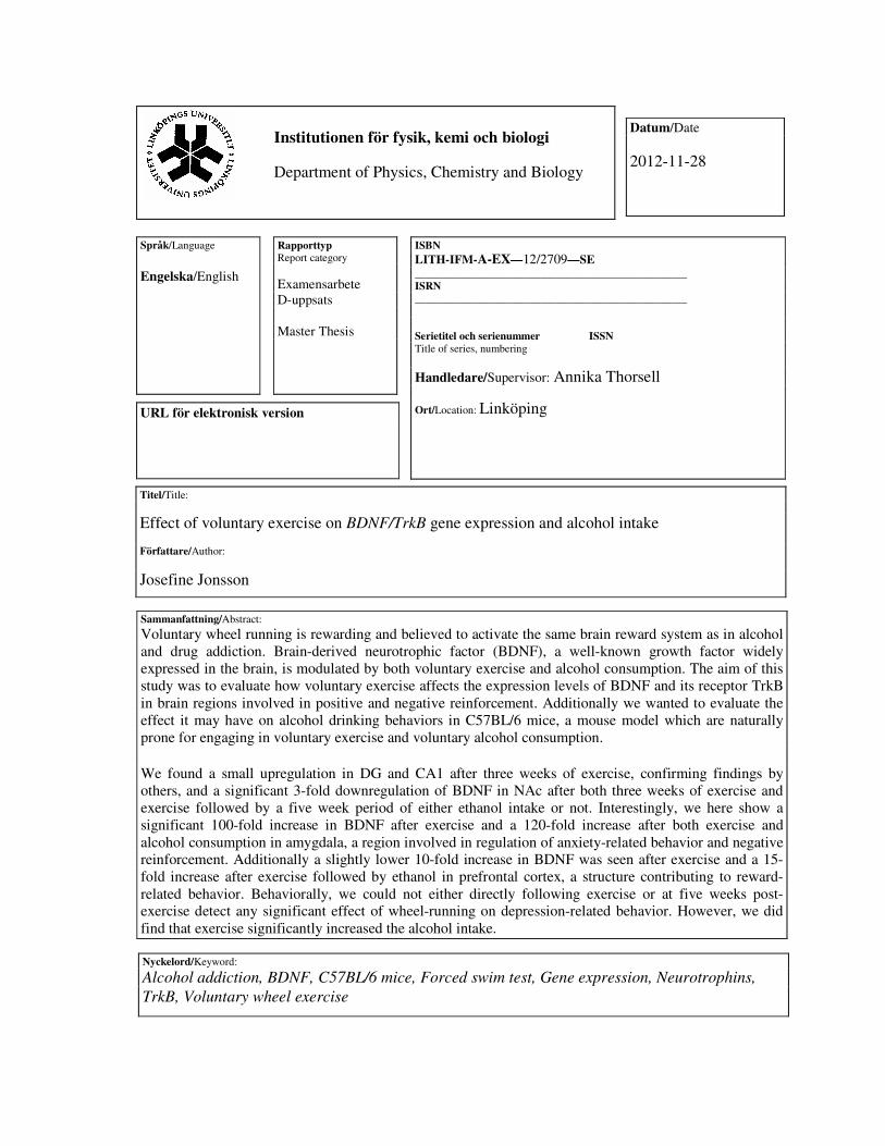
Sammanfattning/Abstract:
Voluntary wheel running is rewarding and believed to activate the same brain reward system as in alcohol
and drug addiction. Brain-derived neurotrophic factor (BDNF), a well-known growth factor widely
expressed in the brain, is modulated by both voluntary exercise and alcohol consumption. The aim of this
study was to evaluate how voluntary exercise affects the expression levels of BDNF and its receptor TrkB
in brain regions involved in positive and negative reinforcement. Additionally we wanted to evaluate the
effect it may have on alcohol drinking behaviors in C57BL/6 mice, a mouse model which are naturally
prone for engaging in voluntary exercise and voluntary alcohol consumption.
We found a small upregulation in DG and CA1 after three weeks of exercise, confirming findings by
others, and a significant 3-fold downregulation of BDNF in NAc after both three weeks of exercise and
exercise followed by a five week period of either ethanol intake or not. Interestingly, we here show a
significant 100-fold increase in BDNF after exercise and a 120-fold increase after both exercise and
alcohol consumption in amygdala, a region involved in regulation of anxiety-related behavior and negative
reinforcement. Additionally a slightly lower 10-fold increase in BDNF was seen after exercise and a 15-
fold increase after exercise followed by ethanol in prefrontal cortex, a structure contributing to reward-
related behavior. Behaviorally, we could not either directly following exercise or at five weeks post-
exercise detect any significant effect of wheel-running on depression-related behavior. However, we did
find that exercise significantly increased the alcohol intake.
ISBN
LITH-IFM-A-EX—12/2709—SE
__________________________________________________ ISRN
__________________________________________________ Serietitel och serienummer ISSN Title of series, numbering
Handledare/Supervisor: Annika Thorsell
Ort/Location: Linköping
Nyckelord/Keyword:
Alcohol addiction, BDNF, C57BL/6 mice, Forced swim test, Gene expression, Neurotrophins,
TrkB, Voluntary wheel exercise
Datum/Date
2012-11-28
URL för elektronisk version
Institutionen för fysik, kemi och biologi
Department of Physics, Chemistry and Biology
Contents
1 Abstract.............................................................................................2
2 List of abbreviations..........................................................................2
3 Introduction.......................................................................................3
4 Material & methods...........................................................................7
4.1 Animals ......................................................................................7
4.2 Experimental lay-out ..................................................................7
4.3 Behavioral testing.......................................................................8
4.3.1 Voluntary exercise......................................................................8
4.3.2 Alcohol intake ............................................................................8
4.3.3 Forced swim test.........................................................................9
4.4 Brain tissue collection and dissection .........................................9
4.5 RNA isolation from homogenized tissue ..................................11
4.6 Reverse Transcriptase PCR ......................................................11
4.7 Quantitative Real-Time PCR ....................................................11
4.8 Data analyses............................................................................12
5 Results ............................................................................................13
5.1 Impact of voluntary exercise on BDNF and TrkB gene
expression.................................................................................13
5.2 Impact of voluntary exercise on depression-like behavior ........15
5.3 Impact of voluntary exercise and ethanol consumption on BDNF
and TrkB gene expression ........................................................16
5.4 Impact of voluntary exercise on ethanol consumption ..............18
5.5 Impact of voluntary exercise and ethanol consumption on
depression-like behavior...........................................................20
6 Discussion.......................................................................................21
6.1 Conclusions ..............................................................................23
7 Acknowledgement ..........................................................................23
8 References.......................................................................................24
2
1 Abstract
Voluntary wheel running is rewarding and believed to activate the same brain reward
system as in alcohol and drug addiction. Brain-derived neurotrophic factor (BDNF), a
well-known growth factor widely expressed in the brain, is modulated by both
voluntary exercise and alcohol consumption. The aim of this study was to evaluate
how voluntary exercise affects the expression levels of BDNF and its receptor TrkB
in brain regions involved in positive and negative reinforcement. Additionally we
wanted to evaluate the effect it may have on alcohol drinking behaviors in C57BL/6
mice, a mouse model which are naturally prone for engaging in voluntary exercise
and voluntary alcohol consumption.
We found a small upregulation in DG and CA1 after three weeks of exercise,
confirming findings by others, and a significant 3-fold downregulation of BDNF in
NAc after both three weeks of exercise and exercise followed by a five week period
of either ethanol intake or not. Interestingly, we here show a significant 100-fold
increase in BDNF after exercise and a 120-fold increase after both exercise and
alcohol consumption in amygdala, a region involved in regulation of anxiety-related
behavior and negative reinforcement. Additionally a slightly lower 10-fold increase in
BDNF was seen after exercise and a 15-fold increase after exercise followed by
ethanol in prefrontal cortex, a structure contributing to reward-related behavior.
Behaviorally, we could not either directly following exercise or at five weeks post-
exercise detect any significant effect of wheel-running on depression-related
behavior. However, we did find that exercise significantly increased the alcohol
intake.
Keywords: Alcohol addiction, BDNF, C57BL/6 mice, Forced swim test, Gene
expression, Neurotrophins, TrkB, Voluntary wheel exercise
2 List of abbreviations
AA- Amygdala AD – Alcohol dependence
BDNF – Brain-derived neurotrophic factor CA1 – Cornu ammonis 1
Ct – Cycle-threshold DG – Dentate Gyrus
EtOH – Alcohol / Ethanol FST – Forced swim test
NAc – Nucleus Accumbens NTC – No template control
PCR – Polymerase Chain Reaction PFC – Prefrontal cortex
RT – Reverse Transcriptase TrkB – Tyrosine kinase B receptor
Trk – Tropomyosin-related kinase receptor qRT – Quantitative Real-Time
7,8-DHF – 7,8-dihydroxyflavone
3
3 Introduction Alcohol abuse and addiction Alcohol dependence (AD) is a big health problem around the world, because of the
difficulty to treat and the high rates of relapse (Davis 2008). According to the World
Health Organization, alcohol is the second largest risk factor for disease in Europe,
and results in 2.5 million deaths each year worldwide. Alcohol abuse is the cause of
many social issues like violence, traffic accidents, child neglect and abuse (World
Health Organization 2012). In addition, AD is often associated with underlying
psychiatric disorders like anxiety, psychosis and mood disorders like depression
(Bosse and Mathews 2011; Zanardini et al. 2011). The development of alcoholism
depends on multiple factors, where the susceptibility to addiction depends on both
genetic and environmental factors (Wong et al. 2011; Zanardini et al. 2011). Within
the central nervous system, numerous neurotransmitters and other factors contribute
to the addicted phenotype. Among those are the brain-derived neurotrophic factor
(BDNF) whose expression can be modulated by alcohol and therefore appears to
regulate reward as well as alcohol consumption behaviors (Bosse and Mathews 2011;
Costa et al. 2011).
An addiction only occurs as a consequence of repeated exposure to an addictive
substance, such as alcohol (Wong et al. 2011). Alcohol addiction is characterized by a
three-step behavioral cycle that includes the anticipation of alcohol intake,
intoxication or the drunken state and withdrawal with the negative effects of
abstinence. In the beginning of an addiction, drug use is associated with positive
reinforcement, where the pleasant effect of the drug leads to a drug-seeking behavior.
After prolonged use, addiction becomes associated with negative reinforcement
because the drug use becomes compulsive and its main effect is to suppress the
emotional states caused by intoxication (Davis 2008). Chronic alcohol consumption
causes neurobiological changes in neurotransmitter and neural signaling systems in
the brain, including GABA, opiates, serotonin and dopamine (Davis 2008; Zanardini
et al. 2011) and can also cause neurodegeneration (Davis 2008).
BDNF - background
Brain-derived neurotrophic factor (BDNF), is a secretory protein and a member of the
neurotrophin growth factor family (Davis 2008; Zanardini et al. 2011). The
neurotrophin family consists of structurally related growth factors and besides BDNF
also include nerve growth factor (NGF), Neurotrophin (NT)-3 and NT-4/5.
Neurotrophins are important to neuronal survival because they influence neuronal
growth and differentiation during the development in both the central nervous system
(CNS) and peripheral nervous system (PNS) (Davis 2008; Murray and Holmes 2011).
BDNF and NT-4 are widely expressed throughout the brain, but most abundant in the
hippocampus, cortex and cerebellum (Davis 2008; Bosse and Mathews 2011;
Zanardini et al. 2011), whereas NGT is particularly important for the survival of
sympathetic and cholinergic neurons in the CNS (Davis 2008). Production and
secretion of BDNF are generally accomplished by neurons (astrocytes) and therefore
regulated by neuronal activity (Bosse and Mathews 2011), but BDNF can also be
produced by lymphocytes, platelets and vascular endothelium (Davis 2008).
4
Neurotrophins signal through and bind with high affinity to tropomyosin-related
kinase (Trk) receptors. Although the Trks are not entirely selective the different
neurotrophins prefer specific types of Trk receptors. NGF has a high affinity for
TrkA, whereas NT-4 and BDNF bind TrkB, and NT-3 prefers TrkC. Neurotrophins
can also bind with low affinity to the tumor necrosis factor receptor p75, inducing cell
death and plasticity (Davis 2008; Murray and Holmes 2011). BDNF is produced as a
pro-peptide that is cleaved by several proteolytic pathways into its mature form. It has
been suggested that only the mature BDNF binds to the TrkB receptor, whereas the
preferred receptor for pro-BDNF is p75 (Davis 2008).
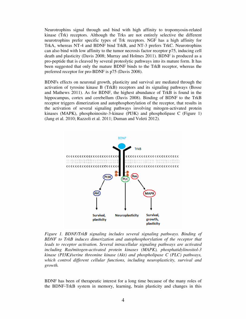
BDNFs effects on neuronal growth, plasticity and survival are mediated through the
activation of tyrosine kinase B (TrkB) receptors and its signaling pathways (Bosse
and Mathews 2011). As for BDNF, the highest abundance of TrkB is found in the
hippocampus, cortex and cerebellum (Davis 2008). Binding of BDNF to the TrkB
receptor triggers dimerization and autophosphorylation of the receptor, that results in
the activation of several signaling pathways involving mitogen-activated protein
kinases (MAPK), phosphoinosite-3-kinase (PI3K) and phospholipase C (Figure 1)
(Jang et al. 2010; Razzoli et al. 2011; Duman and Voleti 2012).
Figure 1. BDNF/TrkB signaling includes several signaling pathways. Binding of
BDNF to TrkB induces dimerization and autophosphorylation of the receptor that
leads to receptor activation. Several intracellular signaling pathways are activated
including Ras/mitogen-activated protein kinases (MAPK), phosphatidylinositol-3
kinase (PI3K)/serine threonine kinase (Akt) and phospholipase C (PLC) pathways,
which control different cellular functions, including neuroplasticity, survival and
growth.
BDNF has been of therapeutic interest for a long time because of the many roles of
the BDNF-TrkB system in memory, learning, brain plasticity and changes in this
5
system is associated with several disorders, like Alzheimer’s disease and Parkinson’s
disease, and also in the development of AD (Jang et al. 2010; Andero et al. 2011).
Since recombinant BDNF trials have not yielded any positive results because of some
limitations like short half-life and poor delivery, there has been several attempts to
find exogenous therapeutic agents that act as BDNF on the TrkB receptor. Recently a
series of flavone compounds were discovered to activate the TrkB receptor, and the
most potent compound being 7,8-dihydroxyflavone (7,8-DHF), an antioxidant found
in fruits and vegetables (Jang et al. 2010; Andero et al. 2011). 7,8-DHF is a specific
TrkB agonist that has the same function as BDNF to activate brain TrkB receptors.
7,8-DHF can activate the TrkB receptors and its signaling pathways in the absence of
BDNF (Andero et al. 2011).
BDNF in addiction BDNF has been shown to play a big role in the regulation of neurodevelopment, in
homeostasis of the adult brain through neurogenesis and plasticity, and addiction
(Davis 2008; Zanardini et al. 2011). There is also evidence to suggest that BDNF may
play a protective role in the regulation of the reinforcing effects of alcohol, in both
humans and animals (Bosse and Mathews 2011). Most studies show that the BDNF
level in hippocampus is decreased with long-term exposure to alcohol. This suggests
that the decrease of BDNF may be the consequence of neurodegeneration. Common
biochemical substrates, like dopamine, that are used in memory-function are also
associated with addiction. They cause long lasting alterations in the circuitry of the
brain and these changes lead to compulsive drug seeking (Davis 2008). BDNF itself is
important for long-term memory (Bekinschtein et al. 2008).
BDNF is an alcohol-responsive gene. In rodents both acute administration and
voluntary consumption of alcohol activates the BDNF signaling pathway and
increases BDNF mRNA levels. This shows that alcohol upregulates BDNF signaling
that subsequently inhibits alcohol drinking. The positive effects of alcohol
reinforcement are mediated mostly through the activation of dopaminergic pathways.
BDNF has been shown to have a modulatory effect on dopamine transmission, and
has the ability to alter the increased extracellular levels of dopamine that are induced
by alcohol. This might be how BDNF can regulate alcohol drinking behavior (Ghitza
et al. 2010; Bosse and Mathews 2011; Costa et al. 2011).
Susceptibility to develop alcohol dependence has been reported to be associated with
polymorphisms of both the BDNF and TrkB gene (Bosse and Mathews 2011;
Zanardini et al. 2011). Inhibition of TrkB receptors has been shown to increase the
alcohol consumption in mice (Bosse and Mathews 2011). One well-known BDNF
polymorphism Val66Met is clearly associated with alcohol use and also with a higher
risk of relapse to drug-taking after treatment for alcoholism (Wojnar et al. 2009;
Bosse and Mathews 2011; Costa et al. 2011; Zanardini et al. 2011). During
withdrawal, BDNF has been suggested to have a role in neuroadaptation, because of
the reports of increased BDNF levels after 24 hours of abstinence. Therefore it is
possible that the levels of BDNF may be important for long-term alcohol abstinence
(Costa et al. 2011).
Neurotrophins regulates the survival of the neurons in the brain, but if this regulated
support does not function properly it may be involved in the susceptibility to alcohol
dependence and in brain damage that often is the outcome from chronic alcohol intake
6
(Zanardini et al. 2011). Chronic alcohol exposure can impair the activation of
transcription factors that regulate the expression of the BDNF gene. This indicates
that the neuroprotective response to alcohol in the brain can be damaged by a
lowering of BDNF levels due to chronic alcohol consumption. The reduced synthesis
of BDNF therefore inhibits the protective mechanism and facilitates alcohol addiction
(Costa et al. 2011; Zanardini et al. 2011). Regulatory impairment of the BDNF
homeostasis may be an underlying factor of the progression and persistence of alcohol
addiction (Crews and Nixon 2009; Costa et al. 2011).
Exercise and reward Exercise has several health benefits, but also neural and cognitive effects in both
humans and in laboratory animals. The brain reward system may play a role in wheel-
running behavior, since voluntary exercise clearly seems to be rewarding. It is
possible that the neural pathways that regulate the rewarding effects of food, sex and
drugs of abuse also may be activated by voluntary exercise (Meeusen and De Meirleir
1995; Novak et al. 2012).
The reward system consists of several brain structures that are involved in mediating
reinforcing and pleasurable behaviors. The most important pathway of the reward
system, the mesocorticolimbic dopamine pathway, is formed in part by the ventral
tegmental area (VTA) and NAc, but also includes prefrontal cortex and amygdala.
Dopamine is released by dopamine-containing neurons in VTA onto neurons in the
NAc and act as a signal for reinforcement. Consumption of alcohol or drugs of abuse
leads to an increased release of dopamine in the VTA-NAc pathway, which leads to
positive reinforcement of the rewarding behavior. A repeated exposure to drugs
results in craving and addiction by producing plasticity and changing the level of
neuronal excitation in this system. Reward and withdrawal affect the level of
excitement by increasing the desire to consume more alcohol. The rewarding
properties of drugs (morphine or cocaine) can be blocked by a direct infusion of
BDNF into VTA, suggesting that BDNF may play a role in the VTA-NAc pathway in
promoting addiction, craving and withdrawal (Davis 2008; Belujon and Grace 2011).
It has been known for a long time that dopaminergic systems play a role in substance
abuse, like alcohol addiction. The same systems are considered to be influenced by
voluntary exercise, since an increase of the dopamine metabolism has been shown
after just thirty minutes of wheel running (Meeusen and De Meirleir 1995; Novak et
al. 2012).
Access to a running wheel also affects depression-like behaviors in rodents, by
working as an antidepressant. This is most often assessed by the forced swim test
(FST) where floating or immobility is seen as a measurement of depression (Cryan et
al. 2005; Duman et al. 2008). It has been reported that voluntary exercise decreases
the immobility, i.e. depression-like behavior, in rodents (Bjornebekk et al. 2005). An
effect in BDNF is also seen after wheel running, where exercise increases the
expression of BDNF mRNA and neurogenesis in hippocampus, the most studied brain
area that are rich in growth factors (Neeper et al. 1996; Adlard et al. 2004; Novak et
al. 2012). A less beneficial condition that also affects the levels of BDNF is stress.
Most studies have demonstrated that stress and depression can lead to loss of neurons
and cells in prefrontal cortex, amygdala and hippocampus, due to a decreased
expression of BDNF (Duman and Monteggia 2006). However it has been shown that
7
chronic stress increases the levels of BDNF in the brain region NAc that are involved
in the reward system (Krishnan et al. 2007).
Experiments The first aim of this study was to look at the reward system after voluntary exercise
and what kind of effects voluntary exercise (wheel running) has on both the BDNF
and TrkB levels in different brain areas, i.e. Prefrontal cortex (PFC), Nucleus
accumbens (NAc), Amygdala (AA), and two regions of the Hippocampus; dentate
gyrus (DG) and cornu ammonis CA1. A second aim was to see if and how this would
have an effect on depression- like behavior in mice. The third aim was to see if BDNF
can be involved in the development of alcohol dependence in C57BL/6 mice, a model
with the highest levels of voluntary ethanol drinking (Davis, 2008). Therefore, we
investigated if the use of voluntary wheel exercise before exposure to alcohol (EtOH)
in drinking water could have a protective effect against a development of alcohol
dependency by an up regulation of BDNF. Really to see if natural reward (e.g. food,
water and sex) from exercise, that induces neurogenesis, can protect the brain from
neurodegeneration that occurs due to AD. The last and fourth aim was to observe if
there were any changes in alcohol consumption after a period of alcohol deprivation
(withdrawal).
4 Material & methods
4.1 Animals
Male C57BL/6 mice were obtained from Nova-SCB AB (Sollentuna, Sweden) and
housed in a climate-controlled environment in the animal research facility at
Linköping University. Mice were obtained at approximately 6-7 weeks of age and
allowed a minimum of 1 week to acclimatize to the new environment. The mice were
housed individually with free access to food and water, and maintained at standard
conditions: 20 ± 2°C temperature, 33 ± 10% relative humidity and under a 12 h
light/dark cycle (lights on at 7:00 am). Animal maintenance and all experimental
procedures were approved by the Animal Care and Use Committee at Linköping
University and performed in accordance to the Swedish regulations for animal
experimentation.
4.2 Experimental lay-out
Experiment 1: Voluntary exercise and brain BDNF/TrkB expression The experimental group (n = 10) were housed in standard rat cages with wire lid
(Makrolon 365 x 205 x 145 mm, floor area 540 cm2) equipped with a running wheel
(13 cm in diameter) and were given continual wheel access for three weeks, whereas
the housing of the control group (n = 10) remained unchanged. The mice were
checked and handled every other day to make sure that the wheels were working
properly and to decrease stress. After three weeks of voluntary exercise, all mice were
assessed for depression in a behavioural test, e.g. forced swim test, FST (see below).
After the FST the mice were allowed back into their home cages for four days of
voluntary exercise to lower their stress, before all mice, one by one, were euthanized
by means of CO2 and cervical dislocation (Figure 2).
8
Experiment 2: Voluntary exercise, alcohol intake and brain BDNF/TrkB expression Two experimental groups were housed individually in rat home cages equipped with
running wheels, and allowed to run for three weeks. After three weeks of voluntary
exercise, one group (n = 12, Exercise + EtOH) were subjected to a two-bottle choice
paradigm of increasing concentrations of EtOH solutions and water (H2O) for five
weeks. The other group (n = 12, Exercise) were not subjected to any EtOH, only two
bottles with H2O. Two additional groups were housed individually in standard mouse
home cages with wire lid (Makrolon 265 x 205 x 140 mm, floor area 350 cm2)
without a running wheel. After three weeks, one group (n = 12, EtOH) were subjected
to a two-bottle choice paradigm of EtOH and H2O, as previously described. The other
group (n = 12, Control) acted as the control group, having no exposure to either a
running wheel or EtOH. All mice were assessed for depression in a FST behavioural
test, and allowed back into their home cage for a few days before being humanely
euthanized by CO2 followed by cervical dislocation (Figure 2). Each brain was
dissected out and quickly frozen in isopentane on dry ice, and stored in a -80°C
freezer.
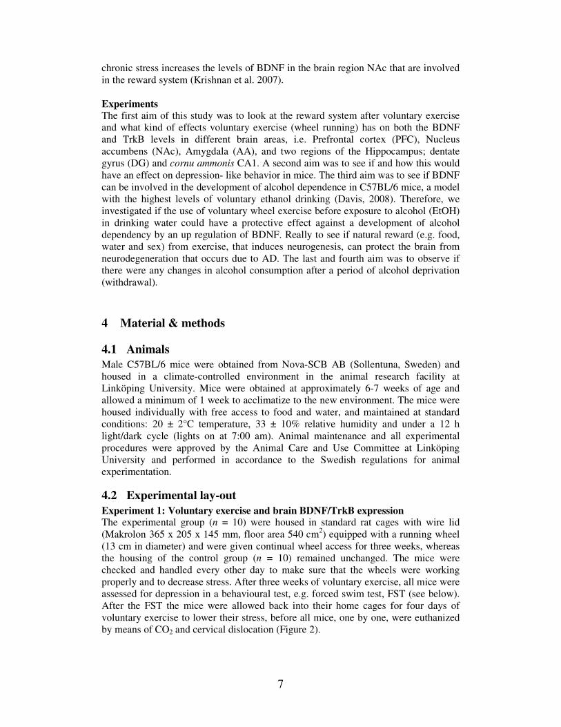
Figure 2. The difference in the experimental lay-out for experiment 1 and experiment
2. In experiment 1, the exercising group had access to a running wheel for 3 weeks
and then both groups were assessed in a FST. Animals were then euthanized directly
following exercise. In experiment 2, the exercised groups had access to a running
wheel for 3 weeks, then all wheels were removed and a five week long two-bottle
choice paradigm was started. All groups were then assessed in a FST, before they
were euthanized.
4.3 Behavioral testing
4.3.1 Voluntary exercise Animals were allowed 24 hours free access to a running wheel in their home cage for
three weeks. They were only deprived running for the short period of time it took to
remove excessive sawdust under the wheel that stopped it from turning.
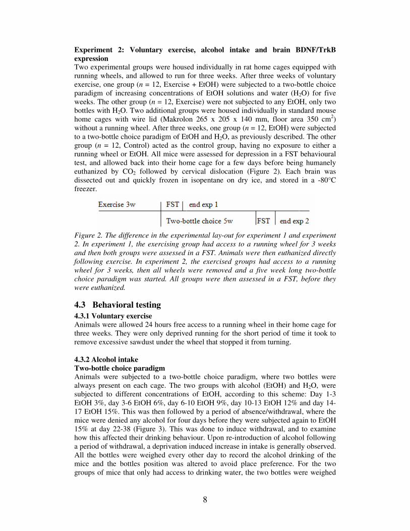
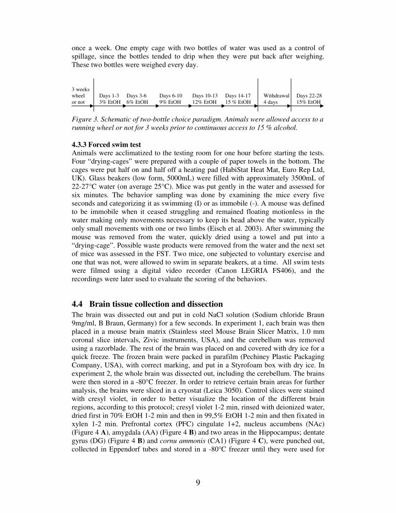
4.3.2 Alcohol intake Two-bottle choice paradigm Animals were subjected to a two-bottle choice paradigm, where two bottles were
always present on each cage. The two groups with alcohol (EtOH) and H2O, were
subjected to different concentrations of EtOH, according to this scheme: Day 1-3
EtOH 3%, day 3-6 EtOH 6%, day 6-10 EtOH 9%, day 10-13 EtOH 12% and day 14-
17 EtOH 15%. This was then followed by a period of absence/withdrawal, where the
mice were denied any alcohol for four days before they were subjected again to EtOH
15% at day 22-38 (Figure 3). This was done to induce withdrawal, and to examine
how this affected their drinking behaviour. Upon re-introduction of alcohol following
a period of withdrawal, a deprivation induced increase in intake is generally observed.
All the bottles were weighed every other day to record the alcohol drinking of the
mice and the bottles position was altered to avoid place preference. For the two
groups of mice that only had access to drinking water, the two bottles were weighed
9
once a week. One empty cage with two bottles of water was used as a control of
spillage, since the bottles tended to drip when they were put back after weighing.
These two bottles were weighed every day.
3 weeks
wheel Days 1-3 Days 3-6 Days 6-10 Days 10-13 Days 14-17 Withdrawal Days 22-28
or not 3% EtOH 6% EtOH 9% EtOH 12% EtOH 15 % EtOH 4 days 15% EtOH
Figure 3. Schematic of two-bottle choice paradigm. Animals were allowed access to a
running wheel or not for 3 weeks prior to continuous access to 15 % alcohol.
4.3.3 Forced swim test Animals were acclimatized to the testing room for one hour before starting the tests.
Four “drying-cages” were prepared with a couple of paper towels in the bottom. The
cages were put half on and half off a heating pad (HabiStat Heat Mat, Euro Rep Ltd,
UK). Glass beakers (low form, 5000mL) were filled with approximately 3500mL of
22-27°C water (on average 25°C). Mice was put gently in the water and assessed for
six minutes. The behavior sampling was done by examining the mice every five
seconds and categorizing it as swimming (I) or as immobile (-). A mouse was defined
to be immobile when it ceased struggling and remained floating motionless in the
water making only movements necessary to keep its head above the water, typically
only small movements with one or two limbs (Eisch et al. 2003). After swimming the
mouse was removed from the water, quickly dried using a towel and put into a
“drying-cage”. Possible waste products were removed from the water and the next set
of mice was assessed in the FST. Two mice, one subjected to voluntary exercise and
one that was not, were allowed to swim in separate beakers, at a time. All swim tests
were filmed using a digital video recorder (Canon LEGRIA FS406), and the
recordings were later used to evaluate the scoring of the behaviors.
4.4 Brain tissue collection and dissection
The brain was dissected out and put in cold NaCl solution (Sodium chloride Braun
9mg/ml, B Braun, Germany) for a few seconds. In experiment 1, each brain was then
placed in a mouse brain matrix (Stainless steel Mouse Brain Slicer Matrix, 1.0 mm
coronal slice intervals, Zivic instruments, USA), and the cerebellum was removed
using a razorblade. The rest of the brain was placed on and covered with dry ice for a
quick freeze. The frozen brain were packed in parafilm (Pechiney Plastic Packaging
Company, USA), with correct marking, and put in a Styrofoam box with dry ice. In
experiment 2, the whole brain was dissected out, including the cerebellum. The brains
were then stored in a -80°C freezer. In order to retrieve certain brain areas for further
analysis, the brains were sliced in a cryostat (Leica 3050). Control slices were stained
with cresyl violet, in order to better visualize the location of the different brain
regions, according to this protocol; cresyl violet 1-2 min, rinsed with deionized water,
dried first in 70% EtOH 1-2 min and then in 99,5% EtOH 1-2 min and then fixated in
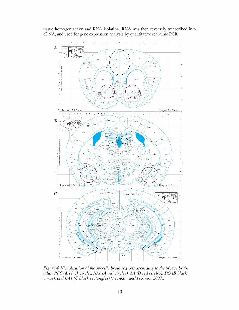
xylen 1-2 min. Prefrontal cortex (PFC) cingulate 1+2, nucleus accumbens (NAc)
(Figure 4 A), amygdala (AA) (Figure 4 B) and two areas in the Hippocampus; dentate
gyrus (DG) (Figure 4 B) and cornu ammonis (CA1) (Figure 4 C), were punched out,
collected in Eppendorf tubes and stored in a -80°C freezer until they were used for
10
tissue homogenization and RNA isolation. RNA was then reversely transcribed into
cDNA, and used for gene expression analysis by quantitative real-time PCR.
Figure 4. Visualization of the specific brain regions according to the Mouse brain
atlas. PFC (A black circle), NAc (A red circles), AA (B red circles), DG (B black
circle), and CA1 (C black rectangles) (Franklin and Paxinos, 2007).
A
B
C
11
4.5 RNA isolation from homogenized tissue Micropunched tissues of left side brain regions of PFC, NAc, AA and DG (as dorsal
sample) and CA1 (as ventral sample) were homogenized with a TissueLyser in order
to isolate and purify RNA by using the RNAeasy® Micro Kit (cat. no. 74004,
Qiagen®), following the manufacturers “Purification of total RNA from animal and
human tissue” protocol. The RNA concentration and quality were determined using a
Nanodrop ND-1000 Spectrophotometer, in order to calculate how much RNA to use
for the transcription of cDNA. The RNA was stored in a -80 freezer.
4.6 Reverse Transcriptase PCR
RNA was reversely transcribed into cDNA using the High Capacity cDNA Reverse
Transcription kit (200 reactions) from Applied Biosystems. Briefly, total RNA was
mixed with RNase-free water, 10X RT Buffer, 10X RT Random Primers, 25X dNTP
Mix (100 mM), and MultiScribe® Reverse Transcriptase. The samples were run, in
the Veriti® 96-well Thermal Cycler, a 9700 GeneAmp® PCR instrument from
Applied Biosystems, according to the BDNF setup; 25°C for 10 min, 37°C for 60
min, 85°C for 5 s. The cDNA was then diluted 1:10 by mixing 20 µL cDNA with
180 µL RNase free water, and stored in a -80 freezer.
4.7 Quantitative Real-Time PCR
Quantitative Real-time PCR was used to measure the levels of gene expression of
BDNF, TrkB and the housekeeping gene β-actin in the different brain areas; PFC,
NAc, AA, DG and CA1. All samples, including NTC as control, were run in
duplicates for each gene on Fast optical 96-well reaction plates. Real-time PCR was
performed on a 7900HT FAST Real-Time PCR system from Applied Biosystems
using TaqMan® Fast universal PCR Master Mix (2X) No AmpEraser® UNG
according to the manufacturer’s instructions. The gene expression assays used were;
BDNF: Mm 04230607_s1; TrkB (Ntrk2): Mm 00435422_m1 and β-actin: Mm
00607939_s1. Dilution curves for all probes used were performed to assure the
quality and specificity of the assay. Gene expression was calculated using the ∆∆Ct
method. The gene expression for each gene was normalized to the corresponding
expression of β-actin, and the 2^-∆∆Ct values for the experimental group(s) were
compared to the values of the control group and demonstrated as fold difference.
4.8 Data analyses
Statistical analysis was performed for each brain area using STATISTICA 10
analytical software. Gene expression data was analyzed with Student’s t-test in
Experiment 1 and one-way ANOVA in Experiment 2 (due to multiple groups).
Drinking data was analyzed using a repeated-measures two-way ANOVA. The level
of significance was set at p<0.05. All data are presented as mean ± SEM.
12
5 Results
5.1 Impact of voluntary exercise on BDNF and TrkB gene expression
We tested how voluntary wheel running activity would affect the mRNA levels of
BDNF and its receptor TrkB following three weeks of exercise. Our hypothesis was
that exercise, that most likely triggers the brain reward system and the dopaminergic
pathways, would upregulate the expression of BDNF, at least in the two hippocampal
areas. TrkB is most likely upregulated if BDNF is downregulated, in order for the
receptor to be able to use the limited supply of BDNF. Some studies have however
shown that TrkB also can be upregulated by exercise (Gomez-Pinilla et al. 2002;
Klintsova et al. 2004).
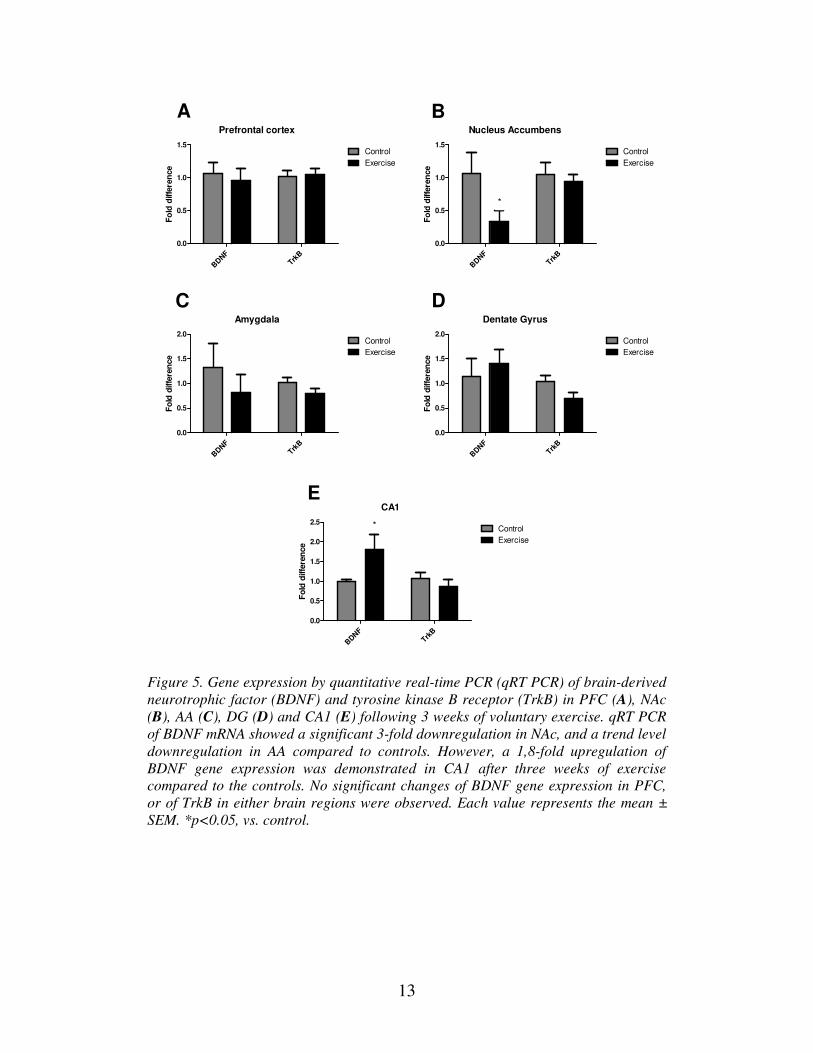
Our expression study showed a significant 3-fold decrease of the BDNF expression in
NAc compared to controls (Figure 5 B). There were no significant difference in the
expression of BDNF in the AA, PFC or DG (Figure 5 A, C and D). For TrkB, no
significant changes could be observed between running and controls in any examined
brain-region (Figure 5 A-E). A trend-level decrease could be seen in the DG, but this
failed to reach statistical significance.
The only upregulation of BDNF expression directly following exercise-exposure
could be seen in the CA1. Here, a 1,8-fold upregulation was demonstrated (Figure 5 E). Hippocampus is rich in growth factors and is therefore the most studied brain
region when it comes to BDNF. Many studies have shown that wheel running has an
effect on BDNF expression, where exercise increases the expression of BDNF mRNA
and neurogenesis in hippocampus, especially in DG, but also in CA1 (Neeper et al.
1996; Adlard et al. 2004; Novak et al. 2012).
These singled housed animals were euthanized directly after exposure to running
wheels, so the results represent direct measurements after voluntary exercise.
13
Prefrontal cortex
Fo
ld d
iffe
ren
ce
BDNF
TrkB
0.0
0.5
1.0
1.5Control
Exercise
Nucleus Accumbens
Fo
ld d
iffe
ren
ce
BDNF
TrkB
0.0
0.5
1.0
1.5Control
Exercise
*
Amygdala
Fo
ld d
iffe
ren
ce
BDNF
TrkB
0.0
0.5
1.0
1.5
2.0Control
Exercise
Dentate Gyrus
Fo
ld d
iffe
ren
ce
BDNF
TrkB
0.0
0.5
1.0
1.5
2.0Control
Exercise
CA1
Fo
ld d
iffe
ren
ce
BDNF
TrkB
0.0
0.5
1.0
1.5
2.0
2.5Control
Exercise
*
A B
C D
E
Figure 5. Gene expression by quantitative real-time PCR (qRT PCR) of brain-derived
neurotrophic factor (BDNF) and tyrosine kinase B receptor (TrkB) in PFC (A), NAc
(B), AA (C), DG (D) and CA1 (E) following 3 weeks of voluntary exercise. qRT PCR
of BDNF mRNA showed a significant 3-fold downregulation in NAc, and a trend level
downregulation in AA compared to controls. However, a 1,8-fold upregulation of
BDNF gene expression was demonstrated in CA1 after three weeks of exercise
compared to the controls. No significant changes of BDNF gene expression in PFC,
or of TrkB in either brain regions were observed. Each value represents the mean ±
SEM. *p<0.05, vs. control.
14
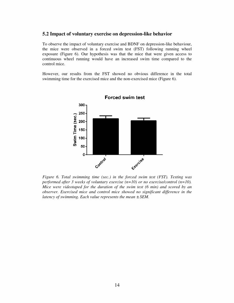
5.2 Impact of voluntary exercise on depression-like behavior
To observe the impact of voluntary exercise and BDNF on depression-like behaviour,
the mice were observed in a forced swim test (FST) following running wheel
exposure (Figure 6). Our hypothesis was that the mice that were given access to
continuous wheel running would have an increased swim time compared to the
control mice.
However, our results from the FST showed no obvious difference in the total
swimming time for the exercised mice and the non-exercised mice (Figure 6).
Figure 6. Total swimming time (sec.) in the forced swim test (FST). Testing was
performed after 3 weeks of voluntary exercise (n=10) or no exercise/control (n=10).
Mice were videotaped for the duration of the swim test (6 min) and scored by an
observer. Exercised mice and control mice showed no significant difference in the
latency of swimming. Each value represents the mean ± SEM.
15
5.3 Impact of voluntary exercise and ethanol consumption on BDNF and TrkB gene expression
In addition to analysing how voluntary wheel exercise would influence the expression
of BDNF and TrkB, we also tested what effect alcohol consumption with or without
preceding running wheel exposure would have on BDNF and TrkB expression in
PFC, NAc and AA. Our hypothesis was that alcohol alone may downregulate
expression of BDNF, as a result of neurodegeneration. As for the mice subjected to
exercise followed by alcohol, our hypothesis was that exercise either would have a
protective effect against any influences by alcohol, or that voluntary exercise and
subsequent alcohol intake would act together to further stimulate the reward system
and therefore upregulate BDNF further.
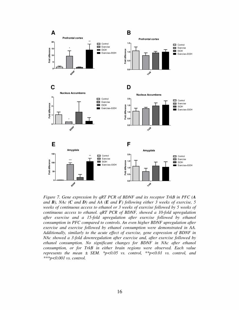
Our results showed a 3-fold downregulation of BDNF in NAc after exercise (Figure 7
C), which is consistent with our previous findings (Figure 5 B). However in PFC, a
10-fold upregulation of BDNF was seen in mice subjected to exercise, and a 15-fold
upregulation in the group with both exercise and alcohol consumption (Figure 7 A).
For AA an even higher upregulation of BDNF expression after exercise as well as
after combined exercise and alcohol consumption could be seen (Figure 7 E). Ethanol
itself did not significantly affect BDNF expression. No significant changes in the
expression levels of TrkB were observed for any of the three brain regions (Figure 7
B, D, and F).
These singled housed mice were euthanized after a five week period from the end of
the voluntary exercise.
16
B
C
Prefrontal cortex
Fo
ld d
iffe
ren
ce
BDNF
0
5
10
15
20Control
Exercise
EtOH
Exercise+EtOH
*
**
Prefrontal cortex
Fo
ld d
iffe
ren
ce
TrkB
0.0
0.5
1.0
1.5Control
Exercise
EtOH
Exercise+EtOH
Nucleus Accumbens
Fo
ld d
iffe
ren
ce
BDNF
0
1
2
3Control
Exercise
EtOH
Exercise+EtOH
*
Nucleus Accumbens
Fo
ld d
iffe
ren
ce
TrkB
0.0
0.5
1.0
1.5
2.0Control
Exercise
EtOH
Exercise+EtOH
Amygdala
Fo
ld d
iffe
ren
ce
BDNF
0
50
100
150Control
Exercise
EtOH
Exercise+EtOH
***
**
Amygdala
Fo
ld d
iffe
ren
ce
TrkB
0.0
0.5
1.0
1.5
2.0Control
Exercise
EtOH
Exercise+EtOH
A
D
E F
Figure 7. Gene expression by qRT PCR of BDNF and its receptor TrkB in PFC (A
and B), NAc (C and D) and AA (E and F) following either 3 weeks of exercise, 5
weeks of continuous access to ethanol or 3 weeks of exercise followed by 5 weeks of
continuous access to ethanol. qRT PCR of BDNF, showed a 10-fold upregulation
after exercise and a 15-fold upregulation after exercise followed by ethanol
consumption in PFC compared to controls. An even higher BDNF upregulation after
exercise and exercise followed by ethanol consumption were demonstrated in AA.
Additionally, similarly to the acute effect of exercise, gene expression of BDNF in
NAc showed a 3-fold downregulation after exercise and, after exercise followed by
ethanol consumption. No significant changes for BDNF in NAc after ethanol
consumption, or for TrkB in either brain regions were observed. Each value
represents the mean ± SEM. *p<0.05 vs. control, **p<0.01 vs. control, and
***p<0.001 vs. control.
17
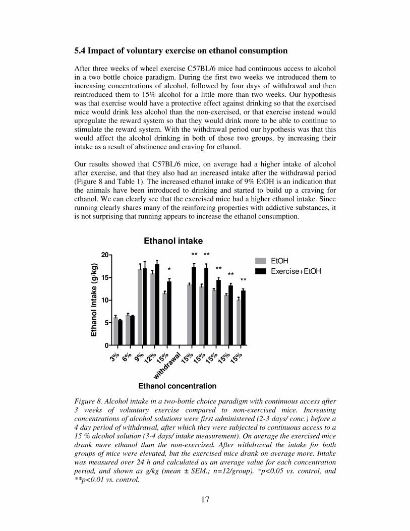
5.4 Impact of voluntary exercise on ethanol consumption
After three weeks of wheel exercise C57BL/6 mice had continuous access to alcohol
in a two bottle choice paradigm. During the first two weeks we introduced them to
increasing concentrations of alcohol, followed by four days of withdrawal and then
reintroduced them to 15% alcohol for a little more than two weeks. Our hypothesis
was that exercise would have a protective effect against drinking so that the exercised
mice would drink less alcohol than the non-exercised, or that exercise instead would
upregulate the reward system so that they would drink more to be able to continue to
stimulate the reward system. With the withdrawal period our hypothesis was that this
would affect the alcohol drinking in both of those two groups, by increasing their
intake as a result of abstinence and craving for ethanol.
Our results showed that C57BL/6 mice, on average had a higher intake of alcohol
after exercise, and that they also had an increased intake after the withdrawal period
(Figure 8 and Table 1). The increased ethanol intake of 9% EtOH is an indication that
the animals have been introduced to drinking and started to build up a craving for
ethanol. We can clearly see that the exercised mice had a higher ethanol intake. Since
running clearly shares many of the reinforcing properties with addictive substances, it
is not surprising that running appears to increase the ethanol consumption.
Ethanol intake
Ethanol concentration
Eth
an
ol in
take (
g/k
g)
3% 6% 9% 12%
15%
with
drawal
15%
15%
15%
15%
15%
0
5
10
15
20EtOH
Exercise+EtOH*
** **
****
**
Figure 8. Alcohol intake in a two-bottle choice paradigm with continuous access after
3 weeks of voluntary exercise compared to non-exercised mice. Increasing
concentrations of alcohol solutions were first administered (2-3 days/ conc.) before a
4 day period of withdrawal, after which they were subjected to continuous access to a
15 % alcohol solution (3-4 days/ intake measurement). On average the exercised mice
drank more ethanol than the non-exercised. After withdrawal the intake for both
groups of mice were elevated, but the exercised mice drank on average more. Intake
was measured over 24 h and calculated as an average value for each concentration
period, and shown as g/kg (mean ± SEM.; n=12/group). *p<0.05 vs. control, and
**p<0.01 vs. control.
18
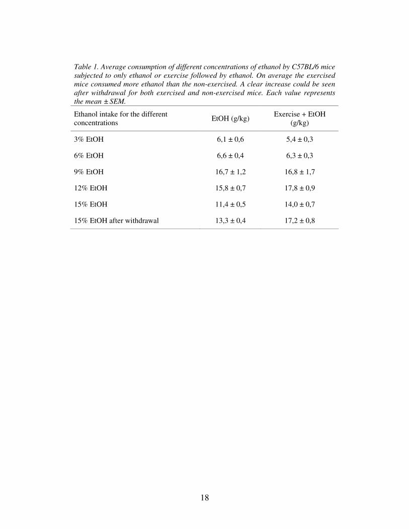
Table 1. Average consumption of different concentrations of ethanol by C57BL/6 mice
subjected to only ethanol or exercise followed by ethanol. On average the exercised
mice consumed more ethanol than the non-exercised. A clear increase could be seen
after withdrawal for both exercised and non-exercised mice. Each value represents
the mean ± SEM.
Ethanol intake for the different
concentrations EtOH (g/kg)
Exercise + EtOH
(g/kg)
3% EtOH 6,1 ± 0,6 5,4 ± 0,3
6% EtOH 6,6 ± 0,4 6,3 ± 0,3
9% EtOH 16,7 ± 1,2 16,8 ± 1,7
12% EtOH 15,8 ± 0,7 17,8 ± 0,9
15% EtOH 11,4 ± 0,5 14,0 ± 0,7
15% EtOH after withdrawal 13,3 ± 0,4 17,2 ± 0,8
19
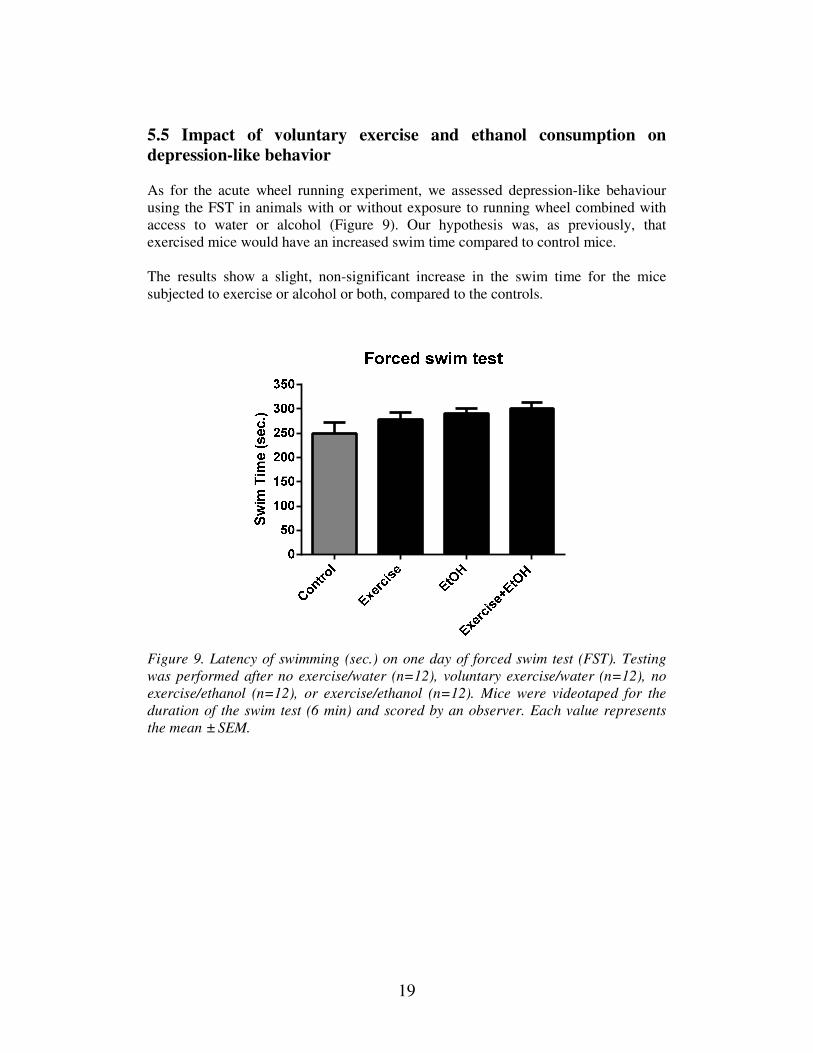
5.5 Impact of voluntary exercise and ethanol consumption on depression-like behavior
As for the acute wheel running experiment, we assessed depression-like behaviour
using the FST in animals with or without exposure to running wheel combined with
access to water or alcohol (Figure 9). Our hypothesis was, as previously, that
exercised mice would have an increased swim time compared to control mice.
The results show a slight, non-significant increase in the swim time for the mice
subjected to exercise or alcohol or both, compared to the controls.
Figure 9. Latency of swimming (sec.) on one day of forced swim test (FST). Testing
was performed after no exercise/water (n=12), voluntary exercise/water (n=12), no
exercise/ethanol (n=12), or exercise/ethanol (n=12). Mice were videotaped for the
duration of the swim test (6 min) and scored by an observer. Each value represents
the mean ± SEM.
20
6 Discussion
Almost all studies of the involvement of voluntary wheel exercise on BDNF mRNA
expression levels have been done in hippocampus, where the main focus has been on
the ability of BDNF to affect neural plasticity, enhance learning and memory, and
regulate stress and depression-like behaviors. There are in fact very limited studies on
exercise-induced effects on BDNF in other brain areas than hippocampus (Davis
2008; Gustafsson et al. 2011). This is not very surprising since hippocampus is a brain
area rich in growth factors, like BDNF, that are highly responsive to wheel running,
thus increasing their expression and neurogenesis in this area (Novak et al. 2012).
This is, to our knowledge, one of very few studies where exercise-induced effects on
mRNA levels of both BDNF and its receptor TrkB has been investigated in different
brain areas other than the hippocampus. Here we have looked at prefrontal cortex,
nucleus accumbens and amygdala as well as two different areas of the hippocampus –
dentate gyrus and CA1.
Our results show that voluntary exercise acutely upregulates BDNF mRNA
expression in the hippocampus (CA1). The expression of TrkB however was not
affected by exercise. BDNF protein can however come from the ventral tegmental
area (VTA) since VTA-NAc forms a pathway that is involved in the reward system
(Eisch, Bolanos et al. 2003).
Mice that are allowed to perform voluntary exercise show less depression-like
behaviors and therefore tend to have a higher latency of swimming than non-
exercised mice. This is due to the positive reinforcement they get from exercising and
they are less likely to feel depressed and give up trying to escape from the water
(Bjornebekk et al. 2005; Cryan et al. 2005; Novak et al. 2012). Maybe this
upregulation of BDNF was too small to affect the depression-like behavior for these
mice. FST is also somewhat difficult to assess and these mice may also have been
more susceptible to stress, that would have influenced the outcome of this test.
BDNF has been reported to be an alcohol-responsive gene and most of the research
on ethanol-induced effects on BDNF has also been done in hippocampus (Davis
2008). Short-term alcohol consumption has been shown to decrease BDNF mRNA
levels, maybe as a result of the system trying to compensate with homeostasis or
repair, whereas withdrawal may cause an upregulation, since presence of alcohol may
inhibit the receptor of transducing its signals mediated by BDNF (Davis 2008).
However, other studies have shown that both acute administration and voluntary
consumption of alcohol activates the BDNF signaling pathway and increases BDNF
mRNA levels (Ghitza et al. 2010; Bosse and Mathews 2011; Costa et al. 2011). The
differential results obtained in these studies point to a complex relationship between
BDNF and reward-related behaviors, with changes in expression being dependent on
time-point as well as brain region examined. Here we directed our attention towards
other brain areas – prefrontal cortex, nucleus accumbens and amygdala – and what
effect voluntary exercise would have on alcohol drinking behaviors in C57BL/6 mice.
Most studies on the effect of voluntary exercise on alcohol consumption, in mice and
other rodents, have been done by introducing both wheel running and alcohol
21
simultaneously, in order to observe the relationship between natural reward and
alcohol drinking. When these mice were denied any access to alcohol, their wheel
running behavior increased. Interestingly, wheel running decreased upon the
reintroduction of alcohol (Ozburn et al. 2008; Ehringer et al. 2009; Novak et al.
2012). We instead first introduced male C57BL/6 mice to three weeks of voluntary
wheel exercise followed by five weeks of continuous access to alcohol, including a
withdrawal period of four days. An interesting part was to see if exercise somehow
would protect against the neurodegenerative effect of alcohol consumption, in terms
of affecting the mRNA levels of BDNF and TrkB.
Our results showed that mice allowed utilizing of a running wheel for three weeks
prior to continuous access to 15 % ethanol have a higher alcohol intake than control
animals. Maybe this increased intake is a way of receiving the same level of reward
after that running has triggered the natural release of dopamine/endorphins (natural
reward). These enormously high expression levels of BDNF could be the result of an
upregulation due to withdrawal. In fact BDNF has been suggested to have a role in
neuroadaptation during withdrawal, because of the reports of increased BDNF levels
after 24 hours of abstinence. It is possible that this may be important for long-term
alcohol abstinence (Costa et al. 2011). Wheel running for rodents is clearly
reinforcing and rewarding, but it may also be addictive as alcohol. Studies in rats have
shown that wheel running can potentiate alcohol preference and increase their ethanol
intake (Werme et al. 2002).
Some considerations must be made, on the difference between our first experiment,
with only exercise, and our second experiment, with both exercise and alcohol
consumption. During the first experiment, the mice were either subjected to wheels or
not for three weeks before their behavior were tested in a FST and then animals were
euthanized directly following access to running wheel. During the second experiment
the mice were after three weeks of exercise, either subjected to a two-bottle choice
paradigm with alcohol or only water for five weeks before being evaluated in a FST
and euthanized (Figure 2). During these five weeks no animals were allowed any
wheel exercise, and this could also be seen as a sort of withdrawal period from natural
reward which could have influenced the high BDNF expression levels in PFC and AA
after exercise in experiment 2.
In order to strengthen our trend results for both the running study and the drinking
study, more samples must be run in qRT-PCR. It would also be of interest to look at
the protein levels by using ELISA since mRNA expression changes may not
necessarily result in changes in protein levels. However, mRNA levels may here be
good indicator, since mRNA expression and protein levels for neuropeptides usually
are comparable. Since we see a huge effect of exercise and exercise followed by
ethanol consumption on BDNF expression in amygdala, it would be interesting to do
an in situ hybridization to look at the neuronal activation and see if this could actually
be the reason for our results. The 100-fold increase in BDNF gene expression is very
high but plausible. It has been shown in other studies using real time PCR that this
level of change can be detected (Dessie et al. 2007), however this has mainly been
done in other kinds of tissues than brain. The results should therefore be interpreted
with caution, although the results are supported by the maintained decrease detected
in the NAc as well as the unchanged level of TrkB gene expression seen in the same
material. This indicates that the very high fold-change is most likely not due to
22
methodological error. Still, the assay-results will need to be confirmed in a new set of
animals.
Another interesting aspect would be to investigate the activity of the TrkB receptor,
by using the agonist 7,8-DHF which activates the high affinity BDNF-receptor TrkB
in the central nervous system (CNS). This would be done to see if the activation of
TrkB receptors by 7,8-DHF would lead to an upregulation of BDNF and if 7,8-DHF
could be a good therapeutic compound for depression, anxiety, or for example other
brain disorders that break down neurons. It would also be interesting to see if
voluntary exercise can increase the BDNF expression levels in heterozygote BDNF
mice (BDNF+/-
), that have a 50% reduction of BDNF expression. Homozygote BDNF-
/- mice die after some weeks because of their inability to perform neurogenesis,
whereas heterozygote BDNF+/-
with a 50 % reduction of BDNF levels can survive
(Bosse and Mathews 2011).
6.1 Conclusions
Our results show a region as well as time-dependent plasticity for BDNF expression
within the brain reward system following exercise as well as following exercise
combined with alcohol intake. This possibility of the involvement of BDNF in
regulation of alcohol reward is further strengthened by the recent finding of Koo et al.
(2012) demonstrating that BDNF is a negative modulator of morphine action. The
possibility of regulation of BDNF expression by exercise and that the changes in
BDNF expression following exercise may contribute to subsequent effects of alcohol
needs to be further established, however the results obtained here do implicate BDNF
to be part of the mechanisms underlying alcohol dependence.
7 Acknowledgement
I would like to thank my supervisor Annika Thorsell for allowing me to perform this
project, and I would also like to thank my examiner Johan Edqvist for your advice
and good feedback. Especially big thanks to my good friend and colleague Anna
Johansson, for all the help and support during the work on my master thesis project. I
really appreciate our many discussions and our happy times at the animal facility
(annexet) and in the lab. Thank you to the whole lab group, Susanne Hilke, Lovisa
Holm, Abdul Maruf Asif Aziz and Daniel Nätt for all the help and support.
23
8 References Adlard, P. A., V. M. Perreau, et al. (2004). "The timecourse of induction of brain-derived
neurotrophic factor mRNA and protein in the rat hippocampus following voluntary
exercise." Neurosci Lett 363(1): 43-48.
Andero, R., S. A. Heldt, et al. (2011). "Effect of 7,8-dihydroxyflavone, a small-molecule
TrkB agonist, on emotional learning." Am J Psychiatry 168(2): 163-172.
Bekinschtein, P., M. Cammarota, et al. (2008). "BDNF is essential to promote persistence of
long-term memory storage." Proc Natl Acad Sci U S A 105(7): 2711-2716.
Belujon, P. and A. A. Grace (2011). "Hippocampus, amygdala, and stress: interacting systems
that affect susceptibility to addiction." Ann N Y Acad Sci 1216: 114-121.
Bjornebekk, A., A. A. Mathe, et al. (2005). "The antidepressant effect of running is associated
with increased hippocampal cell proliferation." Int J Neuropsychopharmacol 8(3):
357-368.
Bosse, K. E. and T. A. Mathews (2011). "Ethanol-induced increases in extracellular
dopamine are blunted in brain-derived neurotrophic factor heterozygous mice."
Neurosci Lett 489(3): 172-176.
Costa, M. A., M. Girard, et al. (2011). "Brain-derived neurotrophic factor serum levels in
alcohol-dependent subjects 6 months after alcohol withdrawal." Alcohol Clin Exp
Res 35(11): 1966-1973.
Crews, F. T. and K. Nixon (2009). "Mechanisms of neurodegeneration and regeneration in
alcoholism." Alcohol Alcohol 44(2): 115-127.
Cryan, J. F., R. J. Valentino, et al. (2005). "Assessing substrates underlying the behavioral
effects of antidepressants using the modified rat forced swimming test." Neurosci
Biobehav Rev 29(4-5): 547-569.
Davis, M. I. (2008). "Ethanol-BDNF interactions: still more questions than answers."
Pharmacol Ther 118(1): 36-57.
Dessie, S. W., F. Rings, et al. (2007). "Dielectrophoretic behavior of in vitro-derived bovine
metaphase II oocytes and zygotes and its relation to in vitro embryonic
developmental competence and mRNA expression pattern." Reproduction 133(5):
931-946.
Duman, C. H., L. Schlesinger, et al. (2008). "Voluntary exercise produces antidepressant and
anxiolytic behavioral effects in mice." Brain Res 1199: 148-158.
Duman, R. S. and L. M. Monteggia (2006). "A neurotrophic model for stress-related mood
disorders." Biol Psychiatry 59(12): 1116-1127.
Duman, R. S. and B. Voleti (2012). "Signaling pathways underlying the pathophysiology and
treatment of depression: novel mechanisms for rapid-acting agents." Trends Neurosci
35(1): 47-56.
Ehringer, M. A., N. R. Hoft, et al. (2009). "Reduced alcohol consumption in mice with access
to a running wheel." Alcohol 43(6): 443-452.
Eisch, A. J., C. A. Bolanos, et al. (2003). "Brain-derived neurotrophic factor in the ventral
midbrain-nucleus accumbens pathway: a role in depression." Biol Psychiatry 54(10):
994-1005.
Ghitza, U. E., H. Zhai, et al. (2010). "Role of BDNF and GDNF in drug reward and relapse: a
review." Neurosci Biobehav Rev 35(2): 157-171.
Gomez-Pinilla, F., Z. Ying, et al. (2002). "Voluntary exercise induces a BDNF-mediated
mechanism that promotes neuroplasticity." J Neurophysiol 88(5): 2187-2195.
Gustafsson, S., W. Liang, et al. (2011). "Effects of voluntary running in the female mice
lateral septum on BDNF and corticotropin-releasing factor receptor 2." Int J Pept
2011: 932361.
Jang, S. W., X. Liu, et al. (2010). "A selective TrkB agonist with potent neurotrophic
activities by 7,8-dihydroxyflavone." Proc Natl Acad Sci U S A 107(6): 2687-2692.
24
Klintsova, A. Y., E. Dickson, et al. (2004). "Altered expression of BDNF and its high-affinity
receptor TrkB in response to complex motor learning and moderate exercise." Brain
Res 1028(1): 92-104.
Koo, J. W., M. S. Mazei-Robison, et al. (2012). "BDNF is a negative modulator of morphine
action." Science 338(6103): 124-128.
Krishnan, V., M. H. Han, et al. (2007). "Molecular adaptations underlying susceptibility and
resistance to social defeat in brain reward regions." Cell 131(2): 391-404.
Meeusen, R. and K. De Meirleir (1995). "Exercise and brain neurotransmission." Sports Med
20(3): 160-188.
Murray, P. S. and P. V. Holmes (2011). "An overview of brain-derived neurotrophic factor
and implications for excitotoxic vulnerability in the hippocampus." Int J Pept 2011:
654085.
Neeper, S. A., F. Gomez-Pinilla, et al. (1996). "Physical activity increases mRNA for brain-
derived neurotrophic factor and nerve growth factor in rat brain." Brain Res 726(1-2):
49-56.
Novak, C. M., P. R. Burghardt, et al. (2012). "The use of a running wheel to measure activity
in rodents: Relationship to energy balance, general activity, and reward." Neurosci
Biobehav Rev 36(3): 1001-1014.
Ozburn, A. R., R. A. Harris, et al. (2008). "Wheel running, voluntary ethanol consumption,
and hedonic substitution." Alcohol 42(5): 417-424.
Razzoli, M., E. Domenici, et al. (2011). "A role for BDNF/TrkB signaling in behavioral and
physiological consequences of social defeat stress." Genes Brain Behav 10(4): 424-
433.
Werme, M., S. Lindholm, et al. (2002). "Running increases ethanol preference." Behav Brain
Res 133(2): 301-308.
Wojnar, M., K. J. Brower, et al. (2009). "Association between Val66Met brain-derived
neurotrophic factor (BDNF) gene polymorphism and post-treatment relapse in
alcohol dependence." Alcohol Clin Exp Res 33(4): 693-702.
Wong, C. C., J. Mill, et al. (2011). "Drugs and addiction: an introduction to epigenetics."
Addiction 106(3): 480-489.
Zanardini, R., A. Fontana, et al. (2011). "Alterations of brain-derived neurotrophic factor
serum levels in patients with alcohol dependence." Alcohol Clin Exp Res 35(8):
1529-1533.
World Health Organization (2012). WHO Media centre. Fact sheets: Alcohol. World Health
Organization, Geneva.