Effect of hyperbaric oxygen therapy combined with …Effect of hyperbaric oxygen therapy combined...
8
Effect of hyperbaric oxygen therapy combined with autologous platelet concentrate applied in rabbit fibula fraction healing Paulo Ce ´ sar Fagundes Neves, I Simone de Campos Vieira Abib, II Roge ´ rio Fagundes Neves, II Oronzo Pircchio, Karen Ruggeri Saad, I Paulo Fernandes Saad, I Ricardo Santos Simo ˜ es, II Marcia Bento Moreira, III Cristiano Frota de Souza Laurino IV I Universidade Federal do Vale do Sa ˜ o Francisco (UNIVASF), Colegiado de Medicina, Petrolina/PE, Brazil. II Universidade Federal de Sa ˜ o Paulo (UNIFESP), Departamento de Cirurgia, Sa ˜ o Paulo/SP, Brazil. III Universidade Federal do Vale do Sa ˜ o Francisco (UNIVASF), Colegiado de Medicina Veterina ´ ria, Petrolina/ PE, Brazil. IV Sociedade Brasileira de Traumatologia Esportiva e Artroscopia, Sa ˜ o Paulo/SP, Brazil. OBJECTIVES: The purpose is to study the effects of hyperbaric oxygen therapy and autologous platelet concentrates in healing the fibula bone of rabbits after induced fractures. METHODS: A total of 128 male New Zealand albino rabbits, between 6–8 months old, were subjected to a total osteotomy of the proximal portion of the right fibula. After surgery, the animals were divided into four groups (n = 32 each): control group, in which animals were subjected to osteotomy; autologous platelet concentrate group, in which animals were subjected to osteotomy and autologous platelet concentrate applied at the fracture site; hyperbaric oxygen group, in which animals were subjected to osteotomy and 9 consecutive daily hyperbaric oxygen therapy sessions; and autologous platelet concentrate and hyperbaric oxygen group, in which animals were subjected to osteotomy, autologous platelet concentrate applied at the fracture site, and 9 consecutive daily hyperbaric oxygen therapy sessions. Each group was divided into 4 subgroups according to a pre-determined euthanasia time points: 2, 4, 6, and 8 weeks postoperative. After euthanasia at a specific time point, the fibula containing the osseous callus was prepared histologically and stained with hematoxylin and eosin or picrosirius red. RESULTS: Autologous platelet concentrates and hyperbaric oxygen therapy, applied together or separately, increased the rate of bone healing compared with the control group. CONCLUSION: Hyperbaric oxygen therapy and autologous platelet concentrate combined increased the rate of bone healing in this experimental model. KEYWORDS: Hyperbaric Oxygenation; Platelet-rich Plasma; Fibula, Fracture Consolidation. Neves PC, Abib SC, Neves RF, Pircchio O, Saad KR, Saad PF, et al. Effect of hyperbaric oxygen therapy combined with autologous platelet concentrate applied in rabbit fibula fraction healing. Clinics. 2013;68(9):1239-1246. Received for publication on February 27, 2013; First review completed on March 25, 2013; Accepted for publication on April 22, 2013 E-mail: [email protected] / [email protected] Tel.: 55 87 2101–6865 & INTRODUCTION Loss of osseous continuity due to fracture is a disabling condition, even momentarily, that often deprives the individual of his or her social and professional lifestyle. Therefore, as a medical specialty, orthopedics has invested in studies that promote reduced bone healing time and, consequently, reduced time required to return to normal or almost normal function. Although most fractures do not present healing problems under normal conditions, in some situations, the poorly vascularized bone tissue does not respond adequately to treatment, resulting in delayed consolidation or non- consolidation (1). Thus, developing new techniques to help the bone healing process and stimulate fracture consolida- tion is very important. Several studies have focused on reducing bone healing time using low-intensity pulsed ultrasound, low-energy lasers, electrical stimulation, and platelet-rich concentrates (2). Platelet-rich concentrate (PRC) is an autologous concen- tration of platelets that releases growth factors. Platelets and the release of these proteins are essential for clot formation in the physiological process of tissue repair. A normal clot consists of 95% red blood cells, 4% platelets, and 1% white blood cells. However, an analysis of platelet-enriched clots Copyright ß 2013 CLINICS – This is an Open Access article distributed under the terms of the Creative Commons Attribution Non-Commercial License (http:// creativecommons.org/licenses/by-nc/3.0/) which permits unrestricted non- commercial use, distribution, and reproduction in any medium, provided the original work is properly cited. No potential conflict of interest was reported. DOI: 10.6061/clinics/2013(09)11 BASIC RESEARCH 1239
Transcript of Effect of hyperbaric oxygen therapy combined with …Effect of hyperbaric oxygen therapy combined...
Effect of hyperbaric oxygen therapy combined withautologous platelet concentrate applied in rabbitfibula fraction healingPaulo Cesar Fagundes Neves,I Simone de Campos Vieira Abib,II Rogerio Fagundes Neves,II Oronzo Pircchio,
Karen Ruggeri Saad,I Paulo Fernandes Saad,I Ricardo Santos Simoes,II Marcia Bento Moreira,III Cristiano
Frota de Souza LaurinoIV
I Universidade Federal do Vale do Sao Francisco (UNIVASF), Colegiado de Medicina, Petrolina/PE, Brazil. II Universidade Federal de Sao Paulo (UNIFESP),
Departamento de Cirurgia, Sao Paulo/SP, Brazil. III Universidade Federal do Vale do Sao Francisco (UNIVASF), Colegiado de Medicina Veterinaria, Petrolina/
PE, Brazil. IV Sociedade Brasileira de Traumatologia Esportiva e Artroscopia, Sao Paulo/SP, Brazil.
OBJECTIVES: The purpose is to study the effects of hyperbaric oxygen therapy and autologous plateletconcentrates in healing the fibula bone of rabbits after induced fractures.
METHODS: A total of 128 male New Zealand albino rabbits, between 6–8 months old, were subjected to a totalosteotomy of the proximal portion of the right fibula. After surgery, the animals were divided into four groups(n= 32 each): control group, in which animals were subjected to osteotomy; autologous platelet concentrategroup, in which animals were subjected to osteotomy and autologous platelet concentrate applied at the fracturesite; hyperbaric oxygen group, in which animals were subjected to osteotomy and 9 consecutive daily hyperbaricoxygen therapy sessions; and autologous platelet concentrate and hyperbaric oxygen group, in which animalswere subjected to osteotomy, autologous platelet concentrate applied at the fracture site, and 9 consecutive dailyhyperbaric oxygen therapy sessions. Each group was divided into 4 subgroups according to a pre-determinedeuthanasia time points: 2, 4, 6, and 8 weeks postoperative. After euthanasia at a specific time point, the fibulacontaining the osseous callus was prepared histologically and stained with hematoxylin and eosin or picrosirius red.
RESULTS: Autologous platelet concentrates and hyperbaric oxygen therapy, applied together or separately,increased the rate of bone healing compared with the control group.
CONCLUSION: Hyperbaric oxygen therapy and autologous platelet concentrate combined increased the rate ofbone healing in this experimental model.
KEYWORDS: Hyperbaric Oxygenation; Platelet-rich Plasma; Fibula, Fracture Consolidation.
Neves PC, Abib SC, Neves RF, Pircchio O, Saad KR, Saad PF, et al. Effect of hyperbaric oxygen therapy combined with autologous platelet concentrateapplied in rabbit fibula fraction healing. Clinics. 2013;68(9):1239-1246.
Received for publication on February 27, 2013; First review completed on March 25, 2013; Accepted for publication on April 22, 2013
E-mail: [email protected] / [email protected]
Tel.: 55 87 2101–6865
& INTRODUCTION
Loss of osseous continuity due to fracture is a disablingcondition, even momentarily, that often deprives theindividual of his or her social and professional lifestyle.Therefore, as a medical specialty, orthopedics has investedin studies that promote reduced bone healing time and,consequently, reduced time required to return to normal oralmost normal function.
Although most fractures do not present healing problemsunder normal conditions, in some situations, the poorlyvascularized bone tissue does not respond adequately totreatment, resulting in delayed consolidation or non-consolidation (1). Thus, developing new techniques to helpthe bone healing process and stimulate fracture consolida-tion is very important.
Several studies have focused on reducing bone healingtime using low-intensity pulsed ultrasound, low-energylasers, electrical stimulation, and platelet-rich concentrates(2).
Platelet-rich concentrate (PRC) is an autologous concen-tration of platelets that releases growth factors. Platelets andthe release of these proteins are essential for clot formationin the physiological process of tissue repair. A normal clotconsists of 95% red blood cells, 4% platelets, and 1% whiteblood cells. However, an analysis of platelet-enriched clots
Copyright � 2013 CLINICS – This is an Open Access article distributed underthe terms of the Creative Commons Attribution Non-Commercial License (http://creativecommons.org/licenses/by-nc/3.0/) which permits unrestricted non-commercial use, distribution, and reproduction in any medium, provided theoriginal work is properly cited.
No potential conflict of interest was reported.
DOI: 10.6061/clinics/2013(09)11
BASIC RESEARCH
1239

reveals 95% platelets, 4% red blood cells, and 1% whiteblood cells (2).
Platelet-derived growth factor (PDGF) has defined peri-ods of activity. PDGF is a major factor during the onset oftissue repair and acts directly on cell differentiation andangiogenesis. According to classic studies by Heldin &Westmark (3), PDGF is the first growth factor that initiatesrepair by stimulating mitogenesis and increasing the localpopulations of mesenchymal stem cells and osteoprogenitorcells. At concentrations of 20 to 100 ng/ml, bone formationand alkaline phosphatase activity increase in the deminer-alized matrices of rats (4).
Transforming growth factor beta (TGF-b) is a cytokinesynthesized in bone tissue and platelets that stimulates theproliferation of osteoblast precursor cells, promotes collagensynthesis, increases the number of cells that express theosteoblast genotype, increases osteoclast apoptosis, andactivates endothelial cells for angiogenesis and chondro-progenitor cells in cartilage formation. However in vitro,TGF-b alone does not induce bone formation in an animalmodel (5).
Methods for obtaining platelet growth factors shouldconsider the platelet concentrate rate, processing technique,concentration of secreted proteins, handling and applica-tion, and especially the clinical application (6). Platelet-richplasma (PRP) is undoubtedly the most widely used serum;however, its indiscriminate use has been criticized, and it isstill not approved by the Food and Drug Administration(FDA) (7).
Autologous platelet concentrate (APC) is a platelet-richconcentrate (PRC) that is similar to PRP but has certainmethodological advantages in terms of how it is obtained. Inaddition to being approved by the FDA (using the double-syringe method), APC can be withdrawn and used during asurgical procedure for fracture fixation and is easilyprocessed in a few minutes, which is important becausePDGF and TGF-b have half-lives of only a few minutesoutside the vascular system (7).
The basic difference between PRP and APC is that PRP isan PRC, with a platelet concentration over 2 times greaterthan that of plasma. APC is a PRC that combines PRP withplatelet-poor plasma (PPP); according to the manufacturer,APC contains 2 times the platelet concentration of bloodplasma. According to studies by Graziane et al. (8), thisplatelet concentration is sufficient to stimulate osteoblastproliferation in vitro. In addition, obtaining APC using thedouble-syringe method eliminates leukocytes, proteases,and hydrolases, which may be able to degrade APC (9).
Hyperbaric oxygen therapy (HBO) is another treatmentthat may have positive effects on the bone healing process.
According to the Undersea and Hyperbaric MedicalSociety, HBO is defined as intermittently inhaling 100%oxygen inside a chamber at pressures greater than 1atmosphere absolute (ATA). This therapy has been first-choice treatment for decompression sickness in divers sincethe 1920s; however, only since the 1950s has it been used totreat gas embolism, carbon monoxide poisoning, gangrenegas, compartment syndrome, soft tissue infection, osteo-myelitis, and osteoradionecrosis (10).
The advantage of HBO is that it is a safe treatment.According to the literature, the complication rate is 1.83%,of which 94% are considered minor complications.The disadvantages of HBO include the high cost of thehyperbaric chamber and the technical training necessary,
considering the number of circumstances under which it canbe used with some level of evidence. However, the delayedconsolidation or non-consolidation of fractures has signifi-cant financial impacts on society and represents a seriousclinical challenge for which few treatment options areavailable. Various, often necessary surgical procedures areaccompanied with patient morbidity and reduced quality oflife (11).
This study was designed to test the hypothesis that manypatients with fractures or delayed consolidations couldbenefit from APC, HBO, or a combination of bothtreatments because the literature indicates that the osteo-genic action of growth factors depends directly on the tissueoxygen tension (12).
& MATERIALS AND METHODS
SampleThis study was approved by the Ethics Committee of
Universidade Federal de Sao Paulo (0163/08). All animalswere handled according to "The Ethical Principles ofAnimal Experiments of the International Union for theProtection of Animals" 11.794/2008 Act of October 8, 2008.
A total of 128 6-8-month-old male New Zealand albinorabbits (Oryctolagus cuniculus), with an average weight of3,000 grams, were subjected to an osteotomy of the rightfibula after an adaptation period.
After surgery, the animals were randomized into 4 groupsof 32 rabbits according to the postsurgical treatment used, asfollows:
N Control Group (CG) - animals subjected only toosteotomy of the right fibula, without further treatment
N APC Group (APCG) - animals subjected to osteotomy ofthe right fibula and treated with APC
N HBO Group (HBOG) - animals subjected to osteotomy ofthe right fibula and treated with consecutive daily HBOsessions, totaling 9 sessions
N APC and HBO group (APC+HBOG) - animals subjectedto osteotomy of the right fibula and treated with APC,combined with consecutive daily HBO sessions, totaling9 sessions
Each group was further divided into 4 subgroups of 8animals each, according to a pre-determined euthanasiatime point at 2, 4, 6, and 8 weeks postoperatively.
Operative procedureAll animals were food- and water-fasted for 6 hours pre-
operatively and then weighed; all the fur was shaved at theleg region.
Pre-anesthesia was induced with ketamine hydrochloridesolution at a dose of 30 mg/kg, xylazine at a dose of 3 mg/kg, and tramadol hydrochloride at a dose of 5 mg/kgintramuscularly in the gluteal region.
The animals were anesthetized with spinal anesthesiausing 2% lidocaine without a vasoconstrictor at a dose of0.5 mg/kg. Venoclysis of the right auricular vein wasperformed for the infusion of the necessary drugs.Antisepsis was then performed with polyvinylpyrrolidone,and the operative field was isolated with sterile field cloths,delimiting a restricted area for the surgery.
Then, a 2-cm lateral longitudinal incision was made alongthe right leg and dissected by following the muscular planes
HBO and APC in fraction healingNeves PCF et al.
CLINICS 2013;68(9):1239-1246
1240

and localization of the fibular diaphyseal region. A completetransverse osteotomy of the fibula was performed using ano. 15 scalpel blade at the proximal third of the tibialtuberosity.
The surgery was completed by suturing the planes,aponeurosis, subcutaneous tissue, and skin with 4-0 nylonthread. Tramadol hydrochloride analgesic was adminis-tered at a dose of 2 mg/kg intramuscularly and maintainedevery 12 hours for 3 consecutive days, in combination with4 intramuscular antibiotic treatments with benzathinepenicillin and G potassium at doses of 20,000 IU/kg every48 hours, in all animals.
Immediately after the osteotomy of the fibula, the animalswere randomized according to the pre-determined treat-ment groups under study and to their respective subgroups.The hind limb did not need to be immobilized because thedistal portion of the fibula in rabbits exhibits fusion with thetibia.
During the post-operative period, at the pre-determinedtime for each subgroup (2, 4, 6, or 8 weeks), the animalswere anesthetized again according to the previous protocol,and the fibulae were resected, including the osseous callus,using a no. 15 scalpel blade. Euthanasia was induced duringdeep anesthesia with xylazine.
Application of autologous platelet concentrateThis procedure was performed during the surgery. Before
the surgical procedure, 2.5 ml of blood was withdrawn fromthe auricular vein using a 10-ml double-syringe (ACP-double syringe, Arthrex, Naples, FL, USA) containing 0.5 mlof acid citrate dextrose anticoagulant, and the syringe wasgently agitated to ensure the adequate mixing of the bloodwith the anticoagulant. The syringe containing the anti-coagulated blood was then stored in a suitable tube andcentrifuged (Rotofix 32A, Arthrex, USA) at 1500 RPM for 5minutes to separate the red blood cells from the plasma.
After centrifugation, the plasma was removed from thetube using the same syringe, while the internal syringecollected only the concentrate, leaving the red blood cells.Using this procedure, which was provided by the manu-facturer, we obtained approximately 1.5 ml of concentrate,with a high concentration of autologous platelets enrichedwith growth factors.
The platelet concentrate was applied directly to thefracture (osteotomy) in the proximal third of the rabbitfibula before closing the muscular fascia.
Hyperbaric oxygen therapyHBO sessions were conducted using a veterinary hyper-
baric chamber model (AN – 500, Sechrist, USA) with a 14-rabbit capacity (average weight of 2,500 grams each). Thesessions were conducted daily, 8 animals at a time, andbegan on the first post-operative day for 90 minutes at 3ATA (13). We opted for 9 sessions in the HBO andAPC+HBO groups because the growth factor acts primarilyduring the first 7 post-operative days (14).
The beginning of the session included a compressionperiod. After 15 minutes, the chamber manometer indicated2 atm of pressure; assuming that the manometer in thehyperbaric chamber measures the pressure differencebetween the chamber and the surrounding environment,the barometric pressure and environmental pressure (1ATA) were added to convert the values into absoluteatmospheres (ATA). Therefore, the pressure applied by the
chamber was 3 ATA. This absolute pressure (3 ATA) wasmaintained for 60 minutes, followed by decompression for15 minutes; thus, each session ended after 90 minutes.
Histomorphometric analysisImmediately after the experiment, the proximal third of
the fibula from each animal, containing the osseous callusregion, was surgically removed and placed in 10% bufferedformalin for 48 hours. After this period, the decalcificationprocess was begun in ethylenediaminetetraacetic acid(EDTA); after 6 days, the pieces were dehydrated inincreasing concentrations of ethyl alcohol, diaphanized inxylene, impregnated in histological paraffin, and embeddedin paraffin.
Paraffin blocks were cut with a Minot microtome (Leica,Model RM 2145) with semi-serial 4 ım cuts. After drying for6–8 hours in an oven at 60 C, some sections were stainedwith hematoxylin and eosin (HE), and others were stainedwith picrosirius red. The slides were observed under a lightmicroscope (Carl ZeissH) at 4 to 1006.
To determine the concentration of bone cells (osteocytes,osteoblasts, and osteoclasts) in the fibula, an 180061600 mMrectangle was initially determined, drawn using a mouse, intwo central regions adjacent to the healing bone; one was onthe diaphysis side, and the other was on the epiphyseal side.Then, the cells were individually counted within therectangles, excluding cells that touched the rectangles. Foreach animal, 6 slides were evaluated. We recorded theaverage number of cells counted in the rectangles for eachanimal.
The thickness of the cartilage region within the remainingosseous callus was obtained at 3 points in each slide (6 slidesfor each animal) using a mouse. We recorded the averagenumber of measurements in mM for each animal.
Determining the percentages of collagenThe cuts stained with picrosirius red were used to
determine the percentage of collagen formed. Initially, thesections were dewaxed in xylene and then hydrated in adecreasing ethanol gradient. Afterward, elastin was blockedwith 0.2% phosphomolybdic acid for 10 minutes. Thesections were washed in distilled water, immersed in a0.1% sirius red solution, and dissolved in saturated aqueouspicric acid for 90 minutes. Then, the sections were washedin 0.01 N HCl for 2 minutes. Later, the sections weredehydrated in an increasing ethanol gradient, cleared inxylene, and mounted with EntellanH.
Photomicrographs of the picrosirius red-stained sectionsfrom the rabbit fibulae of different groups were taken undera polarized light microscope (Axiolab Standard 2.0, CarlZeissH) coupled to a high-resolution video camera(AxionCam, Carl ZeissH) to identify the distribution ofcollagen fibers in the bone matrix. Then, the micrographswere analyzed on a computer with the ImagelabH program.
Statistical analysisFor quantitative variables, descriptive statistics were
calculated. Analysis of variance (ANOVA) and multiplecomparisons by the Tukey test were used as inferentialanalyses to either confirm or refute the evidence found inthe descriptive analysis.
For all the results obtained via inferential analyses, asignificance level of a= 5% was used. All statistical analyseswere performed with the R program software, version 2.11.1.
CLINICS 2013;68(9):1239-1246 HBO and APC in fraction healingNeves PCF et al.
1241

& RESULTS
Information referring to the percentages of collagen,numbers of osteoblasts and osteocytes, and lesion thicknesswere important measurements assessed among groups andeuthanasia time periods (subgroups). Table 1 and Figure 1(A – D) summarize the behavior of these measurementsaccording to the groups and euthanasia periods (subgroup).
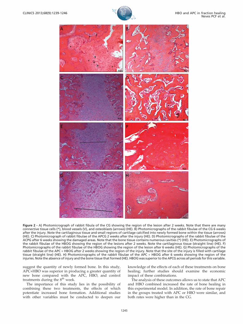
Table 2 shows the results of comparisons of thepercentages of collagen, numbers of osteoblasts andosteocytes, and lesion thicknesses among groups andeuthanasia periods. Figure 2 (A – H) shows the HE slidesfrom the groups and subgroups (2th and 6th weeks).
& DISCUSSION
The results obtained from this experimental model indicatea higher rate of bone healing in response to APC combinedwith HBO. No previous study in the literature was found tohave used this combination in an experimental study.
Qualitative analysis highlighted the accelerated bonehealing in the APC+HBOG compared with the CG duringall weeks studied; additional data on the percentages ofcollagen and lesion thickness confirm this assessment. In1983, a review article found that low oxygen tension reducedthe action of autologous growth factors (12). Similarly, theoutcomes shown here suggest that the higher oxygen tensioncaused by HBO application could potentiate the effects of thegrowth factors offered by APC therapy on bone healing.
Although there was no significant difference between thenumbers of osteoblasts during any of the periods studied orbetween the number of osteocytes counted during the 2nd
and 8th weeks, relative to the groups treated with APC andHBO combined, quantitative data on the average lesionthickness showed that the APCG, HBOG, and APC+HBOGwere superior to the CG. The APC+HBOG was superior tothe other groups across all periods, whereas the HBOG wassuperior to the APCG across all periods for this variable.
The fact that PRC use (in this study, APC) led to asuperior rate of bone healing compared with the CG
corroborates the outcomes of other studies. Marx et al. (15)performed the first and most convincing study using PRPcombined with autogenous bone grafts. The authorsevaluated the effect of PRP on the reconstruction ofmandibular bone defects resulting from tumor removal.The authors stated that bone grafts with PRP showed morethan double the maturity of the controls.
Another study by Anitua produced excellent outcomesusing autogenous bone grafts combined with PRP (16). Theauthors observed a total alveolar bone regeneration in 8 of10 patients who were treated with PRP and bone graft.Biopsies of the respective areas showed compact maturebone with normal morphology and well-organized trabe-cular regions, while the control patients showed connectivetissue, mostly filled with alveoli, and no mature bone.
However, some studies do not support the effect of PRCon the rate of bone healing. Recently, Broggini et al. (17)evaluated bone repairs in the monocortical defects in thecalvaria of 15 New Zealand rabbits. The authors concludedthat PRP, alone or combined with an autogenous graft, didnot lead to a higher rate of bone regeneration comparedwith basal clot during the initial healing.
Currently, the literature is more concerned with quantify-ing the concentration of growth factors present in some ofthe most widely used PRCs, especially PRP. A study usingthis approach stated that the concentration of growth factorsin PRP can reach up to 7 and 30 times the basal levels ofTGF-b and PDGF, respectively, in a basal clot (18).According to the manufacturer’s data on the double-syringemethod used in this study, APC can have up to 4 and 25times the levels of TGF-b and PDGF, respectively. Althoughthe concentrations of growth factors found in APC werelower than in PRP, this study demonstrated that thisdifference does not seem to be a disadvantage. However,it is important to note that functional and non-harmfulamounts of these factors in tissue repair have not been welldetermined.
Regarding our outcomes on the effect of HBO on the rateof bone healing, HBO increased the bone consolidation ratecompared with the CG, which could be observed through
Table 1 - Summary measurements (average ¡ standard deviation) of the percentage of collagen, number of osteoblastsand osteocytes, and lesion thickness, according to groups and euthanasia periods (subgroups).
Group Euthanasia period (weeks) No. of osteocytes No. of osteoblasts % of collagen Lesion thickness
CG 2 (n = 8) 0.13¡0.35 11.25¡0.89 15.59¡0.33 297.97¡0.02
4 (n = 8) 10.38¡0.52 6.38¡0.52 30.26¡0.27 192.30¡0.10
6 (n = 8) 7.25¡0.46 3.50¡0.53 70.39¡0.16 98.34¡0.18
8 (n = 8) 19.38¡0.74 0.13¡0.35 80.83¡0.28 5.54¡0.24
APCG 2 (n = 8) 1.00¡0.00 13.63¡0.52 15.58¡0.29 205.16¡0.02
4 (n = 8) 11.25¡0.71 4.38¡0.52 50.50¡0.31 165.76¡0.28
6 (n = 8) 14.75¡0.71 2.13¡0.35 81.76¡0.28 47.36¡0.13
8 (n = 8) 20.00¡0.76 0.38¡0.52 92.86¡0.55 3.43¡0.28
HBOG 2 (n = 8) 1.25¡0.46 12.38¡0.52 19.81¡0.15 198.23¡0.39
4 (n = 8) 10.75¡0.46 2.25¡0.46 45.78¡0.24 164.94¡1.00
6 (n = 8) 16.00¡0.53 1.25¡0.46 83.24¡0.42 34.99¡0.41
8 (n = 8) 20.63¡0.92 0.25¡0.46 90.58¡0.46 1.10¡0.74
APC+HBOG 2 (n = 8) 1.38¡0.52 12.75¡0.89 20.91¡0.18 165.45¡0.28
4 (n = 8) 14.75¡0.71 2.00¡0.00 65.38¡0.35 110.41¡0.11
6 (n = 8) 20.00¡0.93 0.38¡0.52 95.06¡0.26 24.62¡0.46
8 (n = 8) 21.13¡1.13 0.00¡0.00 98.00¡0.39 0.25¡0.46
CG: Control Group; APCG: Autologous platelet concentrate Group; HBOG: Hyperbaric oxygen therapy Group; APC+HBOG: Autologous platelet
concentrate plus Hyperbaric oxygen therapy Group.
HBO and APC in fraction healingNeves PCF et al.
CLINICS 2013;68(9):1239-1246
1242

the varying percentages of collagen during each week, thenumber of osteocytes during the 6th and 8th weeks, and thelesion thickness across all weeks.
Our search for relevant publications led us to theobservation that the first studies on the effects of HBO onbone healing date back to the 1960s. After almost 50 years,there are still few studies on this subject.
In a Cochrane review study (19), the authors concludedthat there is insufficient evidence to support or refute HBOuse for fracture treatment or as a therapy for non-consolidated fractures within an expected time. Thisconclusion was based on 9 potentially relevant articlesamong the 68 references identified. However, none of thearticles met the review’s inclusive criteria: any randomizedor quasi-randomized studies that compared clinical resultsobtained with HBO use to those obtained without HBO use(no treatment).
Although the literature did not include clinical resultsthat met the methodology described above, our outcomes
and the studies cited indicate that HBO is beneficial to thefracture healing process.
In 1998, Ueng et al. (20) studied bone mineral density andtensile strength in a rabbit tibial-lengthening model. Thebone segments from HBO-treated animals had highermineral densities and superior biomechanical propertiescompared with the control animals. Using the sameexperimental model, this group obtained similar findings,showing that HBO is also advantageous during the earlystages of tibial fracture healing (21).
Chen et al. (22) studied the effect of HBO in rabbits subjectedto fusion of the vertebrae with a bone graft. According to theseauthors, the incidence of bone non-consolidation for thisprocedure in humans ranges from 6–35% of cases and 40–50%in rabbits. After 8 weeks, the HBO group showed a 100%consolidation of the fractures, which were identified radi-ologically and by torsional biomechanical tests.
The vast majority of the studies combining HBO and bonerepair refer to osteoradionecrosis treatment. Radiation
Figure 1 - Distribution of the average percentage of collagen (A), average number of osteoblasts (B) and osteocytes (C), and averagelesion thickness (D), according to groups and euthanasia periods (subgroups).
CLINICS 2013;68(9):1239-1246 HBO and APC in fraction healingNeves PCF et al.
1243

therapy results in progressive obliterative endarteritis,which leads to ischemia and a pathological fracture of thebone with reduced healing ability (23). Muhonen et al. (24)evaluated the angiogenic and osteogenic responses afterradiation and HBO in a rabbit mandibular distractionmodel. The authors concluded that the HBO groupimproved in the bone formation pattern.
Most experimental studies have found a positive effectfrom HBO use in the fracture healing process. This effectmay be directly associated with the increased formation ofnew vessels, which increases blood inflow and, thereby,bone formation. Wu et al. (25) presented the outcomes of anin vitro study that investigated the effects of HBO on theproliferation and differentiation of human osteoblasts andconcluded that HBO also stimulates initial cell proliferationand acts directly on osteogenic differentiation in osteoblasts.
Jan et al. (26) presented the results of a study that assessedthe effect of HBO on a rabbit calvaria fracture model. Afterradiographic and histomorphometric evaluations of thelesion after 6 and 12 weeks, the authors concluded thatbone regeneration was significantly improved in the groupof HBO-treated animals. It is noteworthy that the type oflesion caused in this experiment was considered critical, andbone healing was unlikely to occur.
Yeh et al. (27) studied the effects of HBO on theneovascularization of the tendon-bone junction, on thebehavior of collagen fibers within the graft, and on theincorporation process of the graft into the bone tunnel ofrabbits subjected to knee arthroscopy for the reconstructionof the anterior cruciate ligament with a tendon graft. Thegraft is incorporated into the bone tunnel through callusformation and the posterior ossification of the tunnel (28). Inthe HBO-treated animals, collagen fibers showed greaterorganization surrounding the graft and had an earlymineralization of fibrocartilage compared with the controlanimals. The biomechanical test also indicated that HBO-treated animals exhibited greater resistance to the tensile
strength of the graft at 18 weeks post-operation than thecontrol animals.
The benefits of HBO are related to its proven therapeuticsafety and the possible use of the equipment to treat a widerrange of diseases. Its disadvantages include the high cost ofthe equipment and the low level of scientific evidence forHBO use in treating these diseases (19).
However, in choosing a treatment method, one mustconsider many variables, such as the cost of the equipment,the specialized personnel, the methodological complexity,and the characteristics of the fracture to be treated. Althoughthe mechanisms behind the actions of HBO and APC on bonerepair may be different, the variables in this study showedonly a few differences between the two methods of treatment.Although qualitative assessment highlights APC’s super-iority to HBO in relation to the consolidation rate, furtherstudies should be conducted comparing the two therapiesand their effects on bone healing.
In this study, we observed that osteoblastic activity washigh in the CG during the 4th and 6th weeks but was higherin all other groups during the 2nd week. We also observedthat osteoblastic activity was higher in the APC group thanin the HBO group during the 2nd and 4th weeks, while nodifference was found between these groups in relation to thenumber of osteoblasts during the 6th and 8th weeks. Thesefindings corroborate previous knowledge that osteoblasticactions are strongly associated with higher concentrations ofgrowth factors. We wish to note that this action wasstronger in the first weeks with APC treatment and that itwas superior to treatment with HBO at this stage.
Despite the differences observed in relation to theosteoblastic activity among the subgroups during the firstexperimental weeks, counting the number of osteocytesduring the 8th week does not indicate the quantity of newlyformed bone present in each subgroup, as advocated byother authors (29). We believe that the number of osteocytesassociated with qualitative analysis and other variables,such as lesion thickness and percentage of collagen, better
Table 2 - Multiple comparisons of the average percentage of collagen, average number of osteoblasts and osteocytes,and average lesion thickness, according to groups and euthanasia periods (subgroups).
W Comparisons between groups
% of collagen (p) Lesion thickness (p) No. of osteocytes (p) No. of osteoblasts (p)
2 CG,APCG,HBOG
,APC+HBOG (,0.001)
CG.APCG.HBOG
.APCG+HBOG (,0.001)
CG = APCG = HBOG (.0.05) GC,HBOG,APCG (,0.005)
APC+HBOG.CG (0.025) APC+HBOG.CG (,0.001)
APC+HBOG = APCG =
HBOG(.0.999)
APC+HBOG =
APCG, HBOG (.0.05)
4 CG,HBOG,APCG
,APC+HBOG (,0.001)
CG.APCG.HBOG
.APC+HBOG(,0.001)
CG = APCG = HBOG (.0.05) GC.HBOG.APCG (,0.001)
APC+HBOG.CG, APCG,HBOG
(,0.001)
APC+HBOG,CG, APCG(,0.001)
APC+HBOG = HBOG (.0.999)
6 CG,APCG,HBOG
,APC+HBOG (,0.001)
CG.APCG.HBOG
.APCG+HBOG (,0.001)
CG,APCG,HBOG,APC+HBOG
(,0.05)
CG.APCG, HBOG,
APC+HBOG (,0.001)
HBOG = APCG (0.077)
APC+HBOG,APCG (,0.001)
APC+HBOG = HBOG (0.077)
8 CG,HBOG,APCG
,APC+HBOG (,0.001)
CG.APCG.HBOG
.APCG+HBOG (,0.05)
APCG = CG (0.885) CG = HBOG = APCG = APC+HBOG
(.0.999)
HBOG, APC+HBOG.CG (,0.05)
HBOG = APCG = APC+HBOG (.0.05)
W: Weeks; CG: Control Group; APCG: Autologous platelet concentrate Group; HBOG: Hyperbaric oxygen therapy Group; APC+HBOG: Autologous platelet
concentrate plus Hyperbaric oxygen therapy Group.
HBO and APC in fraction healingNeves PCF et al.
CLINICS 2013;68(9):1239-1246
1244
suggest the quantity of newly formed bone. In this study,APC+HBO was superior in producing a greater quantity ofnew bone compared with the APC, HBO, and controltreatments during the 8th week.
The importance of this study lies in the possibility ofcombining these two treatments, the effects of whichpotentiate increased bone formation. Additional studieswith other variables must be conducted to deepen our
knowledge of the effects of each of these treatments on bonehealing; further studies should examine the economicimpact of these combinations.
The analysis of these outcomes allows us to state that APCand HBO combined increased the rate of bone healing inthis experimental model. In addition, the rate of bone repairin the groups treated with APC or HBO were similar, andboth rates were higher than in the CG.
Figure 2 - A) Photomicrograph of rabbit fibula of the CG showing the region of the lesion after 2 weeks. Note that there are manyconnective tissue cells (*), blood vessels (V), and osteoblasts (arrows) (HE). B) Photomicrographs of the rabbit fibulae of the CG 6 weeksafter the injury. Note the cartilaginous tissue and small regions of cartilage calcified into newly formed bone within the tissue (arrows)(HE). C) Photomicrograph of rabbit fibulae of the APCG 2 weeks after the injury (HE). D) Photomicrographs of the rabbit fibulae of theACPG after 6 weeks showing the damaged areas. Note that the bone tissue contains numerous cavities (*) (HE). E) Photomicrographs ofthe rabbit fibulae of the HBOG showing the region of the lesions after 2 weeks. Note the cartilaginous tissue (straight line) (HE). F)Photomicrographs of the rabbit fibulae of the HBOG showing the region of the lesion after 6 weeks (HE). G) Photomicrographs of therabbit fibulae of the APC + HBOG after 2 weeks showing the region of the injury. Note that the site of the injury is filled with cartilagetissue (straight line) (HE). H) Photomicrographs of the rabbit fibulae of the APC + HBOG after 6 weeks showing the region of theinjuries. Note the absence of injury and the bone tissue that formed (HE). HBOG was superior to the APCG across all periods for this variable.
CLINICS 2013;68(9):1239-1246 HBO and APC in fraction healingNeves PCF et al.
1245
& ACKNOWLEDGMENTS
This study was supported by the Fundacao de Amparo a Pesquisa do
Estado de Sao Paulo (FAPESP #104698).
& AUTHOR CONTRIBUTIONS
Neves PC, Abib SC, Neves RF, and Moreira MB conceived and designed
the study. Neves PC, Neves RF, Pirchio O, Simoes RS, and Laurino CF
conducted the experiments and performed the data analysis. Neves PC,
Saad KR, and Saad PF performed the data analysis and wrote the
manuscript. All authors approved the final version of the manuscript.
& REFERENCES
1. Chiang CC, Su CY, Huang CK, Chen WM, Chen TH, Tzeng YH. Earlyexperience and results of bone graft enriched with autologous plateletgel for recalcitrant nonunions of lower extremity. J Trauma. 2007;63(3):655–61.
2. Taylor MA, Norman TL, Clovis NB, Blaha JD. The response of habbitpatellar tendon after autologous blood injection. Med Sci Sports Exerc.2002;34(1):70–3.
3. Heldin CH, Westemark B. PDGF-like growth factors in autocrinestimulation of growth. J Cell Physiol Suppl. 1987;Suppl 5:31–4, http://dx.doi.org/10.1002/jcp.1041330407.
4. Andrew JG, Hoyland JA, Freemont AJ, March DL. Platelet deliverygrowth factor expression in normally healing human fractures.Bone.1995;16:455–60.
5. Beck LS, Deguzman L, Lee WP, Xu Y, McFratridge LA, Gillett NA, et al.Rapid publication. TGF-beta 1 induces bone closure of skull defects. J BoneMiner Res. 1991;6(11):1257–65.
6. Eppley BL, Pietrzak WS, Blanton M. Platelet-Rich Plasma: A Review ofBiology and Applications in Plastic Surgery. Plast Reconstr Surg.2006;118(6):147e–159e.
7. Nimni ME. Polypeptide growth factors: targeted delivery systems.Biomaterials. 1997;18(18):1201–25, http://dx.doi.org/10.1016/S0142-9612(97)00050-1.
8. Graziani F, Ivanovski S, Cei S, Ducci F, Tonetti M, Gabriele M. The invitro effect of different PRP concentrations on osteoblasts and fibroblasts.Clin Oral Implants Res. 2006;17(2):212–9, http://dx.doi.org/10.1111/j.1600-0501.2005.01203.x.
9. Anitua E, Sanchez M, Orive G, Andıa I. Delivering growth factors fortherapeutics. Trends Pharmacol Sci. 2008;29:37–41, http://dx.doi.org/10.1016/j.tips.2007.10.010.
10. Williams STB. The role of hyperbaric oxygen therapy in trauma. Trauma.2010;12(1):13–20, http://dx.doi.org/10.1177/1460408609354430.
11. Huang KC, Hsu WH, Peng KT, Huang TJ, Hsu RWJ. Hyperbaric oxygentherapy in orthopedic conditions: an evaluation of safety. J Trauma.2006;61(4):913–7.
12. Gospodarowicz D. Growth factors and their action in vivo and in vitro.J Pathol.1983;141(3):201–33, http://dx.doi.org/10.1002/path.1711410304.
13. Tobey RE, Kelly JF. Osteoradionecrosis of the jaws. Otolaryngol. ClinNorth Am. 1979;12(1):183–6.
14. Delgado R, Bonatelli APF, Alves MTS. Estudo sobre a associacao deceramica a Plasma Rico em Plaquetas na coluna vertebral de ratos.Acta Ortop Bras. 2009;17(5):282–5, http://dx.doi.org/10.1590/S1413-78522009000500006.
15. Marx R.E, Garg A.K. Bone structure, metabolism, and physiology: itsimpact on dental implantology. Implant Dent. 1998;7(4):267–75, http://dx.doi.org/10.1097/00008505-199807040-00004.
16. Anitua E. Plasma rich in growth factors: preliminary results of use in thepreparation of future sites for implants. Int J Oral Maxillofac Implants.1999;14(4):529–35.
17. Broggini N, Hofstetter W, Hunziker E, Bosshardt DD, Bornstein MM,Seto I, et al. The Influence of PRP on Early Bone Formation in MembraneProtected Defects. A Histological and Histomorphometric Study in theRabbit Calvaria. Clin Implant Dent Relat Res. 2011;13(1):1–12, http://dx.doi.org/10.1111/j.1708-8208.2009.00266.x.
18. Arora NS, Ramanayake T, Ren YF, Romanos GE. Platelet rich plasma: aliterature review. Implant Dent. 2009;18(4):303–10, http://dx.doi.org/10.1097/ID.0b013e31819e8ec6.
19. Bennett MH, Standford R, Turner R. Hiperbaric Oxigen Therapy forpromoting fracture healing and treating fracture nonunion. ChrocaneDapabase of Systematic Reviews (1) CD004712,2008.
20. Ueng SW, Lee SS, Lin SS, Wang CR, Liu SJ, Yang HF, et al. Bone healingof tibial lengthening is enhanced by hyperbaric oxygen therapy: a studyof bone mineral density and torsional strength on rabbits. J Trauma.1998;44(4):676–81.
21. Wang IC, Wen-Neng Ueng S, Yuan LJ, Tu YK, Lin SS, et al. Earlyadministration of hyperbaric oxygen therapy in distraction osteogenesis-a quantitative study in New Zealand rabbits. J Trauma. 2005;58(6):1230–5.
22. Chen WJ, Lai PL, Chang CH, Lee ML, Chen CH, Tai CL. The effect ofhyperbaric oxygen therapy on spinal fusion: using the model ofposterolateral intertransverse fusion in rabbits. J Trauma. 2002;52(2):333–8.
23. Karamitros AE, Kalentzos VN, Soucacos PN. Electric stimulation andhyperbaric oxygen therapy in the treatment of nonunions. Injury. 2006;37Suppl 1:S63–73.
24. Muhonen A, Haaparanta M, Gronroos T, Bergman J, Knuuti J, Hinkka S,et al. Osteoblastic activity and neoangiogenesis in distracted bone ofirradiated rabbit mandible with or without hyperbaric oxygen treatment.Int J Oral Maxillofac Surg. 2004;33(2):173–8, http://dx.doi.org/10.1054/ijom.2003.0489.
25. Wu D, Malda J, Crawford RW, Xiao Y. Effects of hyperbaric oxygen onproliferation and differentiation of osteoblasts derived from humanalveolar bone. Connect Tissue Res. 2007;48(4):206–13, http://dx.doi.org/10.1080/03008200701458749.
26. Jan AM, Sandor GK, Iera D, Mhawi A, Peel S, Evans AW, et al.Hyperbaric oxygen results in an increase in rabbit calvarial critical sizeddefects. Oral Surg Oral Med Oral Pathol Oral Radiol Endod.2006;101(2):144–9.
27. Yeh WL, Lin SS, Yuan LJ, Lee KF, Lee MY, Ueng SW. Effects ofhyperbaric oxygen treatment on tendon graft and tendon-boneintegration in bone tunnel: biochemical and histological analysis inrabbits. J Orthop Res. 2007;25(5):636–45, http://dx.doi.org/10.1002/jor.20360.
28. Weiler A, Hoffman RF, Bail HG, Rehm O, Sudkamp NP. Tendon Healingin a Bone Tunnel. Part II: Histologic Analysis After BiodegradableInterference Fit Fixation in a Model ofAnterior Cruciate LigamentReconstruction in Sheep. Arthroscopy. 2002;18(2):124–35.
29. Haga M, Nozawa-Inoue K, Li M, Oda K, Yoshie S, Amizuka N, Maeda T.A morphological analysis on the osteocytic lacunar canalicular system inbone surrounding dental implants. Anat Rec (Hoboken). 2011;294(6):1074–82, http://dx.doi.org/10.1002/ar.21391.
HBO and APC in fraction healingNeves PCF et al.
CLINICS 2013;68(9):1239-1246
1246