Effect of Guibi-Tang, a Traditional Herbal Formula, on Retinal ...
11
Article Effect of Guibi-Tang, a Traditional Herbal Formula, on Retinal Neovascularization in a Mouse Model of Proliferative Retinopathy Yun Mi Lee, Yu-Ri Lee, Chan-Sik Kim, Kyuhyung Jo, Eunjin Sohn, Jin Sook Kim and Junghyun Kim * Received: 30 October 2015; Accepted: 9 December 2015; Published: 16 December 2015 Academic Editor: Joseph V. Moxon KM Convergence Research Division, Korea Institute of Oriental Medicine, Daejeon 34054, Korea; [email protected] (Y.M.L.); [email protected] (Y.-R.L.); [email protected] (C.-S.K.); [email protected] (K.J.); [email protected] (E.S.); [email protected] (J.S.K.) * Correspondence: [email protected]; Tel.: +82-42-868-9574; Fax: +82-42-868-9471 Abstract: Ocular pathologic angiogenesis is an important causative risk factor of blindness in retinopathy of prematurity, proliferative diabetic retinopathy, and neovascular macular degeneration. Guibi-tang (GBT) is a frequently used oriental herbal formula in East Asian countries, and is also called Qui-pi-tang in Chinese and Kihi-To in Japanese. In the present study, we investigated the preventive effect of GBT on retinal pathogenic neovascularization in a mouse model of oxygen-induced retinopathy (OIR). C57BL/6 mice were exposed to 75% hyperoxia for five days on postnatal day 7 (P7). The mice were then exposed to room air from P12 to P17 to induce ischemic proliferative retinopathy. GBT (50 or 100 mg/kg/day) was intraperitoneally administered daily for five days (from P12 to P16). On P17, Retinal neovascularization was measured on P17, and the expression levels of 55 angiogenesis-related factors were analyzed using protein arrays. GBT significantly decreased retinal pathogenic angiogenesis in OIR mice, and protein arrays revealed that GBT decreased PAI-1 protein expression levels. Quantitative real-time PCR revealed that GBT reduced vascular endothelial growth factor (VEGF), fibroblast growth factor 2 (FGF2), and plasminogen activator inhibitor 1 (PAI-1) mRNA levels in OIR mice. GBT promotes potent inhibitory activity for retinal neovascularization by decreasing VEGF, FGF2, and PAI-1 levels. Keywords: retinal neovascularization; fibroblast growth factor 2; plasminogen activator inhibitor 1; vascular endothelial growth factor; oxygen-induced retinopathy 1. Introduction Retinal neovascularization, which is the pathological growth of new blood vessels, is associated with many disease processes including diabetic retinopathy, retinopathy of prematurity, central retinal vein occlusion, and branch retinal vein occlusion [1,2]. Vascular endothelial growth factor (VEGF) plays a central role in physiological and pathological angiogenesis [3]. However, despite strong evidence associating VEGF with retinal neovascularization, it is likely that VEGF collaborates with other angiogenic factors such as insulin-like growth factor-I (IGF-I) and fibroblast growth factor 2 (FGF2) to stimulate retinal neovascularization [4]. Experimental evidence indicates that targeting FGF2, much like VEGF, might result in a synergistic angiogenic response for the treatment of angiogenesis-related diseases (in vitro and in vivo)[5–8]. FGF2 has been a candidate retinal angiogenesis factor longer than VEGF, and many studies have investigated its possible role in retinal neovascularization [9]. Moreover, VEGF and FGF2 induced production of uPA and plasminogen activator inhibitor 1 (PAI-1) in cultured bovine Int. J. Mol. Sci. 2015, 16, 29900–29910; doi:10.3390/ijms161226211 www.mdpi.com/journal/ijms
Transcript of Effect of Guibi-Tang, a Traditional Herbal Formula, on Retinal ...

Article
Effect of Guibi-Tang, a Traditional Herbal Formula,on Retinal Neovascularization in a Mouse Model ofProliferative Retinopathy
Yun Mi Lee, Yu-Ri Lee, Chan-Sik Kim, Kyuhyung Jo, Eunjin Sohn, Jin Sook Kim andJunghyun Kim *
Received: 30 October 2015; Accepted: 9 December 2015; Published: 16 December 2015Academic Editor: Joseph V. Moxon
KM Convergence Research Division, Korea Institute of Oriental Medicine, Daejeon 34054, Korea;[email protected] (Y.M.L.); [email protected] (Y.-R.L.); [email protected] (C.-S.K.);[email protected] (K.J.); [email protected] (E.S.); [email protected] (J.S.K.)* Correspondence: [email protected]; Tel.: +82-42-868-9574; Fax: +82-42-868-9471
Abstract: Ocular pathologic angiogenesis is an important causative risk factor of blindnessin retinopathy of prematurity, proliferative diabetic retinopathy, and neovascular maculardegeneration. Guibi-tang (GBT) is a frequently used oriental herbal formula in East Asian countries,and is also called Qui-pi-tang in Chinese and Kihi-To in Japanese. In the present study,we investigated the preventive effect of GBT on retinal pathogenic neovascularization in a mousemodel of oxygen-induced retinopathy (OIR). C57BL/6 mice were exposed to 75% hyperoxia forfive days on postnatal day 7 (P7). The mice were then exposed to room air from P12 to P17to induce ischemic proliferative retinopathy. GBT (50 or 100 mg/kg/day) was intraperitoneallyadministered daily for five days (from P12 to P16). On P17, Retinal neovascularization wasmeasured on P17, and the expression levels of 55 angiogenesis-related factors were analyzed usingprotein arrays. GBT significantly decreased retinal pathogenic angiogenesis in OIR mice, andprotein arrays revealed that GBT decreased PAI-1 protein expression levels. Quantitative real-timePCR revealed that GBT reduced vascular endothelial growth factor (VEGF), fibroblast growth factor2 (FGF2), and plasminogen activator inhibitor 1 (PAI-1) mRNA levels in OIR mice. GBT promotespotent inhibitory activity for retinal neovascularization by decreasing VEGF, FGF2, and PAI-1 levels.
Keywords: retinal neovascularization; fibroblast growth factor 2; plasminogen activator inhibitor 1;vascular endothelial growth factor; oxygen-induced retinopathy
1. Introduction
Retinal neovascularization, which is the pathological growth of new blood vessels, is associatedwith many disease processes including diabetic retinopathy, retinopathy of prematurity, centralretinal vein occlusion, and branch retinal vein occlusion [1,2].
Vascular endothelial growth factor (VEGF) plays a central role in physiological andpathological angiogenesis [3]. However, despite strong evidence associating VEGF with retinalneovascularization, it is likely that VEGF collaborates with other angiogenic factors such asinsulin-like growth factor-I (IGF-I) and fibroblast growth factor 2 (FGF2) to stimulate retinalneovascularization [4]. Experimental evidence indicates that targeting FGF2, much like VEGF, mightresult in a synergistic angiogenic response for the treatment of angiogenesis-related diseases (in vitroand in vivo) [5–8]. FGF2 has been a candidate retinal angiogenesis factor longer than VEGF, and manystudies have investigated its possible role in retinal neovascularization [9]. Moreover, VEGF andFGF2 induced production of uPA and plasminogen activator inhibitor 1 (PAI-1) in cultured bovine
Int. J. Mol. Sci. 2015, 16, 29900–29910; doi:10.3390/ijms161226211 www.mdpi.com/journal/ijms

Int. J. Mol. Sci. 2015, 16, 29900–29910
endothelial cells [10,11]. PAI-1 belongs to the serine proteinase inhibitors (serpin) superfamily [12];PAI-1 is known as an endogenous inhibitor of a major fibrinolytic factor and tissue-type plasminogenactivator [13]. Inhibition or loss of PAI-1 downregulates overall retinal angiogenesis, which suggeststhat PAI-1 is a potential therapeutic target for retinal neovascularization [14].
The traditional herbal medicine Guibi-tang (Guipi-tang in Chinese or Kihi-to in Japanese), is amixture of 12 herbs that are used to treat amnesia, fatigue, poor memory or forgetfulness, anorexia,anemia, insomnia, palpitation, and neurosis [15]. Recent evidence has suggested that Guibi-tang(GBT) has specific bioactivities, including immune regulation [16], anti-stress [17], antioxidanteffects [18], and protective effect of the gastric mucosa [19]. Moreover, GBT is a Chinese patentformula for wet macular degeneration [20]. Decursin, a major ingredient in GBT, inhibited retinalneovascularization in a mouse model of retinopathy of prematurity [21]. Despite the various effectsof GBT, knowledge about the mechanisms of its effect on retinal neovascularization is limited. To thebest of our knowledge, there are no published studies describing the therapeutic effect of GBT onretinal neovascularization. Therefore, the aim of the current study is to examine the pharmacologicaleffects of GBT on retinal angiogenesis in a mouse model of oxygen-induced retinopathy (OIR).
2. Results
2.1. GBT Treatment Significantly Downregulated the Central Non-Perfusion Area and Retinal Tuftsin Flat Mounts
Vascular development and neovascularization patterns were easily observed in the retinalflat-mounts that were prepared after fluorescein-dextran perfusion. The OIR mice that were treatedwith GBT exhibited significant decreases in ischemia retinopathy-induced pathological changes.Oxygen-induced retinal neovascularization was elicited by tissue ischemia because of retinalcentral capillary dropout during hyperoxia. As demonstrated in Figure 1, GBT promoted therevascularization of the central retina in OIR. Mice that were treated with 100 mg/kg GBTsignificantly changed the non-perfusion area in the retina center compared to the OIR group.
Int. J. Mol. Sci. 2015, 16, page–page
2
PAI-1 is known as an endogenous inhibitor of a major fibrinolytic factor and tissue-type plasminogen activator [13]. Inhibition or loss of PAI-1 downregulates overall retinal angiogenesis, which suggests that PAI-1 is a potential therapeutic target for retinal neovascularization [14].
The traditional herbal medicine Guibi-tang (Guipi-tang in Chinese or Kihi-to in Japanese), is a mixture of 12 herbs that are used to treat amnesia, fatigue, poor memory or forgetfulness, anorexia, anemia, insomnia, palpitation, and neurosis [15]. Recent evidence has suggested that Guibi-tang (GBT) has specific bioactivities, including immune regulation [16], anti-stress [17], antioxidant effects [18], and protective effect of the gastric mucosa [19]. Moreover, GBT is a Chinese patent formula for wet macular degeneration [20]. Decursin, a major ingredient in GBT, inhibited retinal neovascularization in a mouse model of retinopathy of prematurity [21]. Despite the various effects of GBT, knowledge about the mechanisms of its effect on retinal neovascularization is limited. To the best of our knowledge, there are no published studies describing the therapeutic effect of GBT on retinal neovascularization. Therefore, the aim of the current study is to examine the pharmacological effects of GBT on retinal angiogenesis in a mouse model of oxygen-induced retinopathy (OIR).
2. Results
2.1. GBT Treatment Significantly Downregulated the Central Non-Perfusion Area and Retinal Tufts in Flat Mounts
Vascular development and neovascularization patterns were easily observed in the retinal flat-mounts that were prepared after fluorescein-dextran perfusion. The OIR mice that were treated with GBT exhibited significant decreases in ischemia retinopathy-induced pathological changes. Oxygen-induced retinal neovascularization was elicited by tissue ischemia because of retinal central capillary dropout during hyperoxia. As demonstrated in Figure 1, GBT promoted the revascularization of the central retina in OIR. Mice that were treated with 100 mg/kg GBT significantly changed the non-perfusion area in the retina center compared to the OIR group.
Figure 1. The effect of GBT on retinal neovascularization in OIR mice. (A) The retinal blood vessels were visualized via fluorescein angiography using FITC-dextran. Con, normal control mice; OIR, saline-treated OIR mice; GBT-50, OIR mice treated with 50 mg/kg of GBT; and GBT-100, OIR mice treated with 100 mg/kg GBT; Scale bar = 500 µm; (B) The quantification results are expressed as a percentage of the central nonperfused area within the total retinal area. The bar graph values represent the mean ± SE (n = 5). * p < 0.05 for OIR group vs. GBT-treated group.
Morphometric analysis of retinal flat mounts stained with TRITC–isolectin B4 was conducted to assess vessel growth (Figure 2). GBT treatment prevented pathogenic retinal neovascularization
Figure 1. The effect of GBT on retinal neovascularization in OIR mice. (A) The retinal blood vesselswere visualized via fluorescein angiography using FITC-dextran. Con, normal control mice; OIR,saline-treated OIR mice; GBT-50, OIR mice treated with 50 mg/kg of GBT; and GBT-100, OIR micetreated with 100 mg/kg GBT; Scale bar = 500 µm; (B) The quantification results are expressed as apercentage of the central nonperfused area within the total retinal area. The bar graph values representthe mean ˘ SE (n = 5). * p < 0.05 for OIR group vs. GBT-treated group.
Morphometric analysis of retinal flat mounts stained with TRITC–isolectin B4 was conductedto assess vessel growth (Figure 2). GBT treatment prevented pathogenic retinal neovascularization
29901
Int. J. Mol. Sci. 2015, 16, 29900–29910
compared with the OIR group on P17. Both GBT doses significantly reduced neovascular tuftformation (by 43.3% and 51.0%, respectively) compared with the OIR group (Figure 2). Consequently,GBT treatment helped maintain a relatively normal retinal structure during ischemic retinopathy.
Int. J. Mol. Sci. 2015, 16, page–page
3
compared with the OIR group on P17. Both GBT doses significantly reduced neovascular tuft formation (by 43.3% and 51.0%, respectively) compared with the OIR group (Figure 2). Consequently, GBT treatment helped maintain a relatively normal retinal structure during ischemic retinopathy.
Figure 2. The effect of GBT on retinal neovascular tufts in OIR mice. (A) The retinal neovascular tufts were visualized using isolectin B4 staining. Con, normal control mice; OIR, saline-treated OIR mice; GBT-50, OIR mice treated with 50 mg/kg of GBT; and GBT-100, OIR mice treated with 100 mg/kg GBT; Scale bar = 500 µm; (B) Quantification results are expressed as neovascular tufts on the retina surface. The bar graph values represent the mean ± SE (n = 5). * p < 0.05 for OIR group vs. GBT-treated group.
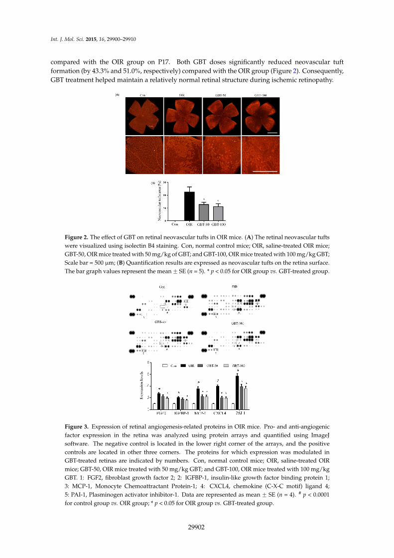
Figure 3. Expression of retinal angiogenesis-related proteins in OIR mice. Pro- and anti-angiogenic factor expression in the retina was analyzed using protein arrays and quantified using ImageJ software. The negative control is located in the lower right corner of the arrays, and the positive controls are located in other three corners. The proteins for which expression was modulated in GBT-treated retinas are indicated by numbers. Con, normal control mice; OIR, saline-treated OIR mice; GBT-50, OIR mice treated with 50 mg/kg GBT; and GBT-100, OIR mice treated with 100 mg/kg GBT. 1: FGF2, fibroblast growth factor 2; 2: IGFBP-1, insulin-like growth factor binding protein 1; 3: MCP-1, Monocyte Chemoattractant Protein-1; 4: CXCL4, chemokine (C-X-C motif) ligand 4; 5: PAI-1, Plasminogen activator inhibitor-1. Data are represented as mean ± SE (n = 4). # p < 0.0001 for control group vs. OIR group; * p < 0.05 for OIR group vs. GBT-treated group.
Figure 2. The effect of GBT on retinal neovascular tufts in OIR mice. (A) The retinal neovascular tuftswere visualized using isolectin B4 staining. Con, normal control mice; OIR, saline-treated OIR mice;GBT-50, OIR mice treated with 50 mg/kg of GBT; and GBT-100, OIR mice treated with 100 mg/kg GBT;Scale bar = 500 µm; (B) Quantification results are expressed as neovascular tufts on the retina surface.The bar graph values represent the mean ˘ SE (n = 5). * p < 0.05 for OIR group vs. GBT-treated group.
Int. J. Mol. Sci. 2015, 16, page–page
3
compared with the OIR group on P17. Both GBT doses significantly reduced neovascular tuft formation (by 43.3% and 51.0%, respectively) compared with the OIR group (Figure 2). Consequently, GBT treatment helped maintain a relatively normal retinal structure during ischemic retinopathy.
Figure 2. The effect of GBT on retinal neovascular tufts in OIR mice. (A) The retinal neovascular tufts were visualized using isolectin B4 staining. Con, normal control mice; OIR, saline-treated OIR mice; GBT-50, OIR mice treated with 50 mg/kg of GBT; and GBT-100, OIR mice treated with 100 mg/kg GBT; Scale bar = 500 µm; (B) Quantification results are expressed as neovascular tufts on the retina surface. The bar graph values represent the mean ± SE (n = 5). * p < 0.05 for OIR group vs. GBT-treated group.
Figure 3. Expression of retinal angiogenesis-related proteins in OIR mice. Pro- and anti-angiogenic factor expression in the retina was analyzed using protein arrays and quantified using ImageJ software. The negative control is located in the lower right corner of the arrays, and the positive controls are located in other three corners. The proteins for which expression was modulated in GBT-treated retinas are indicated by numbers. Con, normal control mice; OIR, saline-treated OIR mice; GBT-50, OIR mice treated with 50 mg/kg GBT; and GBT-100, OIR mice treated with 100 mg/kg GBT. 1: FGF2, fibroblast growth factor 2; 2: IGFBP-1, insulin-like growth factor binding protein 1; 3: MCP-1, Monocyte Chemoattractant Protein-1; 4: CXCL4, chemokine (C-X-C motif) ligand 4; 5: PAI-1, Plasminogen activator inhibitor-1. Data are represented as mean ± SE (n = 4). # p < 0.0001 for control group vs. OIR group; * p < 0.05 for OIR group vs. GBT-treated group.
Figure 3. Expression of retinal angiogenesis-related proteins in OIR mice. Pro- and anti-angiogenicfactor expression in the retina was analyzed using protein arrays and quantified using ImageJsoftware. The negative control is located in the lower right corner of the arrays, and the positivecontrols are located in other three corners. The proteins for which expression was modulated inGBT-treated retinas are indicated by numbers. Con, normal control mice; OIR, saline-treated OIRmice; GBT-50, OIR mice treated with 50 mg/kg GBT; and GBT-100, OIR mice treated with 100 mg/kgGBT. 1: FGF2, fibroblast growth factor 2; 2: IGFBP-1, insulin-like growth factor binding protein 1;3: MCP-1, Monocyte Chemoattractant Protein-1; 4: CXCL4, chemokine (C-X-C motif) ligand 4;5: PAI-1, Plasminogen activator inhibitor-1. Data are represented as mean ˘ SE (n = 4). # p < 0.0001for control group vs. OIR group; * p < 0.05 for OIR group vs. GBT-treated group.
29902

Int. J. Mol. Sci. 2015, 16, 29900–29910
2.2. GBT Significantly Reduced Angiogenesis-Related Factor Protein Expressions
To identify mechanistic insights into the effects of GBT in OIR mice, we analyzed the expressionlevels of 55 angiogenesis-related proteins that play roles in OIR pathogenesis via the protein arrayof the total protein isolated from the retinas. As demonstrated in Figure 3, GBT treatment at a doseof 100 mg/kg significantly decreased the expression of pro-angiogenic factors (i.e., MCP-1, IGFBP-1,PAI-1, and FGF2) compared with saline-treated OIR mice. Consistent with a previous study [22],VEGF protein was not detected in the angiogenesis protein array, possibly due to the low sensitivityto this antibody on the array. Thus, we further investigated changes in VEGF mRNA expressionlevels, which is a key player in angiogenesis.
2.3. GBT Treatment Downregulates VEGF, FGF2 and PAI-1 mRNAs Expression
Retinal VEGF, FGF2, and PAI-1 mRNA expressions were examined using real-time PCR.As expected, we observed a robust induction of VEGF mRNA during oxygen-induced retinopathy.In addition, FGF2 and PAI-1 mRNA levels were elevated in OIR mice compared with normal controlmice. However, VEGF, FGF2 and PAI-1 mRNA levels were significantly decreased in 100 mg/kgGBT-treated OIR mice (Figure 4).
Int. J. Mol. Sci. 2015, 16, page–page
4
2.2. GBT Significantly Reduced Angiogenesis-Related Factor Protein Expressions
To identify mechanistic insights into the effects of GBT in OIR mice, we analyzed the expression levels of 55 angiogenesis-related proteins that play roles in OIR pathogenesis via the protein array of the total protein isolated from the retinas. As demonstrated in Figure 3, GBT treatment at a dose of 100 mg/kg significantly decreased the expression of pro-angiogenic factors (i.e., MCP-1, IGFBP-1, PAI-1, and FGF2) compared with saline-treated OIR mice. Consistent with a previous study [22], VEGF protein was not detected in the angiogenesis protein array, possibly due to the low sensitivity to this antibody on the array. Thus, we further investigated changes in VEGF mRNA expression levels, which is a key player in angiogenesis.
2.3. GBT Treatment Downregulates VEGF, FGF2 and PAI-1 mRNAs Expression
Retinal VEGF, FGF2, and PAI-1 mRNA expressions were examined using real-time PCR. As expected, we observed a robust induction of VEGF mRNA during oxygen-induced retinopathy. In addition, FGF2 and PAI-1 mRNA levels were elevated in OIR mice compared with normal control mice. However, VEGF, FGF2 and PAI-1 mRNA levels were significantly decreased in 100 mg/kg GBT-treated OIR mice (Figure 4).
Figure 4. Real-time PCR analysis of VEGF, FGF2 and PAI-1 mRNA levels in OIR mice. When compared with normal controls, relative VEGF (A); FGF2 (B) and PAI-1 (C) mRNA expression levels were markedly increased in OIR mouse retinas and dramatically reduced after GBT treatment. Con, normal control mice; OIR, saline-treated OIR mice; GBT-50, OIR mice treated with GBT (50 mg/kg); and GBT-100, OIR mice treated with GBT (100 mg/kg). Data are represented as mean ± SE (n = 4). # p < 0.0001 for control group vs. OIR group; * p < 0.05 for OIR group vs. GBT-treated group.
3. Discussion
Retinal neovascularization is a major cause of blindness worldwide. Laser photocoagulation, cryotherapy, and intravitreal injection of anti-VEGF antibodies and VEGFR inhibitors are well-established treatments for retinal neovascularization. However, these retinal neovascularization therapies are limited and involve invasive procedures. Thus, better retinal neovascularization treatments are still very much in demand. In the present study, we evaluated the anti-angiogenic effect of GBT during pathological retinal neovascularization in OIR mice. We demonstrated that GBT has anti-angiogenic effects in this animal model.
Figure 4. Real-time PCR analysis of VEGF, FGF2 and PAI-1 mRNA levels in OIR mice. Whencompared with normal controls, relative VEGF (A); FGF2 (B) and PAI-1 (C) mRNA expression levelswere markedly increased in OIR mouse retinas and dramatically reduced after GBT treatment. Con,normal control mice; OIR, saline-treated OIR mice; GBT-50, OIR mice treated with GBT (50 mg/kg);and GBT-100, OIR mice treated with GBT (100 mg/kg). Data are represented as mean ˘ SE (n = 4).# p < 0.0001 for control group vs. OIR group; * p < 0.05 for OIR group vs. GBT-treated group.
3. Discussion
Retinal neovascularization is a major cause of blindness worldwide. Laser photocoagulation,cryotherapy, and intravitreal injection of anti-VEGF antibodies and VEGFR inhibitors arewell-established treatments for retinal neovascularization. However, these retinal neovascularizationtherapies are limited and involve invasive procedures. Thus, better retinal neovascularizationtreatments are still very much in demand. In the present study, we evaluated the anti-angiogeniceffect of GBT during pathological retinal neovascularization in OIR mice. We demonstrated that GBThas anti-angiogenic effects in this animal model.
29903

Int. J. Mol. Sci. 2015, 16, 29900–29910
The OIR model has been widely used as a valuable tool in retinal neovascularizationpathogenesis research [23]. In the mouse OIR model, the hyperoxic phase (P7–P12) resultsa loss of immature vessels in the central retina and development of vaso-obliteration (VO).When the mice are returned to room air (P12–P17), the retina becomes hypoxic due to theabsence of the retinal vasculature, which stimulates pro-angiogenic factors and results in abnormalneovascularization [24]. Many previous studies using the OIR model have demonstrated valuableeffects and laid the foundation for today’s clinical application of anti-angiogenic treatments in retinalneovascularization [23,24].
Clinical evidence has repeatedly suggested the use of anti-VEGF or VEGFR inhibitors.Bevacizumab, a humanized monoclonal antibody targeting VEGF and the tyrosine kinaseinhibitors sorafenib, a multikinase inhibitor targeting VEGFR/platelet-derived growth factor receptor(PDGFR) represent the first approved anti-angiogenic agents by the FDA that suppress neovesselformation [25]. However, despite anti-VEGF treatment, components of FGF, PDGF, epidermalgrowth factor (EGF), and other pathways may compensate for a VEGF blockade and promoteangiogenesis [26]. Previous studies have reported that tumors may acquire resistance to anti-VEGFtherapy following angiogenesis reactivation, which is induced by compensatory upregulationof FGF2/FGFR system in experimental animals [27] and patients with cancer [28]. Thus,accumulated reports have suggested that FGF has a significant role in retinal pathogenic angiogenesis.Accordingly, there is a strong need to develop new, affordable drugs with different modes ofaction [29]. A combination of multiple herbs has a synergistic effect due to the phytocompoundscontained in the different herbs [30]. Interestingly, GBT, which is a mixture of 12 herbs, inhibitsretinal neovascularization via the suppression of VEGF, FGF2, and PAI-1 in OIR mice. Our array datademonstrate that GBT exerts an inhibitory effect on angiogenesis by downregulating the expressionof FGF2, IGFBP-1, MCP-1 and PAI-1. Although CXLCL4, an anti-angiogenic factor, was alsosignificantly downregulated by GBT treatment, the upregulation of CXCL4 in saline-treated OIR micemay be a defense mechanism against angiogenesis. A decrease of CXCL4 in GBT-treated OIR micemight be an indirect effect as a result of reduced angiogenesis.
FGF2 is a member of heparin-binding growth factors and a potent pro-angiogenic signalingmolecule in vivo and in vitro. FGF2 is expressed in many tissues and cell types and directly affectnew vessel growth through heparan-sulfate proteoglycan tyrosine kinase receptors and integrinsthat are expressed on the surface of vascular endothelial cells [31,32]. Furthermore, blockingboth FGF2 and VEGF is essential to disrupting the interconnection between these two angiogenicfactors and effectively inhibiting early stage angiogenesis [33]. Recently, novel anti-angiogenicdrugs have been developed that block the mechanisms of FGF2 and VEGF action [34]. Previousstudies have demonstrated that anti-FGF2 antibodies either alone or in combination with anti-VEGFantibodies strongly upregulate PAI-1 expression in bovine aortic endothelial cells and bovine vascularendothelial cells and [10,35]. PAl-1 production plays a role in mediating the trophic effects resultingfrom FGF2 receptor activation by FGF2 itself or through the action of the cell adhesion molecule.PAI-1 can both inhibit and promote angiogenesis [13]. PAI-1 inhibited endothelial sprouting in anex vivo study [36]. In contrast, PAI-1 promotes tumor growth via anti-apoptotic effects in cancercells as well as non-cancer cell lines in vitro [37]. It was also demonstrated that a lack of host PAI-1suppresses tumor growth [38] and inhibits cancer metastasis and angiogenesis [39]. In OIR mice,PAI-1 protein and mRNA expression levels are significantly elevated during active phases of newvessel growth [14]. Therefore, VEGF, FGF2, and PAI-1 have been proposed as targets for treatingretinal neovascularization. GBT could reduce VEGF, FGF2, and PAI-1 expression in OIR mice. Theseobservations indicate that the inhibitory effect of GBT on retinal neovascularization might occur viathe partial inhibition of VEGF, FGF2, and PAI-1.
VEGF inhibitors are of great benefit to patients with neovascular ocular diseases [40]. However,an increasing amount of evidence suggests that IGFBP-1 and MCP-1 also have a role in retinalneovascularization. Our array results indicate that GBT inhibits the expression of IGFBP-1 and
29904

Int. J. Mol. Sci. 2015, 16, 29900–29910
MCP-1. MCP-1 expression is highly increased in diabetic retinopathy, while MCP-1 deficiencyprevents the development of subretinal neovascularization in MCP´{´ mice [41]. The enhancedexpression of IGFBP-1 in the retina also plays an important role in the pathogenesis of retinalneovascularization. Vitreal expression levels of IGFBP-1 were increased in patients with ischemiccentral retinal vein occlusion [42], and IGFBP-1 shows large increases in neovascular tufts in ischemicretinopathy [43]. Based on these findings, we can hypothesize that IGFBP-1 and MCP-1 may be validsecondary targets when fighting retinal neovascularization [40]. In the present study, GBT preventedretinal neovascularization through the downregulation of IGFBP-1 and MCP-1.
In OIR animal models, reactive oxygen species (ROS) generation during ischemia plays a crucialrole in retinal neovascularization. Ischemic retina leads to excessive production of ROS, which,in turn, activates NADPH oxidase and contributes to retinal pathogenic neovascularization [44].Natoli et al. reported that oxidative stress-related retinal cell death and photoreceptor dysfunctionswere also observed in OIR animal models [45]. Excess ROS disturbs the normal mitochondrialfunction by widening the permeability transition pore located on an inner mitochondrial membrane,resulting in further ROS overproduction [46], the release of cytochrome c, and the depletion of ATP,leading to cell damage [47]. Cellular ATP is mainly generated in mitochondria. MitochondrialF0F1-ATPase synthesizes ATP during oxidative phosphorylation, which is important for cellularsurvival and proliferation [48]. Recently, Yamamoto et al. reported that several subunits of themitochondrial F0F1-ATPase are expressed on the surface of the human umbilical vein endothelial cells(HUVEC) [49]. Moser et al. showed that angiostatin, a potent anti-angiogenic agent, suppressed theproliferation if HUVECs by reducing ATPase activity [50]. More recently, Calzia et al. reported thatpolyphenolic phytochemicals such as resveratrol and curcumin inhibited the mitochondrial F0F1-ATPsynthase in the rod outer segments of the retina [51]. GBT has various polyphenolic compoundssuch as liquiritin and glycyrrhizin. However, the effect of GBT on F0F1-ATP synthase has not yetbeen investigated.
GBT contains 12 herbal medicines, and its ingredient compounds are two flavonoids (liquiritinand glycyrrhizin), a phenolic (6-gingerol) and five coumarins (nodakenin, nodakenetin, decursinol,decursinol angelate, and decursin) [52,53]. Glycyrrhizin has anti-angiogenic activities such asendothelial cell migration, invasion and tube formation, along with anti-tumor activities such asangiogenesis and tumor growth in mice [54]. Moreover, glycyrrhizin, which is a selective inhibitorof high mobility group box-1, reduced retinal neovascularization in OIR mice [55]. The majorcoumarins decursin, decursinol and decursinol angelate exert inhibitory effects on angiogenesisby reducing VEGF expression [56,57]. Cloricromene, a semi-synthetic coumarin derivative,lowered retinal expressions of VEGF, ICAM-1, and nitrotyrosine in STZ-induced diabetic rats [58].Eriodictyol, a flavonoid compound, protected human retinal pigment epithelial cells from oxidativestress-induced cell death [59] and reduced retinal expressions of TNF-α, ICAM-1, VEGF, and eNOS inSTZ-induced diabetic rats [60]. Flavonoids inhibited VEGF/FGF2-induced angiogenesis to preventVEGF/FGF2-induced uPA expression and the activation of its inhibitor PAI-1 [61].
Herbal medicines usually consist of many compounds. Generally, herbal practitioners rarelyuse single herb alone. Herbal formula has many advantages, such as additive synergic interactionsamong a variety of phytocompounds contained in the different herbs [62]. Although it was stillunclear which compound in GBT might play the most important role in the inhibition of retinalneovascularization, these observations suggest that the prevention of retinal neovascularization byGBT may occur due to a combination of these compounds’ effects. However, the detailed mechanismthat underlies the synergistic effects of GBT on retinal neovascularization remains unknown.
29905

Int. J. Mol. Sci. 2015, 16, 29900–29910
4. Experimental Section
4.1. GBT Preparation
GBT consists of 12 herbal medicines, which were purchased from Baekjedang (Daejeon, Korea).The mixture of crude herbs was boiled with distilled water at 100 ˝C for 2 h in the following ratio:Angelica gigas (3.75 g), Dimocarpus longan (3.75 g), Panax ginseng (3.75 g), Zizyphus jujuba (3.75 g),Polygala tenuifolia (3.75 g), Astragalus membranaceus (3.75 g), Poria cocos (3.75 g), Atractylodes japonica(3.75 g), Zizyphus jujube (3.75 g), Aucklandia lappa (1.875 g), Glycyrrhiza uralensis (1.125 g), andZingiber officinale (6.25 g). The extract was filtered, lyophilized and stored at ´20 ˝C until use.A standardized powder of GBT was provided by Hyeun-Kyoo Shin (Korea Institute of OrientalMedicine, Daejeon, Korea). The HPLC fingerprint and contents of three major compounds ofGBT are described in a previous report [52]. The quantity of the three compounds in GBT were0.70 ˘ 0.01 mg/g (liquiritin), 0.03 ˘ 0.01 mg/g (glycyrrhizin), and 1.43 ˘ 0.01 mg/g (nodakenin).
4.2. A Mouse Model of Oxygen-Induced Retinopathy
Ischemic retinopathy was induced in C57BL/6 mouse pups, as described previously [55].On postnatal day 12 (P12), after being exposed to 75% ˘ 2% oxygen for five days (P7–P12),the mice were randomly assigned to one of three groups: OIR, GBT-50 (50 mg/kg/day), or GBT-100(100 mg/kg/day). The normal control group (Con) was simultaneously raised in normal room air.The GBT was dissolved in saline, and 100 µL of this solution was injected intraperitoneally oncedaily for five days (P12–P16). The OIR and normal control groups were injected with saline solutionfor five days. On P17, after five days of intraperitoneal injections, the mice were anesthetized andsacrificed. These experiments were repeated four times using four animals per group. All animalexperiments were approved by the Korea Institute of Oriental Medicine Institutional Animal Careand Use Committee. (Approval number: 14-024, approval date: 20 February 2014).
4.3. Fluorescein-Dextran Microscopy
On P17, the mice were deeply anesthetized using zolazepam (Zoletil, Virbac, Carros, France),and PBS containing fluorescein-dextran (FD40S, Sigma-Aldrich, St. Louis, MO, USA) was subsequentlyperfused via the left ventricle. The retinas were dissected, flat mounted on to glass slides and viewedusing fluorescence microscopy (BX51, Olympus, Tokyo, Japan). The area of non-perfusion in theretinal center was quantified using ImageJ software (NIH, Bethesda, MD, USA).
4.4. Lectin Staining
The retinas were incubated with 1% bovine serum albumin and 5% Triton X-100 in PBS for threehours at room temperature. The retinas were washed three times with PBS and incubated overnightat 4 ˝C with Bandeiraea simplicifolia isolectin B4 (Sigma-Aldrich, St. Louis, MO, USA), which wasdiluted 1:50 in PBS. The retinas were washed with 0.05% Tween 20 in PBS, followed by incubationwith streptavidin TRITC (1:500, Serotec, Oxford, UK) for 4 h at 37 ˝C. The retinas were flat mountedand viewed using fluorescence microscopy (BX51, Olympus, Tokyo, Japan). ImageJ software (NIH,Bethesda, MD, USA) was used to measure the neovascular tufts on the retinas.
4.5. Angiogenesis-Related Protein Array
To investigate angiogenesis-related proteins in the retinas, a mouse angiogenesis array(R&D Systems Inc., Minneapolis, MN, USA) was performed according to the manufacturer’sprotocol. Briefly, pooled mouse retinas (n = 3) were homogenized in PBS using protease inhibitors,centrifuged at 10,000ˆ g for 5 min, and total protein concentrations were quantified. Lysates wereadded to a membrane and blotted with antibodies against angiogenesis-related proteins. Afterincubation overnight at 4 ˝C, the membranes were treated with streptavidin-horseradish peroxidase
29906

Int. J. Mol. Sci. 2015, 16, 29900–29910
and visualized using an enhanced chemiluminescence detection system (Amersham Bioscience,Piscataway, NJ, USA) on an image analyzer (LAS-3000, Fujifilm, Tokyo, Japan). Optical densitymeasurements were obtained using ImageJ software (NIH, Bethesda, MD, USA).
4.6. Real-Time PCR Analysis
Total RNA was isolated using the TRIzol reagent (Invitrogen, Carlsbad, CA, USA), and0.5 µg of total RNA was reverse transcribed into cDNA with the PrimeScript First Strand cDNASynthesis kit (Bio-Rad, Hercules, CA, USA). Quantitative real-time PCR was performed with specificprimers for VEGF, FGF2, PAI-1 and GAPDH using an iQ5 Continuous Fluorescence Detector System(Bio-Rad, Hercules, CA, USA). The primer sequences were as follows: VEGF, 51-TCCTCCTATCTCCACCACCTATCC-31 and 51-GACCCAGCCAGCCATACCC-31; FGF2, 51-AGAGCGACCCACACGTCAAAC-31 and 51-CCAACTGGAGTATTTCCGTGACC-31; PAI-1, 51-GCCAGATTTATCATCAATGACTGGG-31 and 51-GGAGAGGTGCACATCTTTCTCAAAG-31; GAPDH, 51-TGTCGTCCCAGTTGGTAA-31 and 51-CTTTGCAGCTCCTTCGTT-31. All real-time PCR experiments were run astriplicates. GAPDH mRNA levels were determined for the normalization of VEGF, FGF2 and PAI-1mRNA expression values using the iQ5 optical system software (Bio-Rad, Hercules, CA, USA).
4.7. Statistical Analysis
The results were expressed as the mean ˘ SE and analyzed using one-way analysis of variance(ANOVA) followed by either Tukey’s multiple comparison test or an unpaired Student’s t-test.All analyses were performed using Prism 6.0 software (GraphPad Software, San Diego, CA, USA).
5. Conclusions
In conclusion, we have demonstrated that GBT inhibited ischemic retinopathy-induced retinalpathogenic angiogenesis in OIR mice for the first time. In addition, VEGF, FGF2 and PAI-1overexpression were significantly reduced by GBT treatment. These observations suggest that GBThas anti-angiogenic activities through the partial inhibition of VEGF, FGF2, and PAI-1. Further studiesmust be carried out to figure out the potential clinical use of GBT.
Acknowledgments: This research was supported by a grant of the Korea Institute of Oriental Medicine (K15801).
Author Contributions: Yun Mi Lee performed experiments and wrote the manuscript. Yu-Ri Lee contributedto the revision and reviewed the manuscript. Chan-Sik Kim, Kyuhyung Jo and Eunjin Sohn performedexperiments. Jin Sook Kim contributed to the discussions and reviewed the manuscript. Junghyun Kim designedand supervised the study. All authors read and approved the final manuscript.
Conflicts of Interest: The authors declare no conflict of interest.
References
1. Patz, A. Studies on retinal neovascularization. Friedenwald Lecture. Investig. Ophthalmol. Vis. Sci. 1980, 19,1133–1138.
2. Damico, F.M. Angiogenesis and retinal diseases. Arq. Bras. Oftalmol. 2007, 70, 547–553. [CrossRef][PubMed]
3. Sennlaub, F.; Chemtob, S. VEGFR-1: A safe target for prophylaxis of retinopathy of prematurity?Pediatr. Res. 2004, 55, 1–2. [CrossRef] [PubMed]
4. Smith, L.E.; Kopchick, J.J.; Chen, W.; Knapp, J.; Kinose, F.; Daley, D.; Foley, E.; Smith, R.G.; Schaeffer, J.M.Essential role of growth hormone in ischemia-induced retinal neovascularization. Science 1997, 276,1706–1709. [CrossRef] [PubMed]
5. Asahara, T.; Bauters, C.; Zheng, L.P.; Takeshita, S.; Bunting, S.; Ferrara, N.; Symes, J.F.; Isner, J.M. Synergisticeffect of vascular endothelial growth factor and basic fibroblast growth factor on angiogenesis in vivo.Circulation 1995, 92, 365–371. [CrossRef]
29907

Int. J. Mol. Sci. 2015, 16, 29900–29910
6. Pepper, M.S.; Ferrara, N.; Orci, L.; Montesano, R. Potent synergism between vascular endothelial growthfactor and basic fibroblast growth factor in the induction of angiogenesis in vitro. Biochem. Biophys.Res. Commun. 1992, 189, 824–831. [CrossRef]
7. Goto, F.; Goto, K.; Weindel, K.; Folkman, J. Synergistic effects of vascular endothelial growth factor andbasic fibroblast growth factor on the proliferation and cord formation of bovine capillary endothelial cellswithin collagen gels. Lab. Investig. 1993, 69, 508–517. [PubMed]
8. Hu, D.E.; Fan, T.P. Suppression of VEGF-induced angiogenesis by the protein tyrosine kinase inhibitor,lavendustin A. Br. J. Pharmacol. 1995, 114, 262–268. [CrossRef] [PubMed]
9. D’Amore, P.A. Mechanisms of retinal and choroidal neovascularization. Investig. Ophthalmol. Vis. Sci. 1994,35, 3974–3979.
10. Pepper, M.S.; Ferrara, N.; Orci, L.; Montesano, R. Vascular endothelial growth factor (VEGF)induces plasminogen activators and plasminogen activator inhibitor-1 in microvascular endothelial cells.Biochem. Biophys. Res. Commun. 1991, 181, 902–906. [CrossRef]
11. Fotsis, T.; Pepper, M.; Adlercreutz, H.; Fleischmann, G.; Hase, T.; Montesano, R.; Schweigerer, L. Genistein,a dietary-derived inhibitor of in vitro angiogenesis. Proc. Natl. Acad. Sci. USA 1993, 90, 2690–2694.[CrossRef] [PubMed]
12. Gils, A.; Declerck, P.J. The structural basis for the pathophysiological relevance of PAI-I in cardiovasculardiseases and the development of potential PAI-I inhibitors. Thromb. Haemost. 2004, 91, 425–437. [CrossRef][PubMed]
13. Tashiro, Y.; Nishida, C.; Sato-Kusubata, K.; Ohki-Koizumi, M.; Ishihara, M.; Sato, A.; Gritli, I.;Komiyama, H.; Sato, Y.; Dan, T.; et al. Inhibition of PAI-1 induces neutrophil-driven neoangiogenesis andpromotes tissue regeneration via production of angiocrine factors in mice. Blood 2012, 119, 6382–6393.[CrossRef] [PubMed]
14. Basu, A.; Menicucci, G.; Maestas, J.; Das, A.; McGuire, P. Plasminogen activator inhibitor-1 (PAI-1) facilitatesretinal angiogenesis in a model of oxygen-induced retinopathy. Investig. Ophthalmol. Vis. Sci. 2009, 50,4974–4981. [CrossRef] [PubMed]
15. Hur, J. Donguibogam; Namsandang: Seoul, Korea, 2007; p. 98.16. Busta, I.; Xie, H.; Kim, M.S. The use of Gui-Pi-Tang in small animals with immune-mediated blood
disorders. J. Vet. Clin. 2009, 26, 181–184.17. Eun, J.S.; Song, J.M. Effects of Kwibi-tang on serum levels of hormone and the non-specific immune
response after immobilization stress in mice. Korean J. Orient. Med. Physiol. Pathol. 2004, 18, 172–178.18. Lim, J.W.; Kim, J.W.; Chung, S.Y.; Cho, S.H.; Oh, M.S.; Hwang, W.W. The Antioxidative and
neuroprotective effect of guibi-tang(Guipitang) and guibi-tang gamibang(guipitang jiaweijang) onPC12!cells. J. Orient. Neuropsychiatry 2009, 20, 1–19.
19. Kim, H.J.; Choi, J.H.; Lim, S.W. The Defensive Effect of Keuibi-tang on the Gastric Mucous Membrane ofMouse Injured by Stress and Ethanol. J. Korean Orient. Med. 2003, 24, 155–168.
20. Rosenfarb, A. Healing Your Eyes with Chinese Medicine: Acupuncture, Acupressure, & Chinese Herbs;North Atlantic Books: Berkeley, CA, USA, 2007.
21. Kim, J.H.; Lee, Y.M.; Ahn, E.M.; Kim, K.W.; Yu, Y.S. Decursin inhibits retinal neovascularization viasuppression of VEGFR-2 activation. Mol. Vis. 2009, 15, 1868–1875. [PubMed]
22. Lee, Y.M.; Kim, C.S.; Sohn, E.; Jo, K.; Lim, H.R.; Kim, S.K.; Kim, J.S.; Kim, J. Sipjeondaebo-tang, a traditionalherbal formula, inhibits retinal neovascularization in a mouse model of oxygen-induced retinopathy.Tohoku. J. Exp. Med. 2014, 234, 229–236. [CrossRef] [PubMed]
23. Stahl, A.; Connor, K.M.; Sapieha, P.; Chen, J.; Dennison, R.J.; Krah, N.M.; Seaward, M.R.; Willett, K.L.;Aderman, C.M.; Guerin, K.I.; et al. The mouse retina as an angiogenesis model. Investig. Ophthalmol.Vis. Sci. 2010, 51, 2813–2826. [CrossRef] [PubMed]
24. Stahl, A.; Connor, K.M.; Sapieha, P.; Willett, K.L.; Krah, N.M.; Dennison, R.J.; Chen, J.; Guerin, K.I.;Smith, L.E. Computer-aided quantification of retinal neovascularization. Angiogenesis 2009, 12, 297–301.[CrossRef] [PubMed]
25. Brower, V. Antiangiogenesis research is booming, as questions and studies proliferate. J. Natl. Cancer Inst.2009, 101, 780–781. [CrossRef] [PubMed]
29908

Int. J. Mol. Sci. 2015, 16, 29900–29910
26. Choi, H.J.; Armaiz Pena, G.N.; Pradeep, S.; Cho, M.S.; Coleman, R.L.; Sood, A.K. Anti-vascular therapies inovarian cancer: Moving beyond anti-VEGF approaches. Cancer Metastasis Rev. 2014, 34, 19–40. [CrossRef][PubMed]
27. Casanovas, O.; Hicklin, D.J.; Bergers, G.; Hanahan, D. Drug resistance by evasion of antiangiogenictargeting of VEGF signaling in late-stage pancreatic islet tumors. Cancer Cell 2005, 8, 299–309. [CrossRef][PubMed]
28. Brower, V. How well do angiogenesis inhibitors work? Biomarkers of response prove elusive. J. Natl.Cancer Inst. 2009, 101, 846–847. [CrossRef] [PubMed]
29. Sulaiman, R.S.; Basavarajappa, H.D.; Corson, T.W. Natural product inhibitors of ocular angiogenesis.Exp. Eye Res. 2014, 129, 161–171. [CrossRef] [PubMed]
30. Lee, H.J.; Lee, E.O.; Rhee, Y.H.; Ahn, K.S.; Li, G.X.; Jiang, C.; Lu, J.; Kim, S.H. An oriental herbal cocktail,ka-mi-kae-kyuk-tang, exerts anti-cancer activities by targeting angiogenesis, apoptosis and metastasis.Carcinogenesis 2006, 27, 2455–2463. [CrossRef] [PubMed]
31. Redington, A.E.; Roche, W.R.; Madden, J.; Frew, A.J.; Djukanovic, R.; Holgate, S.T.; Howarth, P.H.Basic fibroblast growth factor in asthma: measurement in bronchoalveolar lavage fluid basally andfollowing allergen challenge. J. Allergy Clin. Immunol. 2001, 107, 384–387. [CrossRef] [PubMed]
32. Yamashita, A.; Yonemitsu, Y.; Okano, S.; Nakagawa, K.; Nakashima, Y.; Irisa, T.; Iwamoto, Y.;Nagai, Y.; Hasegawa, M.; Sueishi, K. Fibroblast growth factor-2 determines severity of joint disease inadjuvant-induced arthritis in rats. J. Immunol. 2002, 168, 450–457. [CrossRef] [PubMed]
33. Chung, S.W.; Bae, S.M.; Lee, M.; Al-Hilal, T.A.; Lee, C.K.; Kim, J.K.; Kim, I.S.; Kim, S.Y.; Byun, Y.LHT7, a chemically modified heparin, inhibits multiple stages of angiogenesis by blocking VEGF, FGF2and PDGF-B signaling pathways. Biomaterials 2015, 37, 271–278. [CrossRef] [PubMed]
34. Cross, M.J.; Claesson-Welsh, L. FGF and VEGF function in angiogenesis: signalling pathways, biologicalresponses and therapeutic inhibition. Trends Pharmacol. Sci. 2001, 22, 201–207. [CrossRef]
35. Mandriota, S.J.; Pepper, M.S. Vascular endothelial growth factor-induced in vitro angiogenesis andplasminogen activator expression are dependent on endogenous basic fibroblast growth factor. J. Cell Sci.1997, 110, 2293–2302. [PubMed]
36. Brodsky, S.; Chen, J.; Lee, A.; Akassoglou, K.; Norman, J.; Goligorsky, M.S. Plasmin-dependentand -independent effects of plasminogen activators and inhibitor-1 on ex vivo angiogenesis. Am. J. Physiol.Heart Circ. Physiol. 2001, 281, H1784–H1792. [PubMed]
37. Kwaan, H.C.; Wang, J.; Svoboda, K.; Declerck, P.J. Plasminogen activator inhibitor 1 may promote tumourgrowth through inhibition of apoptosis. Br. J. Cancer 2000, 82, 1702–1708. [PubMed]
38. Gutierrez, L.S.; Schulman, A.; Brito-Robinson, T.; Noria, F.; Ploplis, V.A.; Castellino, F.J. Tumor developmentis retarded in mice lacking the gene for urokinase-type plasminogen activator or its inhibitor, plasminogenactivator inhibitor-1. Cancer Res. 2000, 60, 5839–5847. [PubMed]
39. Bajou, K.; Noel, A.; Gerard, R.D.; Masson, V.; Brunner, N.; Holst-Hansen, C.; Skobe, M.; Fusenig, N.E.;Carmeliet, P.; Collen, D.; et al. Absence of host plasminogen activator inhibitor 1 prevents cancer invasionand vascularization. Nat. Med. 1998, 4, 923–928. [CrossRef] [PubMed]
40. Campochiaro, P.A. Ocular neovascularization. J. Mol. Med. 2013, 91, 311–321. [CrossRef] [PubMed]41. Ambati, J.; Anand, A.; Fernandez, S.; Sakurai, E.; Lynn, B.C.; Kuziel, W.A.; Rollins, B.J.; Ambati, B.K.
An animal model of age-related macular degeneration in senescent Ccl-2- or Ccr-2-deficient mice. Nat. Med.2003, 9, 1390–1397. [CrossRef] [PubMed]
42. Ehlken, C.; Grundel, B.; Michels, D.; Junker, B.; Stahl, A.; Schlunck, G.; Hansen, L.L.; Feltgen, N.; Martin, G.;Agostini, H.T.; et al. Increased expression of angiogenic and inflammatory proteins in the vitreous ofpatients with ischemic central retinal vein occlusion. PLoS ONE 2015, 10, e0126859. [CrossRef] [PubMed]
43. Lofqvist, C.; Willett, K.L.; Aspegren, O.; Smith, A.C.; Aderman, C.M.; Connor, K.M.; Chen, J.; Hellstrom, A.;Smith, L.E. Quantification and localization of the IGF/insulin system expression in retinal blood vessels andneurons during oxygen-induced retinopathy in mice. Investig. Ophthalmol. Vis. Sci. 2009, 50, 1831–1837.[CrossRef] [PubMed]
44. Li, S.Y.; Fu, Z.J.; Lo, A.C. Hypoxia-induced oxidative stress in ischemic retinopathy. Oxid. Med. Cell. Longev.2012, 2012, 426769. [CrossRef] [PubMed]
29909

Int. J. Mol. Sci. 2015, 16, 29900–29910
45. Natoli, R.; Valter, K.; Barbosa, M.; Dahlstrom, J.; Rutar, M.; Kent, A.; Provis, J. 670nm photobiomodulation asa novel protection against retinopathy of prematurity: evidence from oxygen induced retinopathy models.PLoS ONE 2013, 8, e72135. [CrossRef] [PubMed]
46. Bodrova, M.E.; Brailovskaya, I.V.; Efron, G.I.; Starkov, A.A.; Mokhova, E.N. Cyclosporin A-sensitivedecrease in the transmembrane potential across the inner membrane of liver mitochondria induced bylow concentrations of fatty acids and Ca2+. Biochemistry 2003, 68, 391–398. [PubMed]
47. Greco, T.; Shafer, J.; Fiskum, G. Sulforaphane inhibits mitochondrial permeability transition and oxidativestress. Free Radic. Biol. Med. 2011, 51, 2164–2171. [CrossRef] [PubMed]
48. Panfoli, I.; Ravera, S.; Bruschi, M.; Candiano, G.; Morelli, A. Proteomics unravels the exportability ofmitochondrial respiratory chains. Expert Rev. Proteom. 2011, 8, 231–239. [CrossRef] [PubMed]
49. Yamamoto, K.; Shimizu, N.; Obi, S.; Kumagaya, S.; Taketani, Y.; Kamiya, A.; Ando, J. Involvement of cellsurface ATP synthase in flow-induced ATP release by vascular endothelial cells. Am. J. Physiol. HeartCirc. Physiol. 2007, 293, 1646–1653. [CrossRef] [PubMed]
50. Moser, T.L.; Stack, M.S.; Asplin, I.; Enghild, J.J.; Hojrup, P.; Everitt, L.; Hubchak, S.; Schnaper, H.W.;Pizzo, S.V. Angiostatin binds ATP synthase on the surface of human endothelial cells. Proc. Natl. Acad.Sci. USA 1999, 96, 2811–2816. [CrossRef] [PubMed]
51. Calzia, D.; Oneto, M.; Caicci, F.; Bianchini, P.; Ravera, S.; Bartolucci, M.; Diaspro, A.; Degan, P.; Manni, L.;Traverso, C.E.; et al. Effect of polyphenolic phytochemicals on ectopic oxidative phosphorylation in rodouter segments of bovine retina. Br. J. Pharmacol. 2015, 172, 3890–3903. [CrossRef] [PubMed]
52. Seo, C.S.; Kim, J.H.; Shin, H.K. Simultaneous determination of liquiritin, nodakenin and glycyrrhizininGuibi-tang, a traditional herbal prescription by HPLC-PDA. Pak. J. Pharm. Sci. 2014, 27, 819–824. [PubMed]
53. Liang, C.; Lee, K.J.; Jeong, S.W.; Ha, J.H.; Ma, J.Y. Analysis of Constituents from Guibi-Tang withLactobacillus. Int. J. Biosci. Biochem. Bioinform. 2012, 2, 374–376. [CrossRef]
54. Kim, K.J.; Choi, J.S.; Kim, K.W.; Jeong, J.W. The anti-angiogenic activities of glycyrrhizic acid in tumorprogression. Phytother. Res. 2013, 27, 841–846. [CrossRef] [PubMed]
55. Lee, Y.M.; Kim, J.; Jo, K.; Shin, S.D.; Kim, C.S.; Sohn, E.J.; Kim, S.G.; Kim, J.S. Ethyl pyruvate inhibits retinalpathogenic neovascularization by downregulating HMGB1 expression. J. Diabetes Res. 2013, 2013, 245271.[CrossRef] [PubMed]
56. Jung, M.H.; Lee, S.H.; Ahn, E.M.; Lee, Y.M. Decursin and decursinol angelate inhibit VEGF-inducedangiogenesis via suppression of the VEGFR-2-signaling pathway. Carcinogenesis 2009, 30, 655–661.[CrossRef] [PubMed]
57. Son, S.H.; Kim, M.J.; Chung, W.Y.; Son, J.A.; Kim, Y.S.; Kim, Y.C.; Kang, S.S.; Lee, S.K.; Park, K.K.Decursin and decursinol inhibit VEGF-induced angiogenesis by blocking the activation of extracellularsignal-regulated kinase and c-Jun N-terminal kinase. Cancer Lett. 2009, 280, 86–92. [CrossRef] [PubMed]
58. Bucolo, C.; Ward, K.W.; Mazzon, E.; Cuzzocrea, S.; Drago, F. Protective effects of a coumarin derivative indiabetic rats. Investig. Ophthalmol. Vis. Sci. 2009, 50, 3846–3852. [CrossRef] [PubMed]
59. Johnson, J.; Maher, P.; Hanneken, A. The flavonoid, eriodictyol, induces long-term protection in ARPE-19cells through its effects on Nrf2 activation and phase 2 gene expression. Investig. Ophthalmol. Vis. Sci. 2009,50, 2398–2406. [CrossRef] [PubMed]
60. Bucolo, C.; Leggio, G.M.; Drago, F.; Salomone, S. Eriodictyol prevents early retinal and plasmaabnormalities in streptozotocin-induced diabetic rats. Biochem. Pharmacol. 2012, 84, 88–92. [CrossRef][PubMed]
61. Kim, M.H. Flavonoids inhibit VEGF/bFGF-induced angiogenesis in vitro by inhibiting thematrix-degrading proteases. J. Cell. Biochem. 2003, 89, 529–538. [CrossRef] [PubMed]
62. Cao, P.; Cai, X.; Lu, W.; Zhou, F.; Huo, J. Growth inhibition and induction of apoptosis in SHG-44 gliomacells by Chinese medicine formula “Pingliu Keli”. Evid. Based Complement. Alternat. Med. 2011. [CrossRef][PubMed]
© 2015 by the authors; licensee MDPI, Basel, Switzerland. This article is an openaccess article distributed under the terms and conditions of the Creative Commons byAttribution (CC-BY) license (http://creativecommons.org/licenses/by/4.0/).
29910