Effect of different restorative procedures on the … · Effect of different restorative procedures...
6
Restorative Dentistry Braz Oral Res. 2012 Jan-Feb;26(1):77-82 77 Andiara Ribeiro Roberto (a) Manoel Damião de Sousa-Neto (b) Raqueli Viapiana (a) Alessandro Rogério Giovani (a) Celso Bernardo de Souza Filho (a) Silvana Maria Paulino (a) Yara Teresinha Correa Silva-Sousa (a) (a) Dentistry Course, University of Ribeirão Preto, Ribeirão Preto, SP, Brazil. (b) Department of Restorative Dentistry, School of Dentistry of Ribeirão Preto, University of São Paulo, SP, Brazil. Restorative Dentistry Corresponding author: Manoel Damião de Sousa-Neto E-mail: [email protected] Received for publication on Aug 31, 2011 Accepted for publication on Nov 18, 2011 Effect of different restorative procedures on the fracture resistance of teeth submitted to internal bleaching Abstract: The aim of this study was to evaluate the influence of differ- ent restorative procedures on the fracture resistance of endodontically treated teeth submitted to intracoronal bleaching. Fifty upper central in- cisors were distributed into 5 groups: GI - healthy teeth; GII - endodon- tically treated teeth sealed with Coltosol; GIII - endodontically treated teeth bleached and sealed with Coltosol; GIV - endodontically treated teeth bleached and restored with composite resin; and GV - endodonti- cally treated teeth bleached and restored with a fiberglass post and com- posite resin. In the bleached specimens, a cervical seal was made prior to bleaching with 38% hydrogen peroxide. The gel was applied on the buc- cal surface and in the pulp chamber, and was then light-activated for 45 s. This procedure was repeated three times per session for four sessions, and each group was submitted to the restorative procedures described above. The specimens were submitted to fracture resistance testing in a universal testing machine. There were statistically significant differences among the groups (p < 0.05). The mean value found for GIII was the low- est (0.32 kN) and was significantly different from the values found for GI (0.75 kN), GII (0.67 kN), GIV (0.70 kN), and GV (0.72 kN), which were not significantly different from each other (p > 0.05). The restorative pro- cedures using composite resin were found to successfully restore the frac- ture resistance of endodontically treated and bleached teeth. Descriptors: Tooth Bleaching; Endodontics; Dentistry, Operative; Tooth Fractures. Introduction Intracoronal dental bleaching is commonly recommended for dis- colored teeth submitted to endodontic treatment. 1 However, some stud- ies have described the action of bleaching agents on the morphological structure of dentin as being responsible for making the tooth more sus- ceptible to fracture. 2-6 As to the bleaching procedures, there is a general concern regarding their effects on enamel and dentin, since changes in tooth structure, such as those related to porosity, 7 demineralization 8-10 and reduction in the ad- hesion of restorative materials 5,6 have been associated with the oxidizing agents used in the bleaching process. The reduction in hardness and in tooth fracture resistance promoted by bleaching agents has motivated the development of restorative materials and techniques that aim at reinforc- Declaration of Interests: The authors certify that they have no commercial or associative interest that represents a conflict of interest in connection with the manuscript.
Transcript of Effect of different restorative procedures on the … · Effect of different restorative procedures...

Restorative Dentistry
Braz Oral Res. 2012 Jan-Feb;26(1):77-82 77
Andiara Ribeiro Roberto(a)
Manoel Damião de Sousa-Neto(b)
Raqueli Viapiana(a)
Alessandro Rogério Giovani(a)
Celso Bernardo de Souza Filho(a)
Silvana Maria Paulino(a)
Yara Teresinha Correa Silva-Sousa(a)
(a) Dentistry Course, University of Ribeirão Preto, Ribeirão Preto, SP, Brazil.
(b) Department of Restorative Dentistry, School of Dentistry of Ribeirão Preto, University of São Paulo, SP, Brazil.
Restorative Dentistry
Corresponding author: Manoel Damião de Sousa-Neto E-mail: [email protected]
Received for publication on Aug 31, 2011 Accepted for publication on Nov 18, 2011
Effect of different restorative procedures on the fracture resistance of teeth submitted to internal bleaching
Abstract: The aim of this study was to evaluate the influence of differ-ent restorative procedures on the fracture resistance of endodontically treated teeth submitted to intracoronal bleaching. Fifty upper central in-cisors were distributed into 5 groups: GI - healthy teeth; GII - endodon-tically treated teeth sealed with Coltosol; GIII - endodontically treated teeth bleached and sealed with Coltosol; GIV - endodontically treated teeth bleached and restored with composite resin; and GV - endodonti-cally treated teeth bleached and restored with a fiberglass post and com-posite resin. In the bleached specimens, a cervical seal was made prior to bleaching with 38% hydrogen peroxide. The gel was applied on the buc-cal surface and in the pulp chamber, and was then light-activated for 45 s. This procedure was repeated three times per session for four sessions, and each group was submitted to the restorative procedures described above. The specimens were submitted to fracture resistance testing in a universal testing machine. There were statistically significant differences among the groups (p < 0.05). The mean value found for GIII was the low-est (0.32 kN) and was significantly different from the values found for GI (0.75 kN), GII (0.67 kN), GIV (0.70 kN), and GV (0.72 kN), which were not significantly different from each other (p > 0.05). The restorative pro-cedures using composite resin were found to successfully restore the frac-ture resistance of endodontically treated and bleached teeth.
Descriptors: Tooth Bleaching; Endodontics; Dentistry, Operative; Tooth Fractures.
IntroductionIntracoronal dental bleaching is commonly recommended for dis-
colored teeth submitted to endodontic treatment.1 However, some stud-ies have described the action of bleaching agents on the morphological structure of dentin as being responsible for making the tooth more sus-ceptible to fracture.2-6
As to the bleaching procedures, there is a general concern regarding their effects on enamel and dentin, since changes in tooth structure, such as those related to porosity,7 demineralization8-10 and reduction in the ad-hesion of restorative materials5,6 have been associated with the oxidizing agents used in the bleaching process. The reduction in hardness and in tooth fracture resistance promoted by bleaching agents has motivated the development of restorative materials and techniques that aim at reinforc-
Declaration of Interests: The authors certify that they have no commercial or associative interest that represents a conflict of interest in connection with the manuscript.

Effect of different restorative procedures on the fracture resistance of teeth submitted to internal bleaching
78 Braz Oral Res. 2012 Jan-Feb;26(1):77-82
ing weakened dental structures.2,4
The use of composite resins with intraradicular posts has been recommended by some authors for restoring endodontically treated teeth submitted to dental bleaching,11,12 mainly owing to their abil-ity to increase the fracture resistance of these teeth, whereas other authors claim that intraradicular posts should be recommended only when there is loss of more than half of the dental crown, for the purpose of connecting the prosthetic crown to the remaining root structure, or when intense occlusal forces are at play.13,14
Few studies have investigated different restor-ative procedures to minimize the damage caused by the loss of dental tissue resulting from endodontic treatment, on one hand, and by the changes in den-tal structure resulting from dental bleaching, on the other. Therefore, the aim of this study was to ana-lyze the influence of restorative procedures on the fracture resistance of endodontically treated teeth submitted to intracoronal bleaching with 38% hy-drogen peroxide and light-activated with a LED-laser system.
MethodologyThe study protocol was reviewed and approved
by the local Ethics Committee (#218/07). Fifty healthy upper central incisors without root canal calcification or resorption were radiographically selected and examined at 20× magnification using a stereomicroscope (Leica Microsystems, Wetzlar, Germany) to discard those with fissures or cracks. The specimens were distributed into 5 groups (n = 10): • GI - healthy teeth (control); • GII - endodontically treated teeth sealed with
Coltosol; • GIII - endodontically treated teeth bleached and
sealed with Coltosol; • GIV - endodontically treated teeth bleached and
restored with composite resin; • GV - endodontically treated teeth bleached and
restored with composite resin and fiberglass post.
All teeth, except for those in the control group, were submitted to endodontic treatment. The bio-
mechanical preparation was carried out with NiTi instruments (Endo K3 rotary system; SybronEndo, Glendora, USA), and the teeth were obturated with thermomechanically compacted gutta-percha cones (Tanari, Manacapuru, Brazil) combined with zinc oxide-eugenol sealer (Endofill; Dentsply-Maillefer, Petrópolis, Brazil).
In the bleached specimens (groups III, IV and V), a cervical seal was made with zinc phosphate prior to bleaching. The teeth were stored at 37 °C for 45 min, to allow the zinc phosphate cement to set, then inserted into a metallic rectangular matrix (16.5 mm wide × 31.0 mm long) and further embed-ded in auto-polymerized acrylic resin (Jet Clássico, São Paulo, Brazil) up to the cementoenamel junction.
The bleaching agent used was 38% hydrogen peroxide (Opalescence Xtra Boost; Ultradent Prod-ucts, Inc., South Jordan, USA) activated by a LED-laser system (Brightness; Kondortech, São Carlos, Brazil) at 50 nW. This device combines an infrared diode laser (790 nm) with a set of LEDs (470 nm). Each bleaching procedure consisted of applying the bleaching gel to the buccal surface and in the pulp chamber, followed by light activation for 45 s on both surfaces, with a 5 min interval and reap-plication of light. The bleaching gel was removed by aspiration and the area was then irrigated with 1% sodium hypochlorite. This procedure was repeated 3 times in the same session.
The specimens were temporarily sealed with Coltosol (Vigodent, Rio de Janeiro, Brazil), stored in artificial saliva at room temperature for 7 days, and then submitted to a new bleaching session similar to that described previously.
After four sessions of dental bleaching, the specimens were stored at 37 °C and a 10-day post-bleaching interval was observed before initiating the restoration procedures. At the end of this period, the temporary restoration and the cervical seal were removed, and the teeth were grouped according to the restorative procedures performed, as described below:• GII - Non-bleached teeth sealed with Coltosol;• GIII - Teeth submitted to dental bleaching and
sealed with Coltosol, from 3 mm below the ce-mentoenamel junction (CEJ) up to the tooth
Roberto AR, Sousa-Neto MD, Viapiana R, Giovani AR, Souza Filho CB, Paulino SM, Silva-Sousa YTC
79Braz Oral Res. 2012 Jan-Feb;26(1):77-82
crown; • GIV - Teeth submitted to dental bleaching and
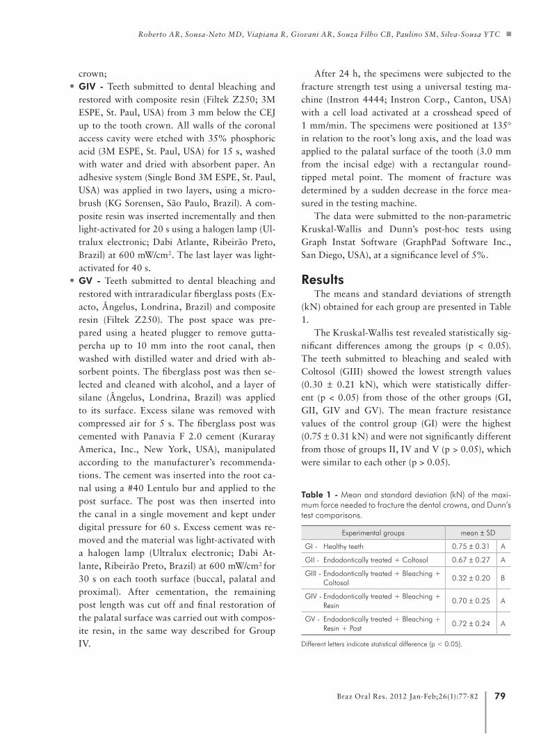
restored with composite resin (Filtek Z250; 3M ESPE, St. Paul, USA) from 3 mm below the CEJ up to the tooth crown. All walls of the coronal access cavity were etched with 35% phosphoric acid (3M ESPE, St. Paul, USA) for 15 s, washed with water and dried with absorbent paper. An adhesive system (Single Bond 3M ESPE, St. Paul, USA) was applied in two layers, using a micro-brush (KG Sorensen, São Paulo, Brazil). A com-posite resin was inserted incrementally and then light-activated for 20 s using a halogen lamp (Ul-tralux electronic; Dabi Atlante, Ribeirão Preto, Brazil) at 600 mW/cm2. The last layer was light-activated for 40 s.
• GV - Teeth submitted to dental bleaching and restored with intraradicular fiberglass posts (Ex-acto, Ângelus, Londrina, Brazil) and composite resin (Filtek Z250). The post space was pre-pared using a heated plugger to remove gutta-percha up to 10 mm into the root canal, then washed with distilled water and dried with ab-sorbent points. The fiberglass post was then se-lected and cleaned with alcohol, and a layer of silane (Ângelus, Londrina, Brazil) was applied to its surface. Excess silane was removed with compressed air for 5 s. The fiberglass post was cemented with Panavia F 2.0 cement (Kuraray America, Inc., New York, USA), manipulated according to the manufacturer’s recommenda-tions. The cement was inserted into the root ca-nal using a #40 Lentulo bur and applied to the post surface. The post was then inserted into the canal in a single movement and kept under digital pressure for 60 s. Excess cement was re-moved and the material was light-activated with a halogen lamp (Ultralux electronic; Dabi At-lante, Ribeirão Preto, Brazil) at 600 mW/cm2 for 30 s on each tooth surface (buccal, palatal and proximal). After cementation, the remaining post length was cut off and final restoration of the palatal surface was carried out with compos-ite resin, in the same way described for Group IV.
After 24 h, the specimens were subjected to the fracture strength test using a universal testing ma-chine (Instron 4444; Instron Corp., Canton, USA) with a cell load activated at a crosshead speed of 1 mm/min. The specimens were positioned at 135° in relation to the root’s long axis, and the load was applied to the palatal surface of the tooth (3.0 mm from the incisal edge) with a rectangular round-tipped metal point. The moment of fracture was determined by a sudden decrease in the force mea-sured in the testing machine.
The data were submitted to the non-parametric Kruskal-Wallis and Dunn’s post-hoc tests using Graph Instat Software (GraphPad Software Inc., San Diego, USA), at a significance level of 5%.
ResultsThe means and standard deviations of strength
(kN) obtained for each group are presented in Table 1.
The Kruskal-Wallis test revealed statistically sig-nificant differences among the groups (p < 0.05). The teeth submitted to bleaching and sealed with Coltosol (GIII) showed the lowest strength values (0.30 ± 0.21 kN), which were statistically differ-ent (p < 0.05) from those of the other groups (GI, GII, GIV and GV). The mean fracture resistance values of the control group (GI) were the highest (0.75 ± 0.31 kN) and were not significantly different from those of groups II, IV and V (p > 0.05), which were similar to each other (p > 0.05).
Table 1 - Mean and standard deviation (kN) of the maxi-mum force needed to fracture the dental crowns, and Dunn’s test comparisons.
Experimental groups mean ± SD
GI - Healthy teeth 0.75 ± 0.31 A
GII - Endodontically treated + Coltosol 0.67 ± 0.27 A
GIII - Endodontically treated + Bleaching + Coltosol
0.32 ± 0.20 B
GIV - Endodontically treated + Bleaching + Resin
0.70 ± 0.25 A
GV - Endodontically treated + Bleaching + Resin + Post
0.72 ± 0.24 A
Different letters indicate statistical difference (p < 0.05).

Effect of different restorative procedures on the fracture resistance of teeth submitted to internal bleaching
80 Braz Oral Res. 2012 Jan-Feb;26(1):77-82
DiscussionTooth weakening is the main side effect of end-
odontic treatment15 and dental bleaching.16 Restor-ing the fracture resistance of endodontically treated teeth is becoming a greater focus of concern in den-tal research.12
The main factors involved in tooth structure loss are caries and root canal access preparation and in-strumentation.17 According to the related literature, there is no consensus regarding reduced fracture re-sistance observed after endodontic treatment.18,19 Al-though this was not the main objective of this study, the present results also revealed that the endodontic treatment did not reduce tooth fracture resistance, since the resistance values of the teeth submitted only to endodontic treatment and sealed with Colto-sol (GII) were not significantly different from those of healthy teeth (GI).
Until recently, increased susceptibility to fracture in endodontically treated teeth had been attributed to an increased brittleness of dentin as a result of moisture loss.20 Today, however, researchers stress that a loss of tooth structure is the key reason be-hind the observed increase in the fracture predis-position of endodontically treated teeth.21 In this study, endodontically treated teeth had a minimal loss of tooth structure and the irrigating solutions remained in contact with the dentin for only a short period of time. As a result, these teeth did not be-come more susceptible to fracture on account of the endodontic treatment.
According to the related literature, differ-ent bleaching agents or whitening techniques can adversely affect the fracture resistance of teeth, likely owing to changes in dental structure, such as those related to porosity, demineralization, de-creased adhesion of restorative materials to dentin, increased dentin permeability, reduced dentin mi-crohardness and decreased dentin diametral ten-sile strength.2,3,6,22 This effect was also observed in this study, since the fracture resistance values of the endodontically treated teeth submitted to den-tal bleaching and sealed with Coltosol (GIII) were the lowest (0.31kN) and statistically different from those of healthy teeth (GI), endodontically treated teeth and sealed with Coltosol (GII), endodontically
treated teeth submitted to dental bleaching and re-stored with composite resin (GIV), and endodonti-cally treated teeth submitted to dental bleaching and restored with composite resin combined with a fiberglass post (GV).
Another factor of great relevance that may be as-sociated with the decrease in fracture resistance of bleached teeth is the number of applications of the bleaching agent to the dental surface. According to Pobbe et al.,4 two or more sessions of dental bleach-ing with 38% hydrogen peroxide, activated by a LED-laser system, reduce the fracture resistance of endodontically treated teeth probably as a result of the action of hydrogen peroxide, which modifies the mechanical and chemical properties of dentin.
According to Kawamoto and Tsujimoto,23 the OH radical resulting from hydrogen peroxide deg-radation is responsible for tooth whitening, and acts on intertubular and peritubular dentin, destroying its organic portion, increasing permeability,24,25 and decreasing its hardness and elasticity modulus,2,3 which can be intensified with a greater exposure time of the tooth to the bleaching agent. This may explain the greater susceptibility to fracture ob-served in the teeth endodontically treated, bleached and only sealed with Coltosol (GIII).
It is also noteworthy to mention that some au-thors have reported that the heat applied to activate the bleaching agent or even produced by chemical reactions during this clinical procedure may cause reversible, or even irreversible, deleterious effects on dental and periodontal tissues.4,11,25 However, some studies have shown that this negative effect may be offset by the thermal insulating capability of dentin, which reduces the amount of heat reaching the pulp chamber significantly.12,18 Another important con-sideration is that low intensity laser irradiation may compensate the cytotoxic effect of a highly concen-trated hydrogen peroxide gel.26
Although the results suggest a decrease in the frac-ture resistance of teeth submitted to dental bleaching (GIII), and restored only with composite resin (GIV) or with fiberglass post (GV), no statistically signifi-cant difference was observed when these teeth were compared to healthy teeth (GI) and to those end-odontically treated and sealed with Coltosol (GII).

Roberto AR, Sousa-Neto MD, Viapiana R, Giovani AR, Souza Filho CB, Paulino SM, Silva-Sousa YTC
81Braz Oral Res. 2012 Jan-Feb;26(1):77-82
It seems that the restorative procedures restored the fracture resistance of endodontically treated teeth submitted to dental bleaching. This observation is in agreement with that of other studies12,15 reporting that composite resin is a suitable restorative material for weakened teeth, because it restores the tooth’s fracture resistance up to values comparable to those of healthy teeth. This may be attributed to the resin’s low polymerization shrinkage rates, and satisfactory characteristics in terms of hardness, and resistance to abrasion and compression. Furthermore, the ad-hesive system used to bond composite resins has the ability of moistening and infiltrating into the dentin after the smear layer has been removed by acid etch-ing, which creates micromechanical retention, pro-moting stress distribution through the dentin, and reducing the chances of fracture.
The association of composite resin with fiber-glass posts did not improve the fracture resistance of teeth. According to some authors,27,28 endodonti-cally treated anterior teeth with thick dentin walls should be restored with composite resin. Addition-ally, it has been reported that the fracture resistance of weakened teeth restored with fiber posts does not improve.13,29 This may be related to the fact that the main role of intracanal posts is to promote the retention of the filling material onto the remaining
dental structure.12,13,29,30
Both restorative procedures tested in this study proved feasible for restoring the fracture resistance of endodontically treated teeth submitted to bleach-ing. Furthermore, the use of intracanal posts did not increase the resistance of bleached teeth. It should be borne in mind that the experiments of the pres-ent study were conducted in healthy teeth; therefore, caution should be taken in extrapolating these re-sults to carious and destroyed teeth. Further studies are necessary to assess other variables involved in the fracture resistance of endodontically treated and bleached teeth.
ConclusionsBased on the methodology and results of this
study, it could be concluded that endodontically treated teeth submitted to dental bleaching and sealed with Coltosol showed the lowest values of fracture resistance. Furthermore, the restorative procedures using composite resin restored the fracture resistance of endodontically treated and bleached teeth, but the association of composite resin with fiberglass posts did not increase fracture resistance more than restoring teeth with composite resin alone.
References 1. Lee GP, Lee MY, Lum SOY, Poh RS, Lim KC. Extraradicular
diffusion of hydrogen peroxide and pH changes associated
with intracoronal bleaching of discoloured teeth using dif-
ferent bleaching agents. Int Endod J. 2004 Jul;37(7):500-6.
2. Chng HK, Yap AUJ, Wattanapayungkul P, Sim CPC. Effect
of traditional and alternative intracoronal bleaching agents
on microhardness of human dentine. J Oral Rehabil. 2004
Aug;31(8):811-6.
3. Pécora JD, Cruz-Filho AM, Sousa-Neto MD, Silva RG. In
vitro action of various bleaching agents on the microhardness
of human dentin. Braz Dent J. 1994; 5(2):129-34.
4. Pobbe POS, Viapiana R, Souza-Grabriel AE, Marchesan MA,
Sousa-Neto MD, Silva-Sousa YTC, Silva RG. Coronal resis-
tance to fracture of endodontically treated teeth submitted to
light-activated bleaching. J Dent. 2008 Nov;36(11):935-9.
5. Swift Jr EJ. Critical Effects of bleaching on tooth structure and
restorations, part II: enamel bonding. J Esthet Restor Dent.
2008;20(1):68-73.
6. Swift Jr. Effects of bleaching on tooth structure and res-
torations, part III: effects on dentin. J Esthet Restor Dent.
2008;20(2):1-7.
7. Shannon H, Spencer P, Gross K, Tira D. Characterization of
enamel exposed to 10% carbamide peroxide bleaching agents.
Quintessence Int. 1993 Jan;24(1):39-44.
8. Worschech CC, Rodrigues JA, Martins LR, Ambrosano GM.
In vitro evaluation of human dental enamel surface rough-
ness bleached with 35% carbamide peroxide and submitted
to abrasive dentifrice brushing. Pesqui Odontol Bras. 2003
Oct-Dec;17(4)342-8.
9. Soldani P, Amaral CM, Rodrigues JA. Microhardness evalu-
ation of in situ vital bleaching and thickening agents on hu-
man dental enamel. Int J Periodontics Restorative Dent. 2010
Apr;30(2):203-11.
10. Rodrigues JA, Marchi GM, Ambrosano GM, Heymann HO,
Pimenta LA. Microhardness evaluation of in situ vital bleach-
Effect of different restorative procedures on the fracture resistance of teeth submitted to internal bleaching
82 Braz Oral Res. 2012 Jan-Feb;26(1):77-82
ing on human dental enamel using a novel study. Dent Mat.
2005 Nov;21(11):1059-67.
11. Tredwin CJ, Naik S, Lewis NJ, Scully C. Hydrogen peroxide
tooth whitening (bleaching) products: review of adverse effects
and safety issues. Br Dent J. 2006 Apr;200(7):371-6.
12. Bonfante G, Kaizer OB, Pegoraro LF, Valle AL. Fracture re-
sistance and failure pattern of teeth submitted to internal
bleaching with 37% carbamide peroxide, with application
of different restorative procedures. J Appl Oral Sci. 2006
Aug;14(4):247-52.
13. Marchi GM, Paulino LA, Pimenta LAF, De Lima FA. Effect of
different filling materials in combination with intraradicular
posts on the resistance to fracture of weakened roots. J Oral
Rehabil. 2003Jun; 30(6):623-9
14. D’Arcangelo C, De Angelis F, Vadini M, D’Amario M, Ca-
puti. Fracture Resistance and Deflection of Pulpless Anterior
Teeth Restored with Composite or Porcelain Veneers. J Endod.
2010;36(1):153-6
15. Soares PV, Santos PCF, Martins LRM, Soares CJ. Influence
of restorative technique on the biomechanical behavior of end-
odontically treated maxillary premolars. Part I: Fracture resis-
tance and fracture mode. J Prosthet Dent. 2008 Jan;99(1):30-
7.
16. Attin T, Müller T, Patyk A, Lennon AM. Influence of differ-
ent bleaching systems on fracture toughness and hardness of
enamel. Oper Dent. 2004 Mar-Apr;29(2):188-95.
17. Hannig C, Westphal C, Becker K, Attin T. Fracture resis-
tance of endodontically treated maxillary premolars restored
with CAD/CAM ceramic inlays. J Prosthet Dent. 2005
Oct;94(4):342-9.
18. Hansen EK, Asmussen E. In vivo fractures of endodontically
treated posterior teeth restored with enamel-bonded resin.
Endod Dent Traumatol. 1990 Oct;6(5):218-25.
19. Papa J, Cain C, Messer HH. Moisture content of vital vs
endodontically treated teeth. Endod Dent Traumatol. 1994
Apr;10(2):91-3.
20. Helfer AR, Melnick S, Schilder H. Determination of the mois-
ture content of vital and pulp less teeth. Oral Surg Oral Med
Oral Pathol 1972 Oct;34(4):661-70.
21. Kishen A, Asundi A. Photomechanical investigations on
post-endodontically rehabilitated teeth. J Biomed Opt. 2002
Apr;7(2):262-70.
22. Tam LE, Kuo VY, Noroozi A. Effect of prolonged direct end
indirect peroxide bleaching and fracture toughness of human
dentin. J Esthet Restor Dent. 2007;19(2):100-9.
23. Kawamoto K, Tsujimoto Y. Effects of the hydroxyl radi-
cal and hydrogen peroxide on tooth bleaching. J. Endod.
2004 Jan;30(1):45-50.
24. Carrasco LD, Guerisoli DMZ, Pécora JD, Froner IC. Evalu-
ation of dentin permeability after light active internal dental
bleaching. Dent Traumatol. 2007 Feb;23(1):30-4.
25. Sydney GB, Barletta FB, Sydney RB. In vitro analysis of effects
of heat used in dental bleaching on human dental enamel. Braz
Dent J. 2002;13(3):166-9.
26. Dantas CM, Vivan CL, Ferreira LS, Freitas PM, Marques
MM. In vitro effect of low intensity laser on the cytotoxicity
produced by substances released by bleaching gel. Braz Oral
Res. 2010 Oct-Dec;24(4):460-6.
27. Pontius O, Hutter JW. Survival rate and fracture strength of
incisors restored with different post and core systems and
endodontically treated incisors without coronoradicular re-
inforcement. J Endod. 2002 Oct;28(10):710-5.
28. Azevedo RA, Silva-Sousa YT, Souza-Gabriel AE, Messias DC,
Alfredo E, Silva RG. Fracture resistance of teeth subjected to
internal bleaching and restored with different procedures.
Braz Dent J. 2011; 22(2):117-21.
29. Garbin CA, Spazzin AO, Meira-Júnior AD, Loretto SC, Lyra
AM, Braz R. Biomechanical behaviour of a fractured maxil-
lary incisor restored with direct composite resin only or with
different post systems. Int Endod J. 2010 Dec;43(12):1098-
107.
30. Bonfante G, Kaizer OB, Pegoraro LF, do Valle AL. Tensile
bond strength of glass fiber posts luted with different cements.
Braz Oral Res. 2007 Apr-Jun;21(2):159-64.