EEG IN CHILDREN: NORMAL AND ABNORMAL - … ¾Survey some abnormal and normal patterns ¾Maturation...
102
EEG IN CHILDREN: NORMAL AND ABNORMAL Warren T. Blume, MD,FRCPC EEG Course FSNC/CNSF JUNE 2007
Transcript of EEG IN CHILDREN: NORMAL AND ABNORMAL - … ¾Survey some abnormal and normal patterns ¾Maturation...

EEG IN CHILDREN: NORMAL AND ABNORMAL
Warren T. Blume, MD,FRCPCEEG CourseFSNC/CNSFJUNE 2007

OBJECTIVES
Survey some abnormal and normal patternsMaturation characteristicsArtefact recognitionPatterns from superimposed componentsNormal/abnormal not always distinctPaediatric EEG knowledge improves adult EEG interpretation

5 QUESTIONS ABOUT PAEDIATRIC EEG
In what state is the patient? Drowsiness and sleep occupy a high percentage of children’s recordings; their features are in some aspects distinct from those of adults.Is the electrical maturation for each stage adequate?Are there any persistent, marked, and non-artefactualasymmetries which are not accepted for the waveform in question?Are there any spikes? These must be distinguished from other sharply contoured waves.Does focal or diffuse excess delta activity exist for this patient’s age and state?


Passive Eye Closure Demonstrates Background Activity
A: With eyes open: 5 Hz better left
B,C: Passive eye closure: 9 Hz better right.
Fig. 1a

Fig. 1a

Passive Eye Closure Demonstrates Background Activity
A: With eyes open: 5 Hz better left
B,C: Passive eye closure: 9 Hz better right.
Fig. 1b

Fig. 1b

Fig. 1c

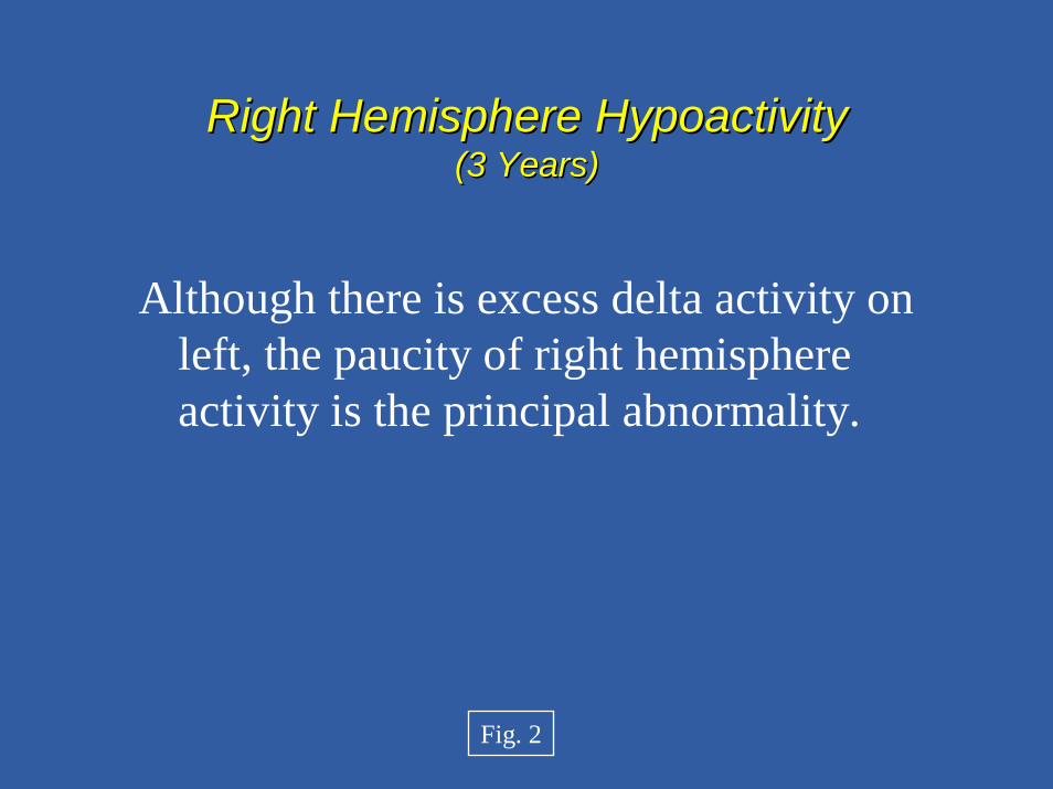
Right Hemisphere HypoactivityRight Hemisphere Hypoactivity(3 Years)(3 Years)
Although there is excess delta activity on left, the paucity of right hemisphere activity is the principal abnormality.
Fig. 2

Fig. 2

Right Hemisphere HypoactivityRight Hemisphere Hypoactivity(3 Years)(3 Years)
Right temporal hypoactivity is principal abnormality.Right central-parietal (C4-P4) delta.Mild excess left hemisphere delta.
Fig. 3

Fig. 3

Test
POSTERIOR DELTAPOSTERIOR DELTA

Posterior-Accentuated DeltaPosterior-Accentuated Delta(6 Years)(6 Years)
Although delta is most prominent at 01,02, P4; excess delta appears moreanteriorly.
Fig. 4

Fig. 4


POSTERIOR DELTAPOSTERIOR DELTACLINICAL SIGNIFICANCECLINICAL SIGNIFICANCE
Recent seizureMild to moderate recent trauma

RHYTHMIC POSTERIOR DELTA
• Normal, or• Hidden spike-waves


FOCAL DELTA

ARTEFACT NORMAL ABNORMAL


Focal DeltaFocal Delta(7 Years)(7 Years)
2-3 Hz arrhythmic delta at F4-C4.
Fig. 5


Fig. 5
NORMAL POSTERIOR
RHYTHMS AND FEATURES

• Anything maintaining an invariably regular frequency is likely to be normal.
• Sharp elements that clearly grow out of an ongoing baseline rhythm should be considered normal.
Engel 1984


POLYPHASIC POTENTIALS– 250-500 msec– among alpha– “polyphasic morphology”– often more abundant on right
RHYTHMIC WAVES– 3-4 Hz– upon eye closure


POLYPHASIC POTENTIALS– 250-500 msec– among alpha– “polyphasic morphology”– often more abundant on right
RHYTHMIC WAVES– 3-4 Hz– upon eye closure


LAMBDA• Apiculate• Positive• Eyes open
OCCIPITAL SPIKES• Apiculate• Negative• Eyes closed


CENTRAL PHENOMENA


Epileptiform Abnormalities:ROLANDIC SPIKES
• Abundant and stereotyped.
• Unilateral, bilaterally independent, and/or bilaterally synchronous.
• Background normal when spikes absent.• Seizures in 54 - 84%


EPILEPTIFORM:GENERALISED


Generalised Epileptiform Phenomena:GENERALISED SPIKE-WAVE
• Bilaterally synchronous spike-wave complexes with repetition rate of 2.5 to 4 Hz.
• Bursts begin and end abruptly.
• Repetition rate slows during long paroxysms.
• Maximum amplitude usually at F3, F4; occasionally posterior (P3,4; 01,2).


Generalised Epileptiform Phenomena:POLYSPIKES
• Burst of spikes repeating at 10-25 Hz.• Irregular discharge rate.• Generalised, maximum frontally.• 40-350 µV.• Duration 1-8 seconds.• Tonic seizures or absence in association.• May occur on eye closure.


Epileptiform Abnormalities:SLOW SPIKE-WAVES (SSW)
• Bilaterally synchronous sinusoidal waves, each accompanied by a sharp wave or spike forming a complex.
• Repetition rate less than 2.5 per second.• Onset and offset less abrupt than spike waves.• Occupy high percentage of recording.• Often no discernible clinical alteration.• Associated wtih a slow background.
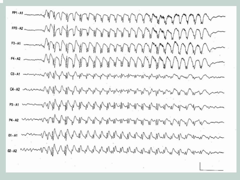

Fast rhythmic waves• Bursts of 8- to 30-Hz widespread or
generalised waves.• Usual clinical accompaniment is tonic
seizure when hypsarrhythmia or slow spike-waves appear in same record.
• Usual clinical accompaniment is absence attack when spike-wave complexes appear in same record.


LENNOX-GASTAUT SYNDROME
CRITERIA
• Generalised seizures– Tonic– Atypical absence– Others
• EEG: slow spike-wavesepileptic recruiting rhythm =
fast rhythmic waves

SECONDARY BILATERAL SYNCHRONY
• Bilaterally synchronous discharge• Can be shown to arise from a unilateral
cortical focus• Origin usually from most “active” spike
focus• Usually frontal


Normal Drowsiness, Sleep, And Arousal: BURST DROWSY
• Bursts of 2- to 5-Hz sinusoidal waves, usually maximum frontocentrally.
• Superimposed on other drowsy patterns.• Begin at 14 to 18 months; most common at
3 to 5 years; seen until 11 years.


Normal Drowsiness, Sleep, And Arousal: V-WAVES
• Higher voltage and briefer than in adults, therefore, spike-like.
• Variable morphology and polarity.• May occur sequentially.• Shifting asymmetries.• Begin at 3 to 4 months, maximum at 3 to 4
years.


SPINDLES
• First clearly expressed at 3 to 4 months.
• More numerous and longer at 3 to 9 months than later.
• Asynchrony common in first year.• Central-parietal location in early
childhood.• May be comb shaped.


Absent Spindles 5 mos

AROUSAL
• 4-6 Hz rhythmic waves diffusely• 1-3 Hz diffuse delta• Principally < 5 years


5 QUESTIONS ABOUT PAEDIATRIC EEG
In what state is the patient? Drowsiness and sleep occupy a high percentage of children’s recordings; their features are in some aspects distinct from those of adults.Is the electrical maturation for each stage adequate?Are there any persistent, marked, and non-artefactualasymmetries which are not accepted for the waveform in question?Are there any spikes? These must be distinguished from other sharply contoured waves.Does focal or diffuse excess delta activity exist for this patient’s age and state?

END


Subtle Focal DeltaSubtle Focal Delta(9 Years)(9 Years)
Abnormal delta quantity right frontal-central (F4-C4) region.Normal delta amount on left.


Epileptiform Abnormalities:ROLANDIC SPIKES
• High voltage at C3 or C4 using 10-20 System.
• Involve principally lower Rolandic area (C5,6) using closely spaced electrodes.
• Marked downward deflection at F3-C3 or F4-C4 suggests dipole.
• Principal parasagittal spread of negative component usually parietal, occasionally frontal.


•Spike wave quantity =amount of absence seizures