
EEG, ECG Measurement and Data Analysis Carlo Lucignoli ATSiP Teesside 2006.
31
-
Upload
antony-milhouse -
Category
Documents
-
view
218 -
download
0
Transcript of EEG, ECG Measurement and Data Analysis Carlo Lucignoli ATSiP Teesside 2006.


EEG, ECG Measurement andData Analysis
Carlo Lucignoli
ATSiP Teesside 2006

What are we trying to achieve ?
The psychologists wish to measure patients’ anxiety.
The measurement is shown to patients so they can see their
anxiety before they feel it.
This enables patients to learn how to monitor and control
anxiety when it is still in the early stages.
Thus, anxious patients can learn to manage their anxiety
before it becomes a problem.
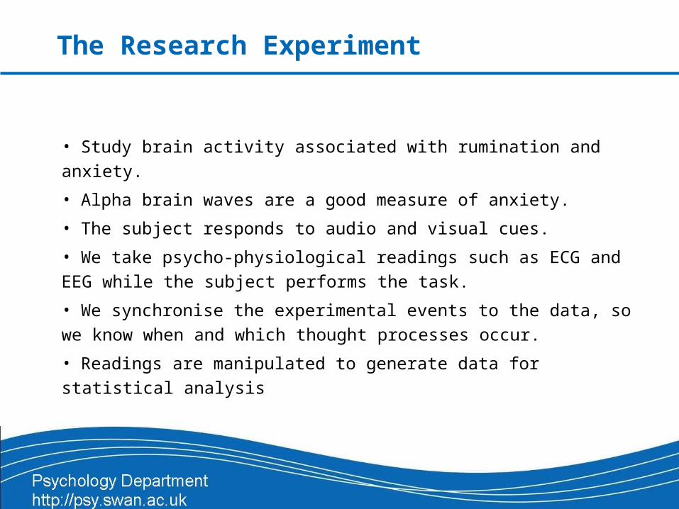
• Study brain activity associated with rumination and anxiety.
• Alpha brain waves are a good measure of anxiety.
• The subject responds to audio and visual cues.
• We take psycho-physiological readings such as ECG and EEG
while the subject performs the task.
• We synchronise the experimental events to the data, so we know
when and which thought processes occur.
• Readings are manipulated to generate data for statistical analysis
The Research Experiment

What do we need to do?
• Measurements
– Electroencephalograph (EEG) for Alpha-wave brain activity
– Electrocardiogram (ECG) for stress level
– Skin Conductance Response (SCR) arousal level
• Health & Safety
– The tester or the participant should not be at risk at any time
– Good hygiene, avoid cross contamination
– Electrical protection eg. isolation
• Participants
– Gather participants that meet the required criteria

The Solution
• Choose equipment which will make the task as easy as possible.
• EEG is obtained using a BioSemi 128 channel Active Electrode
system, recorded with ActiView program.
• ECG and SCR recorded using Cambridge Electronic Design
(CED) equipment and Spike program.
• Data analysis handled by BESA program.
• Timing and control is handled by E-Prime program.

The Participants Criteria
• No age restrictions

The Participants Criteria
• No age restrictions
• University students

The Participants Criteria
• No age restrictions
• University students

The Participants Criteria
• No age restrictions
• University students
• Free from excessive alcohol previous day
• Refrain from caffeine and nicotine 2hrs before
• Had a good nights sleep
• Right handed
• Accept £8 payment

The Participants Criteria
• No age restrictions
• University students
• Free from excessive alcohol previous day
• Refrain from caffeine and nicotine 2hrs before
• Had a good nights sleep
• Right handed
• Accept £8 payment

Health and Safety
• Electrical Safety and Shock Prevention for medical research– Extra Low Voltage (battery) for the EEG System– BioSemi Active Electrodes have built in current limit system with auto shutoff– Remaining electrodes have electrical isolation from their amplifiers– Amplifiers are powered by Isolated Power Supply Units
• Cross Contamination– Traditionally, electrodes required skin to be scratched to create better electrical
contact. This can lead to cross infection– The BioSemi Active Electrodes require no skin preparation, making things safer
and quicker– Separate washing area was needed to ensure electrodes were kept away from
food preparation areas– Disposable electrode pads were used for the remaining signals

Synchronisation
• The experiment is controlled by the PC running E-Prime.
• E-Prime presents a series of visual and audible cues to the participant.
• In synchrony to these cues, triggers are sent to BioSemi (EEG) and CED
(ECG and SCR).
• Triggers are sent out of the printer port using its 8 data channels.
• These may appear as binary, levels, or as a number.
• Trigger information contains Record/Pause command and task identification
• In this way the data indicates exactly where tasks begins and end
Speed
• Commissioning Speed
– Plan ahead: Consider how many PCs, monitors, key-boards, amps, etc. there will be, and where will they go. Is there sufficient access to prepare and connect up the participant?
– This experiment requires the participant be in a separate room
– 3 PCs, two fitted with a pair of monitors, keyboards and mice, speakers
– Assembly: Hole through wall, are cables long enough?
– Setup correct settings before you start
– Read the instruction manual before you start no matter how unnatural it may seem

Speed
•For those of you who’ve
never seen one, here is an
example.

Speed
• Subject Preparation Time
– No skin preparation needed for the EEG Active Electrodes.
– Head cap is simple to assemble. A little gel and push-fit the 128 electrodes in place.
– Takes approx 20 minutes. Time saved 20-30 minutes. Number of participants 100.
– Fitting ECG and SCR self-adhesive electrodes takes little time.

EEG with BioSemi and ActiView
• BioSemi Active Electrodes each have
a tiny electronic circuit built in, just a
couple of mm’s from the point of
contact.
• No actual GND Reference point.
• Eliminates 50 Hz interference pickup
by the electrode wires
• Works within a high impedance range• 128 channels give detailed coverage (256 max capacity).
• 8 channels for auxiliary electrodes (eg. Left and right eye-blink)

EEG with BioSemi and ActiView
• Check EEG electrode
connections using the Electrode
Offset screen in the ActiView.
• CM in Range safety feature:
shuts off all Active Electrodes if
the CMS or DRL electrodes are
mis-fitted, or if any electrode
defects are detected. Indicated
by the blue LED flashing (top
right).

EEG with BioSemi and ActiView
• Check EEG electrode
connections using the Electrode
Offset screen

EEG with BioSemi and ActiView
• Record EEG with ActiView.
• Simple and straightforward to use.
• Data is sent to the PC via a fibre optic cable (electrically isolated).
• Settings can be stored in a configuration file (.cfg) and loaded each
time ActiView is run.
• Other uses of the configuration file include:
– Enables the remote record function ‘pause save’.
– Stores the file location where data file will be stored.
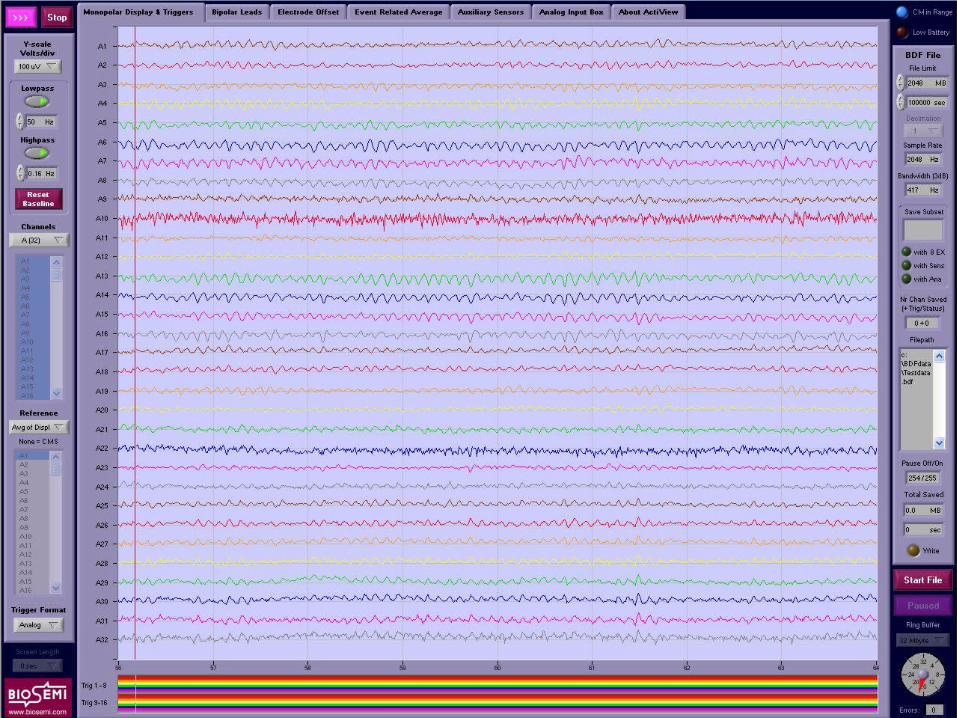
EEG with BioSemi and ActiView
• Record EEG signals with ActiView Program
• Simple and straight forward to use
•The settings for your particular experiment can be stored in a configuration file (.cfg) and loaded each time you open ActiView through a desktop shortcut
•Other uses of the configuration file
– Enables the remote record function ‘pause save’
– Holds the file path name to where BDF data file will be stored
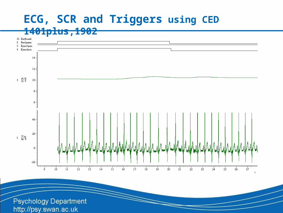
ECG, SCR and Triggers using CED 1401plus,1902

ECG, SCR and Triggers using CED 1401plus,1902

Data Analysis
BESA Software:
• BESA is a highly versatile and user-friendly program
• Widely used for source analysis in EEG research
• 2D and 3D images
• Supports scripts for automating repetitive tasks
• What does it look like?

BESA Data Analysis
Data collected
from a different
research project.
RAW Data from
ActiView
displayed in
BESA.
An example of 50
Hz interference at
Cz, the top line.

BESA Data Analysis
One without the artefact using a 50 Hz notch filter.
This includes an example of a Fast Fourier Transform (FFT) on all channels whilst eyes closed.
Shows some paroxysmal non-epileptic spike activity at T3 (left temporal lobe), which is related to emotional explosive behaviour and memory problems.

BESA Data Analysis
Using TAL as the so-called ‘source reference’. This is a unique feature of BESA.
This way, you can look at activity generated in different areas of the brain.
Example here shows the source of the spike activity you saw earlier on T3 and its neighbourhood. (Left anterior temporal lobe)
BESA Data Analysis
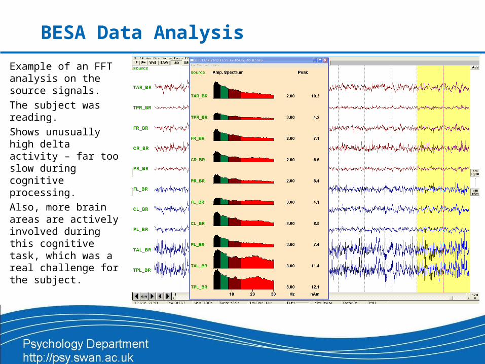
Example of an FFT analysis on the source signals.
The subject was reading.
Shows unusually high delta activity – far too slow during cognitive processing.
Also, more brain areas are actively involved during this cognitive task, which was a real challenge for the subject.

BESA Data Analysis
User defined
montage (snap-
shot at a selected
instant in time)
using a standard
3D brain.

Summary
What did we do?
• Stayed safe electrically and medically
• Gathered approximately 100 participants
• Used two rooms
• Recorded EEG on PC1 with BioSemi Active Electrode equipment
• Analysed EEG on PC1 with BESA
• Recorded & analysed ECG and SCR data on PC 2 with CED hardware and Spike
• Controlled the experiment on PC 3 with E-Prime and connected it to the other two PCs
• Connect systems between both rooms using 5 monitors, 4 keyboards and mice etc.
• Benefits of using BioSemi Active Electrodes:
– Low noise levels due to first amplifier stage being in the electrode
– Saves approx 30 minutes as there is no need to prepare the skin surface
– Almost no risk of infection
• Experiment documented for future reference

Thank you all for listening
THE END
Some images from ActiView, BESA and CED