Editorial Dilated cardiomyopathy: genetic approach · Heart 1997;77:185-188 HEART Editorial Dilated...
4
Heart 1997;77:185-188 HEART Editorial Dilated cardiomyopathy: a genetic approach Dilated cardiomyopathy is a myocardial disease charac- terised by impaired systolic function and dilatation of the left or both ventricles. The disease is not rare, affecting about one in 2500 individuals' and, despite the develop- ment of new treatments, it remains an important cause of mortality and morbidity and is a leading indication for heart transplantation. The cause of dilated cardiomyo- pathy is generally unknown and the identification of the aetiological and pathogenetic mechanisms underlying the disease is regarded a research priority.2 An important advance in the search for the aetiology has been the recognition of hereditary transmission in a subset of patients, which indicates that in these cases the origin of the disease must be an altered gene product. The recognition of dilated cardiomyopathy as a genetic disease has clinical implications. Furthermore, the development of molecular genetic techniques has provided the tools for the identification of the gene, or genes, causing the dis- ease. Clinical genetics A careful evaluation of the familial history of patients with dilated cardiomyopathy and the examination of relatives with a suspected cardiac disease showed that genetic fac- tors were more frequent than previously recognised.3 Controlled surveys based on the systematic screening of relatives, irrespective of family history, showed a familial form in 20-25% of patients with dilated cardiomyopa- thy.45 These data probably underestimate the real preva- lence, because affected individuals are likely to be missed, particularly in small pedigrees, and because of the absence of early markers of disease and reduced penetrance (the proportion of carriers who manifest the disease). Penetrance is reduced and age-related in dilated cardio- myopathy: as a consequence young family members in particular can appear clinically normal though they carry the disease gene. Different patterns of transmission and variable clinical features suggest that familial dilated cardiomyopathy (FDC) is the final common pathway of a heterogeneous group of disorders. These may include an autosomal dom- inant and an autosomal recessive FDC; conduction defects with later development of dilated cardiomyopathy; dilated cardiomyopathies associated with subclinical myopathy, such as the X-linked form; and possibly a mito- chondrial dilated cardiomyopathy. Molecular genetics of familial dilated cardiomyopathies Molecular genetics offers several approaches to study inherited diseases: functional cloning, which allows the identification of gene mutations when the protein defect is known; positional cloning (or reverse genetics), based on linkage analysis which allows the chromosomal localisa- tion of the disease genes to be mapped; and the positional candidate approach, based on the co-segregation of candi- date genes (genes that are candidates for causing the disease) with the disease within families. These approaches have led to impressive advances in the study of primary myocardial diseases during the past few years. The most frequent form of inherited dilated cardiomyo- pathy is autosomal dominant FDC which is characterised by development of ventricular dilatation and dysfunction, usually in the second to third decade of life, with progres- sive heart failure and ventricular arrhythmias. Segregation analysis suggests a monogenic disorder.4 The premature mortality, the reduced penetrance of the disease, and the absence of early clinical markers have always been serious obstacles in collecting large enough families for molecular genetic studies. Only recently, two research groups reported the results of linkage analysis performed in families with autosomal dominant FDC. First, a single large kindred was studied by our group: affected family members typically had a poorly contracting left ventricle, often associated with ventricular arrhyth- mias. Strict diagnostic criteria, based on full invasive eval- uation of the affected family members, were used to assign clinical status.2 After many of the candidate genes were excluded, a whole-genome random screening with more than 250 polymorphic markers was undertaken. This allowed the analysis of about 95% of the genome. Linkage of autosomal dominant FDC was eventually found with chromosome 9 (9q13-q22) in this kindred and in two other families6 (fig 1). A second locus for autosomal dominant FDC was then localised on chromosome lq32 in a large Utah family7 (fig 1). The analysis of the candidate genes mapping in these regions is currently under investigation. A rare form of FDC is characterised by autosomal dom- inant cardiac conduction system disease and later develop- ment of myocardial dysfunction (called conduction disease and dilated cardiomyopathy, CDDC). The affected family members manifest arrhythmias and atrio- ventricular block in the second to third decade of life and a progressive cardiomegaly and heart failure in the fifth to sixth decade. A first linkage study carried out in a single large family from Ohio mapped the CDDC disease gene in the centromere of chromosome 1 (lpl-lql)8 (fig 1). Recently, in another family of Swiss-German ancestry and similar clinical features, linkage was found with the short arm of chromosome 3 (3p22-p25)9 (fig 1). The disease genes are unknown and are under investigation. Till now the only inherited dilated cardiomyopathy for which the disease gene was known is X-linked dilated car- diomyopathy (XLDC), which generally presents in teenage boys as rapidly progressive congestive heart failure 185 on July 5, 2020 by guest. Protected by copyright. http://heart.bmj.com/ Heart: first published as 10.1136/hrt.77.3.185 on 1 March 1997. Downloaded from
Transcript of Editorial Dilated cardiomyopathy: genetic approach · Heart 1997;77:185-188 HEART Editorial Dilated...

Heart 1997;77:185-188
HEART
Editorial
Dilated cardiomyopathy: a genetic approach
Dilated cardiomyopathy is a myocardial disease charac-terised by impaired systolic function and dilatation of theleft or both ventricles. The disease is not rare, affectingabout one in 2500 individuals' and, despite the develop-ment of new treatments, it remains an important cause ofmortality and morbidity and is a leading indication forheart transplantation. The cause of dilated cardiomyo-pathy is generally unknown and the identification of theaetiological and pathogenetic mechanisms underlying thedisease is regarded a research priority.2An important advance in the search for the aetiology
has been the recognition of hereditary transmission in asubset of patients, which indicates that in these cases theorigin of the disease must be an altered gene product. Therecognition of dilated cardiomyopathy as a genetic diseasehas clinical implications. Furthermore, the developmentof molecular genetic techniques has provided the tools forthe identification of the gene, or genes, causing the dis-ease.
Clinical geneticsA careful evaluation of the familial history of patients withdilated cardiomyopathy and the examination of relativeswith a suspected cardiac disease showed that genetic fac-tors were more frequent than previously recognised.3Controlled surveys based on the systematic screening ofrelatives, irrespective of family history, showed a familialform in 20-25% of patients with dilated cardiomyopa-thy.45 These data probably underestimate the real preva-lence, because affected individuals are likely to be missed,particularly in small pedigrees, and because of the absenceof early markers of disease and reduced penetrance (theproportion of carriers who manifest the disease).Penetrance is reduced and age-related in dilated cardio-myopathy: as a consequence young family members inparticular can appear clinically normal though they carrythe disease gene.
Different patterns of transmission and variable clinicalfeatures suggest that familial dilated cardiomyopathy(FDC) is the final common pathway of a heterogeneousgroup of disorders. These may include an autosomal dom-inant and an autosomal recessive FDC; conductiondefects with later development of dilated cardiomyopathy;dilated cardiomyopathies associated with subclinicalmyopathy, such as the X-linked form; and possibly a mito-chondrial dilated cardiomyopathy.
Molecular genetics of familial dilatedcardiomyopathiesMolecular genetics offers several approaches to studyinherited diseases: functional cloning, which allows theidentification of gene mutations when the protein defect is
known; positional cloning (or reverse genetics), based onlinkage analysis which allows the chromosomal localisa-tion of the disease genes to be mapped; and the positionalcandidate approach, based on the co-segregation of candi-date genes (genes that are candidates for causingthe disease) with the disease within families. Theseapproaches have led to impressive advances in the study ofprimary myocardial diseases during the past few years.The most frequent form of inherited dilated cardiomyo-
pathy is autosomal dominant FDC which is characterisedby development of ventricular dilatation and dysfunction,usually in the second to third decade of life, with progres-sive heart failure and ventricular arrhythmias. Segregationanalysis suggests a monogenic disorder.4The premature mortality, the reduced penetrance of the
disease, and the absence of early clinical markers havealways been serious obstacles in collecting large enoughfamilies for molecular genetic studies. Only recently, tworesearch groups reported the results of linkage analysisperformed in families with autosomal dominant FDC.First, a single large kindred was studied by our group:affected family members typically had a poorly contractingleft ventricle, often associated with ventricular arrhyth-mias. Strict diagnostic criteria, based on full invasive eval-uation of the affected family members, were used to assignclinical status.2
After many of the candidate genes were excluded, awhole-genome random screening with more than 250polymorphic markers was undertaken. This allowed theanalysis of about 95% of the genome. Linkage of autosomaldominant FDC was eventually found with chromosome 9(9q13-q22) in this kindred and in two other families6(fig 1). A second locus for autosomal dominant FDC wasthen localised on chromosome lq32 in a large Utah family7(fig 1). The analysis of the candidate genes mapping inthese regions is currently under investigation.A rare form ofFDC is characterised by autosomal dom-
inant cardiac conduction system disease and later develop-ment of myocardial dysfunction (called conductiondisease and dilated cardiomyopathy, CDDC). Theaffected family members manifest arrhythmias and atrio-ventricular block in the second to third decade of life and aprogressive cardiomegaly and heart failure in the fifth tosixth decade. A first linkage study carried out in a singlelarge family from Ohio mapped the CDDC disease genein the centromere of chromosome 1 (lpl-lql)8 (fig 1).Recently, in another family of Swiss-German ancestry andsimilar clinical features, linkage was found with the shortarm of chromosome 3 (3p22-p25)9 (fig 1). The diseasegenes are unknown and are under investigation.
Till now the only inherited dilated cardiomyopathy forwhich the disease gene was known is X-linked dilated car-diomyopathy (XLDC), which generally presents inteenage boys as rapidly progressive congestive heart failure
185 on July 5, 2020 by guest. P
rotected by copyright.http://heart.bm
j.com/
Heart: first published as 10.1136/hrt.77.3.185 on 1 M
arch 1997. Dow
nloaded from
Editorial
3
CDDC2
x
24232221
13
121111
12
1321.121.221.322.122.222.3
313233
34.134.234.3
9 22.322.222.121.321.221.111.411.3
11.2311.2211.111
12.112.213
21.121.221.322.1
2324
25
26
27
28
] FDC1
| LDMD/I_ XLDC
Figure 1 Diagrams of the four chromosomes containing known disease gene locifor differentforms of dilated cardiomyopathy. The disease loci, identifiedby square brackets, were mapped on chromosome 1, containing the CDDC1 locus (iql-p)' and the FDC2 locus (Iq31-q32)7, chromosome 3 with theCDDC2 locus (3p22-p25)9, chromosome 9 with the FDC1 locus (9ql3-q22)', and chromosome X with the dystrophin gene (DMD), cause ofXLDC(Xp2l) (reprintedfrom Mestroni et al,l" with the permission ofArchives des Maladies du Coeur et Vaisseaux).
in the absence of clinical signs of skeletal myopathy.Affected family members may have a mild increase ofmuscle creatine kinase (MM-CK). Female carriers have a
later onset and slower progression of the disease. Thiscondition is characterised by the transmission of the disor-der with the X chromosome (no male-to-male transmis-sion) in a dominant or recessive fashion.Two independent studies showed that the dystrophin
gene, the same one that causes Duchenne and Beckermuscular dystrophies, was also responsible for XLDC'0 11(fig 1). The identification of deletions in the region of themuscle promoter-first muscle exon" and recently of a
point mutation (G/T) in the 5' splice site of the firstintron'2 (fig 2) leads to the hypothesis that the integrity ofthe 5' region of the gene is critical for the expression ofdystrophin in the heart. Immunocytochemical studies fullyexplain the phenotype of these patients, showing that dys-trophin is reduced in quantity but normally distributed inskeletal muscle, whereas it is undetectable in the cardiacmuscle.'2 Studies of the expression of the major dys-trophin mRNA isoforms show none in the myocardium,
whereas brain and Purkinje cell (but not muscle) isoformsare detectable in the skeletal muscle.'2
Furthermore, deletions of exons 48 and 49 were foundto be frequently associated with cardiomyopathy inpatients with Becker muscular dystrophy.'3 Our data indi-cate that carriers of these deletions can present as isolateddilated cardiomyopathy in absence of overt signs ofmyopathy (unpublished data).By analogy, other cytoskeletal proteins could be
involved in the aetiology of dilated cardiomyopathy.Deficiency of adhalin, a dystrophin-associated glycopro-tein, has recently been described in a patient with dilatedcardiomyopathy associated with signs of muscle dystro-phy.'4 Furthermore, alterations of the vinculin gene, codingfor a protein of the intercalated disk, have been reportedin a patient with dilated cardiomyopathy.'5
Dilated cardiomyopathy is found in mitochondrial dis-eases, usually complex syndromes caused by mitochondrialDNA (mtDNA) defects and inherited by maternal trans-mission. Multiple deletions ofmtDNA have been describedin a single family with dilated cardiomyopathy,16 as well as in
1
36.336.2
36.135
34.334.234.133
32.332.232.131.331.2
31.1
22.322.222.1
21
13.313.213.1121 11 1
12
21.121.221.32223
24
25
31
32.1
32.232.341
42.142.242.34344
2625
24
2322
21.3
21.221.114.314.214.113
12
1111.111.212
13.113.213.3
212223
2425.125.225.326.126.226.3272829
I CDDC1
] FDC2
186 on July 5, 2020 by guest. P
rotected by copyright.http://heart.bm
j.com/
Heart: first published as 10.1136/hrt.77.3.185 on 1 M
arch 1997. Dow
nloaded from

Editorial
XLDC1
A 2
1 2 3 7 4
N0
B
a--
c bdd
C
Muscleexon 1
G
A
G
G
A
C
T
G
T
T
G
T
A
A
GIntron T
A
C
A
A Normal Patient
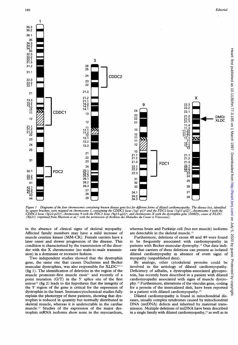
Figure 2 XLDC caused by a dystrophin gene point mutation. (A) Pedigree offamily XLDCJ. Individuals are indicated by generation and pedigree number.Their status is indicated by solid symbols (affected), open symbols (unaffected), and a circle with a dot (carrier). (B) Mutation analysis: SSCP analysis of thePCR product encompassing thefirst muscle exon-intron junction. Individual ssDNA strands are indicated by arrows (a to d). Individuals II-1 and II-2(affected) carry the same dystrophin aUlele, which is differentfrom the one of individual II-3 (normal). The mother (I-1) is heterozygous, as expected. (C)Sequence analysis of the first muscle exon-intron junction ofdystrophin gene. In the patient II-2 a GITpoint mutation eliminates the 5' splice site consensus
sequence. The normal control is represented by the healthy brother (II-3) (reprintedfrom Milasin et al,'2 with the permission of Oxford University Press).
11
0
NY.
Muscleexon 1
Intron
G
A
G
G
ACT
GT
TT
A
AG
T
A
C
A
A
187
6 A.0'
,..Il
on July 5, 2020 by guest. Protected by copyright.
http://heart.bmj.com
/H
eart: first published as 10.1136/hrt.77.3.185 on 1 March 1997. D
ownloaded from

Editorial
sporadic dilated cardiomyopathy, however, their primaryrole as pathogenetic factors is still controversial.
Clinical implications ofthe genetic and molecularstudiesThe fact that several surveys show that dilated cardiomyo-pathy is a genetic disease in a significant proportion ofpatients and that a gene defect should be considered as thecause of the disease in at least one third of patients hasimportant clinical implications.The management of a potentially inherited disorder
requires an accurate investigation of the familial history anda systematic screening of first degree relatives (parents, sib-lings, and offspring). Family studies allow early clinicaldiagnosis and for treatment to be started before symptomsdevelop. Moreover, a molecular diagnosis ofXLDC and ofthe carrier status is now possible. In the future, linkageanalysis could be used as a diagnostic procedure for thedetection of family members at risk of the disease.Moreover, genetic counselling should be offered to patientswith dilated cardiomyopathy to inform them about thecharacteristics of the disease, the risk in their relatives, thescope of early treatment, and the likelihood of future devel-opments.On the other hand, the experience in family studies raises
new questions about the diagnostic criteria. Frequently, rel-atives show minor signs of myocardial disease, such as seg-mental wall motion abnormalities, dilatation withoutventricular dysfunction, frequent and repetitive arrhyth-mias, and conduction defects that do not satisfy thetraditionally accepted diagnostic criteria of dilated cardio-myopathy.These signs in the context of a hereditary diseasestrongly suggest early manifestations of the disorder. Followup studies and, eventually, a molecular diagnosis willresolve these questions. The possibility of subclinical skeletalmuscle involvement should always be considered, and insuspected cases supplementary diagnostic procedures, suchas skeletal muscle immunocytochemistry and quantitativeelectromyography, should be performed.The identification of the genes and the understanding of
the molecular mechanisms causing dilated cardiomyopathywill provide a means not only of diagnosing and preventingthe disease but also of developing genotype-based treat-ments to modify the primary defect underlying the disease.
Recently, another locus for autosomal dominant FDC has beenreported on the short arm of chromosome 10 (I0q21-23)."8 This formofFDC appears to be characterised by the association with mitral valveprolapse and by a high penetrance.
LUISA MESTRONI
International Centre for Genetic Engineering and Biotechnology,AREA Science Park and Department of Cardiology,Hospital and University of Tnieste, Padriciano 99 34012 Trieste (Italy)I acknowledge the Heart Muscle Disease Study Group: Fulvio Camerini,Bruno Pinamonti, Gianfranco Sinagra, Andrea Di Lenarda, Chiara Rocco,Snjezana Miocic, Furio Silvestri, Rossana Bussani, Dario Gregori, MillaDavanzo, Cristiana Zanchi (Hospital and University of Trieste, Trieste, Italy)Mauro Giacca, Arturo Falaschi, Jelena Milasin, Matteo Vatta, Maja Matulic(International Centre for Genetic Engineering and Biotechnology, Trieste,Italy). Studies on muscle histopatology are performed in collaboration with DrFrancesco Muntoni, Royal Postgraduate Medical School, HammersmithHospital, London, UK. The grants of the Associazione Amici del Cuore ofTrieste, of the National Research Council (CNR 95.04378.CT04,95.00824.CT04, 95.01670.CT04, AI95.00346.04), and of Telethon-Italy(Grant No.E.291) are gratefully acknowledged.
1 Codd MB, Sugrue DD, Gersh BJ, Melton U. Epidemiology of idiopathicdilated and hypertrophic cardiomyopathy. A population-based study inOlmsted County, Minnesota, 1975-1984. Circulation 1989;80:564-72.
2 Manolio TA, Baughman KL, Rodeheffer R, Pearson TA, Bristow JD,Michels VV, et al. Prevalence and etiology of idiopathic dilated cardiomyo-pathy (summary of a National Heart, Lung and Blood InstituteWorkshop). Am 7 Cardiol 1992;69:1459-66.
3 Mestroni L, Miani D, Di Lenarda A, Silvestri F, Bussani R, Filippi G, et al.Clinical and pathologic study of familial dilated cardiomyopathy. Am 7Cardiol 1990;65:1449-53
4 Michels VV, Moll PP, Miller FA, Tajik AJ, Chu JS, Driscoll DJ, et al. Thefrequency of familial dilated cardiomyopathy in a series of patients withidiopathic dilated cardiomyopathy. NEnglJ Med 1992;326:77-82.
5 Keeling PJ, Gang G, Smith G, Seo H, Bent SE, Murday V, et al. Familialdilated cardiomyopathy in the United Kingdom. Br Heart J 1995;73:417-21.
6 Krajinovic M, Pinamonti B, Sinagra G, Vatta M, Severini GM, Milasin J, etal. Linkage of familial dilated cardiomyopathy to chromosome 9. Am JHum Genet 1995;57:846-52.
7 Durand J-B, Bachinski LL, Bieling LC, Czemuszewicz GZ, Abchee AB,Yu QT, et al. Localization of a gene responsible for familial dilated car-diomyopathy to chromosome 1q32. Circulation 1995;92:3387-9.
8 Kass S, MacRae C, Graber HL, Sparks EA, McNamara D, Boudoulas H, et al.A gene defect that causes conduction system disease and dilated cardio-myopathy maps to chromosome lpl-lql. Nature Genet 1994;7:546-51.
9 Olson TM, Keating MT. Mapping a cardiomyopathy locus to chromosome3p22-p25. J Clin Invest 1996;97:528-32.
10 Towbin JA, Hejtmancik F, Brink P, Gelb BD, Zhu XM, Chamberlain JS, etal. X-linked cardiomyopathy (XLCM): molecular genetic evidence oflinkage to the Duchenne muscular dystrophy (dystrophin) gene at theXp2l locus. Circulation 1993;87: 1854-65.
11 Muntoni F, Cau M, Ganau A, Congiu R, Arvedi G, Mateddu A, et al.Deletion of the dystrophin muscle-promoter region associated with X-linked dilated cardiomyopathy. NEngl3JMed 1993;329:921-5.
12 Milasin J, Muntoni F, Severini GM, Bartoloni L, Vatta M, Krajinovic M, etal. A point mutation in the 5' splice site of the dystrophin gene first intronresponsible for X-linked dilated cardiomyopathy. Hum Mol Genet1996;5:73-9.
13 Melacini P, Fanin M, Danieli GA, Fasoli G, Villanova C, Angelini C, et al.Cardiac involvement in Becker muscular dystrophy. J Am Coll Cardiol1993;22: 1927-34.
14 Fadic R, Sunada Y, Waclawik AJ, Buck S, Lewandoski PJ, Campbell KP, etal. Brief report: deficiency of a dystrophin-associated glycoprotein(adhalin) in a patient with muscular dystrophy and cardiomyopathy. NEnglJ Med 1996;334:362-6.
15 Maeda M, Holder E, Lowes B, Bies RD. Dilated cardiomyopathy associ-ated with deficiency of the cytoskeletal protein metavinculin. Circulation1997;95: 17-20.
16 Suomalainen A, Paetau A, Leinonen H, Majander A, Peltonen L, SomerH. Inherited dilated cardiomyopathy with multiple deletions of mito-chondrial DNA. Lancet 1992;340:1319-20.
17 Mestroni L, Milasin J, Vatta M, Pinamonti B, Sinagra G, Rocco C, et al.Genetic factors in dilated cardiomyopathy. Arch Mal Coeur 1996;89(ii):15-20.
18 Bowles KL, Gajarski R, Porter P, Goytia V, Bachinski L, Roberts R, et al.Gene mapping of familial autosomal dominant dilated cardiomyopathyto chromosome 10q21-23. J Clin Invest 1996;98: 1355-60.
188 on July 5, 2020 by guest. P
rotected by copyright.http://heart.bm
j.com/
Heart: first published as 10.1136/hrt.77.3.185 on 1 M
arch 1997. Dow
nloaded from