edition - St. Louis Children's Hospital with innovative pediatric nursing care related to endoscopic...
12
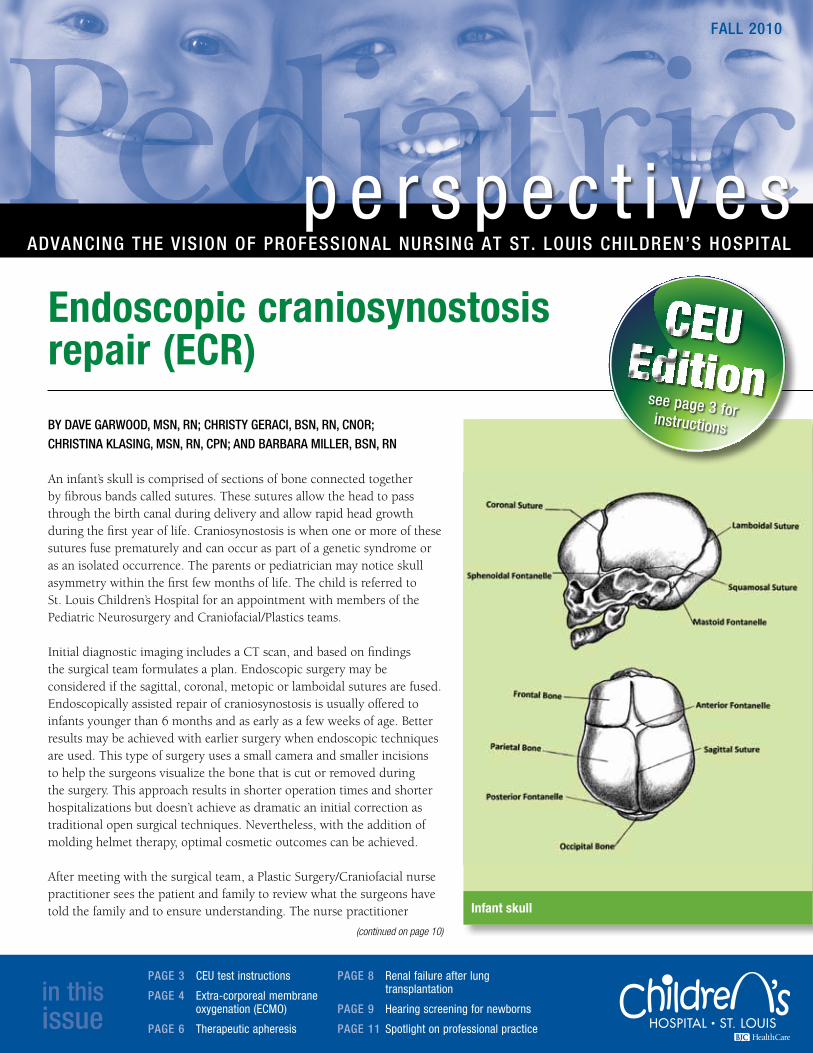
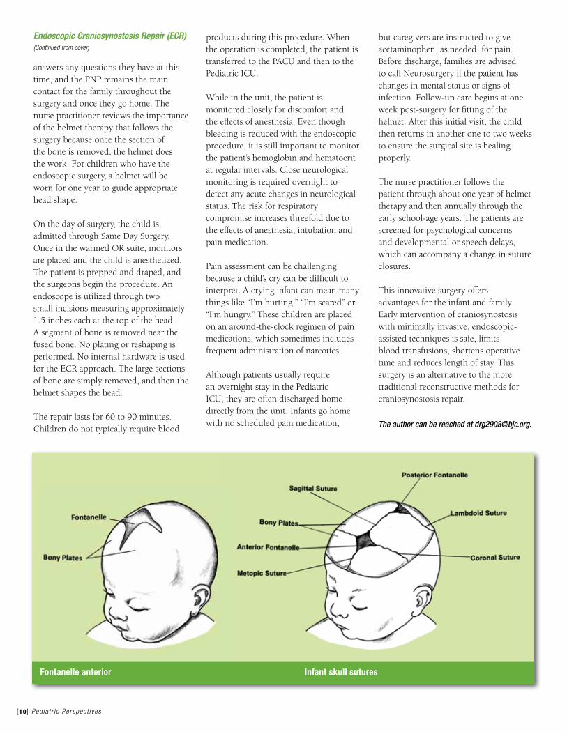
BY DAVE GARWOOD, MSN, RN; CHRISTY GERACI, BSN, RN, CNOR; CHRISTINA KLASING, MSN, RN, CPN; AND BARBARA MILLER, BSN, RN An infant’s skull is comprised of sections of bone connected together by fibrous bands called sutures. These sutures allow the head to pass through the birth canal during delivery and allow rapid head growth during the first year of life. Craniosynostosis is when one or more of these sutures fuse prematurely and can occur as part of a genetic syndrome or as an isolated occurrence. The parents or pediatrician may notice skull asymmetry within the first few months of life. The child is referred to St. Louis Children’s Hospital for an appointment with members of the Pediatric Neurosurgery and Craniofacial/Plastics teams. Initial diagnostic imaging includes a CT scan, and based on findings the surgical team formulates a plan. Endoscopic surgery may be considered if the sagittal, coronal, metopic or lamboidal sutures are fused. Endoscopically assisted repair of craniosynostosis is usually offered to infants younger than 6 months and as early as a few weeks of age. Better results may be achieved with earlier surgery when endoscopic techniques are used. This type of surgery uses a small camera and smaller incisions to help the surgeons visualize the bone that is cut or removed during the surgery. This approach results in shorter operation times and shorter hospitalizations but doesn’t achieve as dramatic an initial correction as traditional open surgical techniques. Nevertheless, with the addition of molding helmet therapy, optimal cosmetic outcomes can be achieved. After meeting with the surgical team, a Plastic Surgery/Craniofacial nurse practitioner sees the patient and family to review what the surgeons have told the family and to ensure understanding. The nurse practitioner perspectives ADVANCING THE VISION OF PROFESSIONAL NURSING AT ST. LOUIS CHILDREN’S HOSPITAL FALL 2010 in this issue PAGE 3 CEU test instructions PAGE 4 Extra-corporeal membrane oxygenation (ECMO) PAGE 6 Therapeutic apheresis Endoscopic craniosynostosis repair (ECR) PAGE 8 Renal failure after lung transplantation PAGE 9 Hearing screening for newborns PAGE 11 Spotlight on professional practice (continued on page 10) see page 3 for instructions Infant skull
-
Upload
nguyenngoc -
Category
Documents
-
view
216 -
download
0
Transcript of edition - St. Louis Children's Hospital with innovative pediatric nursing care related to endoscopic...
By Dave GarwooD, MSN, rN; ChriSty GeraCi, BSN, rN, CNor; ChriStiNa KlaSiNG, MSN, rN, CPN; aND BarBara Miller, BSN, rN
An infant’s skull is comprised of sections of bone connected together by fibrous bands called sutures. These sutures allow the head to pass through the birth canal during delivery and allow rapid head growth during the first year of life. Craniosynostosis is when one or more of these sutures fuse prematurely and can occur as part of a genetic syndrome or as an isolated occurrence. The parents or pediatrician may notice skull asymmetry within the first few months of life. The child is referred to St. Louis Children’s Hospital for an appointment with members of the Pediatric Neurosurgery and Craniofacial/Plastics teams.
Initial diagnostic imaging includes a CT scan, and based on findings the surgical team formulates a plan. Endoscopic surgery may be considered if the sagittal, coronal, metopic or lamboidal sutures are fused. Endoscopically assisted repair of craniosynostosis is usually offered to infants younger than 6 months and as early as a few weeks of age. Better results may be achieved with earlier surgery when endoscopic techniques are used. This type of surgery uses a small camera and smaller incisions to help the surgeons visualize the bone that is cut or removed during the surgery. This approach results in shorter operation times and shorter hospitalizations but doesn’t achieve as dramatic an initial correction as traditional open surgical techniques. Nevertheless, with the addition of molding helmet therapy, optimal cosmetic outcomes can be achieved.
After meeting with the surgical team, a Plastic Surgery/Craniofacial nurse practitioner sees the patient and family to review what the surgeons have told the family and to ensure understanding. The nurse practitioner
p e r s p e c t i v e saDvaNCiNG the viSioN of ProfeSSioNal NurSiNG at St. louiS ChilDreN’S hoSPital
fall 2010
in this issue
PaGe 3 CEU test instructions
PaGe 4 Extra-corporeal membrane oxygenation (ECMO)
PaGe 6 Therapeutic apheresis
Endoscopic craniosynostosis repair (ECR)
PaGe 8 Renal failure after lung transplantation
PaGe 9 Hearing screening for newborns
PaGe 11 Spotlight on professional practice
(continued on page 10)
CEU Edition
see page 3 for instructions
Infant skull
As the kids are back in school and we publish our second annual continuing education
edition of Pediatric Perspectives, the topic of learning comes to mind. The beginning
of the school year always feels like a time of promise – new books, notebooks and
backpacks, a blank report card with no grades to be pulled up, and the possibility of
new relationships with teachers who see a child’s promise, rather than his or her past.
Once we are out of formal schooling, some of this academic-year rhythm is lost. Yet,
taking that approach to new patients, new situations, and new co-workers can be
quite energizing. Looking at every situation as a blank notebook and being curious
rather than “knowing” is extremely helpful in almost every situation in health care. It is
especially important when working with people from different backgrounds.
How often do we approach patients with a particular diagnosis or social history with
pre-conceived ideas of who they are and what their issues will be? As pediatric nurses,
we do know a lot about the common illnesses that affect our patient population, and
with increasing experience we become experts in some areas. According to Dr. Patricia
Benner, author of the landmark work From Novice to Expert: Excellence and Power
in Clinical Nursing Practice, being an expert means we are able to integrate large
quantities of information, subtle cues and past experience to guide our thinking about
the patient in front of us. This expertise enables us to work more quickly and often
identify problems earlier and more accurately so that they can be resolved.
However, that same expertise can lead us into stereotyping or clinical “tunnel vision,”
causing us to miss other important cues if we are not careful. It also can lead to
something called “confirmation bias” where we see something for what we expect it to
be and miss important details that could indicate another problem or an error. Always
questioning “what could I be missing here?” is the antidote to the downside of “expert
thinking.”
As you read this issue, each of you will be learning about some area of nursing practice
here at SLCH that you have not likely encountered yet. When we are placed in that
situation in the actual clinical environment, we consciously go into learner mode. I
would ask you to consider whether you also treat your initial encounter with a new
patient and family as a similarly new experience where you have much to learn. Being
a learner in every encounter is a very useful approach to building relationships as well
as skills. Try it out! You will be surprised where it will take you.
From Peggylearning our way through life
Peggy Gordin, MS, RNC, NEA-BC, FAAN, is SLCH’s Vice President of Patient Care Services. She can be reached at [email protected].
About St. Louis Children’s Hospital’s Nursing VisionA vision helps everyone focus on a common direction for the future, and Pediatric Perspectives charts progress in achieving our vision:
St. Louis Children’s Hospital nurses are committed to providing innovative, evidence-based practice, which improves the health of children. As nationally recognized leaders, we will have a sense of ownership of nursing practice and will be challenged and supported to do what’s right for kids and families. The unique contribution of each individual will be respected within an environment of collaboration and teamwork with colleagues, families and the community.
p e r s p e c t i v e s
[2 ] Ped ia t r i c Perspec t i ves
fall 2010 volume 7, No. 4
Editorial boardterry Bryant, MBa, BSN, rN, Ne-BC Professional Practice and Systemslisa Chapman, BSN, rN Emergency UnitDiane Dubois, MSN, rN, rNC Answer Lineangie eschmann, rN Operating Roomheidi fields, MSN, rN, CPNP-PC Professional Practice and Systemsrobin foster, MSN, rN, CPNP-PC, CDe, CPN General Medicinelinda Gauvain, MSw, BSN, rN 7 EastJeanne Giebe, MSN, NNP-BC, rNC Newborn ICUPeggy Gordin, MS, rN, Nea-BC, faaN Vice President, Patient Care ServicesBeth hankamer, MSN, BS, rN, CaPa Same Day Surgery, PACUlisa henry, MSN, rN, PNP-BC Healthy Kids ExpressKim hume, MSN, rN Family Resource Centerlisa isenberg, BSN, rN, CPN Adolescent CenterDora o’Neil, BSN, rN, CCrN Cardiac ICUChristina Patrick, MSN, rN, CPN Clinical Educationlara Smith, MSN, rN, CPNP-aC, CPNP-PC Pediatric ICU
John twombly Managing editor/photographerKristine Brooks-Quinn Design
to aDD or reMove a MailiNG aDDreSS, CoNtaCt arvella roBiNSoN, [email protected]

Ped ia t r i c Perspec t i ves [3 ]
CEU test instructionsIn order to receive continuing education credit:
General Purpose: To familiarize the registered professional nurse with innovative pediatric nursing care related to endoscopic craniosynostosis repair, extracorporeal membrane oxygenation (ECMO), apheresis, renal failure after lung transplantation and hearing screening for newborns.
Learning Objectives: After reading all of the articles in this issue and taking the online test (in Cornerstone/Online Learning Center) the nurse will be able to: 1. Describe general principles of ECMO.2. Explain why renal function can be
comprised after lung transplantation. 3. List methods for monitoring and
measuring renal function. 4. Define apheresis. 5. Describe patient conditions that can
benefit from apheresis therapy.
6. Identify three risk factors for hearing loss in the newborn.
7. List three reasons a newborn may not initially pass his/her hearing screen.
8. Explain the differences between endoscopic craniosynostosis repair and traditional reconstructive methods for craniosynostosis repair.
Provider Accreditation:This continuing nursing education activity was approved for one contact hour by St. Louis Children’s Hospital (SLCH). SLCH is an approved provider of continuing nursing education by the Missouri Nurses Association, accredited approver by the American Nurses Credentialing Center’s Commission on Accreditation.
Disclosures:• Successful Completion: Participants
must complete an evaluation form to receive a certificate of attendance. One contact hour is available to those who meet the successful completion requirements.
• Sponsorship and Commercial Support: This activity has received no sponsorship or commercial support
• Conflict of interest: No conflicts of interest were identified
• Non-endorsement: Accreditation approval refers only to MONA’s continuing education activities and does not imply MONA or ANCC Commission on Accreditation endorsement of any commercial products
• off label use: There will be no discussion of uses of products other than what is approved by the FDA
Complete steps 1-41. Read the fall 2010 issue of Pediatric Perspectives pages
1-12.
2. Log into the Online Learning Center (Cornerstone) with your username and password.
3. Type: SLCH-Pediatric Perspectives CE Edition Fall 2010 into the search bar to locate the test.
4. Complete online test and evaluation questions. • A passing score for this test is 11 correct answers. • CE certificates provided by the Clinical Education Office
will be awarded for your earned contact hour. • Test completion deadline: April 1, 2011. • Contact hours currently only available to St. Louis
Children’s Hospital staff.
KIDDOS super user Jennifer Cook, BSN, RN, left, worked with Brandi Gregory, RN, and other nurses to ensure the new clinical information system functioned effectively.
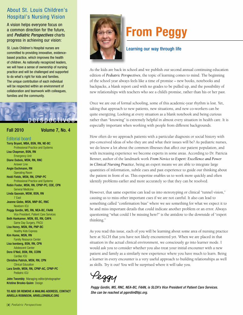
These successful outcomes
are directly related to the
dedication of the multidisciplinary team involved in the ECMO
program.
Extra-corporeal membrane oxygenation (ECMO)
By Mary MeheGaN, BSN, rN
Extra-corporeal membrane oxygenation (ECMO) is a method of providing cardiopulmonary support outside the body with a machine consisting of a pump, a circuit warmer and an oxygenator. ECMO is used for critically ill infants, children and adults as a means to help them recover from acute, reversible pulmonary or cardiac failure.
Delivery of oxygen to tissues is accomplished by creating an extension of the patient’s circulation outside of the body where blood is pumped and warmed through a circuit, oxygen is delivered and carbon dioxide is removed. There are two distinct methods of ECMO: veno-arterial and veno-venous.
In veno-arterial ECMO, two cannulae are used. One is placed in a large vein, typically the superior vena cava, to allow for the gravitational flow of
blood to the ECMO circuit. The second cannula is placed in a large artery to infuse the blood back into the patient after re-oxygenation and carbon dioxide removal.
Veno-venous ECMO is used exclusively for patients in respiratory failure. A new single bi-caval (double lumen) venous cannulae is used, which reduces the risk factors (for example, potential for thrombus bleeding) associated with cannulation of a large artery.
Both methods of ECMO provide an opportunity to allow the patient’s heart and/or lungs to “rest” and recover. As the function of the organs improve, the ECMO support is slowly weaned to a level where the patient’s own heart and lungs assume full function, and the extracorporeal support is discontinued.
This year is the 25th anniversary of the ECMO program. To date, 800 infants and children have been placed
[4 ] Ped ia t r i c Perspec t i ves
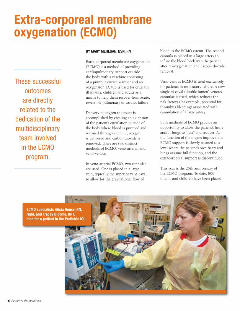
ECMO specialists Alicia House, RN, right, and Tracey Bloome, RRT, monitor a patient in the Pediatric ICU.
on ECMO. Based on data extracted from the national Extracorporeal Life Support Organization (ELSO) database, the clinical outcomes of these patients are above-average. In 2009, for example, SLCH had a 100 percent survival-to-discharge for neonates with respiratory failure who were placed on ECMO. The national average of survival-to-discharge for this population was only 76 percent as reported by ELSO.
These successful outcomes are directly related to the dedication of the multidisciplinary team involved in the ECMO program. The team consists of CT surgeons, general surgeons, perfusionists, operating room nurses, ECMO specialists, respiratory therapists and critical care physicians. The ECMO team is prepared to offer ECMO around-the-clock and responds quickly to the ECMO activation call. Support is offered in all three intensive care units – cardiac, newborn and pediatric.
The author can be reached at [email protected].
Ped ia t r i c Perspec t i ves [5 ]
A 12-year-old girl, “Lydia,” was found unconscious in a house
fire by the St. Louis Fire Department. She was then immediately
brought to the Emergency Department at St. Louis Children’s
Hospital. Upon arrival to the ED, she was having agonal respirations
with significant hypoxia and hypotension. She was immediately
intubated and placed on a ventilator, as she had smoke inhalation
injury that caused severe hypoxemia and carbon monoxide
poisoning. She was then transferred to the Pediatric ICU for further
management. The oxygen level in her body was dangerously low
even with maximum ventilatory support.
Lydia’s pO2 level was 39 pO
2, and her oxygen saturations were
50-60 percent. After her ventilator function did not improve
with high-frequency oscillatory ventilation, the ECMO team was
activated. She was placed on ECMO less than four hours after
arrival to SLCH. Her first pO2 level after ECMO initiation was
329. After five days of therapy, she was successfully weaned
from ECMO. Three days after ECMO was discontinued she was
extabated, and three days after that she was transferred to the
inpatient unit and was discharged from the hospital after six days
there. She spent 17 days in the hospital and had a full recovery.
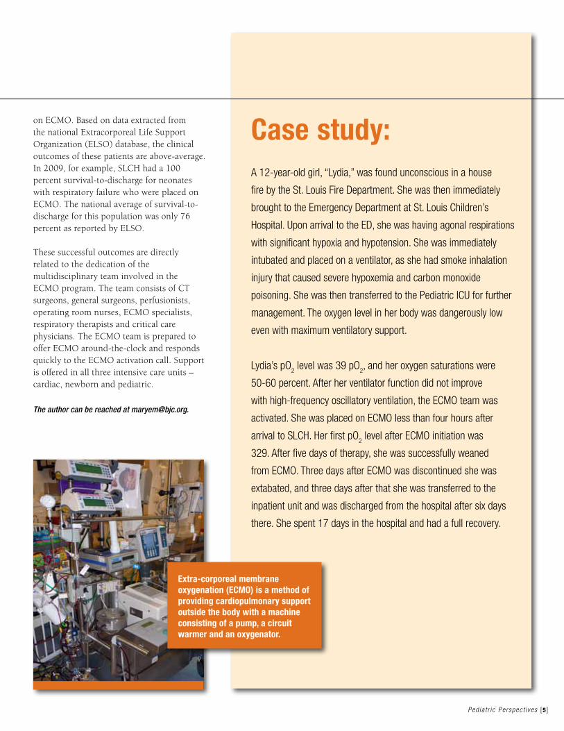
Case study:
Extra-corporeal membrane oxygenation (ECMO) is a method of providing cardiopulmonary support outside the body with a machine consisting of a pump, a circuit warmer and an oxygenator.
[6 ] Ped ia t r i c Perspec t i ves
By Kira Geile, MSN, rN, CPNP
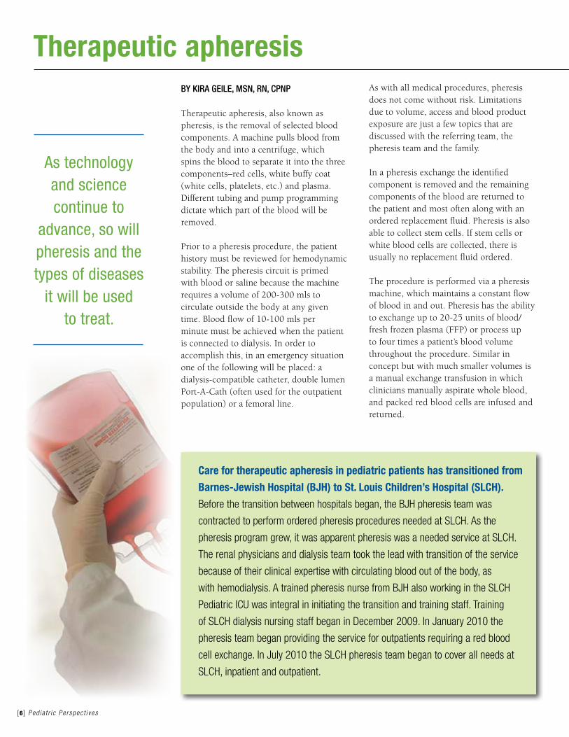
Therapeutic apheresis, also known as pheresis, is the removal of selected blood components. A machine pulls blood from the body and into a centrifuge, which spins the blood to separate it into the three components–red cells, white buffy coat (white cells, platelets, etc.) and plasma. Different tubing and pump programming dictate which part of the blood will be removed.
Prior to a pheresis procedure, the patient history must be reviewed for hemodynamic stability. The pheresis circuit is primed with blood or saline because the machine requires a volume of 200-300 mls to circulate outside the body at any given time. Blood flow of 10-100 mls per minute must be achieved when the patient is connected to dialysis. In order to accomplish this, in an emergency situation one of the following will be placed: a dialysis-compatible catheter, double lumen Port-A-Cath (often used for the outpatient population) or a femoral line.
As with all medical procedures, pheresis does not come without risk. Limitations due to volume, access and blood product exposure are just a few topics that are discussed with the referring team, the pheresis team and the family.
In a pheresis exchange the identified component is removed and the remaining components of the blood are returned to the patient and most often along with an ordered replacement fluid. Pheresis is also able to collect stem cells. If stem cells or white blood cells are collected, there is usually no replacement fluid ordered.
The procedure is performed via a pheresis machine, which maintains a constant flow of blood in and out. Pheresis has the ability to exchange up to 20-25 units of blood/fresh frozen plasma (FFP) or process up to four times a patient’s blood volume throughout the procedure. Similar in concept but with much smaller volumes is a manual exchange transfusion in which clinicians manually aspirate whole blood, and packed red blood cells are infused and returned.
Therapeutic apheresis
Care for therapeutic apheresis in pediatric patients has transitioned from
Barnes-Jewish Hospital (BJH) to St. Louis Children’s Hospital (SLCH).
Before the transition between hospitals began, the BJH pheresis team was
contracted to perform ordered pheresis procedures needed at SLCH. As the
pheresis program grew, it was apparent pheresis was a needed service at SLCH.
The renal physicians and dialysis team took the lead with transition of the service
because of their clinical expertise with circulating blood out of the body, as
with hemodialysis. A trained pheresis nurse from BJH also working in the SLCH
Pediatric ICU was integral in initiating the transition and training staff. Training
of SLCH dialysis nursing staff began in December 2009. In January 2010 the
pheresis team began providing the service for outpatients requiring a red blood
cell exchange. In July 2010 the SLCH pheresis team began to cover all needs at
SLCH, inpatient and outpatient.
As technology and science continue to
advance, so will pheresis and the types of diseases
it will be used to treat.

Ped ia t r i c Perspec t i ves [7 ]
“Makayla” is a 14-year-old black female diagnosed with Sickle Cell
Disease during her newborn screening. When she was 8 years old,
the family noted coordination difficulties and left-sided weakness.
A stroke was confirmed by radiologic imaging studies. She had a
pheresis catheter placed and pheresis procedure performed upon
admission. Red blood cell exchanges were continued every four
weeks to keep her sickle cell percentage below 30 percent. Although
Makayla received some manual exchanges, the automatic exchanges
(pheresis) are much more efficient and keep her sickle cell level in
goal ranges. After two years, Makayla continued to do well and had
no new strokes. Makayla is now receiving exchanges at six-week
intervals to keep her sickle cells below 50 percent. She recently tried
out for and made her school’s dance team.
Case study:Some sickle cell patients who have had a stroke receive erythrocytopheresis (red blood cell exchange). In this kind of pheresis, sickle-shaped cells are removed and replaced with packed red blood cells. For those patients who have had a previous stroke, pheresis has shown positive outcomes and reduced risk of another stroke.
Therapeutic Plasma Exchange (TPE or plasmapheresis) removes the plasma component of blood, which includes antibodies, and replaces the blood component with albumin or fresh frozen plasma. This procedure may be considered before and/or after a transplant. Removing the antibodies “attacking” the transplanted organ can allow for time for the body to adjust or allow for immunosuppression drugs to take effect – or both. TPE may also be considered for inappropriate antibody function disorders in acute phases, such as atypical Hemolytic Uremic Syndrome and Goodpasture’s syndrome.
Since pheresis was first successfully performed more than 50 years ago, the procedure has continued to develop. Pheresis offers other possibilities for collecting specific components of the blood’s buffy coat. Leukocytapheresis may be considered in leukemia when white cells are in extreme excess. Thrombocytapheresis is used with thrombocythemia when platelets are in extreme excess. A process called extracorporeal photopheresis is used in graft-versus-host disease and other conditions. In this procedure, specific cells are removed, manipulated with ultraviolet radiation and then re-infused to the patient. As technology and science continue to advance, so will pheresis and the types of diseases it will be used to treat.
The author can be reached at [email protected].
Tessa Patterson, RN, checks the vital signs of Tayler Campbell, 14, of Cape Girardeau, Mo., during her pheresis treatment.
By Carole BraNCh, MSN, rN, PNP-BC
Susan underwent bilateral lung transplantation at the age of 11 years for complications of cystic fibrosis (CF). Her postoperative course was complicated by episodes of acute rejection, lymphoproliferative disease (a type of cancer seen in patients receiving immunosuppression), medication-related hearing loss and chronic renal failure requiring dialysis. Susan was evaluated and found to be a candidate for a kidney transplant, was listed and–when a suitable donor was found–underwent transplantation.
Renal dysfunction is one of the most common and well-described complications following lung transplantation. It is a multi-factorial process seen in both adults and pediatrics and can occur following any solid organ transplant. The class of drugs known as calcineurin inhibitors (CNI), such as
cyclosporine and tacrolimus, are a major contributing factor. The nephrotoxic side effects of these medications have been well recognized since the introduction of cyclosporine in the early 1980s. The CNIs cause afferent arteriolar vasoconstriction and reduced renal blood flow leading to potentially irreversible structural changes that may ultimately lead to chronic renal failure.
As an inpatient pediatric nurse practitioner for the Lung Transplant Program, I noticed that a number of my patients developed renal dysfunction after transplant. I questioned why some recovered and others progressed to chronic renal failure requiring dialysis –and in some patients like Susan–kidney transplantation. The desire to understand led me to embark on a research project to:• determine the extent of decreased
renal function following pediatric lung transplantation
• characterize differences in primary diagnoses, co-morbidities and treatment between children who develop decreased renal function from those who did not
• identify which characteristics are predictive of decreased renal function
A retrospective data collection from the Pediatric Lung Transplant Database was obtained on 195 children who survived a minimum of one year following a primary lung transplant performed between Jan. 1, 1990, through Dec. 31, 2008. Of the sample, 107 patients were female patients (55 percent) and 90 had a primary diagnosis of CF (46 percent). Repeated measures analysis of variance (ANOVA) was used to assess the patients’ glomerular filtration rate over time. Preliminary results revealed that virtually all of the subjects developed decreased renal function based on decreasing GFR; however, only 7.69 percent or four of the
[8 ] Ped ia t r i c Perspec t i ves
Renal failure after lung transplantation
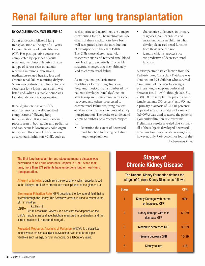
Stages of Chronic Kidney Disease
The National Kidney Foundation defines the stages of Chronic Kidney Disease as follows:
(continued on back cover)
the first lung transplant for end-stage pulmonary disease was performed at St. louis Children’s hospital in 1990. Since that time, more than 371 patients have undergone lung or heart-lung transplantation.
Afferent arterioles branch from the renal artery, which supplies blood to the kidneys and further branch into the capillaries of the glomerulus.
Glomerular Filtration Rate (GFR) describes the flow rate of fluid that is filtered through the kidney. The Schwartz formula is used to estimate the GFR in children.
where k is a constant that depends on the child’s muscle mass and age, height is measured in centimeters and the serum creatinine is measured in mg/dL.
Repeated Measures Analysis of Variance (ANOVA) is a statistical model where the same subject is evaluated over time for multiple variables such as age, gender, diagnosis, or a laboratory value.
Stage Description CFR
1 Kidney Damage with normal 90+ or increased OFR
2 Kidney damage with mild 60-89 decrease GFR 3 Moderate decreases GFR 30-59
4 Severe decrease GFR 15-29
5 Kidney failure <15
eGFR=k x Height
——————Serum Creatinine
By KriStiNa ShiltS, MS, CCC-a
Statistics indicate that approximately four out of 1,000 infants will be born with a significant hearing loss affecting speech and language development. This rate increases to 40 per 1,000 for those infants who spend more than 48 hours in the Newborn ICU. Other important risk factors for hearing loss include: Extracorporeal Membrane Oxygenation (ECMO), cytomegalovirus (CMV), ototoxic medications (for example, furosemide or gentamicin), ventilatory support, low birthweight and low Apgar scores.
St. Louis Children’s Hospital’s (SLCH) Audiology department has been performing hearing screens on infants with high risk factors since 1987 and screening all infants in the Newborn ICU since 1994. Universal newborn hearing screening in all nurseries became law in 2002 for Missouri and 2003 for Illinois. Those infants not passing the newborn hearing screen will be referred for further testing. Typical referral rates are 1 to 2 percent for infants in a well-baby nursery and approximately 10 percent for infants in the Newborn ICU.
The American Speech-Language-Hearing Association’s Joint Committee on Infant Hearing 2007 Position Statement recommends that a newborn hearing screening program should screen infants by 1 month of age, diagnose by 3 months of age and intervene by 6 months of age. There are two different types of hearing screens that may be used:1. Otoacoustic emissions measure the
function of the cochlea or inner ear and Automated Auditory Brainstem Response (AABR). Commonly done at the bedside, this screen measures responses of the auditory nerve from the cochlea into the brainstem. An infant may not pass (“refer”) a hearing screen for multiple reasons including but not limited to:• permanent hearing loss• transitory middle ear fluid
• state of the child at time of screening (crying, fussing)
2. The diagnosis of hearing loss in infancy is established with a diagnostic Auditory Brainstem Response (ABR). During the initial evaluation, the audiologist will determine the severity of the hearing loss and whether intervention or amplification is required. If hearing loss is diagnosed, infants are referred to a parent-infant program that provides hearing aid fitting, speech and language therapy, aural or hearing rehabilitation, and family support.
In 2009, 590 infants had a newborn hearing screen in the SLCH Newborn ICU. Of the 60 infants who did not pass–either in one or both ears:
• 37 infants were referred for diagnostic evaluation; 21 of these infants had ABR results indicative of normal hearing sensitivity, and 16 of these infants had hearing loss that required intervention either due to transient middle ear pathology or permanent hearing loss;
• 21 infants were lost to follow-up;• 2 infants expired.
The high percentage of infants lost to follow-up is a significant concern for the program. Both Missouri and Illinois monitor newborn hearing screens, which must be reported to the Department of Health and Senior Services. The family and primary care provider receive a letter indicating the rationale and importance of follow-up. Infants 12-18 months of age who have had repeated screenings since birth are referred to an audiologist for the first time for diagnostic evaluation. Late diagnosis and intervention have been shown to have a significantly negative impact on long-term prognosis.
Early identification and management of hearing loss optimizes speech, language and social development. With early and appropriate intervention, many children are able to develop normally in this area and be mainstreamed into preschool or kindergarten.
The author can be reached at [email protected]
Hearing screening for newborns
Hearing and Understanding Development Chart Birth to 3 months• Startles to loud sounds• Quiets or smiles when spoken to• Seems to recognize your voice and quiets if crying• Increases or decreases sucking behavior in
response to sound
4-6 months• Moves eyes in direction of sounds• Responds to changes in tone of voice• Notices toys that make sounds• Pays attention to music
7 months to 1 year• Enjoys games like peek-a-boo and pat-a-cake• Turns and looks in direction of sounds• Listens when spoken to• Recognizes words for common items like “cup” or
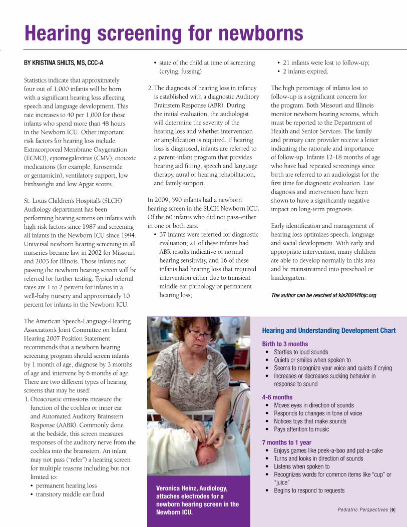
“juice”• Begins to respond to requestsVeronica Heinz, Audiology,
attaches electrodes for a newborn hearing screen in the Newborn ICU. Ped ia t r i c Perspec t i ves [9 ]
answers any questions they have at this time, and the PNP remains the main contact for the family throughout the surgery and once they go home. The nurse practitioner reviews the importance of the helmet therapy that follows the surgery because once the section of the bone is removed, the helmet does the work. For children who have the endoscopic surgery, a helmet will be worn for one year to guide appropriate head shape.
On the day of surgery, the child is admitted through Same Day Surgery. Once in the warmed OR suite, monitors are placed and the child is anesthetized. The patient is prepped and draped, and the surgeons begin the procedure. An endoscope is utilized through two small incisions measuring approximately 1.5 inches each at the top of the head. A segment of bone is removed near the fused bone. No plating or reshaping is performed. No internal hardware is used for the ECR approach. The large sections of bone are simply removed, and then the helmet shapes the head.
The repair lasts for 60 to 90 minutes. Children do not typically require blood
products during this procedure. When the operation is completed, the patient is transferred to the PACU and then to the Pediatric ICU.
While in the unit, the patient is monitored closely for discomfort and the effects of anesthesia. Even though bleeding is reduced with the endoscopic procedure, it is still important to monitor the patient’s hemoglobin and hematocrit at regular intervals. Close neurological monitoring is required overnight to detect any acute changes in neurological status. The risk for respiratory compromise increases threefold due to the effects of anesthesia, intubation and pain medication.
Pain assessment can be challenging because a child’s cry can be difficult to interpret. A crying infant can mean many things like “I’m hurting,” “I’m scared” or “I’m hungry.” These children are placed on an around-the-clock regimen of pain medications, which sometimes includes frequent administration of narcotics.
Although patients usually require an overnight stay in the Pediatric ICU, they are often discharged home directly from the unit. Infants go home with no scheduled pain medication,
but caregivers are instructed to give acetaminophen, as needed, for pain. Before discharge, families are advised to call Neurosurgery if the patient has changes in mental status or signs of infection. Follow-up care begins at one week post-surgery for fitting of the helmet. After this initial visit, the child then returns in another one to two weeks to ensure the surgical site is healing properly.
The nurse practitioner follows the patient through about one year of helmet therapy and then annually through the early school-age years. The patients are screened for psychological concerns and developmental or speech delays, which can accompany a change in suture closures.
This innovative surgery offers advantages for the infant and family. Early intervention of craniosynostosis with minimally invasive, endoscopic-assisted techniques is safe, limits blood transfusions, shortens operative time and reduces length of stay. This surgery is an alternative to the more traditional reconstructive methods for craniosynostosis repair.
The author can be reached at [email protected].
[10 ] Ped ia t r i c Perspec t i ves
Endoscopic Craniosynostosis Repair (ECR) (Continued from cover)
Fontanelle anterior Infant skull sutures
P r o f e s s i o n a l a c c o m P l i s h m e n t s a n d r e c o g n i t i o n
Spotlight on Professional Practice
Ped ia t r i c Perspec t i ves [11 ]
PresentationsAmerican Academy of Ambulatory Care Nursing, Las VegasFISH! Improving Morale in the Telehealth Setting•SuzanneWells,BSN,RN Answer Line
Midwest Nursing Research SocietyKansas City, Mo.Promotion of Physical Activity in Children with Asthma and Prevention of Obesity•ChristinaMahl,BSN,RN Child Health Advocacy and Outreach
Child Health Corporation of America (CHCA) WebinarImproving Labor Productivity at St. Louis Children’s Hospital•LisaCoakley,MSN,RN,FNP-BC 8 West General Medicine•LeroyLove,MS,BS,ChEEngr, CSSMBB Performance Excellence•PeggyGordin,MS,RNC,NEA-BC, faaN Patient Care Services•SusanHibbits,OTL/R,FACHE Neurosciences•TomChiarelli,BS Finance
Poster PresentationsSigma Theta Tau Iota Chapter, Improving Patient Outcomes through Research and Evidence-Based Practice, St. LouisAn Outcome of Evidence-Based Practice Education: Sustained Clinical Decision-Making Among Bedside Nurses•KarenBalakas,PhD,RN,CNE•LisaSteurer,MSN,RN,CPNP-PC, CPN•TerryBryant,MBA,BSN,RN,NE-BC Professional Practice and Systems
Third Evidence-Based Practice on the Frontline: Building a Culture of Quality, Safety and Nursing Professionalism,Columbia, Mo.Supporting Parental Attachment:
Newborn Intensive Care Unit to Home•AshleyStrobach,BSN,RN Float Pool Specialty
Active Computer Games as Exercise for Children with Cystic Fibrosis•JeffFilipiak,BSN,RN,CPN 7 East General Medicine
Published articlesJournal of Nursing AdministrationPediatric Falls Benchmarking Collaborative•TerryBryant,MBA,BSN,RN,NE-BC Professional Practice and Systems
DegreesBachelor’s of Science in Nursing, Goldfarb School of Nursing at Barnes-Jewish College•JulieBubash,BSN,RN•TamikaSmith,BSN,RN Newborn Intensive Services•SaraOwens,BSN,RN,CPN Vascular Access Team•CrystalClinch,BSN,RN Float Pool Specialty
Bachelor’s of Science in Nursing, Saint Louis University•LindsaySaylor,BSN,RN Newborn Intensive Services
Bachelor’s of Science in Nursing, University of Missouri-St. Louis•CrystalBuesking,BSN,RN,CPN 7 West Cardiology
Bachelor’s of Science in Nursing, Webster University•PatriciaRelleke,BSN,RN•CarrieLeone,BSN,RN Operating Room
Bachelor’s of Science in Nursing, Chamberlain College of Nursing•LisaChapman,BSN,RN Emergency Unit
Master’s of Science in Nursing, Webster University•BethHankamer,MSN,RN,CAPA Clinical Education
Master’s of Science in Nursing, McKendree University•ChristinaKlasing,MSN,RN,CPN Neurosciences/Rehabilitation
Master’s of Science in Nursing, University of Missouri-St. Louis•ChristineClune,MSN,CPNP-PC,RN Float Pool
National CertificationRegistered Nurse Certification-Neonatal Intensive Care•BeckieFrohock,BSN,RN,RNC•KarleneBessler,BSN,RN,RNC•KelleyKostich,BSN,RN,RNC•LauraDow,BSN,RN,RNC•RebeccaJones,BSN,RN,RNC Newborn Intensive Services
International Board-Certified Lactation Consultant•JoanEigelberger,BSN,RN,RNC, iBClC Newborn Intensive Services
Certified Pediatric Nurse Practitioner•ChristineClune,MSN,CPNP-PC,RN
Registered Nurse, Board Certified: Ambulatory Care Nursing•DianeDuBois,MSN,RN-BC Answer Line
elected officersAmerican Academy of Ambulatory Care Nursing, Treasurer•SuzanneWells,BSN,RN Answer Line
other Professional honors Finalists in St. Louis Magazine’s 2010 Excellence in Nursing Awards•KathyBlanke,RN Washington University School of Medicine•CaroleBranch,MSN,RN,PNP-BC Transplant Services•CaroleCampbell,BSN,RNC Answer Line and Children’s Direct
CondolencesBridgetKurz,BSN,RN,CPNCharge nurse, 8 West General Medicine
Bridget, 24, passed away after an auto accident in August. She had served as a charge nurse since joining SLCH in 2006. She was the preceptor on the night shift, participated in several quality improvement projects and wrote a diabetes teaching tool.
“You couldn’t ask for a better person to know or work with,” said Natalie Amighetti, RN, 8 West. “Even when times got stressful, you could always turn to Bridget because she was loyal and dependable. She brightened everyone’s day.”
Nursing Spotlight reflects achievements through July 2010.
Please submit more recent accomplishments to the nursing intranet home page, Spotlight on Professional Practice.
St. Louis Children’s HospitalOne Children’s PlaceSt. Louis, MO 63110
Non-ProfitOrganizationU.S. Postage
PAIDSt. Louis, MO
Permit No. 617
Pediatric Perspectives is published by the St. Louis Children’s Hospital Communications and Marketing department. Please direct inquiries to:
600 S. Taylor Ave., Suite 202 St. Louis, MO 63110
314.286.0417 314.286.0420 (fax)
© 2010, St. Louis Children’s Hospital
St.LouisChildren’sHospitalisrecognized
among america’s best children’s hospitals
by Parents magazineandU.S.News & World
Report. for more information about nursing
opportunities at a Magnet hospital, visit
StLouisChildrens.org/jobs
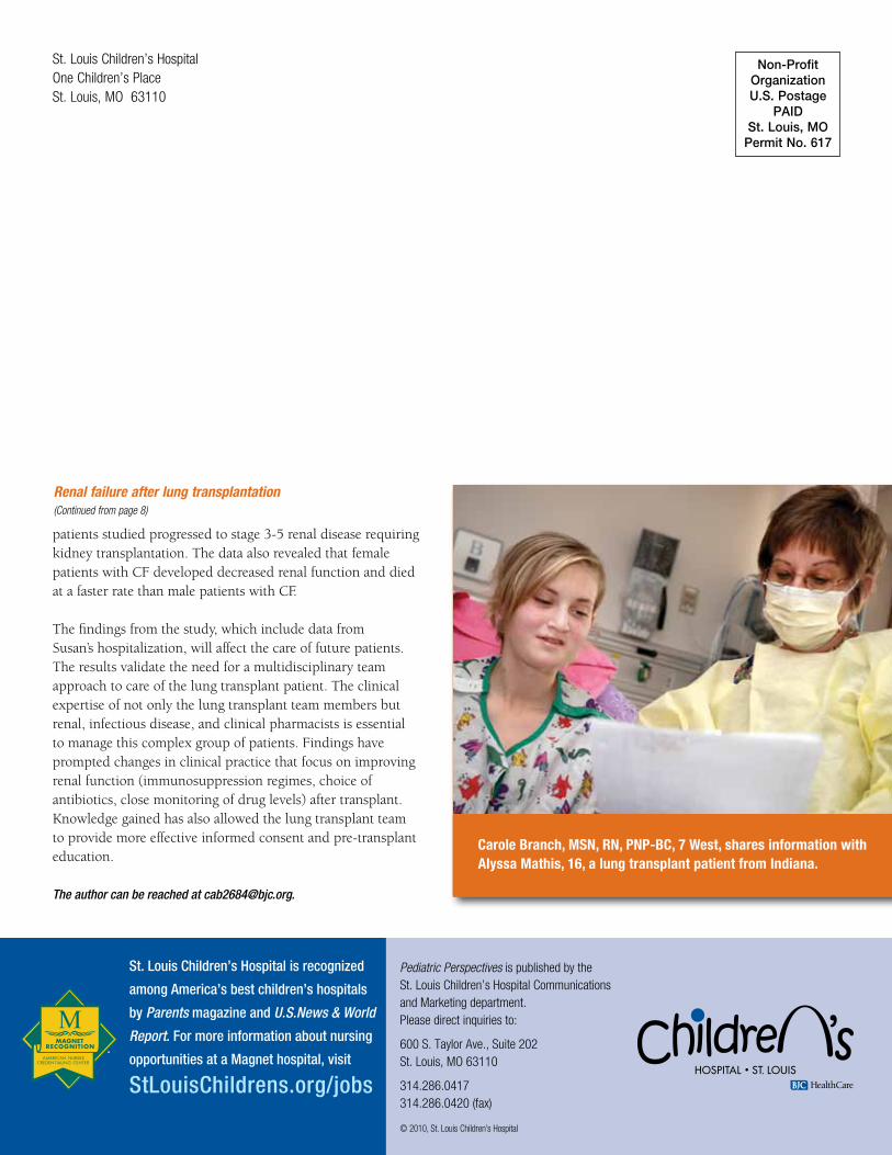
patients studied progressed to stage 3-5 renal disease requiring kidney transplantation. The data also revealed that female patients with CF developed decreased renal function and died at a faster rate than male patients with CF.
The findings from the study, which include data from Susan’s hospitalization, will affect the care of future patients. The results validate the need for a multidisciplinary team approach to care of the lung transplant patient. The clinical expertise of not only the lung transplant team members but renal, infectious disease, and clinical pharmacists is essential to manage this complex group of patients. Findings have prompted changes in clinical practice that focus on improving renal function (immunosuppression regimes, choice of antibiotics, close monitoring of drug levels) after transplant. Knowledge gained has also allowed the lung transplant team to provide more effective informed consent and pre-transplant education.
The author can be reached at [email protected].
Renal failure after lung transplantation (Continued from page 8)
Carole Branch, MSN, RN, PNP-BC, 7 West, shares information with Alyssa Mathis, 16, a lung transplant patient from Indiana.