Edexcel IAL Biology A Level Topic 2 : Membranes, Proteins ... · The tertiary structure of proteins...
15
Edexcel IAL Biology A Level Topic 2 : Membranes, Proteins, DNA and Gene Expression Notes www.pmt.education
Transcript of Edexcel IAL Biology A Level Topic 2 : Membranes, Proteins ... · The tertiary structure of proteins...

Edexcel IAL Biology A Level
Topic 2 : Membranes, Proteins, DNA and Gene Expression
Notes
www.pmt.education

Gas exchange Exchange surfaces Multicellular organisms require gas exchange systems in order to obtain sufficient oxygen for respiring cells, and to expel the carbon dioxide created by these cells. In order to maximise the rate of exchange of substances, gas exchange surfaces are adapted to have a:
● Large surface area to volume ratio - the larger the ratio, the greater the surface area for the organism to carry out exchange, so a faster transfer of substances across the surface.
● Short diffusion pathway - a short distance for substances to move across means they move faster.
● Steep concentration gradient - a large difference in concentrations between 2 areas means diffusion of particles from an area of high concentration to an area of lower concentration occurs faster.
Fick’s Law The rate of diffusion is also dependent on the above features. This is shown by Fick’s law that states that the rate of diffusion is proportional to the surface area multiplied by difference in concentration, divided by the length of diffusion pathway.
The mammalian lung When we breathe, air enters the mouth, passes into the trachea which splits into 2 bronchi; one on the left side and one on the right side of the body. Each bronchus branches out into smaller bronchioles which end in tiny air sacs called alveoli, where gas exchange takes place. Deoxygenated blood flows into the alveoli, where carbon dioxide diffuses out of the capillary, through the alveolar membrane and into the surrounding air, down a concentration gradient. Oxygen moves in the opposite direction from the surrounding air and into the blood steam, making the blood oxygenated. The extensive branching of vessels in the lungs means there are many alveoli - over 300 million in an average adult - this gives them a very large surface area which increases the rate of diffusion of oxygen into the blood, and carbon dioxide out of the blood. The alveoli are adapted further. They have a rich blood supply from surrounding capillaries which maintains a steep concentration gradient between the blood in the capillaries and the air entering the lungs, which again increases the rate of diffusion.
www.pmt.education

Alveoli also have a moist outer lining, allowing gases to dissolve and move across their membrane faster. Finally, alveoli have a wall of only one cell thick and pores in the endothelium, this creates a short distance for the gases to travel over so they can diffuse quickly.
Cell membranes All cells and organelles are surrounded by a partially permeable membrane composed of a sea of phospholipids with protein molecules between the phospholipid molecules. The main function of the membrane is controlling the movement of substances in and out of the cell/organelle. However, it also contains receptors for other molecules, such as hormones, and enables adjacent cells to stick together. The main structure of a membrane is the phospholipid bilayer - 2 rows of phospholipids (lipids made from 1 molecule of glycerol, 1 phosphate group and 2 fatty acid chains). The phosphate groups are hydrophilic (water loving), so form the outside of the bilayer. Whereas the fatty acid chains are hydrophobic (water hating), so lie in between the 2 rows of phosphate heads. The fatty acid chains are non-polar, allowing non-polar molecules like carbon dioxide to pass straight through the phospholipid bilayer. While polar substances, like water, have to move through channel proteins, since they aren’t soluble in the fatty acid tails. Fluid Mosaic Model The fluid mosaic model is the name given to the model suggested for the structure of the cell membranes. It is described as ‘fluid’ due to the fluidity of the phospholipid bilayer which allows all molecules to move freely within it. The cell membrane also contains a ‘mosaic’ of transport proteins, receptor proteins, enzymes, structural and recognition proteins of varying shapes and sizes. Cholesterol molecules are also found in the bilayer, these give the membrane stability and reduce its fluidity. Scientific models such as the fluid-mosaic model are based on data and results of investigations by scientists. Models like this may be updated over time, the way the model of the atom was updated numerously, as new data and discoveries are made. Transport of substances The movement of molecules through cell membrane depends on the properties of the molecule (for instance its size and whether it is polar or nonpolar) as well as the requirements of the cell. Movement can be passive (require no energy) or active (requires energy released from respiration). There are several types of movement:
www.pmt.education

● Diffusion is the passive movement of molecules down a concentration gradient, from an area of high concentration to an area of lower concentration through a partially permeable membrane. There are specific types of diffusion - simple diffusion, facilitated diffusion and osmosis.
● Simple diffusion is the passive movement of small, non-polar lipid soluble molecules,
such as carbon dioxide and oxygen, from an area of high concentration to an area of low concentration. The molecules move directly through the phospholipid bilayer.
● Facilitated diffusion requires a membrane protein to transport polar molecules, charged and water soluble molecules across the membrane. Since these molecules cannot pass through the non-polar inside of the bilayer.
● Osmosis is the diffusion of water molecules.
● Active transport can transport all types of molecules through carrier proteins from an area of low concentration to an area of high concentration. This process moves particles against the concentration, and so requires energy in the form of ATP.
● Exocytosis and endocytosis transport large particles. The particles are enclosed in vesicles made from the cell surface membrane and transported into the cell / organelle in endocytosis. In exocytosis, vesicles containing large particles are fused with the membrane and leave the cell/organelle.
Osmosis - a special type of diffusion Osmosis is the net diffusion of water molecules from an area of higher water potential to an area of lower water potential, through a partially permeable membrane. In the case of osmosis, ‘water potential’ is used to describe the relative concentration of water molecules. Water potential is the tendency of a solution to gain or lose water. A high water potential means there is a low concentration of solute, in other words a high concentration of water. Pure water has the highest water potential possible. Water potential is measured in pascals, pure water has a value of 0 pascals and all other values are negative, as water potential becomes lower. When comparing 2 solution’s water potentials, they can be described as:
● Isotonic - the 2 solutions have the same water potential.
www.pmt.education
● Hypertonic - the solution with a lower water potential relative to another solution (think ‘hyper’ - it has lots of sugars/salts in it like a hyper child, so has lower concentration of water, therefore a lower water potential).
● Hypotonic - the solution with a higher water potential relative to another solution. Membrane proteins Different types of protein in a membrane are involved in different types of transport:
● Carrier proteins - can move particles through the membrane by both active transport and facilitated diffusion.
● Channel proteins - forms pores in the membrane for polar particles to move through by facilitated diffusion.
● Extrinsic proteins - a membrane protein that goes through only 1 layer of the bilayer.
● Intrinsic proteins - a membrane protein that goes through both layers of the bilayer.
Proteins
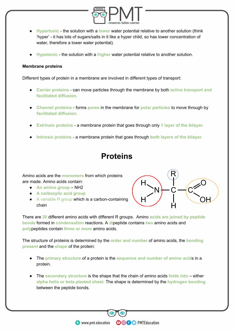
Amino acids are the monomers from which proteins are made. Amino acids contain:
● An amino group – NH2 ● A carboxylic acid group ● A variable R group which is a carbon-containing
chain
There are 20 different amino acids with different R groups. Amino acids are joined by peptide bonds formed in condensation reactions. A dipeptide contains two amino acids and polypeptides contain three or more amino acids. The structure of proteins is determined by the order and number of amino acids, the bonding present and the shape of the protein:
● The primary structure of a protein is the sequence and number of amino acids in a protein.
● The secondary structure is the shape that the chain of amino acids folds into – either alpha helix or beta pleated sheet. The shape is determined by the hydrogen bonding between the peptide bonds.
www.pmt.education

● The tertiary structure of proteins is the 3D shape of the protein, it can be globular or fibrous. Globular proteins, such as enzymes, are compact. Whereas fibrous proteins, such as keratin, are long and thus can be used to form fibres. The shape of the protein is determined by hydrogen, ionic and disulphide bonds between the R groups of amino acids.
● Some proteins have a quaternary structure, this is when 2 or more polypeptide chains are joined together, sometimes with the addition of a non-protein prosthetic group.
KEY EXAMPLE: Collagen is a fibrous protein of great strength due to presence of both hydrogen and covalent bonds in its structure. Collagen molecules wrap around each other and form fibrils which form strong collagen fibres. Collagen forms the structure of bones, cartilage and connective tissue and is a main component of tendons which connect muscles to bones. KEY EXAMPLE 2: Haemoglobin is a water soluble globular protein which consists of two beta polypeptide chains, 2 alpha polypeptide chains and 4 haem groups. It carries oxygen in the blood as oxygen can bind to the haem (Fe2+) group, and oxygen is then released when required. Haemoglobin is made of 4 polypeptide chains as well as 4 prosthetic groups, so it has a quaternary structure. Enzymes Enzymes are biological catalysts that increase the rate of reaction by lowering the activation energy (the energy needed for a reaction to occur) of the reactions they catalyse, including both intracellular (within cells) and extracellular (outside of cells) reactions. Part of the enzyme is known as the active site, this is where the reaction with the substrate takes place. Since enzymes are proteins, they have a very specific 3D shape due to the bonding in their tertiary structure. This means enzymes have a specific and complementary shape to the substrates they bind to, meaning that only one type of substrate fits into the active site of the enzyme. When the enzyme and substrate form a complex (an enzyme-substrate complex), the tertiary structure of the enzyme is altered so that the active site of the enzyme fits around the substrate. This is called the induced fit model. Once an enzyme-substrate complex is formed and there is sufficient energy, the reaction can take place, often breaking down the substrate or combining 2 to build up larger molecules.
www.pmt.education

Nucleotides
Both deoxyribonucleic acid (DNA) and ribonucleic acid (RNA) are polymers of nucleotides. Nucleotides consist of:
1. A pentose sugar - a 5 carbon sugar 2. A nitrogen containing organic base 3. A phosphate group
The components of a DNA nucleotide are a pentose sugar called deoxyribose, a phosphate group and one of the organic bases adenine, cytosine, guanine or thymine. Adenine and guanine both have double ring structure and are classified as purine bases. The components of an RNA nucleotide are a pentose sugar called ribose, a phosphate group and one of the organic bases adenine, cytosine, guanine or uracil. Thymine, uracil and cytosine all have single ring structure and are classified as pyrimidine bases. Nucleotides join together by phosphodiester bonds formed in condensation reactions. The phosphodiester bonds are formed between the phosphate groups and a carbon on the pentose sugar of adjacent nucleotides. Complementary base pairing A DNA molecule is a double helix composed of two polynucleotides chains running antiparallel to each other, joined together by hydrogen bonds between the 2 strands. 2 hydrogen bonds form between the complementary bases adenine and thymine, and 3 hydrogen bonds form between the complementary bases cytosine and guanine. This bonding is essential for DNA to maintain a stable structure, as it prevents the chemical bases being corrupted by other outside chemicals or forces. The bonding is also what twists the strands into the double helix shape. RNA is a single-stranded polynucleotide chain, so does not have hydrogen bonding between the bases within it, however its bases are still complementary to each other - cytosine and guanine, and adenine and uracil are complementary.
DNA replication
The semi-conservative replication of DNA ensures genetic continuity between generations of cells, meaning that genetic information is passed on from one generation from the next. The process is described as ‘semi-conservative’ because each strand formed contains 1 new strand,
www.pmt.education

and 1 original strand. The steps of semiconservative replication of DNA are as follows:
1. The double helix unwinds and the enzyme DNA Helicase breaks the hydrogen bonds between the complementary bases, separating the two strands of DNA.
2. One of the strands is used as the template and complementary base pairing occurs between the template strand and free nucleotides that attach to the exposed strands.
3. The enzyme DNA polymerase moves along the strands, joining the adjacent nucleotides by forming phosphodiester bonds in condensation reactions.
Meselson and Stahl’s experiment 2 scientists named Matthew Meselson and Franklin Stahl did an experiment to prove that DNA replicated semi-conservatively. This was what they did:
1. They began by growing bacteria in a broth containing only one isotope of nitrogen - Nitrogen-15, a heavier isotope than the more abundant Nitrogen-14. After allowing the bacteria to grow and take the heavy nitrogen into their DNA they isolated the DNA and centrifuged it; the DNA settles at a point equal to its density.
2. The bacteria containing only 15N DNA were
transferred to a broth now containing only a 14N medium. Since DNA replicates semi-conservatively, after the first replication of DNA, each double
helix contained 1 14N strand, and 1 15N strand , so settled at a different point when centrifuged. The scientists observed this was
between the settling points of 15N only DNA, and 14N only DNA, so concluded the DNA must contain one strand of each.
3. They then carried out a second replication of the DNA, still in the 14N medium, the results of the centrifuge confirmed the semi-conservative hypothesis still, with half of the DNA settling out at
the 14N only point, and half of the DNA settling with a combing 14N and 15N point.
www.pmt.education

The genetic code
The order of nucleotide bases on DNA makes up the genetic code which consists of triplets of bases; each triplet of bases codes for a particular amino acid and is known as a codon. For instance, the codon GCA codes for the amino acid alanine. In ribosomes during protein synthesis the amino acids are joined together by peptide bonds and form a polypeptide chain. Therefore, a gene is a sequence of bases on a DNA molecule that codes for a sequence of amino acids in a polypeptide chain. However, not all of the genome codes for proteins – the non-coding sections of DNA are called introns and the coding regions are called exons. Features of the genetic code:
● The genetic code is non-overlapping meaning that each triplet is only read once and triplets don’t share any bases.
● Genetic code is also degenerate meaning that more than one triplet codes for the same amino acid. This reduces the number of mutations which are mistakes in the base sequence such as base deletion, insertion or substitution. For instance, GCA, GCC, GCG and GCU all code for the amino acid alanine.
● The genetic codes contains start and stop codons which either start or stop protein synthesis.
Protein synthesis There are 2 stages which convert the sequence of bases on DNA into a polypeptide chain; the DNA must first be copied into mRNA (translation) which then leaves the nucleus and travels to a ribosome. This is where the RNA is read and translated into amino acids which are joined to make a polypeptide (translation). Transcription During transcription, a molecule of mRNA is made in the nucleus:
1. The enzyme RNA polymerase attaches to the DNA at the start of the gene (which is signified by a start codon). It breaks the hydrogen bonds between the complementary bases that hold the 2 strands together, this uncoils and separates the 2 strands so they are exposed.
www.pmt.education

2. One of the DNA strands is used as a template to make the mRNA molecule, the template is called the antisense strand.
3. RNA polymerase lines up free nucleotides which attach to the template strand by complementary base pairing; adjacent nucleotides are joined by phosphodiester bonds, thus forming a single-stranded molecule of mRNA.
4. Once the RNA polymerase reaches a stop codon, the mRNA detaches from the template strand. It then moves out of the nucleus through a pore and attaches to a ribosome in the cytoplasm, which is the site of next stage of protein synthesis called translation.
Translation During translation, a polypeptide sequence is translated from the mRNA strand:
1. mRNA attaches to a ribosome and transfer RNA collects amino acids from the cytoplasm and carries them to the ribosome. tRNA is a single stranded molecule with an amino acid binding site at one end (so it can only carry one type of amino acid), and a triplet of bases at the other (called an anticodon) which is complementary to a triplet of bases on the mRNA strand (the codon) produced during transcription.
2. The tRNA molecule attaches itself to mRNA by complementary base pairing – within the ribosome two molecules of tRNA attach to the mRNA strand at a time.
3. The amino acids attached to two the tRNA molecules join by forming a peptide bond; then the tRNA molecules detach themselves from the amino acids, leaving them behind as more amino acids attach to the chain.
4. This process is repeated leading to the formation of a polypeptide chain until a stop codon is reached on mRNA, which ends the process of protein synthesis.
www.pmt.education

Genetic Mutations
Since proteins are coded for by the sequence of bases on DNA, any errors made in copying the DNA during replication can affect the polypeptide produced, since a different amino acid means different folding and bonding so a different tertiary structure and 3D shape. In some cases this can result in non-functional proteins and lead to cancer, for instance if a protein that slows cell division is non-functional, this can lead to the formation of a cancerous tumour. Genetic disorders are also often caused by mutation, such as the mutation which leads to production of sticky mucus and causes cystic fibrosis or sickle cell anaemia, in which a mutated form of haemoglobin distorts the shape of red blood cells. The majority of mutations are not harmful however, due to the nature of the genetic code, and the presence of non-coding introns. As introns do not code for proteins so if a mutation occurs here, no polypeptide will be affected. There are several types of mutation:
● Substitution - this is where one base is substituted for another. For instance, the sequence ATGGCA to ATCGCA. In some cases, the mutation may not change the amino acid coded for, since the degenerate nature of the genetic code means multiple codons code for the same amino acid. Sometimes however it does result in a different amino acid.
● Insertion - this is where an extra base (or bases) is inserted into the DNA sequence. For instance GGCTAC to GGCTTTAC. This mutation often results in a non-functional protein as all of the codons after the insertion are affected as the code is non-overlapping. This is known as frame shift.
● Deletion - this is where a base (or bases) is removed from the sequence. For instance, TGCAAC to TGAAC. This again results in frame shift and a likely non-functional protein as all codons after the deletion are changed.
Genetic disorders
Important definitions: In order to understand the genetics topic fully, you must be confident with the following definitions:
● Gene - a sequence of bases on a DNA molecule that codes for a sequence of amino acids in a polypeptide chain
● Allele – a form of a gene. For instance, one form of a gene for eye colour may code for blue eyes, another may code for brown eyes
● Genotype – The genetic constitution of an organism, the alleles they have
www.pmt.education

● Phenotype – The physical characteristics expressed by an organism due to its genotype
● Recessive – The allele that must be present twice in the genotype to be expressed in the phenotype
● Dominant – The allele that only needs to be present once in the genotype to be expressed in the phenotype
● Codominance – Alleles in the genotype that both contribute to the phenotype. For example the genotype AB for blood group results in the phenotype AB
● Homozygote – When an organism has 2 of the same alleles for a characteristic ● Heterozygote - When an organism has 2 different alleles for a characteristic
Inheritance of genetic disorders Monohybrid inheritance is when we consider the inheritance of 1 gene at a time when 2 parents have offspring. There are multiple combinations possible, since during meiosis only 1 chromosome from each homologous pair goes into the gamete cell. Here are some common genetic crosses: Key: B : Dominant allele for brown eyes b : Recessive allele for blue eyes Cross 1: Homozygous dominant parent x homozygous recessive parent Parent genotypes: BB, bb Possible gametes: B,B and b,b
b b
B Bb - brown eyes Bb - brown eyes
B Bb - brown eyes Bb - brown eyes
Phenotypes: Brown eyes Phenotype ratio: 100% Cross 2 Heterozygous parent x heterozygous parent Parent genotypes: Bb, Bb Possible gametes: B,b and B,b
B b
B BB - brown eyes Bb - brown eyes
b Bb - brown eyes bb - blue eyes
Phenotypes: Brown eyes, blue eyes Phenotype ratio: 3:1
www.pmt.education

Cross 3 Heterozygous parent x homozygous recessive parent Parent genotypes: Bb, bb Possible gametes: B,b and b,b
b b
B Bb - brown eyes Bb - brown eyes
b bb - blue eyes bb - blue eyes
Phenotypes: Brown eyes, blue eyes Phenotype ratio: 1:1 Genetic pedigree diagrams These diagrams are essentially family trees that look at the occurrence of a genetic disorder. They can be used to work out an individual’s genotype and a couple’s chance of passing on the disorder to offspring. For instance, if 2 parents both exhibiting a dominant characteristic had a child expressing the recessive characteristic, the parents must both be heterozygous for the trait and the child is homozygous recessive for the trait. Sex-linked characteristics The 23rd pair of chromosomes in a human can be XX, coding for a female, or XY, coding for a male. The Y chromosome is much smaller than the X chromosome and is described to be almost ‘genetically empty’ as it contains much fewer genes than the X chromosome. This means in males some genes are only present once, meaning recessive disorders more common in males, as they only need one copy of the recessive gene, whereas females would need 2 copies on both X chromosomes, which is less likely. These disorders are known as ‘sex-linked disorders’ as they are caused by genes present on the sex chromosomes. A common example of one such disorder is red-green colour blindness, which is much more common in men and affects the sufferer’s ability to distinguish red and green colours. Although their ability to distinguish other colours such as blue and yellow is, for most people, unaffected. On average 8% of men suffer with it, and only 0.5% of women; providing evidence that the disorder is sex-linked. Cystic fibrosis Cystic fibrosis is a genetic disorder caused by a faulty, recessive allele on chromosome 7. The faulty gene produces a non-functional CFTR protein which is responsible for transporting chloride ions out of the cell.
www.pmt.education

Gaseous exchange In the respiratory tract, chloride ions in a healthy person are transported out of cells into mucus lining the epithelium, this lowers the water potential of the mucus causing water to also move out of the cell, via osmosis. This makes the mucus sticky enough to trap pathogens that pass through the respiratory tract when we breath. People with cystic fibrosis do not have the movement of chloride ions out of the cell, so water does not move out of the cell either, this makes the mucus too thick and sticky meaning the cilia cannot move. This results in pathogens getting stuck and remaining in the tract and often causing infections. Reproduction In the reproductive system mucus is also thickened. For women this can thicken their cervical mucus and make it harder for them to get pregnant, and in men the mucus can block the tube (vas deferens) that transports sperm to the end of the penis, also reducing their fertility. Digestion In the digestive system, thick mucus can block the pancreatic duct, out of which digestive enzymes pass. This means less enzymes enter the small intestine, so less food is broken down and absorbed, preventing normal growth. In the long term, the digestive enzymes that are not released from the pancreas can start to breakdown the pancreas, if the cells producing insulin are affected, patients can also develop diabetes.
Genetic Screening Genetic screening is a branch of gene technology which involves testing people’s DNA for the presence of certain genetic disorders, like cystic fibrosis. Since some disorders have different types (such as breast cancers being caused by different genes) by identifying the specific gene causing a disorder, the most effective treatment can be chosen for the individual. There are further uses to genetic screening:
● The identification of carriers - a carrier is someone who has a recessive allele for a genetic trait in their genes, but does not usually express the trait or show symptoms for the disorder as they have a dominant, working copy of the gene too. It is important to be aware if you are a carrier of a genetic disease, as carriers do not suffer themselves but can still pass on the disorder to children. For instance, 2 carriers of cystic fibrosis have a 25% chance of having a child with the disorder, so it can help parents make informed decisions about having children.
● Preimplantation genetic diagnosis (PGD) - If 2 parents are at risk of passing on a severe genetic disorder to their child, and want to avoid doing so, they can have PGD. The embryos must be produced via in-vitro fertilisation (IVF) in a lab, so they can be screened for the disorder to identify healthy embryos to be implanted into the mother.
www.pmt.education

● Prenatal testing - prenatal testing tests for the presence of genetic disorders and incorrect numbers of chromosomes once the fetus is already growing. It can help parents make decisions on whether to continue the pregnancy and also how to prepare if they are going to have a child who does have a genetic disorder. There are 2 main ways prenatal screening can be done:
○ Amniocentesis - This involves extracting a small sample of amniotic fluid from the amniotic sac. The fluid contains fetal tissue so can be screened for disorders.
○ Chorionic villus sampling - This involves taking a small sample of cells from the placenta and testing those.
Ethics of genetic screening Religion It is likely that a religious person’s views on genetic screening differ on an individual basis, but a common argument is that most religious groups view life as a sacred gift from God, so screening for disorders is irrelevant as the baby should live regardless. Some religious people do believe, however, that the life of the mother is worth more than the life of the unborn baby, so should the disorder cause any risk to the mother then they may agree with aborting it. Moral Genetic screening can sometimes result in false-positive or false-negative results, meaning potentially a foetus could be aborted due to an incorrect diagnosis, leading to unnecessary loss of lives. People who believe quality of life is extremely important may argue in favour of genetic screening, as they believe it’s good to end a life that would likely otherwise be filled with pain and suffering. However, understanding that your child will have a certain disorder can help parents best prepare for it. Social Socially, the acceptance of testing for disorders and sometimes aborting babies with those disorders can encourage the idea that disabled lives are worth less in society, so encourage prejudice and discrimination against those groups. Yet, genetic screening can increase public awareness and understanding about gene technology and genetic disorders. Further ethical concerns include:
● There’s a risk of harm to the foetus of miscarriage ● The outcome of testing might lead to an abortion ● Right to life ● The cost of bringing up a baby with a genetic disorder ● Emotional and mental issues surrounding the birth of a baby with a disorder
www.pmt.education