Edexcel (B) Biology...
11
Edexcel (B) Biology A-level Topic 2: Cells, Viruses and Reproduction of Living Things Notes www.pmt.education
Transcript of Edexcel (B) Biology...

Edexcel (B) Biology A-level
Topic 2: Cells, Viruses and Reproduction of
Living Things
Notes
www.pmt.education
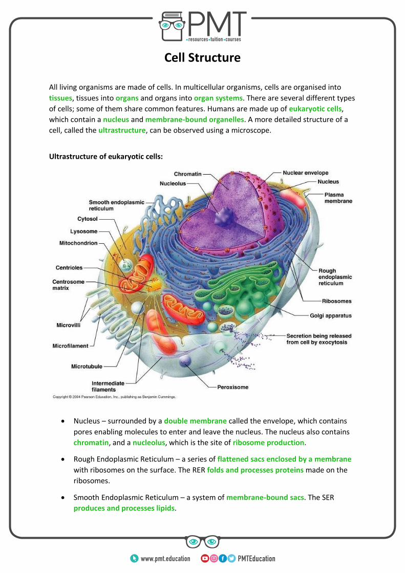
Cell Structure
All living organisms are made of cells. In multicellular organisms, cells are organised into
tissues, tissues into organs and organs into organ systems. There are several different types
of cells; some of them share common features. Humans are made up of eukaryotic cells,
which contain a nucleus and membrane-bound organelles. A more detailed structure of a
cell, called the ultrastructure, can be observed using a microscope.
Ultrastructure of eukaryotic cells:
• Nucleus – surrounded by a double membrane called the envelope, which contains
pores enabling molecules to enter and leave the nucleus. The nucleus also contains
chromatin, and a nucleolus, which is the site of ribosome production.
• Rough Endoplasmic Reticulum – a series of flattened sacs enclosed by a membrane
with ribosomes on the surface. The RER folds and processes proteins made on the
ribosomes.
• Smooth Endoplasmic Reticulum – a system of membrane-bound sacs. The SER
produces and processes lipids.
www.pmt.education
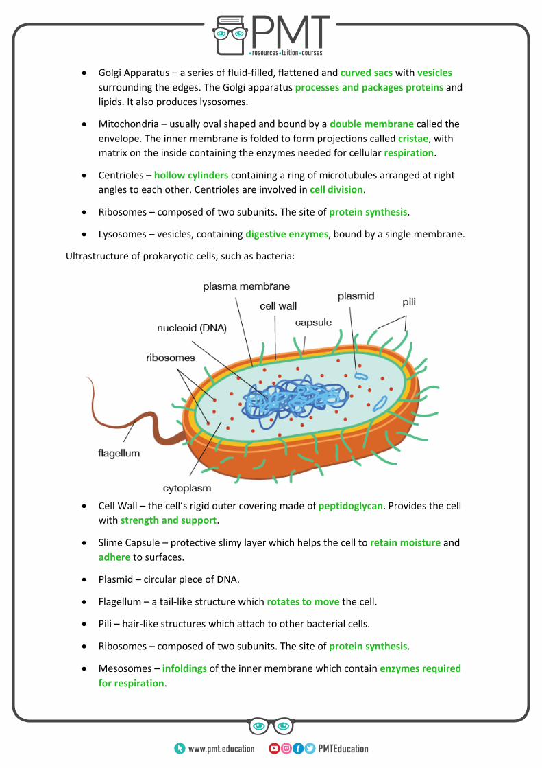
• Golgi Apparatus – a series of fluid-filled, flattened and curved sacs with vesicles
surrounding the edges. The Golgi apparatus processes and packages proteins and
lipids. It also produces lysosomes.
• Mitochondria – usually oval shaped and bound by a double membrane called the
envelope. The inner membrane is folded to form projections called cristae, with
matrix on the inside containing the enzymes needed for cellular respiration.
• Centrioles – hollow cylinders containing a ring of microtubules arranged at right
angles to each other. Centrioles are involved in cell division.
• Ribosomes – composed of two subunits. The site of protein synthesis.
• Lysosomes – vesicles, containing digestive enzymes, bound by a single membrane.
Ultrastructure of prokaryotic cells, such as bacteria:
• Cell Wall – the cell’s rigid outer covering made of peptidoglycan. Provides the cell
with strength and support.
• Slime Capsule – protective slimy layer which helps the cell to retain moisture and
adhere to surfaces.
• Plasmid – circular piece of DNA.
• Flagellum – a tail-like structure which rotates to move the cell.
• Pili – hair-like structures which attach to other bacterial cells.
• Ribosomes – composed of two subunits. The site of protein synthesis.
• Mesosomes – infoldings of the inner membrane which contain enzymes required
for respiration.
www.pmt.education
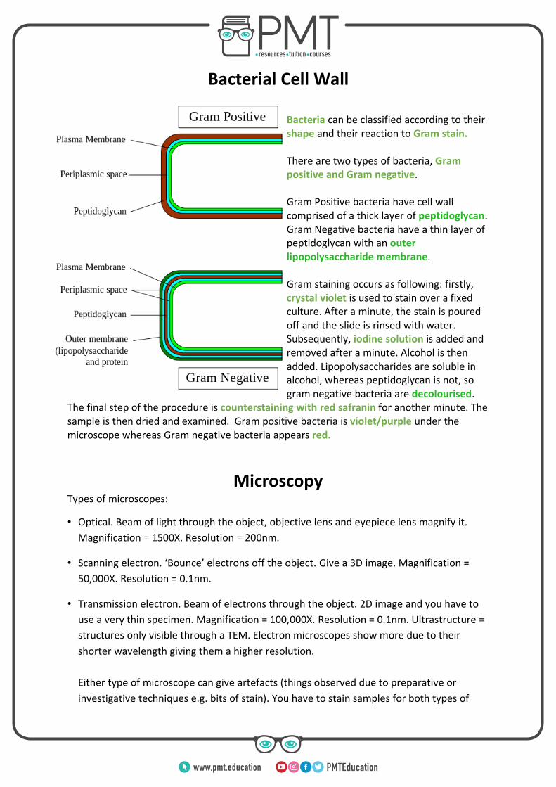
Bacterial Cell Wall
Bacteria can be classified according to their shape and their reaction to Gram stain. There are two types of bacteria, Gram positive and Gram negative. Gram Positive bacteria have cell wall comprised of a thick layer of peptidoglycan. Gram Negative bacteria have a thin layer of peptidoglycan with an outer lipopolysaccharide membrane. Gram staining occurs as following: firstly, crystal violet is used to stain over a fixed culture. After a minute, the stain is poured off and the slide is rinsed with water. Subsequently, iodine solution is added and removed after a minute. Alcohol is then added. Lipopolysaccharides are soluble in alcohol, whereas peptidoglycan is not, so gram negative bacteria are decolourised.
The final step of the procedure is counterstaining with red safranin for another minute. The sample is then dried and examined. Gram positive bacteria is violet/purple under the microscope whereas Gram negative bacteria appears red.
Microscopy Types of microscopes:
• Optical. Beam of light through the object, objective lens and eyepiece lens magnify it.
Magnification = 1500X. Resolution = 200nm.
• Scanning electron. ‘Bounce’ electrons off the object. Give a 3D image. Magnification =
50,000X. Resolution = 0.1nm.
• Transmission electron. Beam of electrons through the object. 2D image and you have to
use a very thin specimen. Magnification = 100,000X. Resolution = 0.1nm. Ultrastructure =
structures only visible through a TEM. Electron microscopes show more due to their
shorter wavelength giving them a higher resolution.
Either type of microscope can give artefacts (things observed due to preparative or
investigative techniques e.g. bits of stain). You have to stain samples for both types of
www.pmt.education
microscope. Electron microscopes = heavy metals (they absorb electrons). Light
microscopes = methylene blue, acetocarmine, haematoxylin. You stain to improve
contrast so you can see cell structures one from another.
• Advantages of electron microscopes:
o Can magnify and resolve better.
• Disadvantages of electron microscopes:
o You have to put the sample in a vacuum, so you can’t magnify living things.
o Very expensive and not portable.
Viruses
Viruses are non-living structures which consist of a nucleic acid (either DNA or RNA)
enclosed in a protective protein coat called the capsid, sometimes covered with a lipid layer
called the envelope. As viruses are non-living, antivirals must work by inhibiting virus
replication. Viruses can be difficult to treat once infection has occurred therefore the focus
of disease control should be on preventing the spread. This idea is exemplified by the 2014
Ebola outbreak in West Africa.
In the 2014 Ebola outbreak, new drugs were needed quickly. Some vaccines were fast-tracked - many doses made so if one passed Phase I it could be given to many people - and an experimental drug, ZMapp was given to seven people. It had never been given to humans before. Some of the seven recovered but some died. There are ethical implications to using, or not using, untested drugs: • Difficult to obtain informed consent • Side effects • May not be as effective as the currently accepted treatment There are two cycles of viral reproduction, the lytic cycle and the lysogenic cycle:
• Lysogenic viruses insert DNA in the form of a provirus into the DNA of the host cell
which enables the viral DNA to be replicated via cell division of the host cell. The
provirus can stay dormant if the virus produces repressor proteins which inhibit the
transcription of the provirus.
• Whereas lytic viruses insert DNA/RNA into the cytoplasm of the host cell, therefore
the viral genome is replicated independently of the host cell genome. Eventually,
www.pmt.education

this leads to lysis of the host cell when a large number of viruses are assembled and
ready to infect more cells.
• In a case where the lysogenic host cells become damaged or the immune system
becomes weak, the dormant viruses can enter the lytic pathway which leads to lysis
of the cell and spread of the viral infection.
Eukaryotic Cell Cycle and Division
The cell cycle is a regulated process in which cells divide to produce two genetically identical daughter cells for growth, repair and asexual reproduction. As all the cells produced by mitosis are genetically identical, mitosis does not give rise to genetic variation. There are three stages of the cell cycle:
• Mitosis – prophase, metaphase, anaphase and telophase.
• Cytokinesis – during cytokinesis the cytoplasm divides, thus producing two daughter
cells.
Figure: Wikipedia - Gram-negative bacteria
www.pmt.education
• Interphase – during this stage the cell grows, DNA replicates and the cell prepares
to divide – chromosomes and some organelles are replicated, chromosomes also
begin to condense to form chromatin.
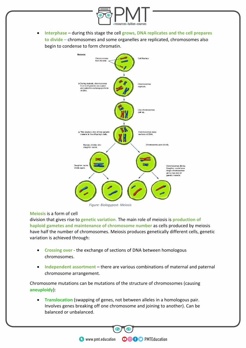
Meiosis is a form of cell division that gives rise to genetic variation. The main role of meiosis is production of haploid gametes and maintenance of chromosome number as cells produced by meiosis have half the number of chromosomes. Meiosis produces genetically different cells, genetic variation is achieved through:
• Crossing over - the exchange of sections of DNA between homologous
chromosomes.
• Independent assortment – there are various combinations of maternal and paternal
chromosome arrangement.
Chromosome mutations can be mutations of the structure of chromosomes (causing
aneuploidy):
• Translocation (swapping of genes, not between alleles in a homologous pair. Involves genes breaking off one chromosome and joining to another). Can be balanced or unbalanced.
Figure: Biologypost- Meiosis
www.pmt.education
• Duplication
• Deletion
• Inversion
Chromosome mutations can also change the number of chromosomes. Aneuploidy happens as a result of non-disjunction. This is where either homologous chromosomes or sister chromatids fail to separate. This can result in:
• More than two chromosomes in a pair. This is called polysomy.
o An example of polysomy is Down’s Syndrome – trisomy-21
• Less than two chromosomes in a pair, called monosomy.
o An example is Turner’s Syndrome, which is monosomy of the sex chromosomes where the one chromosome present is an X.
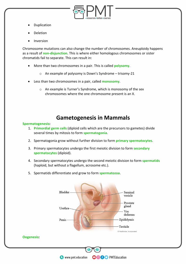
Gametogenesis in Mammals Spermatogenesis:
1. Primordial germ cells (diploid cells which are the precursors to gametes) divide several times by mitosis to form spermatogonia.
2. Spermatogonia grow without further division to form primary spermatocytes.
3. Primary spermatocytes undergo the first meiotic division to form secondary spermatocytes (diploid).
4. Secondary spermatocytes undergo the second meiotic division to form spermatids (haploid, but without a flagellum, acrosome etc.).
5. Spermatids differentiate and grow to form spermatozoa.
Oogenesis:
www.pmt.education
1. Primordial germ cells divide several times by mitosis to form oogonia.
2. Only one oogonium continues to grow to form a primary oocyte.
3. The first meiotic division forms one secondary oocyte and one polar body (small cells that bud off the oocyte, stick to the oocyte and do not develop into gametes).
4. The second meiotic division of the secondary oocyte forms one haploid ootid and one polar body. The second meiotic division of the polar body forms two more polar bodies. They degenerate and die as the ootid develops. This meiotic division starts in utero but is halted at prophase and occurs only in response to fertilisation to form the mature ovum.
Mammalian Gametes and Fertilisation
Ovum:
• It contains the zona pellucida
which is a protective coating which
the sperm have to penetrate in order
for fertilisation to occur, the main
purpose of zona pellucida is to prevent
polyspermy.
• It contains a haploid nucleus so
that a full set of chromosomes is
restored at fertilisation.
• Cortical granules release substances which cause the zona pellucida to harden.
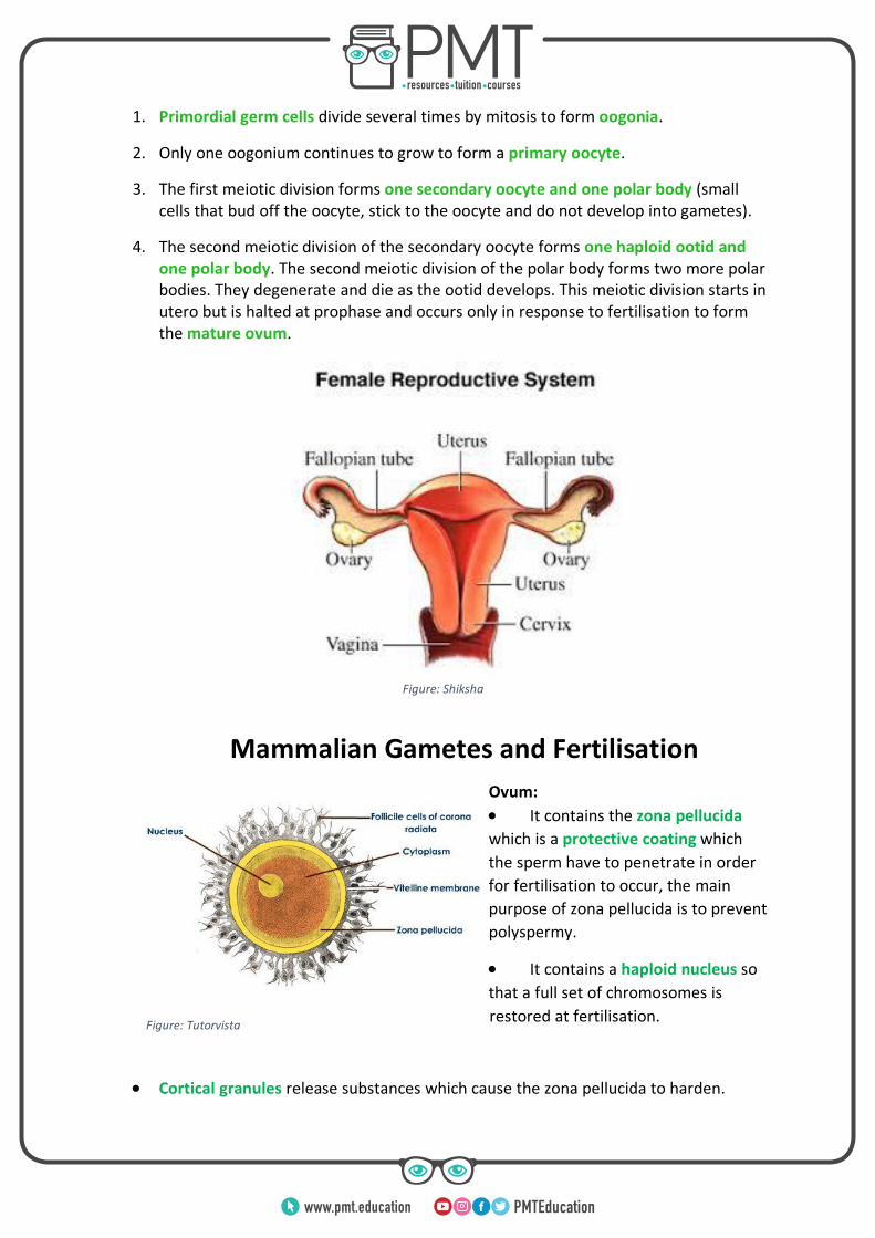
Figure: Shiksha
Figure: Tutorvista
www.pmt.education
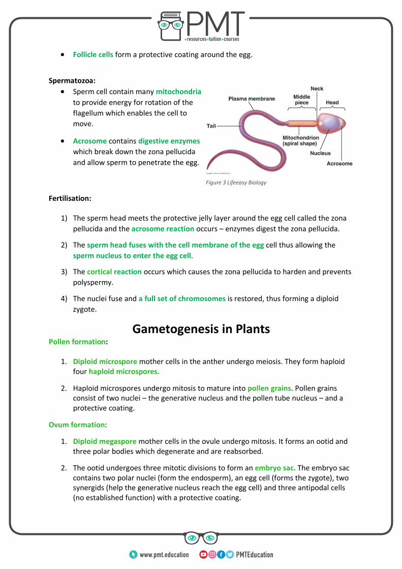
• Follicle cells form a protective coating around the egg.
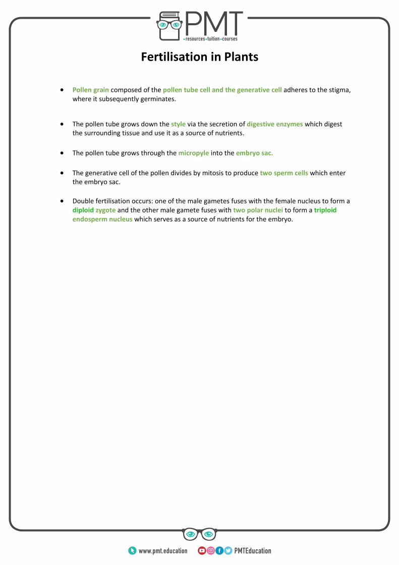
Spermatozoa:
• Sperm cell contain many mitochondria
to provide energy for rotation of the
flagellum which enables the cell to
move.
• Acrosome contains digestive enzymes
which break down the zona pellucida
and allow sperm to penetrate the egg.
Fertilisation:
1) The sperm head meets the protective jelly layer around the egg cell called the zona
pellucida and the acrosome reaction occurs – enzymes digest the zona pellucida.
2) The sperm head fuses with the cell membrane of the egg cell thus allowing the
sperm nucleus to enter the egg cell.
3) The cortical reaction occurs which causes the zona pellucida to harden and prevents
polyspermy.
4) The nuclei fuse and a full set of chromosomes is restored, thus forming a diploid
zygote.
Gametogenesis in Plants Pollen formation:
1. Diploid microspore mother cells in the anther undergo meiosis. They form haploid four haploid microspores.
2. Haploid microspores undergo mitosis to mature into pollen grains. Pollen grains consist of two nuclei – the generative nucleus and the pollen tube nucleus – and a protective coating.
Ovum formation:
1. Diploid megaspore mother cells in the ovule undergo mitosis. It forms an ootid and three polar bodies which degenerate and are reabsorbed.
2. The ootid undergoes three mitotic divisions to form an embryo sac. The embryo sac contains two polar nuclei (form the endosperm), an egg cell (forms the zygote), two synergids (help the generative nucleus reach the egg cell) and three antipodal cells (no established function) with a protective coating.
Figure 3 Lifeeasy Biology
www.pmt.education
Fertilisation in Plants
• Pollen grain composed of the pollen tube cell and the generative cell adheres to the stigma, where it subsequently germinates.
• The pollen tube grows down the style via the secretion of digestive enzymes which digest the surrounding tissue and use it as a source of nutrients.
• The pollen tube grows through the micropyle into the embryo sac.
• The generative cell of the pollen divides by mitosis to produce two sperm cells which enter the embryo sac.
• Double fertilisation occurs: one of the male gametes fuses with the female nucleus to form a diploid zygote and the other male gamete fuses with two polar nuclei to form a triploid endosperm nucleus which serves as a source of nutrients for the embryo.
www.pmt.education