Echocardiography in Systemic Diseases - asecho.org · 4/17/2018 1 Echocardiography in Systemic...
32
4/17/2018 1 Echocardiography in Systemic Diseases Sunil Mankad, MD, FACC, FCCP, FASE Associate Professor of Medicine Mayo Clinic College of Medicine Director, Transesophageal Echocardiography Associate Director, Cardiology Fellowship Mayo Clinic, Rochester, MN [email protected] @MDMankad DISCLOSURE Relevant Financial Relationship(s) Advisory Council: Siemens Healthcare, Ultrasound Off Label Usage None
-
Upload
vuongxuyen -
Category
Documents
-
view
222 -
download
0
Transcript of Echocardiography in Systemic Diseases - asecho.org · 4/17/2018 1 Echocardiography in Systemic...

4/17/2018
1
Echocardiography in Systemic Diseases
Sunil Mankad, MD, FACC, FCCP, FASE
Associate Professor of Medicine
Mayo Clinic College of Medicine
Director, Transesophageal Echocardiography
Associate Director, Cardiology Fellowship
Mayo Clinic, Rochester, MN
@MDMankad
DISCLOSURE
Relevant Financial Relationship(s)
Advisory Council: Siemens Healthcare, Ultrasound
Off Label Usage
None

4/17/2018
2
• Systemic diseases with secondary
cardiac involvement are uncommon
• Echo can identify unique, characteristic
features and echo may be the first clue
to the underlying systemic illness
But
Echo in Systemic Diseases
Cardiac Involvement in Systemic Diseases
• Autoimmune
• Endocrine
• Collagen Vascular Diseases
• Malignancy
• Amyloid/Infiltrative Diseases
• Radiation Induced Heart Disease
• Drug Induced Valvulopathy

4/17/2018
3
Case• 27 y/o female who presents with dyspnea, chest pain,
and fatigue– NYHA class III
• Abnormal nuclear perfusion stress test led to coronary arteriography – Normal coronaries but LV gram suggestive of “Hypertrophic
CM” (EF 75%)
• Elevated Sedimentation Rate
• Referred to Mayo Clinic → Echo performed

4/17/2018
4
Apical 4 Chamber Views
Diastolic Function
• MV Dec. Time = 105 msec
• MV Emax = 1.1 m/sec
• e’ = 0.04 m/sec
• E/e’ 28

4/17/2018
5
What is the Diagnosis?
1. Hypertrophic Cardiomyopathy (Apical Variant)
2. Amyloidosis
3. Eosinophilic Endomyocardial Disease
4. LV Noncompaction
5. LV Myxoma
RV Biopsy (H&E Stain)

4/17/2018
6
Hypereosinophilic SyndromeCardiac Manifestations
• Persistent increase in eosinophil count eosinophil count > 1500 cells/mm3
• CHF (dyspnea)– Restrictive Cardiomyopathy
– Mitral regurgitation
• Systemic embolization
Eosinophilic Heart Disease
1) Acute inflammatory myocarditis
2) Eosinophil rich thrombus deposition
- Mediated by injured endothelium
3) Endocardial thickening- Valve involvement
4) Fibrosis
4 Stages:
Adapted from Hirota Y: In AbelmannWH, Braunwald E [eds]: Atlas of Heart Diseases. Vol 2. 1995
Allergic reaction Autoimmune disease
Parasitic or Protozoal infections
Malignancy Idiopathic
Overproduction of cytotoxic eosinophils
Infiltration of myocardium by eosinophils
Degranulation of eosinophilic granules
Tissue damage
Acute pericarditis, myocarditis, or endocarditis
Necrotic phase
Thrombotic phase
Fibrotic phase
Formation of intramural thrombiAdjacent to injured endocardium
Localized or extensive replacement fibrosis

4/17/2018
7
Hypereosinophilic Syndrome (HES) Cardiac Involvement: 40-60% of patients
LV > RV inflow apical thrombo-obliteration,
endocardial thickening
Subvalvular thrombosis, leaflet entrapment MV > TV
Leaflets; MR&TR
Restrictive diastolic dysfunction
2-D Echo & Doppler Findings
Ommen, Am J Cardiol 2000
Clot
RA LA
LVRV VS
Scar
Natural HistoryHypereosinophilic Syndrome
Myocarditis Thrombus FibrosisImage courtesy of Leslie Elvert RDCS

4/17/2018
8
Basal LV Fibrosis with Mitral Posterior Leaflet Tethering
- Courtesy of Dr. Natesa Pandian
Eosinophilic Heart DiseaseContrast Helpful

4/17/2018
9
Hypereosinophilic Syndrome
• Medical therapy – Corticosteroids
– Hydroxyurea
– Interferon
– CHF Meds
• Surgical Therapy– Palliative
Treatment Echo Differential Diagnosis• Apical hypertrophic CM
• LV Noncompaction
• LV tumor– Myxoma
– Papillary fibroelastoma
• Ischemic LV dysfunction with apical thrombus
Differential Diagnosis: LV Myxoma

4/17/2018
10
Differential Diagnosis: Apical HCM
Our Case:TTE after 2 months of anticoagulation
and 1 month of prednisone therapy

4/17/2018
11
Patient with CREST Syndrome: Dyspnea and Edema
RVSP: 75 mmHg
Scleroderma and Pulmonary HTN
• PH present in 8-12% of scleroderma patients– Higher risk in CREST patients
• Accounts for 30% of deaths
• Screening for PH recommended
• RV dysfunction, cardiac index and pericardial effusion are markers of poor prognosis in PH

4/17/2018
12
• ANA positive and Antiphospholipid antibodies present
• Libman-Sacks endocarditis
33 Year Old Female Multiple Strokes
TEE TEE
Systemic Lupus ErythematosusCardiac Involvement
• Pericarditis (fluid ANA+)
• 50-60% of cases have pericardial effusions
• Lupus anticoagulant
• Anticardiolipin or Antiphospholipid Abs
• Myocarditis
• Coronary arteritis
• Libman-Sacks (Marantic) vegetations

4/17/2018
13
• Lupus anticoagulant + antiphospholipid antibodies present
• Libman-Sacks endocarditis
18 y.o. Female with Occipital StrokeTEE
Not only the mitral valve!
4/17/2018
14
Antiphospholipid SyndromeDiagnosis confirmed at surgery
• IgG and IgM
Antiphsopholipid antibody
• Importance of recognition– Unlikely repair
– Choice of prosthesis??
– Anticoagulation??
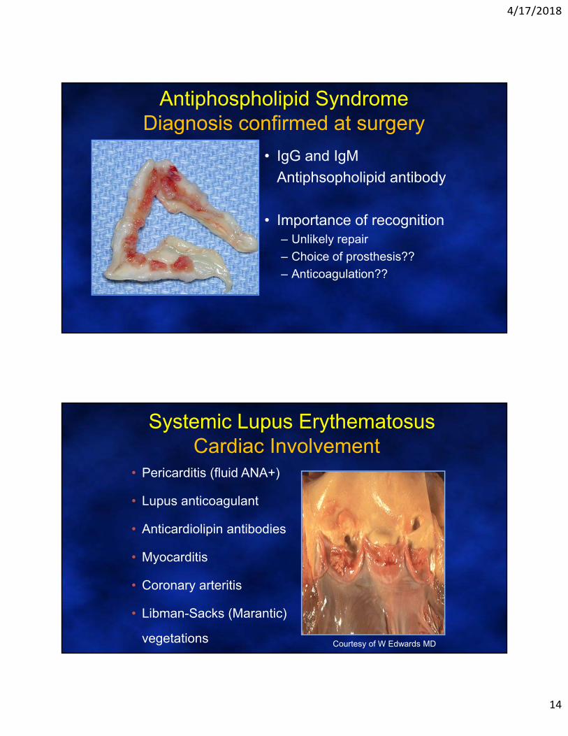
Systemic Lupus ErythematosusCardiac Involvement
• Pericarditis (fluid ANA+)
• Lupus anticoagulant
• Anticardiolipin antibodies
• Myocarditis
• Coronary arteritis
• Libman-Sacks (Marantic)
vegetations Courtesy of W Edwards MD

4/17/2018
15
39 year old male with diarrhea, flushing and weight loss
Carcinoid Syndrome

4/17/2018
16
Carcinoid: Echo FeaturesTricuspid valve• Thickened leaflets
• Retracted leaflets
• Fixed semi-open position
Pulmonary valve• Thickened cusps
• Retracted and rigidConnolly HM. Curr Cardiol Rep. 2006
RA
RV
Systolic RV RA pressure equalization
TR CW Doppler
Severe (Torrential) Tricuspid Regurgitation
Courtesy of Dr. WK Freeman

4/17/2018
17
Pulmonary Valve Involvement
Adapted from Mayo Image Data Base, William Edwards, MD
Pulmonary Valve Involvement

4/17/2018
18
Carcinoid Heart Disease• Carcinoid tumors: 1-2/100,000
• Carcinoid syndrome in 20-30%
• Deposition of a matrix-like material on the valves and endocardium of the right side of the heart
• Treatment of tumor does not cause regression of valve disease
Connolly HM. Curr Cardiol Rep. 2006
Eur Heart J Cardiovasc Imaging 2012Eur Heart J Cardiovasc Imaging 2012
Courtesy of Denisa Muraru, MD, PhDPadua, Italy
Carcinoid Syndrome: 3D TTE

4/17/2018
19
Carcinoid Heart Disease
Flushing
Wheezing
Diarrhea
Over 50% of patients with Carcinoid
Syndrome develop cardiac involvement
Courtesy of Dr. Heidi Connolly

4/17/2018
20
Cardiac Surgery for Carcinoid
• Indications– Symptomatic carcinoid HD
– Controlled carcinoid symptoms
– RV size or dysfunction
– Prior to hepatic surgery
• Replace TV and PV – Improved RV remodeling
Carcinoid Tumor: : Liver Metastases

4/17/2018
21
58 yo woman with weight loss, tremor and HR of 125
Hyperthyroidism
• Atrial fibrillation – difficult to rate control
• Decreased Peripheral resistance – hypotension
• Exacerbation of underlying CAD• increased myocardial O2 demand
• Tachycardia induced cardiomyopathy

4/17/2018
22
Tachycardia Mediated Cardiomyopathy
• 25% of patients w/ LV dysfunction & AF will have improved EF with rate control
• Usually unaware of rhythm
• Resting heart rate - poor indicator of overall rate control
• Consider in all pts with AF & LV dysfunction
Grogan M et al. AJC, 1992.
2 Years after Cardioversion and Treatment of Hyperthyroidsin

4/17/2018
23
Hypothyroidism: Large Pericardial Effusion
43 year old man

4/17/2018
24
43 year old man with amyloidosis
Primary Hyperoxaluria
• Rare metabolic disorder with autosomal recessive inheritance
• PHO type 1 (0.11 - 0.26 per 100,000 live births)
• Enzymatic defect resulting in enhanced conversion of glyoxalate to poorly soluble oxalate which is excreted in the urine

4/17/2018
25
Hydroxychloroquine-induced Cardiotoxicity
Renal Failure

4/17/2018
26
A 60 year old male farmer is referred for evaluation of dyspnea
• NYHA Class III symptoms• PMH: Type 2 DM• Abnormal LFT’s• Physical Exam:
• 110/70 mmHg, HR 70 BPM• S3 gallop• Bronze skin
EKG

4/17/2018
27
CXR
Apical Images

4/17/2018
28
Coronary Angiography
What is the most likely diagnosis?
a. Cardiac hemochromatosis
b. Cardiac amyloidosis
c. Cardiac sarcoidosis
d. Fabry’s Disease
e. Carcinoid syndrome
60 year old male farmer with Type 2 DM, bronze skin, and abnormal LFT’s

4/17/2018
29
Cardiac MRI
Cine-MRI
No Delayed hyperenhancement
• T2* sequence is used to evaluate iron deposition in the myocardium
• 1 – ROI in the myocardium• 2 – ROI in the liver
• On board software then calculates the T2* value (ms)
• This patient had a T2* relaxation time of 20ms that suggests hemochromatosis
• The evaluation of the T2* relaxation time is an excellent noninvasive correlate of myocardial iron deposition and is a useful technique to follow response to iron‐chelation therapy.
• Myocardial T2* has been shown to have no relation to serum ferritin and liver iron overload.
• T2* relaxation time predicts CHF and Arrhythmias
Circulation 2009;120:1961‐8Eur Heart J 2001;22:2171‐9.

4/17/2018
30
Hemochromatosis
Intracellular iron – directly toxic to myocytes
• total body iron – intracellular deposits in heart, liver, pituitary, pancreas, gonads, skin
• Think of this when DCM seen in setting of hepatic dysfunction; diabetes, tanned skin
• Diagnosis is critical, since reversible– Males 9:1– 2-3/1000 population– Ferritin usually > 500, transferrin >
50%• Normal wall thickness• Arrhythmias, conduction abnormalities Courtesy of William Edwards, MD
26 year old with Hemochromatosis
LV RV

4/17/2018
31
After Tx with Deferoxamine
Heomochromatosis: Take Home Points
• The Iron Heart is a weak heart…
• Hemochromatosis may be a cause of idiopathic dilated cardiomyopathy– Reversible with treatment
• Cardiac MRI (T2 relaxation time) is important in helping to establish diagnosis and monitoring treatment effects

4/17/2018
32
Conclusions: Systemic Diseases and the Echo Boards
• Carcinoid Syndrome
• Hypereosinophilic endomyocardial disease
• Sarcoidosis
• Systemic Lupus Erythematosus
• Scleroderma/Crest: Pulm Hypertension
• Amyloidosis
• Hyper or Hypothyroidism
• Radiation Heart Disease
• Drug Induced Valve Disease
• Hemochromatosis
Thank [email protected]
Acknowledgements:Dr. Thais CouthinoDr. Mark Callahan


![Quantification of systemic right ventricle by echocardiography · 2017-02-26 · of systemic right ventricle by echocardiography ... with dobutamine stress [17]. These data were confirmed](https://static.fdocuments.net/doc/165x107/5ecb2f51d4cb202a22168cb3/quantification-of-systemic-right-ventricle-by-echocardiography-2017-02-26-of-systemic.jpg)