ECHOCARDIOGRAPHY IN PATIENTS WITH CARDIOMEGALY …
91
ECHOCARDIOGRAPHY IN PATIENTS WITH CARDIOMEGALY IDENTIFIED ON CHEST X-RAY AT AN ACADEMIC HOSPITAL IN ZAMBIA BY NCHIMUNYA GWABA STUDENT No: 213105810 A dissertation submitted in fulfilment of the requirements for the degree of Master of Science in Radiography: Department of Medical Imaging and Therapeutic Sciences. Faculty of Health and Wellness Sciences at Cape Peninsula University of Technology Principal Supervisor: Ms Ferial Isaacs Co-supervisor: Ms. Maria Hartnick Collaborator: Dr. Bright Nsokolo Bellville Campus November 2018
Transcript of ECHOCARDIOGRAPHY IN PATIENTS WITH CARDIOMEGALY …
ECHOCARDIOGRAPHY IN PATIENTS WITH CARDIOMEGALY IDENTIFIED ON CHEST X-RAY AT AN ACADEMIC HOSPITAL IN ZAMBIA
BY
NCHIMUNYA GWABA
STUDENT No: 213105810
A dissertation submitted in fulfilment of the requirements for the degree of Master
of Science in Radiography: Department of Medical Imaging and Therapeutic
Sciences. Faculty of Health and Wellness Sciences at Cape Peninsula University
of Technology
Principal Supervisor: Ms Ferial Isaacs
Co-supervisor: Ms. Maria Hartnick
Collaborator: Dr. Bright Nsokolo
Bellville Campus
November 2018
1

2
Dedication
I dedicate this research to my wife, children, mother and brothers. Your encouragement
and love have always been amazing to me. Thank you all.
“What is written without effort is in general read without pleasure.”
Samuel Johnson (1709-1784)

3
Acknowledgement
I acknowledge the following people for their support and encouragement towards the
successful completion of this research.
1. My Principal Supervisor, Ms F Isaacs guided me throughout this study.
2. The Co-supervisor Mrs M Hartnick was very instrumental in guiding me especially
on issues related to ECHO examinations.
3. The Head of the Medical Imaging & Therapeutic Sciences Department, Mr A
Speelman, for his assistance.
4. The Collaborator, Dr B Nsokolo is a specialist in internal medicine and he was
instrumental in making sure that the data I collected was factual.
5. The Director of Research for Postgraduate Studies at Apex Medical University, Dr
T Sukwa, for proof reading and corrections of my thesis. Dr. Sukwa is also a former
country director for the World Health Organization.
6. The Statisticians, Dr C Uys (CPUT) and Mr M Sinkala helped in analysing the data
collected. Without their assistance, this project would not have been completed.
7. The Management and staff at Levy Mwanawasa Hospital, for permitting me to
research at their facility and for their help during the course of the study.
8. My utmost gratitude and appreciation also to the patients whose clinical information was used in carrying out this research.

4
Abstract
Introduction: Cardiomegaly is a sign that there is an underlying cardiovascular disease.
It is a medical indication in which the heart is enlarged. This indication is strongly
associated with high blood pressure or coronary heart disease. When enlarged, the heart
may pump blood ineffectively, and this can lead to congestive heart failure. Common
underlying causes of cardiomegaly are heart failure, heart muscle disease
(cardiomyopathy), coronary heart disease, high blood pressure (hypertension),
congenital heart disease, heart valve disease, thyroid disease, and obesity.
Cardiovascular diseases are very often accompanied by an enlarged heart and these
diseases are the world’s leading cause of death. Early detection of cardiovascular
diseases in cardiomegaly patients is particularly important for the prevention of fatalities
as cardiovascular diseases are the number one cause of diseases worldwide. Although
the chest X-ray (CXR) is the number one imaging modality for cardiomegaly,
echocardiography (ECHO), a dynamic ultrasound imaging modality of the heart can also
be used to assess the functioning of the heart. In Zambia, no documented study has been
done to establish the association between cardiomegaly identified on a chest x-ray and
the results of the echocardiography reports of the same patients. The aim of this research
study, therefore, was to establish whether there is an association between cardiomegaly
identified on the CXR and the ECHO reports of the same patients.
Methodology: This retrospective cross-sectional study involved the retrieving of data
from 124 patients who had cardiomegaly identified on the CXR and had undergone an
ECHO examination. The study was performed at Levy Mwanawasa General Hospital
(LMGH) in Lusaka, Zambia.
Findings: Cardiomegaly was detected in 124 patients (n = 124) on the CXR using the
cardio-thoracic-ratio. Cardiomegaly was more prevalent in females (67.7% of participants
were female) compared to males (32.3% of participants were male). All age groups were
affected, however the prevalence increased with age; 60% of the patients were aged 60-
80 years. More than 50% of the patients had severe cardiomegaly. There was no
significant difference between males and females with severe cardiomegaly.
The ECHO findings showed left ventricular diastolic dysfunction as the most common
condition (presented in 71% of the patients) followed by left atrium dilation (presented in

5
29.1% of the patients) and left ventricular systolic dysfunction (presented in 29% of the
patients). Doppler ultrasound was used to detect abnormal blood flow patterns within the
heart. It revealed a significant correlation between severe cardiomegaly and severely
increased blood flow patterns (p=0.004); and between cardiomegaly (minimal, moderate,
and severe) and increased blood flow patterns (p=0.002, p=0.000 and p=0.04
respectively). Other abnormal ECHO findings included: ejection fraction (presented in
36.3% of the patients), fractional shortening (presented in 31.5% of the patients) left atrial
dilation (presented in 28.2% of the patients), tricuspid valve regurgitation (presented in
25.8% of the patients), right atrium dilation (presented in 24.2% of the patients), posterior
wall thickness (presented in 23.4% of the patients), inter-ventricular septal thickness
(presented in 22.5% of the patients), pericardial effusion (presented in 21% of the
patients), left ventricular dilation (presented in 20.2% of the patients ), right ventricular
outflow tract (presented in 16.9% of the patients), mitral valve regurgitation (presented in
15.5% of the patients), aortic root dilation (presented in 8.8% of the patients) and pleural
effusion (presented in 8.1% of the patients).
There was a strong positive association between severely increased blood pressure and
cardiomyopathy (p=0.023), inter-ventricular hypertrophy (p=0.017), left atrial dilation
(p=0.007) left ventricular diastolic dysfunction (p=0.045), left ventricular dilation
(p=0.003), left ventricular hypertrophy (p=0.028), left ventricular systolic dysfunction (p=0.
048) and pericardial effusion (p=0.001).
Conclusion: Cardiomegaly detected on the plain CXR of patients was found to be a
helpful marker for cardiac diseases, as well as an index of its severity. While 7.2% and
4.8% of patients with minimal and moderate cardiomegaly had normal ECHO findings
respectively, all patients with severe cardiomegaly were identified with cardiovascular
diseases. Hence, the ECHO made an important contribution to the diagnosis of specific
cardiac anomalies in patients identified with severe cardiomegaly. ECHO may be
considered a useful screening tool for patients identified with cardiomegaly on the CXR
in adults.

6
Contents
Dedication ....................................................................................................................... 2
Acknowledgement ........................................................................................................... 3
Abstract ........................................................................................................................... 4
List of Abbreviations ........................................................................................................ 9
CHAPTER ONE
INTRODUCTION AND BACKGROUND ………………………………………………….. 10
1.1 Overview ................................................................................................................. 10
1.2 Background ………………………………………………………………………………..10
1.3 Problem Statement ……………………………………………………………………….11
1.4 Justification of the study ......................................................................................... 12
1.5 Benefits of the study ................................................................................................ 12
1.6 Research question .................................................................................................. 13
1.7 Aim of the study....................................................................................................... 13
1.8 Objectives of the study …………………………………………………………………..13
1.9 Rationale ................................................................................................................. 13
CHAPTER TWO
LITERATURE REVIEW ................................................................................................. 15
2.1. Introduction ............................................................................................................ 15
2.2. Cardiomegaly ......................................................................................................... 15
2.3. Pathophysiology of Cardiomegaly ......................................................................... 17
2.4. Prevalence of cardiomegaly ................................................................................... 22
2.5. Common indications associated with cardiomegaly ............................................... 23
2.6. Imaging modalities for cardiomegaly ...................................................................... 24
2.6.1. Chest X-Ray ................................................................................................. 25

7
2.6.2. Heart size on a chest X-Ray ......................................................................... 27
2.6.3. Echocardiography ......................................................................................... 28
2.6.4 Principles of operation for equipment used .................................................... 30
2.7 Common echocardiography prognostic markers in patients with cardiomegaly ...... 32
2.8. Impact of cardiac diseases on households in Zambia ............................................ 38
2.9. Summary of Literature Review ............................................................................... 39
CHAPTER THREE
METHODOLOGY .......................................................................................................... 40
3.1 Study design ........................................................................................................... 40
3.2 Research site ............................................................ Error! Bookmark not defined.
3.3 Study population...................................................................................................... 40
3.4 Inclusion criteria ...................................................................................................... 40
3.5 Exclusion criteria ..................................................................................................... 41
3.6 Sample size calculation ........................................................................................... 41
3.7 Data collection ......................................................................................................... 42
3.8 Data management ................................................................................................... 42
3.9 Data analysis ........................................................................................................... 43
3.10 Data validity and reliability ..................................................................................... 43
3.11 Ethics .................................................................................................................... 44
CHAPTER FOUR
PRESENTATION OF FINDINGS .................................................................................. 46
4.1 Introduction ………………………………………………………………………………..46
4.2 Gender distribution in cardiomegaly ........................................................................ 46
4.3 Age distribution in cardiomegaly ............................................................................. 47
4.4 Cardiomegaly .......................................................................................................... 48

8
4.5 Common diseases identified in the study ……………………………………………...51
4.6 Cardiac disease distribution by gender ….…………………………………………… 52
4.7 Blood pressure in cardiomegaly ………………………………………………………. 52
4.8 Variations in blood pressure and cardiac conditions fond on ECHO ...…………… 54
4.9 Doppler ultrasound results ..…………………………………………………………… 55
4.10 Cardiac abnormalities found on ECHO .……………………………………………. 57
4.11 Summary of results …………………………………………………………………… 71
CHAPTER FIVE
DISCUSSION AND CONCLUSION............................................................................... 72
5.1 Introduction ............................................................................................................. 72
5.2 Cardiomegaly findings ............................................................................................. 72
5.3 Risk by age ............................................................................................................. 73
5.4 Risk by gender ........................................................................................................ 74
5.5 Disease distribution in cardiomegaly ....................................................................... 74
5.6 Limitation of the study ............................................................................................. 75
5.7 Recommendations .................................................................................................. 75
REFERENCES .............................................................................................................. 77
LISTOF APPENDICES .................................................................................................. 84
Appendix A: Form for capturing patients data-CXR report ............................................ 84
Appendix B: Form for capturing patients data- ECHO report ........................................ 85
Appendix C: Participant Information Leaflet- for Research Assistants of the study. ...... 86
Appendix D: Permission to Carry out Research ............................................................ 89
Appendix E: Ethics Certificate ....................................................................................... 90

9
List of Abbreviations
BP- Blood Pressure CPUT-Cape Peninsula University of Technology CT – Computed Tomography CHF-Congestive Heart Failure CTR- Cardiac Thoracic Ratio CXR- Chest X-Ray DCM- Dilated Cardiomyopathy ECHO – Echocardiography / Echocardiogram EF - Ejection Fraction IHD - Ischemic Heart Disease LMGH - Levy Mwanawasa General Hospital MR - Mitral Regurgitation LVH - Left Ventricular Hypertrophy LVEF - Left Ventricular Ejection Fraction MRI – Magnetic Resonance Imaging PA - Posterior Anterior PW - Posterior Wall WHO - World Health Organization

10
CHAPTER ONE
INTRODUCTION AND BACKGROUND
1.1. Overview
Cardiomegaly is an indication that there is an underlying cardiovascular disease. Tavora
et al. (2012) explain that among adults with a main age of about 50 years, cardiomegaly
is a frequent finding in cardiovascular diseases that may cause sudden cardiac death.
Mckee and Ferrier (2017) indicate that a cardiothoracic ratio (CTR) greater than 50% on
a posterior-anterior (PA) chest radiograph (CXR) is representative of cardiomegaly.
However, authors like Brakohiapa et al. (2017) indicate that a cardiothoracic ratio of 55%
may not have any underlying cardiovascular disease and may thus be considered normal
for blacks and Asians.
The World Health Organization (WHO, 2016) estimates that more than 17 million people
(representing 31% of all annual global deaths) die annually from cardiovascular diseases,
globally. In America, a study by Aksut (2015) estimated that about 5 million Americans
have symptomatic heart failure. Cardiovascular diseases are among the top ten leading
causes of death in Zambia in 2016. Stroke is the fifth leading cause of death in Zambia
while ischemic heart disease is the eighth. These two diseases account for 4.3% and 2.6%
of the total deaths recorded respectively, in Zambia (World Health Ranking, 2016). It
should be noted that the total prevalence of cardiovascular diseases in Zambia is 8%
(WHO, 2014).
This study was designed to investigate the association between cardiomegaly diagnosed
on the chest X-ray (CXR) and the echocardiography (ECHO) findings of the same patients.
The recognition of cardiomegaly was based on the cardiothoracic ratio calculations on
the CXR, and cardiac pathologies were investigated using ECHO. The investigated
individuals were aged between 18 to 80 years (mean age was 56.78).
1.2 Background
An enlarged heart may be accompanied by cardiovascular diseases. More than 17 million
(31%) people die worldwide, annually from cardiovascular diseases (WHO, 2016). Many
of these people have been exposed to unhealthy behaviours, including tobacco use,

11
eating foods containing too much salt and inadequate physical activity. Most of these
people could be saved by better access to medical care for high blood pressure
(responsible for the bulk of heart disease-related deaths annually), high blood cholesterol
and other conditions that raise the risk for cardiovascular diseases.
At Levy Mwanawasa General Hospital (LMGH), records show that cardiovascular
diseases were the third leading cause of death (8.6%) in 2015. A total of 3044 patients
(8.5% of total patients seen) were seen with various cardiovascular diseases at LMGH in
2015. Out of these 1760 patients had hypertension, 470 had heart failure, 320 had ill-
defined heart conditions, 284 had cerebral vascular accidents, 146 had cardiomyopathy,
55 had angina pectoris, 13 had chronic ischemic heart disease, 3 had non-rheumatic
mitral valve disease, 3 had tachycardia and 1 had cardiac arrhythmia (LMGH, 2016). Most
of these cardiovascular diseases were accompanied with cardiomegaly. As shown by
records from LMGH (2016), it can be estimated that about 16.7% of all CXR examinations
had cardiomegaly.
The association between an enlarged heart diagnosed on the CXR and the ECHO
findings of the same patient, have not yet been documented at this hospital. With the
projected rise in the incidence of cardiovascular diseases worldwide, it is expected that
the incidence of cardiomegaly will continue to rise in Zambia, in particular at LMGH.
Experts have explained that there is a strong clinical association between cardiomegaly
and cardiovascular diseases. Monfared et al (2015), explain that cardiomegaly is caused
by different diseases, such as valvular heart diseases, Ischemic heart disease (IHD), and
cardiomyopathy as well as pericardial diseases.
Cardiomegaly is thus a serious indication of cardiovascular diseases and early diagnosis
of the pathological conditions found in cardiomegaly can be of great benefit to both the
patients as the underlying disease can be treated. It was thus important to investigate the
associate pathological conditions found in patients with cardiomegaly at LMGH in Lusaka,
Zambia, as this can help to put measures in place that can reduce incidences for these
diseases. The findings may also help identify the gaps in equipment and qualified
manpower needed to diagnose and treat these conditions.
This research study was conducted at LMGH in Lusaka, Zambia. The study is the first of
its kind at the research site in particular and Zambia in general. The study was
quantitative, retrospective, and descriptive. Files for patients with an enlarged heart on a

12
CXR who had undergone an ECHO examination, were used to determine the association
between patients diagnosed with an enlarged heart on CXR and ECHO findings of the
same patients.
1.3 Problem statement
Cardiomegaly is one of the most common findings on CXR and ECHO examinations in
the world (Monfared et al, 2015; Tomaszewki, 2012; Elliot et al, 2000; Tavora et al, 2012).
It was estimated that 16.7% of the CXR examinations done at the study site showed
cardiomegaly and some patients with an enlarged heart are referred for the ECHO
examination, which provides a more detailed assessment of the structure and function of
the heart. It is however unclear to what the association is between cardiomegaly and
ECHO findings of the same patient at this study site. Thus, this retrospective study aimed
to establish whether there is an association between cardiomegaly identified on the CXR
with pathological findings of echocardiography.
1.4 Justification of the study
The justification for this study was that identifying the association between cardiomegaly
diagnosed on a CXR and the ECHO results of the same patients, who presented at the
study site, may increase awareness of the association and thus the common
cardiovascular diseases found in such patients among clinicians, radiographers as well
as patients. These findings may help re-asses the need for further management of
patients identified with cardiomegaly on a CXR and may also lead to the early referral for
ECHO examinations for such patients in communities serviced by the research site.The
expected findings may also help determine the prevalence of actual cardiovascular
diseases at LMGH and may help stimulate interest in further research to identify gaps in
equipment and or qualified man power needed to diagnose and treat prognostic markers
of cardiomegaly.
1.5 Benefits of the study
This study may benefit clinicians, radiographers and patients because the study is
seeking to confirm that cardiomegaly identified on a CXR can be an indicator of underlying
cardiovascular diseases. This may lead to awareness of the possible need for an early
CXR and ECHO in the diagnosis and treatment of cardiovascular diseases.

13
This may help channel resources to prevention measures like awareness programs on
radio, training of more manpower, and also in the purchase of medicines and equipment
needed to manage these conditions. Increased awareness of the frequent pathological
findings in cardiomegaly patients may also have important implications when prioritizing
funding for future research and treatment of patients with cardiomegaly/cardiovascular
diseases.
1.6 Research question
Is there an association between cardiomegaly identified on the CXR and ECHO results
of the same patient?
1.7 Aim of the study
This study aimed to establish whether there is an association between the CXR reports
of patients diagnosed with cardiomegaly and the ECHO reports of the same patients.
1.8 Objectives of the study
The objectives of the study were:
1. to establish whether there was an association between cardiomegaly (cardiac-
thoracic ratio) diagnosed on a CXR and ECHO findings,
2. to establish whether there was a significant difference and correlation in the CXR
and ECHO findings, between males and females, and between different age
groups respectively, and
3. to establish the prevalence of pathological conditions found on the ECHO in
patients diagnosed with an enlarged heart on the CXR
1.9 Rationale
The prevalence of cardiovascular diseases in Zambia is 8% (WHO, 2014) and 8.5% at
the research site (LMGH, 2016). Monfared et al. (2015), Tomaszewki (2012), Elliot et al.
(2000) and Tavora et al. (2012) have all shown that cardiomegaly is the most prominent
diagnosis on a CXR and ECHO globally. In Zambia, the frequent pathological findings in
cardiomegaly patients are not well documented. Therefore, this study sort to investigate,
the association between cardiomegaly diagnosed on the CXR and the ECHO reports of
the same patient. The study was conducted at an academic hospital in Lusaka, Zambia.

14
The results, including recommendations, are reported in the thesis format to the
educational institution Cape Peninsula University of Technology (CPUT), and the study
site. Thus, the consulting cardiologist, physicians, clinical educators and the broader
research community have access to the results of this research, from the above-
mentioned institutions (e.g. via their web sites).
15
CHAPTER TWO
LITERATURE REVIEW
2.1. Introduction
The literature reviewed focused on establishing the various chest x-rays (CXR) and
echocardiography (ECHO) patterns in patients with cardiomegaly. The abstracts of all
articles were reviewed, and the full manuscript of the relevant articles retrieved. The
databases that were consulted for this literature review included Google scholar, Science
direct, Promed, Wiley online, Elsevier, Sciverse, Medline, Springer link, Medscape,
Hospital management information systems for Levy Mwanawasa General Hospital,
United Nations website, Ministry of Health (Zambia) website and some medical textbooks.
What is important in the case of cardiomegaly is to be able to demonstrate the presence
of underlying diseases that could lead to the occurrence of cardiomegaly. Such
information could be obtained from a CXR and ECHO. The CXR provided information on
lung pathology (that may be the cause of the cardiomegaly) and the size of the heart,
while the ECHO provided information on the structural and functional changes in parts of
the heart. Arguments are presented, from the literature, on the need for Levy Mwanawasa
General Hospital the CXR and ECHO examination in the diagnosis of cardiovascular
diseases.
2.2. Cardiomegaly
Cardiomegaly means that the heart is enlarged. It is usually a sign of another
cardiovascular condition. There are two main types of cardiomegaly and these are:
I. Dilated - Dilated cardiomyopathy is characterized by a severe, irreversible form of
heart diseases with left ventricular systolic dysfunction and dilation. In dilated
cardiomegaly, the heart becomes enlarged due to dilation of the myocardium (De
Luca, et.al 2018).
Dilated cardiomyopathy ranks among the most common causes of heart failure in
the world. It has highly variable clinical presentation and prognosis and is the
second leading cause of left ventricular dysfunction. Clinical causes are many and
are highly heterogeneous, ranging from patients who are asymptomatic to those

16
suffering from sudden cardiac death due to arrhythmias or intractable heart failure.
A cardiovascular mortality of up to 40% has been reported in developed countries
from previous studies, mainly due to sudden cardiac death or advanced heart
failure. Thus, dilated cardiomyopathy prognosis depends on multiple risk factors
and is variable. While symptoms and signs of dilated cardiomyopathy may be
common, some are incidental, for example, by identifying cardiomegaly on a CXR
(Schild et al., 2019).
It can be said that dilated cardiomyopathy is the final common response of
myocardium to environmental and diverse genetic insults. Alternative causes of
left ventricular dilation and dysfunction can be excluded by a rigorous work-up, this
can also help identify etiologies that may guide family screening and respond to
specific treatments. Most of the dilated cardiomyopathy cases have an underlying
inflammatory or genetic basis. Although other aspects of cardiac remodelling
inform prognosis and carry therapeutic implications, measurement of ejection
fraction and left ventricular size remains central to diagnosis, stratification, and
treatment. Examining for myocardial fibrosis predicts both risk of likelihood of left
ventricular functional recovery and sudden cardiac death. In depth assessment of
the mitral valve is likely to assume increasing importance with the emergence of
percutaneous interventions for functional mitral regurgitation. Identifying of pre-
clinical dilated cardiomyopathy could greatly reduce morbidity and mortality by
allowing early instigation of cardio protective therapy (Japp, 2016).
II. Hypertrophic – In hypertrophic cardiomyopathies, the walls of the heart are
abnormally thickened and the chambers are small. The heart is thus not able to
supply adequate blood to the whole body (Sanders & Terracciano, 2016).
Hypertrophic cardiomyopathy (HCM) is a primary and mostly familiar cardiac
disorder with a diverse clinical course, heterogeneous expression, and unique
pathophysiology. Clinically, hypertrophic cardiomyopathy has a hypertrophied
non-dilated left ventricle without any other cardiac or systemic disease that could
produce the extent of hypertrophy observed. In most patients dying from HCM,
cardiomegaly has been observed to be in the range of twice the normal heart
weight. The common histological features in HCM are the presence of myofiber
disarray, marked myocyte hypertrophy, intramural coronary abnormalities, left

17
ventricular outflow tract plaque, interstitial fibrosis and intramural coronary
abnormalities. The pathophysiology of HCM is complex and consists of multiple
abnormalities, including diastolic dysfunction, ventricular outflow tract obstruction,
myocardial ischemia, mitral regurgitation and arrhythmia (Sakamoto et al., 2018).
Most patients with HCM have a relatively benign course. But HCM is an important
cause of sudden cardiac death, particularly in young adults and adolescents. None
sustained syncope, ventricular tachycardia; severe cardiac hypertrophy and
sudden cardiac death are sudden major risk factors for sudden cardiac death
(Marian & Braunwald, 2017).
2.3. Pathophysiology of Cardiomegaly
There are a lot of changes to how the body and its systems work when cardiomegaly
occurs. This is because some changes in the heart lead it to pump much harder than
usual, or these changes cause damage to the heart muscles. Some of the most common
causes of an enlarged heart and the changes they cause to the body and its systems are:
➢ heart failure due to heart attack
➢ cardiomyopathy (heart muscle disease)
➢ hypertension (high blood pressure)
➢ pericardial effusion
➢ thyroid disease
➢ congenital heart disease
• Heart failure- Heart failure is a common clinical syndrome usually identified with
fatigue, dyspnoea, and signs of overload, which may include pulmonary rales
and peripheral oedema. Heart failure has high mortality and morbidity rates,
especially in persons with advanced age. The vast majority of conditions, such
as hypertension, coronary artery disease, diabetes mellitus and valvular heart
disease, can lead to or cause chronic heart failure. Between 40 to 50% of
patients with heart failure have diastolic heart failure with preserved left
ventricular function, and their overall mortality is similar to that of systolic heart
failure. Their initial evaluation includes an assessment of history and physical
examination, the CXR and ECHO to identify precipitating factors or causes. A

18
third heart sound, displaced cardiac apex, and CXR findings of interstitial
oedema or venous congestion are useful in identifying heart failure. ECHO is
the diagnostic standard to confirm diastolic or systolic heart failure through
examination of the left ventricular ejection fraction. Evaluation for ischemic
heart disease is important in patients with heart failure, especially if angina is
present, given that coronary artery disease is the most common cause of heart
failure. The CXR should be the initial test of choice to evaluate for heart failure
because it can identify pulmonary causes of dyspnoea. Findings, such as
cardiomegaly or pleural effusion, may slightly increase the possibility of heart
failure. Another diagnostic tool that can be used to identify other causes in
patients with suspected heart failure is electrocardiography (ECG). Changes
such as left ventricular hypertrophy, bundle branch block, acute or previous
myocardial infarction or arterial fibrillation can be identified and may warrant
further investigation by stress testing, ECHO, or cardiology consultation (King
et al., 2012).
• Cardiomyopathy - Cardiomyopathy literally means disease of the heart.
Functionally, the heart has decreased ability to pump blood to the body. The most
common types are hypertrophic and dilated cardiomyopathies. In hypertrophic
cardiomyopathy, the walls of the heart are abnormally thickened and the chambers
are small. Thus, the heart is not able to supply adequate blood to the entire body
(Sanders & Terracciano, 2016). Pathologically, hypertrophic cardiomyopathy is
characterized by ventricular hypertrophy, which may be localized or diffuse to the
septum, ventricular free wall, or apex. Patients with septal hypertrophy are
classified further into those without or those with evidence of dynamic obstruction
to left ventricular outflow. The most common form of hypertrophic cardiomyopathy
is associated with septal hypertrophy. The left ventricular cavity usually appears
small and the ventricular apex may be completely obliterated in systole. The mitral
valve may appear normal, but mostly, there are subtle anomalies of the mitral
apparatus. Mitral regurgitation most often relates to the anatomy apparatus and to
a degree of outflow tract obstruction (Boxt & Abbara, 2016).
19
Dilated cardiomyopathy is a cardiac disorder defined by the presence of a poorly
functioning and dilated left ventricle. In this condition, the heart becomes
weakened and enlarged. It is characterized by impaired systolic function of one or
both ventricles with normal left ventricular wall thickness. (De Luca et al, 2018).
Typically, all chambers of the heart enlarged thus both the right and left ventricles
appear diffusely hypokinetic. In some patients, regional dysfunction may be seen
because of preservation of systolic function at the base of the left ventricle or
because of the presence of left bundle branch block, which causes paradoxical
septal motion (Boxtn & Abbara, 2016). Transthoracic ECHO should be performed
on all patients with suspected cardiomyopathy to confirm its presence, assess
etiology, and determine its extent.
• Pericardial effusion and pericardial tamponade - Pericardial effusion is a
common finding on CXR, either as an incidental finding or as a manifestation of a
systemic or cardiac disease. The range of pericardial effusion can be from mild
asymptomatic effusions to cardiac tamponade. Moreover, pericardial effusion can
accumulate slowly or suddenly. When a pericardial effusion is identified, the first
step is to evaluate its size and haemodynamic importance and its possible
association with concomitant diseases. Pericardial effusion may be identified
based on its distribution (circumferential vs loculated), haemodynamic impact
(none, cardiac tamponade, effusive-constrictive), onset (acute, sub-acute and
chronic when dating > three months), composition (exudates, transudate, blood,
rarely air, or gas from bacterial infections), based on a simple semi quantitative
ECHO assessment has been demonstrated useful to estimate the risk of specific
aetiology and complications during follow up (Imazio & Adler, 2013).
Pericardial effusion is a common cause of death worldwide. It can be due to
diseases of the pleural or extra pleural, mostly cardiopulmonary, though
anatomical variations are common disorders. Most pericardial effusions in
developed countries (more than 90%) are due to congestive heart failure,
pneumonia, malignancy and pulmonary embolism. Tuberculosis is another
common cause in endemic countries (Thomas & Lee, 2013). ECHO is a sensitive
diagnostic method for the diagnosis and localization of pericardial effusion. The
size of the pericardial effusion is mostly described semi-quantitatively as being

20
small, moderate or large. When large, the heart swings freely in the pericardial sac.
In some circumstances, the image may suggest the presence of a specific
pericardial abnormality such as fibrin, tumour or organized hematoma (Boxt &
Abbara, 2016).
• Congenital heart disease - Congenital heart diseases are cardiac anomalies that
one is born with; these may include anomalies that may enlarge the heart.
Pulmonary and cardiac pathophysiology are closely related, which complicates the
management of patients with congenital heart diseases. Pulmonary complications
of congenital heart diseases can be structural due to compression causing airway
atelectasis of the lungs or airway malacia. Surgical repair of congenital heart
disease can also lead in structural trauma to the respiratory system, for example,
sub glottic stenosis, chylothorax, or diaphragmatic paralysis. Disruption of the
starling forces in the pulmonary vascular system in certain types of congenital
heart disease lead to pulmonary oedema and alveolar-capillary membrane
damage. This in turn leads to poor compliance of the lungs with a restrictive lung
function pattern that can deteriorate to cause hypoxemia. The circulation of post
single ventricle palliative surgery poses a unique spectrum of pulmonary
pathophysiology with restrictive lung function and a low pulmonary blood flow state
that predisposes to plastic bronchitis and thromboembolic complications (Healy et
al. 2012).
• Thyroid disease-Cardiac function has an intimate relationship with thyroid
hormones. It has a profound effect on the cardiovascular system and the heart.
Some of the most significant symptoms and clinical signs of thyroid disease are
cardiac manifestations. In both hyperthyroidism and hypothyroidism, the
characteristic physiological effects of thyroid hormone can be understood from the
actions at the molecular and cellular level. Common clinical features of
hyperthyroidism include tachycardia, goitre, and cardiomegaly (Klein &Danzi,
2016).
Specifically, thyroid hormones like triiodothyronine have significant effects on the
cardiovascular system and the heart. Hyperthyrodism, hypothyroidism, subclinical
thyroid disease and low triiodothyronine syndrome each cause cardiovascular and
cardiac abnormalities through both genomic and non-genomic effects on cardiac

21
myocytes and vascular smooth muscle cells. In compromised health, such as it
occurs in heart diseases, alteration in thyroid hormone metabolism may further
impair cardiovascular and cardiac functions. Diagnosis and treatment of cardiac
diseases may benefit from including analysis of thyroid hormone status, including
serum total triiodothyronine levels (Danzi& Klein, 2014). It should be noted that
thyroid hormones modulate all the components of the cardiovascular system
necessary for normal cardiovascular function and development, including heart
size.
• Pulmonary hypertension
Pulmonary hypertension (PH) is identified by an average pulmonary artery
pressure ≥25mm Hg at rest, measured during right heart catheterization.
Pulmonary arterial hypertension (PAH) describes a subpopulation of patients with
PH characterized hemodynamically by the presence of pre-capillary PH including
an end-expiratory pulmonary artery wedge pressure (PAWP) ≤15mm Hg and a
pulmonary vascular resistance >3 wood units (Hoeper et al., 2013).
Despite the notion that left heart diseases are believed to represent the most
common type of pulmonary hypertension, Tudar et al. (2013) explain that while PH
primarily affects the arteries, venous diseases are increasingly recognized as an
important entity. Moreover, the prognosis is that PH is identified largely by the
status of the right ventricle, rather than the levels of pulmonary artery pressures. It
is increasingly clear that although vasospasm plays a role, PH is an obstructive
lung pan vasculopathy. Disordered metabolism and mitochondrial structure,
inflammation and deregulation of growth factors lead to a proliferative, apoptosis-
resistant state. These abnormalities may be genetically mediated as a result of
mutation in bone morphogenetic protein receptor-2, acquired or active in-like
kinase-1, or epigenetically inherited (as a result of epigenetic silencing of genes
such as superoxide dismutase-2). The pulmonary circulation is a central
determinant of right ventricular after load and an increase in right ventricular
ejection impedance can rapidly result in right ventricular failure, tricuspid
regurgitation and central venous pressure rise.
Conditions such as cor pulmonale, associated with right ventricular hypertrophy
and dilation secondary to pulmonary hypertension caused by respiratory disorders

22
are common in PH. ECHO is an important screening tool in the diagnostic
algorithm of such conditions. ECHO also provides estimates of arterial pressure,
either during exercise or at rest and is useful in ruling out secondary causes of PH.
Furthermore, ECHO is important in assessing treatment options and prognosis,
monitoring the efficiency of specific therapeutic and detecting preclinical stages of
diseases.
• Hypertension
Hypertension is a one of the major risk factors for cardiovascular mortality and
morbidity, including heart failure with both preserved and reduced ejection fraction.
Hypertensive heart disease (HHD) denotes the complex and diverse perturbations
of cardiac structures and functions occurring secondary to hypertension. Left
ventricular hypertrophy (LVH) is one of the recognized findings. Beyond LVH, left
ventricular geometry provides additional data regarding the cardiac response to
hypertension. Studies from larger cohorts of hypertensive patients reveal a wide
variability in the prevalence of LVH and left ventricular geometric patterns, with the
prevalence of concentric LVH similar to that of eccentric LVH. Hypertension is also
related with concomitant impairments in LV diastolic and systolic function as well
as an increase in left-sided filling pressures (Santos & Shah, 2014).
In addition, Guazzi et al. (2011) explain that multifaceted response to
phosphodiesterase-5 inhibition in heart failure with preserved ejection fraction
includes improvement in pulmonary vasomotility and pressure, right ventricular
function and dimension, left ventricular relaxation and distensibility (structural
changes and ventricular interdependence) and lung interstitial water metabolism.
These increase our knowledge of heart failure with preserved ejection fraction.
2.4. Prevalence of cardiomegaly
Cardiovascular diseases, including heart attacks and strokes, are the world’s leading
cause of death. More than 17 million people die annually from cardiovascular diseases
(World Health Organization, 2016). According to Aksut (2015), approximates 5 million
Americans have symptomatic heart failure, but it has been estimated that 50 million
23
Americans fulfils the American Heart Association–American College of Cardiology’s
definitions of heart failure.
In Zambia, cardiovascular diseases are among the top ten causes of death. In this vain,
stroke is the fifth leading cause of death (4.3%) while ischemic heart disease is the
eighth (2.6%) (World Atlas.com, 2016). On the other hand, in Lusaka, Zambia, records
from the study site (2016) indicate that cardiovascular diseases are the third leading
cause of death (8.6% of the total deaths recorded), surpassed only by tuberculosis and
Immune suppression diseases. It should be noted that most cardiovascular diseases
are very often accompanied by an enlarged heart; therefore, cardiovascular diseases
are quite common in patients with cardiomegaly.
Studies by different researchers have confirmed that cardiomegaly is prominent around
the world and the CXR and ECHO have been used to provide evidence of this fact. For
example, in a 170 patients-study conducted by Ryszard (2012) at the Madonna University
Teaching Hospital in Nigeria, cardiomegaly was revealed by chest radiographs. In this
study, patients also underwent echocardiography. Arterial hypertension was found to be
most frequently associated with heart enlargement (39.4%), followed by dilated
cardiomyopathy (21.76%), endomyocardial fibrosis (14.1%), valvular defects (9.4%) and
cardiac enlargement in the course of sickle-cell anaemia (6.47%).
Tomaszewski (2012) estimates that cardiomegaly is found in 5-7% of CXR film
evaluations in tropical Africa. Furthermore, according to a study conducted from 57
hospitals in seven countries around the world, Elliot et al. (2000) found that the most
common chest radiographic interpretations were cardiac enlargement (27%). In addition,
Tavora et al. (2012) carried out a research in which it was discovered that among adults
with a mean age of about 50 years, cardiomegaly is a frequent cause of sudden cardiac
death. The prevalence of cardiomegaly was estimated to be 16.7%, as shown by records
from the X-ray department register at LMGH (LMGH, 2015).
2.5. Common indications associated with cardiomegaly
According to Monfared et al. (2015), cardiomegaly is caused by different diseases,
including valvular heart diseases, ischemic heart disease (IHD), and cardiomyopathy as
24
well as pericardial diseases. Tomaszewski (2012) agrees with this point and further
explains that the most common causes of an enlarged heart are:
• left ventricular hypertrophy
• heart muscle disease (cardiomyopathy)
• endomyocardial fibrosis
• valvular defects
• sickle cell anaemia
• pericardial effusion
Cardiomegaly, especially in the middle-aged and elderly patients, is correlated with IHD
and increased rate of morbidity and mortality. An enlarged heart size is, therefore, an
independent predictor of death, and an increased cardiothoracic ratio (CTR) on a CXR,
irrespective of its aetiology, is associated with poor prognosis in middle-aged patients.
This issue is indicative of great importance and necessity of early diagnosis, especially in
older patients (Lavie et al. 2009, Artham et al. 2009 and Screaton et al. 2010). Causes of
cardiomegaly on a CXR can be due to left ventricular dilatation, right ventricular dilatation,
left, right or bi arterial enlargement, underlying valvular heart disease-causing such
chamber dilatation and or pericardial effusion (Tam, 2006).
Risk factors for cardiomegaly include hypertension, cardiac diseases, diabetes,
hypercholesterolemia, cigarette smoking, illicit drug use, alcoholism and pulmonary
diseases. Other lifestyle-based risk factors are obesity, lack of physical activity and poor
diet (World Heart Federation, 2017).
2.6. Imaging modalities for cardiomegaly
In the modern clinical setting, the practitioner can request a variety of imaging procedures
for the evaluation of cardiomegaly patients. Modern diagnostic imaging offers a vast
spectrum of modalities and techniques, which enables us to study the function and
morphology of the human heart in detail. However, it should be noted that even in the
most advanced imaging department in the economically privileged parts of the world, all
clinically relevant questions about the heart may be solved by using the two main
cornerstones of diagnostic imaging, which are radiography (CXR) and echocardiography.

25
Diagnostic X-rays of the chest and echocardiogram examinations were the two diagnostic
tools that were used during this research study.
2.6.1. Chest X-Ray
In radiology, a CXR is used to diagnose conditions affecting the chest, its contents, and
nearby structures. Chest radiographs are the most common imaging in medicine. Like all
methods in radiography, chest radiographs employ ionizing radiation in the form of x-
rays to generate images of the chest. The CXR provides information about heart size.
Misra et al. (2007) explain that heart size is one of the important and effective parameters
in the CXR interpretation. Therefore, a good quality posterior-anterior (PA) chest
radiograph is an important indicator of the cardiac size. Assessment of cardio-medial
steno contour involves the right side (superior vena cava and right atrium), the anterior
aspect (right ventricle, cardiac aspect and left ventricle) and left side (left ventricle, left
arterial appendage, pulmonary trunk and the aortic arch).
Figure 1: Chest x-ray image showing the heart shadow (courtesy of LMGH)
A Wang Dong medical (WDM) digital X-Ray machine was used at the study site to capture
chest X-ray images.
Therefore, right-sided heart strain and resulting hypertrophy manifest as cardiomegaly on
imaging. Right ventricle enlargement is observed as an obliteration of clear space on
lateral CXR, whereas right atrium enlargement is seen as prominence of right heart

26
border on a posterior anterior projection; dilation of central pulmonary arteries; pruning
(loss) of peripheral blood vessels. Pericardial effusions can also be seen on the CXR and
is characterized by an enlarged cardiac silhouette; lateral views may outline the divides
among the pericardial fluid, epicardial and pericardial fat. The CXR can also be helpful in
supporting the diagnosis of left-sided heart disease and for evaluating lung parenchymal
diseases such as interstitial lung disease and emphysema (Ascha et al., 2017).
Monfared et al. (2015) indicate that a well taken quality posterior-anterior (PA) chest
radiograph is thus a reliable method for assessing cardiac size. However, the CXR is now
being supplemented by more advanced approaches such as echocardiography, magnetic
imaging resonance (MRI) and computed tomography (CT) scanners. But authors like
Gollub et al. (2012) explain that in patients with cancer and undergoing routine ECHO,
the CTR at routine CT scans was highly associated with that of a CXR.
While a CXR can help steer the further diagnostic path, it suffers limitations. Some of the
major limitations are the nonspecific nature of the findings and lack of correlation of
disease severity with the extent of radiographic abnormalities (Ascha et al., 2017).
Recent technological innovations in CT and MRI of the heart have vastly expanded the
clinical utility of these modalities allowing them to complement and in some ways surpass
the capabilities of more traditional methods. Cardiac MRI has an unrivalled ability to
assess contractile function, characterize tissue, and detect minute areas of scar. In turn,
cardiac MRI can reliably risk stratifying ischemic heart disease and has emerged as a
non-invasive gold standard technique for imaging non-ischemic cardiomyopathies (Parsai
et al, 2012). However, Morales et al. (2012) indicates that a CXR is still considered a
reliable technique in predicting left ventricular dilatation by accurately measuring the
transverse diameter of the heart shadow as compared to MRI.
Cardiac CT, by comparison, reveals the cardiac structure and, in particular, coronary
anatomy with remarkable sub-millimetre detail. For the first time, coronary stenosis can
be directly and reliably visualized non-invasively. Owing to its very high negative
predictive value for the detection of significant coronary obstruction, cardiac CT can
accurately exclude coronary disease as a cause of chest pain in low to intermediate-risk
populations (Greenwood, 2012).

27
However, CXR is still more accessible, feasible and cost-effective, and thus remains the
most common imaging examination of the heart. At the research site, the CXR and ECHO
examinations are preferred in the diagnosis of cardiomegaly as these are readily available,
accessible and cheap.
2.6.2. Heart size on a chest X-Ray
Heart size is an important and helpful evaluation parameter on a CXR (Monfared et al.,
2015). Revannasidaiah (2013) defines cardiomegaly by a simple and time-tested method,
using a posterior-anterior chest radiograph obtained in mid-inspiration, as a
‘cardiothoracic ratio greater than 0.5’. The cardiothoracic ratio is in turn calculated by
measuring the distance from the midline to the most lateral aspect of the left and right
cardiac silhouette borders and dividing the sum by the maximum horizontal measurement
of the thorax, from the left to right pleural surface at the level of the diaphragmatic apices.
Figure 2: Image showing the CTR measurements (Patel, 2010)
Mensah et al (2015) agrees with the above-mentioned authors and goes on to explain
that CTR is a simple method in the calculation of heart size and is a useful index of cardiac
size evaluation. A value of 50% is considered to be the upper limit of normal. The above-

28
mentioned calculation (A+B/C) was used to measure the CTR at the research site (figure
2 above).
It can be said that cardiothoracic ratio and the transverse cardiac diameter on a plain
CXR are the two parameters commonly used to identify cardiomegaly and diagnose heart
disease. A CTR which is greater than 50% on a PA CXR film is abnormal and normally
signifies cardiac or pericardial disease; while an enlargement of transverse cardiac
diameter from 1.5 to 2cm on two consecutive CXRs, taken at short interval, suggests
possible cardiac pathology (Brakohiapa, 2017). Furthermore, it should be noted that CTR
is a simple method in the estimation of heart size. It is an important index of cardiac size
evaluation, and a value of 50% is generally considered to indicate the upper limit of normal
(Mensah et.al, 2015). The CTR is thus a clinical metric of heart size on a CXR and is a
key indicator of cardiomegaly (Dong et al., 2018).
2.6.3. Echocardiography
ECHO is fundamental in the management of patients with cardiovascular pathology
(Kaddoura, 2009). ECHO is now being used to confirm the heart size measured on the
CXR in most hospitals. It provides information on heart anatomy and an estimate of
haemodynamics and biventricular remodelling and function. Furthermore, ECHO is
valuable in assessing prognosis and monitoring the efficacy of therapy (D’Alto, 2016).
ECHO can provide important information throughout the whole patient pathway, having
been shown to change therapy in 60-80% of patients in the pre-hospital setting, improve
diagnostic accuracy and efficiency in the emergency room, reveal the aetiology of
unexplained hypotension in 48% of hospital intensive care patients and provide additional
information to that obtained from the pulmonary artery catheter. ECHO is thus now
included in the universal definition of acute myocardial infarction (AMI), and in
international guidelines on how to manage cardiac arrest. In critical care setting, ECHO
may be used to measure/monitor cardiac output and to determine abnormalities of cardiac
physiology and coronary perfusion, as well as providing more standard anatomical data
related to diagnosis (Lancellotti et al., 2014).

29
Tam (2006) describes the echocardiogram as a non-invasive ultrasound assessment of
the heart and the big vessels. It differs from an ordinary ultrasound scan by providing
information on:
I. function, both dynamic systolic and diastolic
II. hemodynamic
III. anatomy of the heart and related big vessels
In addition, ECHO is used in the evaluation of valvular dysfunction and abnormal left
ventricular function and to estimate left ventricular ejection fraction. Other uses include
the assessment of the structural cause of arterial fibrillation, its risk of thrombus embolism
and the diagnosis of congenital heart disease and cardiomyopathy (Tsang, 2000). It can
thus be said that ECHO allows the visualization of cardiac structures, cardiac walls and
the velocity of blood flow at certain points in the heart. Figure 2 below shows the ECHO
appearance of the heart as was captured at the research site.
Fig 3: ECHO of the heart: subcostal view demonstrating four chambers of the heart: left and
right ventricle, left and right atrium (Patel, 2010)
RA
RV
LV
LA
30
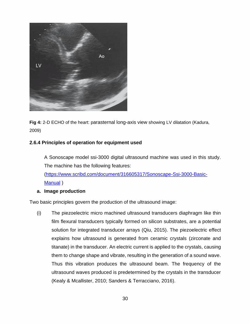
Fig 4: 2-D ECHO of the heart: parasternal long-axis view showing LV dilatation (Kadura,
2009)
2.6.4 Principles of operation for equipment used
A Sonoscape model ssi-3000 digital ultrasound machine was used in this study.
The machine has the following features:
(https://www.scribd.com/document/316605317/Sonoscape-Ssi-3000-Basic-
Manual )
a. Image production
Two basic principles govern the production of the ultrasound image:
(i) The piezoelectric micro machined ultrasound transducers diaphragm like thin
film flexural transducers typically formed on silicon substrates, are a potential
solution for integrated transducer arrays (Qiu, 2015). The piezoelectric effect
explains how ultrasound is generated from ceramic crystals (zirconate and
titanate) in the transducer. An electric current is applied to the crystals, causing
them to change shape and vibrate, resulting in the generation of a sound wave.
Thus this vibration produces the ultrasound beam. The frequency of the
ultrasound waves produced is predetermined by the crystals in the transducer
(Kealy & Mcallister, 2010; Sanders & Terracciano, 2016).

31
(ii) The pulse-echo principle explains how the image is generated. Ultrasound
waves are produced in pulses, not continuously, because the same crystals
are used to generate and receive sound waves, and they cannot do both at the
same time. The ultrasound beam penetrates tissue and is either reflected to
the transducer, transmitted through the tissue (patient), absorbed by the tissue
or scattered. The reflected sound waves or echoes form the ultrasound image.
The reflected echoes cause the crystals in the transducer to change shape
again and produce an electrical signal that is then converted into an image
displayed on the monitor. (Nyland et al., 2002; Sanders & Terracciano, 2016).
b. B-Mode real-time imaging is a grey-scale presentation of the 2-dimension view
of tissue in real-time. The main advantages are:
(i) it is dynamic e.g., movement of cardiac valves can be seen, and
(ii) the grey scale allows for good soft tissue differentiation.
At the study site, 2-D ECHO was used to give snapshots in real time of a cross section of
the heart. These sections were produced in rapid succession and displayed on a monitor,
showing ‘real time imaging’ of the heart chambers, valves and blood vessels.
c. M-mode echo
Motion or M-mode ECHO produces a graph of depth and strength of reflection with time;
changes in motion (e.g. valve opening and closing or ventricular wall movement) can be
displayed. The ultrasound signal should be perpendicular to the structure being examined.
Measurements of the size and thickness of the cardiac chambers can be made either
manually on paper print outs or on the screen monitor using computer software (Kaddoura,
2009). At the study site, M-mode echo was used to produce a graph of strength and depth
of reflection with time of moving structures. Changes in motion, including valve opening
and closing or ventricular wall movement, were displayed and analysed.
d. Doppler imaging
Doppler ultrasound provides information regarding the identification of blood vessels;
direction of blood flow as well as the measurement of blood-flow velocity. The Doppler
Effect is calculated using the frequency change between the transmitted and reflected
sound waves in moving fluids (Sanders & Terracciano, 2016).

32
e. Continuous- wave Doppler
This is a sensitive application in which the sound beam continuously emitted from the
transducer crystal is received by a second. Both transducers are encased in one housing
(Sanders & Terracciano, 2016). This method is useful for measuring high velocities but
its ability to localize a flow signal precisely is limited since the signal can originate at any
point along the width or length of the ultrasound beam (Kaddoura, 2009).
f. Pulsed wave Doppler
In pulsed wave Doppler, a Doppler sound beam is sent and received (pulsed) over a short
period. This is used to measure the blood flow velocity within a small area at a specified
tissue depth (Sanders & Terracciano, 2016). During the study, this was used to assess
ventricular in-flow patterns, intracardiac shunts, and to make precise measurements of
blood flow at valve orifices.
g. Color-flow mapping
This is used to measure the direction and velocity of blood flow to superimpose a colour
pattern. Colour flow mapping was used to measure the velocity and direction of blood
flow during the study (Kaddoura, 2009).
2.7 Common echocardiography prognostic markers in patients with cardiomegaly
i. Left ventricular ejection fraction (LVEF)
Lang et al. (2016) describe the assessment of the left ventricular ejection fraction as a
cornerstone of risk evaluation and management in cardiac pathology. LVEF is a
measurement of the blood that is being pumped out of the left ventricle of the heart, which
is the main pumping chamber, with each contraction. Ejection fraction is an indication of
left ventricular size. A normal LVEF is from 55-75%. For example, a LVEF of 55% means
that 55% of the total amount of blood in the left ventricle is pumped out with each
heartbeat. An EF of less than 35% increases the risk of life-threatening causes of sudden
cardiac arrest.
The ability of echocardiography to quantify EF, therefore, makes it a good baseline
predictor of knowing the cause of cardiomegaly (Hsich, 2014). This makes the use of
ECHO an important tool in the management of cardiomegaly, as is the case in this study.
33
ii. Left ventricular hypertrophy
Left ventricular hypertrophy (LVH) is the thickening of the myocardium of the left ventricle
of the heart, frequently referred to as a pathological reaction to cardiovascular disease,
or high blood pressure. While LVH itself is not a disease, it is usually a marker for diseases
involving the heart which include aortic stenosis, aortic insufficiency and hypertension
(Meijs, 2007). Of these diseases marked by LVH, hypertension stands out as a critical
risk factor for cardiomegaly. ECHO is used to image the left ventricle. The thickness of
the left ventricle as visualized in ECHO correlates with its actual mass. Normal thickness
of the left ventricular myocardium is from 0.6 to 1.1 cm as measured at the very end of
diastole. If the myocardium is more than 1.1 cm thick, the diagnosis of LVH can be made
(Peterson, 2014).
Camici et al (2012) explains that there are two distinct types of left ventricular hypertrophy
and these are:
• “physiologic” - this type of hypertrophy is normally found in athletes, and
• “pathologic” - this is a type of LVH which is found in patients with inherited heart
muscle pathologies such as hypertrophic cardiomyopathy (HCM) or patients with
cardiac and systemic diseases characterized by pressure or volume overload.
Patients with pathologic LVH often have symptoms and signs suggestive of
myocardial ischemia despite normal coronary angiograms. Under these
circumstances ischemia is due to microvascular dysfunction. The abnormalities of
coronary microcirculation may be unrelated to the degree of LVH and cause a
reduction in maximum myocardial blood flow which, in the absence of epicardial
stenosis, is suggestive of microvascular dysfunction. There is no method that
enables direct visualization of coronary microcirculation in vivo in humans. Hence,
its investigation relies on the measurement of parameters which reflect its
functional status, such as myocardial blood flow and coronary flow reserve which
is an integrated measure of flow through both the large epicardial coronary arteries
and microcirculation.
At the site of this study, its standard practice that a diagnosis of LVH on the ECHO is
based on the thickness of the ventricular myocardium.
34
iii. Mitral Valve Stenosis
One of the uses of ECHO is in the diagnosis of valvular heart disease, particularly mitral
stenosis. The mitral valve is found in between the left atrium and left ventricle. The mitral
valve opens during ventricular diastole when blood flows from the left atrium into the left
ventricle. During ventricular systole, the mitral valve closes as blood is ejected from the
left ventricle through the aortic valve (Kaddoura, 2009).
Mitral valve stenosis refers to a narrowing of the mitral valve orifice resulting in impairment
of filling of the left ventricle in diastole and is usually caused by rheumatic heart disease.
An ECHO may reveal evidence of left atrial enlargement and a more advanced stage of
arterial fibrillation or right ventricular hypertrophy consistent with pulmonary hypertension
may be present. Characteristic findings of mitral valve stenosis include valve thickening,
restricted valve opening and anterior leaflet doming. Transthoracic ECHO also allows
assessment of pulmonary artery pressures, detection of other valve disease and
visualization of left arterial thrombus (Nashimura, 2014).
Calcifications of the mitral annulus can also be detected on ECHO and is more common
in the elderly (Boxt & Abbara, 2016). At the site of this study, transthoracic
echocardiography is what is performed at all times and therefore was useful in this study
in assessing the occurrence of mitral valve stenosis in cardiomegaly patients.
iv. Mitral regurgitation
Mitral regurgitation (MR) is the leakage of blood from the left ventricle back into the left
atrium during systole. It is caused by various mechanisms related to structural or
functional abnormalities of the mitral apparatus or the left ventricle. An ECHO may reveal
evidence of left arterial enlargement. In more advanced disease, arterial fibrillation or
hypertrophy consistent with pulmonary hypertension may be present. Transthoracic
ECHO is indicated for all patients with suspected MR to confirm its presence, assess
aetiology, and determine its severity (Zoghbi, 2003). The possibility of cardiomegaly is
high in MR, therefore the use of ECHO and the CXR played a critical role in assessing
how MR affected the heart size. Such findings were of important prognostic significance.

35
v. Aortic valve stenosis
Aortic valve stenosis is a progressive pathology in which the end-stage is characterized
by obstruction of left ventricular outflow, resulting in inadequate cardiac output, decreased
exercise capacity, heart failure and death from cardiac causes (Otto & Prendergast,
2014). ECHO has become the key technique for the diagnosis and evaluation of aortic
valve stenosis. Most of the clinical decision making are based on ECHO assessment of
severity of aortic valve stenosis (Baumgartner et al, 2009). The three cardinal features of
aortic stenosis are leaflet thickening, decreased mobility or doming of the leaflets and
decrease in size of the valve orifice. In patients with severe aortic stenosis, left ventricular
hypertrophy is usually seen (Boxt & Abbara, 2016).
vi. Cardiomyopathy
Cardiomyopathy means disease of the heart. Functionally, the heart has decreased ability
to pump blood to the body. The most common types are hypertrophic and dilated
cardiomyopathies. In hypertrophic cardiomyopathy, the walls of the heart are abnormally
thickened, and the chambers are small. The heart is thus unable to supply adequate blood
to the body (Sanders & Terracciano, 2016).
Dilated cardiomyopathy is a heart disorder defined by the presence of a dilated and poorly
functioning left ventricle. In this condition, the heart becomes weakened and enlarged. It
is characterized by the impaired systolic function of one or both ventricles with normal left
ventricular wall thickness (Luscher, 2016). Transthoracic ECHO is indicated for all
patients with suspected cardiomyopathy to confirm its presence, assess aetiology, and
determine its severity.
i. Aortic root dilation
The dilated aortic root may be linked with underlying aortic valve abnormalities. These
anomalies may lead to complications such as aortic dissection, aortic rupture or
congestive heart failure from aortic insufficiency. Transthoracic echocardiography is used
in serial measurement of aortic root dimensions, proximal aortic segments and
consequently used for thoracic aortic aneurism screening (Saura et al, 2017).

36
ii. Left ventricular dilation
The phyno type of left ventricular dilatation can be the result of a range of pathological
conditions such as toxins, infections, or autoimmune diseases. Dilated cardiomyopathy is
a disease that is characterized by chamber enlargement and contractile dysfunction of
the left ventricle in the absence of chronic pressure and volume overload. ECHO is the
first line imaging test in the assessment of patients with dilated left ventricle. It provides
important information for diagnosis, stratification and guides treatment (Mathew et al,
2017). At the study site, two- and three-dimension echo was used to analyse and measure
the left ventricular chamber size.
iii. Left ventricular diastolic dysfunction
Left ventricular diastolic dysfunction is mostly the result of impaired left ventricular
relaxation with or without reduced restoring forces (and early diastolic suction), and
increased left ventricle chamber stiffness, which increases cardiac filling pressures. Left
ventricular filling pressure should be estimated because elevated left ventricular diastolic
pressure in the absence of raised left ventricular end diastolic volume is strong evidence
of well-developed diastolic dysfunction. In most clinical studies, left ventricular filling
pressures and diastolic function grade can be determined reliably by a few simple ECHO
parameters with a high feasibility. Thus, when performing an ECHO, in patients with
potential diastolic dysfunction, one should search for signs of impaired LV relaxation,
reduced restoring force and increased diastolic stiffness (Nagueh et al., 2016). Diastolic
heart failure is a common form of congestive heart failure that is responsible for most
morbidity and mortality (Shammas et al., 207). In patients with left ventricular failure,
pulmonary hypertension and right ventricular dysfunction are frequent and have an impact
on disease progression, morbidity and mortality (Rosentranz et al., 2015). Therefore, in
hypertension, activation of the sympathetic nervous system may add not only to the blood
pressure elevation but also at the development of left ventricular diastolic dysfunction.
In this study, diastolic function was examined by pulse wave Doppler examination of mitral
flow (before and during Valsalva manoeuvre), pulmonary venous flow, and Doppler
imaging of the medial mitral annulus.

37
iv. Left ventricular systolic dysfunction
Left ventricular systolic function is one of the most important prognostic markers of
patients with cardiac diseases. It is the most often used parameter of left ventricular
systolic function and is measured with two-dimensional (2D) ECHO, by measuring left
ventricular end diastolic and end systolic volumes but does not take into consideration
ultra-structural changes that may occur at the myocardial level and that may impair left
ventricular systolic performance (Tops et al., 2016). At the research site, left ventricular
systolic function was examined with the use of 2-D echocardiography.
v. Fractional shortening
The left ventricle ejects its stroke volume during systole using a combination of
circumferential and longitudinal shortening associated with twisting of the ventricle. The
apex remains relatively stationary; hence, the longitudinal shortening results in mitral
annular motion towards the apex. This shortening produces redial thickening causing an
inward displacement of the endocardium and a reduction in left ventricular cavity volume.
For any given end-diastolic volume, ejection fraction is predominantly determined by
absolute wall thickening rather than relative wall thickening (i.e., radial strain). In addition,
absolute wall thickening is determined by both end-diastolic wall thickness and redial
strain. During diastole, myocardial fibre relaxation, untwisting and lengthening occurs,
resulting in ventricular wall thinning during refilling of the ventricle.
Normally, midwall fractional shortening (FSm) and longitudinal fractional shortening (FSl)
are similar (21%). However, in hypertensive-hypertrophic left ventricular disease, both
FSm and FSl are significantly reduced despite a normal ejection fraction. In aortic stenosis,
FSl is also reduced while ejection fraction is maintained. There is a reduced FSm and FSl
in concentric left ventricular hypertrophy despite a normal ejection fraction. Reduced FSm
occurs even though endocardial fractional shortening (FSen) is normal in hypertensive
heart disease due to a relatively greater contribution of ventricular wall thickening. In
patients with heart failure and preserved ejection fraction, FSm is significantly lower than
in control patients despite mean endocardial fractional shortening not being altered.
Concentric left ventricular hypertrophy with a normal ejection fraction is common in heart

38
failure. A reduced FSm and FSl, and yet normal ejection fraction, may be best explained
by an increase in end-diastolic wall thickness. Furthermore, epicardial FS (FSep) is lower
and FSen is higher than FSm (Maclver, 2012)
In this study, M‐mode cardiac sonographic two-dimensional, Doppler and 3D
echocardiography were all used to assess the function of left ventricle, both during systole
as well as in diastole. All modalities of echocardiography were used to assess left
ventricular systolic function either quantitatively or qualitatively.
vi. Cardiac chamber size
Heart chamber size dimensions can be determined by two dimensional views to allow
quantitative assessment of all the chambers. Quantitative examination of ventricular
function is done by estimating the ejection fraction, determined by calculating the change
in volume of the ventricle between diastole and systole. Left ventricular size enlargement
may be caused by aortic and mitral valve disease, ischemic heart disease, heart failure
and dilated cardiomyopathy. Right ventricular enlargement may be due to volume
overload from tricuspid or pulmonary regurgitation, right ventricular failure secondary to
pulmonary hypertension and cardiomyopathies (Boxt & Abbara, 2016).
Left atrium enlargement may be because of either an increase in arterial pressure
resulting from mitral stenosis or elevated left ventricular end-diastolic pressure, an
increase in volume as in mitral regurgitation or as a result of primary atrial dysfunction, as
in arterial fibrillation. Assessment of the right atrium is mostly made qualitatively by
comparing it to the left atrium in the apical four chamber view. Right atrium enlargement
can result from multiple conditions including tricuspid regurgitation, pulmonary
hypertension and congenital heart defects including atrial septal defects (Boxt & Abbara,
2016).
During this study, heart chamber dimensions were determined by two dimensional views
to allow quantitative examination of each chamber.
2.8. Impact of cardiac diseases on households in Zambia
A study carried out to determine the socio-economic impact of cardiac diseases on
households in Livingstone, Zambia, established that cardiac diseases like cardiac
vascular accidents resulted in the loss of employment, reduced business activity, loss of

39
income and inability to pay for school fees and rentals (Mapulanga, 2010). Cardiomegaly
can thus result in social impacts such as depression, divorce, neglect, fear and apathy
within families. These social issues have the potential to further worsen the condition of
a patient who suffers from cardiac diseases (like cardiomegaly associated diseases) such
that the resulting outcomes make recovery exceedingly difficult. The ability to use imaging
studies in determining the possible physical and functional outcome of cardiomegaly
patients could assist in planning the alleviation of these resultant social issues, for
instance, appropriate counselling and support services.
2.9. Summary of Literature Review
The assessment of the common pathological findings in cardiomegaly patients at LMGH
encompasses everything from complete diagnosis of all symptoms and signs of
cardiomegaly to the putting in place of interventions to prevent catastrophic
consequences of diseases associated with cardiomegaly. Various studies by scholars like
Monfared et al. (2015), Tomaszewki (2012), Elliot et al. (2000) and Tavora et al. (2012),
have all indicated that there is relative consensus that patients presenting with
cardiomegaly should be evaluated with a CXR and an ECHO. The CXR can detect
cardiomegaly and any lung infections while an ECHO can help assess the heart and the
big vessels to detect the cardiac pathology found in cardiomegaly patients. The CXR and
ECHO are important tools in the diagnosis and management of cardiomegaly.
Based on the various findings, by researchers like Monfared et al. (2015), that are
obtainable from these imaging studies, it was important to ascertain if a relationship exists
between these findings in ascertaining the common pathological findings in patients who
present with an enlarged heart on CXR. This was then used to come up with evidence-
based decision making on cardiomegaly diagnosis and management.
40
CHAPTER THREE
METHODOLOGY
The research methods and design for this study involved the selection of the study
population, data collection, statistical analysis, organization and interpretation of results
and ethical statement. This retrospective study involved the retrieving of data for 124
patients who had cardiomegaly identified on a CXR and had undergone an ECHO
examination.
3.1 Study design
The study was a quantitative, retrospective, cross sectional study that was carried out at
a single site in Lusaka. It was appropriate to use this design because it enabled the
systematic collection, numerical analysis and presentation of data (Muranda, 2004). A
retrospective study was used as the data was readily available. Furthermore, this study
was descriptive and it involved the collection of information without changing the
environment (i.e. nothing was manipulated).
3.2 Research site
The study was undertaken at an academic hospital, in Lusaka, Zambia. Prior, no such
formal study had ever been done at this hospital or in Zambia. The research site had a
well-established radiological and cardiovascular department with experts in the field that
were used for consultation regarding the study.
3.3 Study population
The study population included 124 adult male and female patients who were diagnosed
with cardiomegaly on a CXR and had ECHO examinations at the research site.
3.4 Inclusion criteria
The following served as the inclusion criteria:
i. Patients diagnosed with cardiomegaly on PA erect CXR and had a follow up
ECHO examination done.
ii. Male and female patient aged between 18 and 80 years.

41
3.5 Exclusion criteria
. The following served as the exclusion criteria:
i. Patients who did not have cardiomegaly on a CXR
ii. Patients where CXR and/or ECHO reports were not available
iii. Patients who were below 18 years old
iv. Patients who were above 80 years old
3.6 Sample size calculation
The sample size was calculated according to the sample size formula (Isreal, 1992):
𝒏 =𝒁²(𝒑 × 𝒒)
𝒆²
𝒏 = sample size
𝒁 = standard error associated with the chosen level of confidence (1.96)
𝒑 = estimated percent in the population (8% as estimated for Zambia by WHO, 2014)
𝒒 = 100 – p
𝒆 = ± 5 acceptable sample error
𝒏 =𝒁²(𝒑 × 𝒒)
𝒆²
𝒏 =1.96²(8 × 92)
5²
𝒏 =3.8416 (736)
25
𝒏 = 113.2
Therefore, a minimum of 113 patients’ information was supposed to be retrieved for this
study as calculated using 8% as the estimated prevalence rate for cardiovascular
diseases in Zambia (WHO, 2014). However, the study included 124 participants. In
addition, a convenient sampling method was used. All patients who met the inclusion
criteria from the 30th August 2017, dating backwards, formed the sample until the target
was reached.
42
3.7 Data collection
Patients’ files were retrieved by the research assistant. Data was retrieved by the
researcher, a qualified radiographer, from the retrieved files. Results for the CXR and
ECHO were in the form of reports. The results of the CXR and ECHO were entered on
data collection sheets (Appendix A and Appendix B). The data collection sheets used for
the study had the following sections:
i. Patients demographic information: age and gender
ii. Chest X-ray:
a. Cardiac thoracic ratio (CTR)
b. Any lung disease
iii. Echocardiography:
a. Left and right ventricle
b. Left and right atrium
c. Aortic valve (size, systolic and diastolic function)
d. Inter-ventricular septum (thickness measured in cm)
e. Posterior wall of the left ventricle (thickness measured in cm)
f. Mitral valve (regurgitation)
g. Ejection Fraction (normal: 50-70%)
h. Pericardial effusion
i. Colour Doppler
iv. Blood pressure parameters (mm/Hg)
3.8 Data management
Data management involves converting masses of data into smaller, manageable
segments. It involves the organization of data from its entry to the research cycle through
the dissemination and achieving valuable results (Polit & Beck, 2012). The data collected
was coded with the use of specially designed numbers as opposed to individual names
(see appendix A & B).
The data collected was in the care of the principal investigator and was kept on collection
sheets (see appendix A & B) which were locked in a safe box in the office of the head of

43
the radiology department at the study site. Keys for the safe box were kept by the principal
investigator and a spare key was given to the head of the radiology department.
The data will remain stored for the appropriate number of years required by the academic
and health institutions (study site) after graduation and subsequent publications of articles.
Individuals not involved in the study did not have access to the raw data collected. Only
the principal investigator, registry clerk (who was used as a research assistant), the head
of the radiology department, the supervisor and the statistician had access to the
collected data.
3.9 Data analysis
The data was captured on a data spread sheet that was designed by the researcher. The
data was quality controlled, assured and analysed by a statistician using SPSS version
24. Descriptive statistics (mean, median, percentages, standard deviation and variance)
were used for prevalence in age, gender and pathological conditions.
Inferential statistics were also used with a level of statistical significance set at p<0.05.
Spearman’s correlation test was conducted to assess the relationship between age and
cardiomegaly. Chi-squared test was used to assess whether there was a significant
association between 2 sets of variables (Willis, 2004) e.g. gender and cardiomegaly,
Doppler findings and cardiomegaly, cardiac conditions, and blood pressure readings.
3.10 Data validity and reliability
The following aspects of data validity and reliability were applied to this research study:
i. Validity
According to Watkins (2012), validity is concerned with the extent to which the research
findings accurately represent what is happening or more specifically, whether the
information is a true picture of what is being studied. According to Cooper and Schindler
(2006), three major forms of validity can be identified. These are ‘content validity’,
‘criterion-related validity’ and ‘construct validity’. The appropriate research design,
research methodology and the right data collecting tools (as explained in the appropriate
corresponding headings) were used in this study to ensure validity. The data collecting

44
sheets, which were verified by the radiologist and cardiologist, (appendix A & B) contains
sections that were used to meet the objectives of the study.
ii. Reliability
Reliability (sometimes referred to as trustworthiness), is concerned with the findings of
the research (Watkins, 2012). The findings are considered reliable if the research is
repeated and similar results are obtained. This can be achieved by repeating the same
research (at any similar institution), using similar tools and appropriate expert personnel.
For reliability, it is expected that similar results would be obtained.
3.11 Ethics
According to Watkins (2012) in research, ethics refers to the appropriateness of your
behaviour about the rights of those who become the subject of your work, or are affected
by it. Ethics provides rules and guidelines to the researcher about behavioural
expectations and the expected conducts towards participants in the study, co-researchers,
research assistants, fieldworkers, institution and sponsors.
Therefore, before conducting this research, a research proposal was submitted to the
Cape Peninsula University of Technology’s Ethics Committee for approval. A copy of the
proposal and ethical approval (appendix E) were then submitted to the study site, head
of the institution, to seek permission to conduct the study at their site. Thus, this research
was conducted according to the Code of Ethics of the World Medical Association
(Declaration of Helsinki).
Being a retrospective study, particular attention was paid to issues of confidentiality by
ensuring that all information that came into the possession of the researcher, research
supervisor, head of the radiology department and research assistant during the study was
not shared with any unauthorized person. To help achieve this, the patient’s names were
not used in this study and each patient’s file was assigned a code. Additionally, the people
involved in this study were informed of the need to ensure confidentiality and were given
an information leaflet about the research (appendix C). People involved in this research
played the following roles:
i. The research assistant assisted in retrieving files. The research assistant was
a qualified registry clerk.

45
ii. The research supervisor and co-supervisor assisted the researcher on how to
go about the research.
iii. The statistician analysed the data.
iv. The cardiologist who managed the patient was approached to explain some
unfamiliar conditions.
v. The radiologist responsible for reporting the CXR images and conducting the
ECHO was approached to explain some conditions.
vi. The head of the radiology department at the study site ensured that the
researcher and his team were accorded a conducive environment to conduct
the study.
The researcher did not request informed consent from patients as the study was
retrospective. However, additional permission to allow the researcher to research the
study site was obtained from the Medical Superintendent (appendix D and E) at Levy
Mwanawasa General Hospital.
By adhering to strict confidentiality of data retrieved, no harm was done to the patients
(non-maleficence). There may be no direct benefit for the patients as this is a
retrospective study, however, the outcome of the study will be published, and this may
assist in improving patient management in cardiac disease.
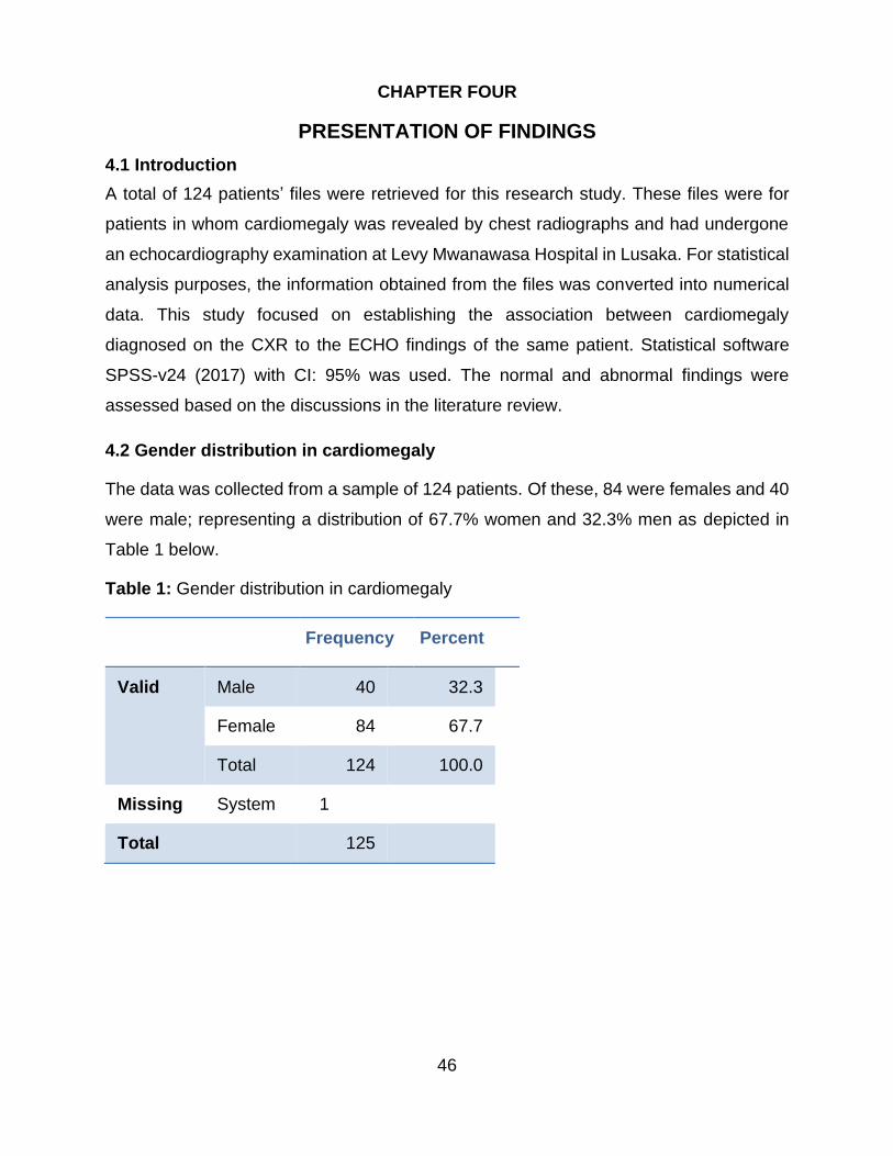
46
CHAPTER FOUR
PRESENTATION OF FINDINGS
4.1 Introduction
A total of 124 patients’ files were retrieved for this research study. These files were for
patients in whom cardiomegaly was revealed by chest radiographs and had undergone
an echocardiography examination at Levy Mwanawasa Hospital in Lusaka. For statistical
analysis purposes, the information obtained from the files was converted into numerical
data. This study focused on establishing the association between cardiomegaly
diagnosed on the CXR to the ECHO findings of the same patient. Statistical software
SPSS-v24 (2017) with CI: 95% was used. The normal and abnormal findings were
assessed based on the discussions in the literature review.
4.2 Gender distribution in cardiomegaly
The data was collected from a sample of 124 patients. Of these, 84 were females and 40
were male; representing a distribution of 67.7% women and 32.3% men as depicted in
Table 1 below.
Table 1: Gender distribution in cardiomegaly
Frequency Percent
Valid Male 40 32.3
Female 84 67.7
Total 124 100.0
Missing System 1
Total 125
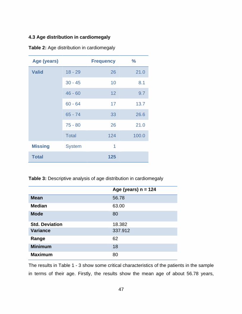
47
4.3 Age distribution in cardiomegaly
Table 2: Age distribution in cardiomegaly
Age (years) Frequency %
Valid 18 - 29 26 21.0
30 - 45 10 8.1
46 - 60 12 9.7
60 - 64 17 13.7
65 - 74 33 26.6
75 - 80 26 21.0
Total 124 100.0
Missing System 1
Total 125
Table 3: Descriptive analysis of age distribution in cardiomegaly
Age (years) n = 124
Mean 56.78
Median 63.00
Mode 80
Std. Deviation 18.382
Variance 337.912
Range 62
Minimum 18
Maximum 80
The results in Table 1 - 3 show some critical characteristics of the patients in the sample
in terms of their age. Firstly, the results show the mean age of about 56.78 years,
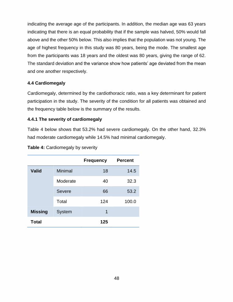
48
indicating the average age of the participants. In addition, the median age was 63 years
indicating that there is an equal probability that if the sample was halved, 50% would fall
above and the other 50% below. This also implies that the population was not young. The
age of highest frequency in this study was 80 years, being the mode. The smallest age
from the participants was 18 years and the oldest was 80 years, giving the range of 62.
The standard deviation and the variance show how patients’ age deviated from the mean
and one another respectively.
4.4 Cardiomegaly
Cardiomegaly, determined by the cardiothoracic ratio, was a key determinant for patient
participation in the study. The severity of the condition for all patients was obtained and
the frequency table below is the summary of the results.
4.4.1 The severity of cardiomegaly
Table 4 below shows that 53.2% had severe cardiomegaly. On the other hand, 32.3%
had moderate cardiomegaly while 14.5% had minimal cardiomegaly.
Table 4: Cardiomegaly by severity
Frequency Percent
Valid Minimal 18 14.5
Moderate 40 32.3
Severe 66 53.2
Total 124 100.0
Missing System 1
Total 125

49
4.4.2 Cardiomegaly and age
Table 5 below shows that as the age of the patients increases, the severity of the condition
also worsens. Specifically, more patients from the young age group <35 years (n=8) were
found to have minimal cardiomegaly while more in the older age group >75 years (n=30)
were found to have the severe condition.
Table 5: The severity of cardiomegaly by age groups
AGE RANGE
(years)
CARDIOMEGALY
Minimal Moderate Severe Total
Less than 35 8 9 3 20
35-45 1 13 6 20
46-54 3 2 7 12
55-64 2 9 6 17
65-74 3 2 14 19
75+ 1 5 30 36
Total 18 40 66 124
The correlation between cardiomegaly and age
The relationship between cardiomegaly and the age of patients was computed to identify
any pattern. This involved the use of Pearson’s Rank Correlation (r). As r > 0.5, i.e. r =
0.58, it is considered a strong positive correlation.
Table 6: Correlation between cardiomegaly and age (p<0.05)
Pearson Correlation
Cardiomegaly Age
Cardiomegaly 1 0.58
Age 0.58 1

50
4.4.3 Cardiomegaly and gender
Table 7: The severity of cardiomegaly and gender
Gender
Male Female
Cardiomegaly Minimal 5 13
12.5% 15.5%
Moderate 15 25
37.5% 29.8%
Severe 20 46
50.0% 54.8%
Total
40 84
100.0% 100.0%
The Chi-square test was conducted to assess the significance of the relationship between
cardiomegaly and gender testing the null hypothesis that cardiomegaly is non-gender
selective. Pearson’s Chi-square test yielded a p-value of 0.676 which is far greater than
the standard 0.05 level of significance, thus leading to the acceptance of the null
hypothesis. Therefore, there is no sufficient evidence to suggest an association between
gender and cardiomegaly. This is supported by the evidence in the table below which
shows that at every level of severity of cardiomegaly, both women and men were affected.
This, therefore, leads to the conclusion that both females and males were at risk to
cardiomegaly.
Table 8: The Chi-square test across gender and cardiomegaly (p<0.05)
Value Df p-value
Pearson Chi-Square .784 2 .676
Likelihood Ratio .777 2 .678
Linear-by-Linear Association .016 1 .899
N of Valid Cases 124

51
4.4.4 Normal and abnormal ECHO findings in cardiomegaly
Table 9 below shows that 87.9% of patients with cardiomegaly had an abnormal ECHO
finding while 12.1% showed normal ECHO reading. This means that 87.9% of patients
with cardiomegaly diagnosed on chest X-ray had one or more cardiovascular diseases
accompanying the cardiomegaly.
Table 9: Normal and abnormal ECHO findings in cardiomegaly
Frequency Percent
Valid Normal 15 12.1
Abnormal 109 87.9
Total 124 100.0
Missing System 1
Total 125
4.5 Common diseases identified in the study
Table 10 displays some of the diseases which were common in the study. As the results
indicate, left ventricular diastolic dysfunction was the most common disease found in the
study population. Left ventricular diastolic dysfunction was found in more than half of the
study participants (71%). Thus, left ventricular diastolic dysfunction was the overall most
common disease among the patients. Other common diseases include left atrium dilation
and left ventricular systolic dysfunction, with 29% distribution each.

52
Table 10: Common diseases found in cardiomegaly
DISEASE PERCENT OF OCCURRENCE
Seen Not seen Total
Cardiomyopathy 16 84 100
Inter Ventricular Hypertrophy 23 77 100
Left Atrium Dilation 29 71 100
Left Ventricular Diastolic Dysfunction
71 29 100
Left Ventricular Dilation 21 79 100
Left Ventricular Hypertrophy 24 76 100
Left Ventricular Systolic Dysfunction 29 71 100
Pericardial Effusion 21 79 100
Pulmonary Hypertension 16 84 100
Right Atrium Dilation 25 75 100
Right Ventricular Dilation 18 82 100
4.6 Cardiac disease distribution by gender
Table 11 below shows the prevalence of diseases based on gender. Some of the
diseases with high prevalence among males include left ventricular diastolic dysfunction
(65%), right atrium dilation (32.5%) and left atrium dilation (37.5%). Among female
patients, left ventricular systolic dysfunction (26.2%), left ventricular hypertrophy (23.8%),
left atrium dilatation (23.8%) and left ventricular diastolic dysfunction (73.8%) were among
the common diseases. However, left ventricular diastolic dysfunction is the most common
in both males and females.

53
Table 11: Common cardiac diseases found by gender
DISEASE MALE FEMALE
Seen Not
seen
Tota
l
See
n
Not
seen
Total
Cardiomyopathy 10 30 40 9 75 84
Inter Ventricular Hypertrophy 11 29 40 17 67 84
Left Atrium Dilation 15 25 40 20 64 84
Left Ventricular Diastolic
Dysfunction
26 14 40 62 22 84
Left Ventricular Dilation 13 27 40 12 72 84
Left Ventricular Hypertrophy 9 31 40 20 64 84
Left Ventricular Systolic
Dysfunction
14 26 40 22 62 84
Pericardial Effusion 4 36 40 7 78 84
Pulmonary Hypertension 6 34 40 13 71 84
Right Atrium Dilation 13 27 40 17 67 84
Right Ventricular Dilation 11 29 40 11 73 84

54
4.7 Blood pressure in cardiomegaly
After testing for blood pressure, the result indicated that 42.7% of the patients had severe
blood pressure, 14.5% had moderate blood pressure while 12.9% had minimal. Normal
blood pressure was shown in 29,8% of the patients (Table 12).
Table 12: Blood pressure in cardiomegaly
Blood Pressure (mm Hg) Frequency Percent
Valid up to 120/60-80 37 29.8
121-140/81-90 (minimal) 16 12.9
141-159/91-99 (moderate) 18 14.5
151 or more/100 or more (severe) 53 42.7
Total 124 100.0
Missing System 1
Total 125
4.8 Variations in blood pressure and cardiac conditions found on ECHO
The blood pressure of patients with various conditions was tested and results showed
that most of the patients with cardiac diseases had severe blood pressure. There was a
significant association between severe blood pressure and cardiomyopathy (p = 0.023),
inter-ventricular hypertrophy (p = 0.017), left atrium dilation (p = 0,007), left ventricular
diastolic dysfunction (p = 0.045), left ventricular dilation (p = 0.003), left ventricular
hypertrophy (p = 0,028), left ventricular systolic dysfunction (p = 0.048) and pericardial
effusion (p = 0,001) (Table 12). On the other hand, conditions like pulmonary hypertension,
right atrium dilation and right ventricular dilation were associated with normal blood
pressure.

55
Table 13: The relationship between common cardiac diseases and blood pressure (Chi-
squared test, *p<0.05)
DISEASE Severity of Blood Pressure in patients with disease
p-value
Cardiomyopathy Generally Severe 0.023*
Inter Ventricular Hypertrophy Generally Severe 0.017*
Left Atrium Dilation Generally Severe 0.007*
Left Ventricular Diastolic Dysfunction
Generally Severe 0.045*
Left Ventricular Dilation Generally Severe 0.003*
Left Ventricular Hypertrophy Generally Severe 0.028*
Left Ventricular Systolic Dysfunction
Generally Severe 0.048*
Pericardial Effusion Generally Severe 0.001*
Pulmonary Hypertension Generally Normal 0.044*
Right Atrium Dilation Generally Normal 0.006*
Right Ventricular Dilation Generally Normal 0.009*
4.9 Doppler ultrasound results
Doppler readings of cardiac cycles were used to evaluate the blood flow of patients in the
study. At the mitral valve, recordings of the peak velocity (E) and the peak atrial velocity
(A) were analysed. The Doppler results showed 60% of patients in the sample had
increased blood flow and 6% had severely increased blood flow. On the other hand, 22%
of patients showed normal blood flow. However, blood flow could not be measured on 12%
of patients in the sample because of the absence of the A-wave (Table 14).
The results in Table 15 showed that increased blood flow was significantly associated
with minimum (p=0.002), moderate (p=0.000) and severe (p=0.04) cardiomegaly.
Severely increased blood flow was significantly associated with severe cardiomegaly
(p=0.004).
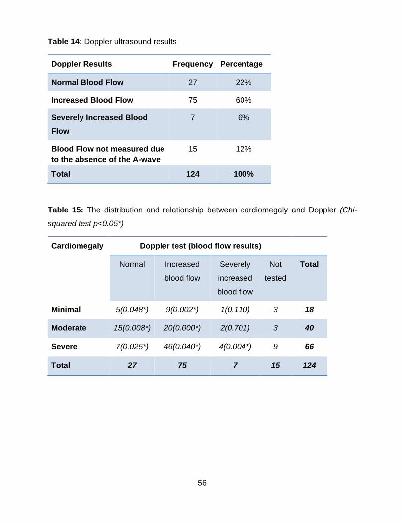
56
Table 14: Doppler ultrasound results
Doppler Results Frequency Percentage
Normal Blood Flow 27 22%
Increased Blood Flow 75 60%
Severely Increased Blood
Flow
7 6%
Blood Flow not measured due
to the absence of the A-wave
15 12%
Total 124 100%
Table 15: The distribution and relationship between cardiomegaly and Doppler (Chi-
squared test p<0.05*)
Cardiomegaly Doppler test (blood flow results)
Normal Increased
blood flow
Severely
increased
blood flow
Not
tested
Total
Minimal 5(0.048*) 9(0.002*) 1(0.110) 3 18
Moderate 15(0.008*) 20(0.000*) 2(0.701) 3 40
Severe 7(0.025*) 46(0.040*) 4(0.004*) 9 66
Total 27 75 7 15 124

57
4.10 Cardiac abnormalities found on ECHO
4.10.1 Aortic root dilation
A normal aortic root diameter was shown in 91.1% of patients, while 4.8% had minimally
dilated, 2.4% had moderately dilated and 1.6% had severely dilated aortic root
diameter.
Table 16: Aortic root dilatation in cardiomegaly
Aortic root diameter (mm) Frequency Percent
Valid up - 30 mm (normal) 113 91.1
31 - 33 mm (minimum) 6 4.8
34 - 36 mm (moderate) 3 2.4
37 mm or more (severe) 2 1.6
Total 124 100.0
Missing System 1
Total 125
4.10.2 Left atrial dilation
A normal left atrial diameter was shown in 71% of patients while 10.5% had minimally
dilated, 11.3% moderately dilated and 7.3% had severely dilated atrial diameter.
Table 17: Left atrial dilation in cardiomegaly
Left atrial diameter (mm) Frequency Percent
Valid up to 38 mm (normal) 88 71.0
39 - 42 mm (minimal) 13 10.5
43 - 46 mm (moderate) 14 11.3
47 mm or more (severe) 9 7.3
Total 124 100.0

58
4.10.3 Left ventricular dilation
A normal left ventricular diameter was shown in 79.8% of the patients, 6.5% had
minimally dilated left ventricle, 9.7% moderate while 4% had severely dilated left
ventricle diameter.
Table 18: Left ventricular dilation in cardiomegaly
Left ventricular diameter (mm) Frequency Percent
Valid up to 53 mm (normal) 99 79.8
54 - 57 mm (minimal) 8 6.5
58 - 61 mm (moderate) 12 9.7
62 mm or more (severe) 5 4.0
Total 124 100.0
Missing System 1
Total 125
4.10.4 Fractional shortening
Normal fractional shortening was reported in 68.5% of patients; 3.2% had minimal, 21.8%
moderate and 6.5% had severe fractional shortening.
Table 19: Fractional shortening in cardiomegaly
Fractional shortening Frequency Percent
Valid 27 - 45% (normal) 85 68.5
22 - 26% (minimal) 4 3.2
17 - 21% (moderate) 27 21.8
16% or less (severe) 8 6.5
Total 124 100.0
Missing System 1
Total 125

59
4.10.5 Mitral valve regurgitation
No mitral valve regurgitation was reported in 83.1% of patients, while16.9% had minimal
mitral valve regurgitation.
Table 20: Mitral valve regurgitation in cardiomegaly
Mitral valve regurgitation Frequency Percent
Valid Normal 103 83.1
Minimal regurgitation 21 16.9
Total 124 100.0
Missing System 1
Total 125
4.10.6 Inter-ventricular septal thickness
Normal inter-ventricular septal thickness was reported in 77.4% of patients; 17.7% had
minimally thickened, while 4% had moderate and 0.8% had severely thickened inter-
ventricular septum.
Table 21: Inter-ventricular septal thickness in cardiomegaly
Inter-ventricular septal thickness
(mm)
Frequency Percent
Valid 6 - 11 mm (normal) 96 77.4
12 - 14 mm (minimal) 22 17.7
15 - 17mm (moderate) 5 4.0
18 mm or more (severe) 1 .8
Total 124 100.0
Missing System 1
Total 125

60
4.10.7 Posterior wall thickness
Normal posterior wall thickness was reported in 76.6% of patients; 20.2% had minimally
thickened wall, while 2.4% had moderate and 0.8% had severely thickened posterior
wall.
Table 22: Posterior wall thickness in cardiomegaly
Posterior wall thickness (mm) Frequency Percent
Valid 6 - 11 mm (normal) 95 76.6
12 - 14 mm (minimal) 25 20.2
15 - 17mm (moderate) 3 2.4
18 mm or more (severe) 1 .8
Total 124 100.0
Missing System 1
Total 125
4.10.8 Right ventricular outflow tract
Normal right ventricular outflow tract was reported in 83.1% of patients; 8.9% had
minimal, while 4% hade moderate and 4% had severe right ventricular outflow tract.
Table 23: Right ventricular outflow tract and cardiomegaly
Right ventricular outflow tract Frequency Percent
Valid up to 29 mm (normal) 103 83.1
30 – 32 mm (minimum) 11 8.9
33 – 35 mm (moderate) 5 4.0
36 mm or more (severe) 5 4.0
Total 124 100.0
Missing System 1
Total 125

61
4.10.9 Ejection fraction
Normal ejection fraction was reported in 63.7% of patients, 8.9 had minimal, while
24.2% had moderate and 3.2% had severe ejection fraction.
Table 24: Ejection fraction and cardiomegaly
Ejection fraction Frequency Percent
Valid 55 - 75% (normal) 79 63.7
45 - 54% (minimal) 11 8.9
30 - 44% (moderate) 30 24.2
29% or less (severe) 4 3.2
Total 124 100.0
Missing System 1
Total 125
4.10.10 Thrombus
No thrombus was detected in any of the patients.

62
4.10.11 Aortic root regurgitation
No aortic valve regurgitation was reported in 94.4% of patients; 0.8% had minimal, while
4% hade moderate and the other 0.8% had severe aortic valve regurgitation.
Table 25: Aortic root regurgitation
Aortic root
regurgitation
Frequency Percent
Valid Not seen 117 94.4
Minimal 1 .8
Moderate 5 4.0
Severe 1 .8
Total 124 100.0
Missing System 1
Total 125
4.10.12 Calcified aortic valve
No aortic calcifications were reported in 97.6% of patients while 2.4% had aortic
calcifications.
Table 26: Calcified aortic valve and cardiomegaly
Calcified aortic valve Frequency Percent
Valid Not seen 121 97.6
Seen 3 2.4
Total 124 100.0
Missing System 1
Total 125

63
4.10.13 Cardiomyopathy
No cardiomyopathy was reported in 84.7% of patients, while 15.3% had
cardiomyopathy.
Table 27: Cardiomyopathy and cardiomegaly
Cardiomyopathy Frequency Percent
Valid Not seen 105 84.7
Seen 19 15.3
Total 124 100.0
Missing System 1
Total 125
4.10.14. Cor pulmonale
No cor pulmonale was reported in 97.6% of patients, while 2.4% had cor pulmonale.
Table 28: Cor pulmonale and cardiomegaly
Cor pulmonale Frequency Percent
Valid Not seen 121 97.6
Seen 3 2.4
Total 124 100.0
Missing System 1
Total 125
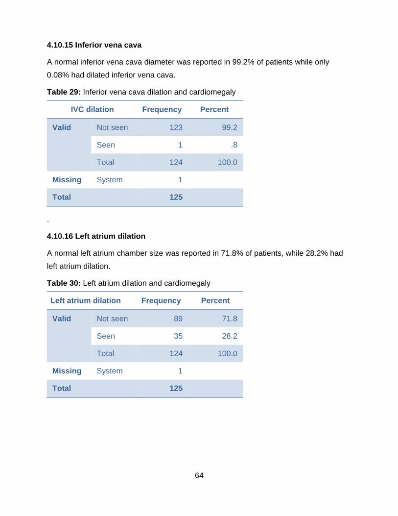
64
4.10.15 Inferior vena cava
A normal inferior vena cava diameter was reported in 99.2% of patients while only
0.08% had dilated inferior vena cava.
Table 29: Inferior vena cava dilation and cardiomegaly
IVC dilation Frequency Percent
Valid Not seen 123 99.2
Seen 1 .8
Total 124 100.0
Missing System 1
Total 125
.
4.10.16 Left atrium dilation
A normal left atrium chamber size was reported in 71.8% of patients, while 28.2% had
left atrium dilation.
Table 30: Left atrium dilation and cardiomegaly
Left atrium dilation Frequency Percent
Valid Not seen 89 71.8
Seen 35 28.2
Total 124 100.0
Missing System 1
Total 125
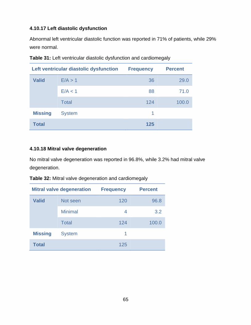
65
4.10.17 Left diastolic dysfunction
Abnormal left ventricular diastolic function was reported in 71% of patients, while 29%
were normal.
Table 31: Left ventricular diastolic dysfunction and cardiomegaly
Left ventricular diastolic dysfunction Frequency Percent
Valid E/A > 1 36 29.0
E/A < 1 88 71.0
Total 124 100.0
Missing System 1
Total 125
4.10.18 Mitral valve degeneration
No mitral valve degeneration was reported in 96.8%, while 3.2% had mitral valve
degeneration.
Table 32: Mitral valve degeneration and cardiomegaly
Mitral valve degeneration Frequency Percent
Valid Not seen 120 96.8
Minimal 4 3.2
Total 124 100.0
Missing System 1
Total 125

66
4.10.19 Mitral valve regurgitation
No mitral valve regurgitation was reported in 84.6% of patients, while 5.7% had minimal,
6.5% moderate and 3.3% had severe mitral valve regurgitation.
Table 33: Mitral valve regurgitation and cardiomegaly
Mitral valve regurgitation Frequency Percent
Valid Not seen 104 84.6
Minimal 7 5.7
Moderate 8 6.5
Severe 4 3.3
Total 123 100.0
Missing System 2
Total 125
4.10.20 Myocardial infarction
No myocardial infarction was reported in 96% of patients, while 4% had a myocardial
infarction.
Table 34: Myocardial infarction and cardiomegaly
Myocardial infarction Frequency Percent
Valid Not seen 119 96.0
Seen 5 4.0
Total 124 100.0
Missing System 1
Total 125

67
4.10.21 Pericardial effusion
Pericardial effusion was reported in 21% of patients, while 79% had no pericardial
effusion.
Table 35: Pericardial effusion and cardiomegaly
Pericardial effusion Frequency Percent
Valid Not seen 98 79.0
Seen 26 21.0
Total 124 100.0
Missing System 1
Total 125
4.10.22 Pleural effusion
Pleural effusion was reported in 8.1% of patients, while 91.9% had no pleural effusion.
Table 36: Pleural effusion and cardiomegaly
Pleural effusion Frequency Percent
Valid Not seen 114 91.9
Seen 10 8.1
Total 124 100.0
Missing System 1
Total 125

68
4.10.23 Pulmonary hypertension
Pulmonary hypertension was reported in 15.3% of patients, while 84.7% had no
pulmonary hypertension.
Table 37: Pulmonary hypertension and cardiomegaly
Pulmonary hypertension Frequency Percent
Valid Not seen 105 84.7
Seen 19 15.3
Total 124 100.0
Missing System 1
Total 125
4.10.24 Right atrium dilation
A dilated right atrium was reported in 24.2% of patients, while 75.8% of the patients had
normal chamber size of the right atrium.
Table 38: Right atrium dilation and cardiomegaly
Right atrium dilation Frequency Percent
Valid Not seen 94 75.8
Seen 30 24.2
Total 124 100.0
Missing System 1
Total 125

69
4.10.25 Right ventricular dilation
A dilated right ventricle was reported in 17.7% of patients, while 82.3% had normal
chamber size of the right ventricle.
Table 39: Right ventricular dilation and cardiomegaly
Right ventricular dilation Frequency Percent
Valid Not seen 102 82.3
Seen 22 17.7
Total 124 100.0
Missing System 1
Total 125
4.10.26 Tricuspid degeneration
No patients had tricuspid valve degeneration.
4.10.27 Tricuspid valve regurgitation
Tricuspid valve regurgitation was reported in patients as follows: 9.7% (minimal
regurgitation), 11.3% (moderate) and 4.8% (severe regurgitation).
Table 40: Tricuspid valve regurgitation and cardiomegaly
Tricuspid valve regurgitation Frequency Percent
Valid Not seen 92 74.2
Minimal 12 9.7
Moderate 14 11.3
Severe 6 4.8
Total 124 100.0
Missing System 1
Total 125

70
4.10.28 Paradoxical motion of the IVC
A paradoxical motion of the IVC was reported in 1.6% of patients, while 98.4% had no
paradoxical motion of the IVC.
Table 41: Paradoxical motion of the IVC and cardiomegaly
Paradoxical motion of IVC Frequency Percent
Valid Not seen 122 98.4
Seen 2 1.6
Total 124 100.0
Missing System 1
Total 125
4.10.29 Paradoxical septal motion
No paradoxical septal motion was reported in 99.2%, while 0.8% had paradoxical septal
motion.
Table 42: Paradoxical septal motion and cardiomegaly
Paradoxical septal motion Frequency Percent
Valid Not seen 123 99.2
Seen 1 .8
Total 124 100.0
Missing System 1
Total 125

71
4.11 Summary of results
The study population (n = 125) were all diagnosed with cardiomegaly by measurement of
the cardio-thoracic ratio on the CXR. Cardiomegaly was more prevalent in females
(67.7%) compared to males (32.3%). All age groups were affected, however, the
prevalence increased with age; 60% of the patients were aged 60-80 years. More than
50% of the cardiomegaly cases were severe. There was no significant difference between
males and females with severe cardiomegaly.
The ECHO findings showed left ventricular diastolic dysfunction as the most common
disease (71%) followed by left ventricular systolic dysfunction (29%) and left atrium
dilation (29.1%). Doppler ultrasound revealed a significant association between severe
cardiomegaly and severely increased blood flow patterns (p=0.004); and between
cardiomegaly (minimal, moderate and severe) and increased blood flow patterns
(p=0.002, p=0.000 and p=0.04 respectively). Other abnormal ECHO findings included:
ejection fraction (36.3%), fractional shortening (31.5%), left atrial dilation (28.2%),
tricuspid valve regurgitation (25.8%), right atrium dilation (24.2%), posterior wall thickness
(23.4%), interventricular septal thickness (22.5%), pericardial effusion (21%), left
ventricular dilation (20.2%), left ventricular dilation (17.7%), right ventricular outflow tract
(16.9%), minimum right ventricular outflow tract (16.9%), mitral valve regurgitation
(15.5%), aorta root dilation (8.8%) and pleural effusion (8.1%)
There was a significant association between severely increased blood pressure and
cardiomyopathy (p=0.023), interventricular hypertrophy (p=0.017), left atrial dilation
(p=0.007), left ventricular diastolic dysfunction (p=0.045), left ventricular dilation
(p=0.003), left ventricular hypertrophy (p=0.028), left ventricular systolic dysfunction
(p=0.048) and pericardial effusion (p=0.001).
Results showed a significant association (p<0.05) between cardiomegaly diagnosed on
the CXR and abnormal findings on the ECHO.

72
CHAPTER FIVE
DISCUSSION AND CONCLUSION
5.1 Introduction
In this chapter, emphasis is placed on those results that answer the research questions
in general and those that meet the aim and specific objectives of this study.
Studies have confirmed that cardiomegaly is very prominent globally and the CXR and
ECHO have been used to prove this fact. A study in Iraq from 2006 to 2007, found
24.9% and 50.8% of patients had cardiomegaly on CXR and ECHO respectively
(Monafred et al., 2015). Tomaszewski (2012) estimated that cardiomegaly is found in
5-7% of CXR in tropical Africa. Tavora et al. (2012) discovered that among adults with
a mean age of ±50 years, cardiomegaly is a frequent cause of sudden cardiac death.
At the research site in Zambia, the prevalence of cardiomegaly was estimated to be at
16.7%, as shown by records from the X-ray department register (LMGH, 2015). It
should be noted that most cardiovascular diseases are very often accompanied by an
enlarged heart. Therefore, cardiovascular diseases are very common in patients with
cardiomegaly. Considering all the complications to the human body due to this disease,
cardiomegaly is a disease of great concern worldwide. Drawing on the findings of this
study, this chapter presents a discussion on the association between cardiomegaly
diagnosed on the CXR and the ECHO results of the same patients. This study provides
evidence that there is a strong association between cardiomegaly diagnosed on a CXR
and the ECHO results of the same patient.
5.2 Cardiomegaly findings
The main objective of this study was to establish whether there was an association
between the CXR reports of patients diagnosed with cardiomegaly to the ECHO reports
of the same patients. Cardiomegaly is an independent prognostic factor for mortality, and
increased CTR, which is associated with poor prognosis in older patients.
Hypertrophy determined by ECHO is associated with 1.5 to 4 times increase in mortality,
which is indicative of great importance and necessity of early diagnosis, especially in old
or middle-aged patients as the results from the preceding chapter showed. Cardiomegaly

73
could result in mortality due to some mechanisms such as higher oxygen need by larger
ventricular mass, fatal ventricular arrhythmias, and endothelial dysfunction (Elhendy et
al., 2003).
The results of this study show that more than half of the cardiomegaly patients (53%) had
severe cardiomegaly. On the other hand, 32 percent had moderate while 15 percent had
minimal cardiomegaly. Of all the patients with cardiomegaly diagnosed by the CTR on the
CXR, only 12% had normal ECHO findings while 88% had one or more cardiovascular
disease accompanying the cardiomegaly.
The study results also show that most conditions of cardiomegaly such as
cardiomyopathy (15.3% prevalence), pulmonary hypertension (15.3% prevalence) and
left atrium dilation (28.2% prevalence) were present in patients. These findings are
consistent but lower than the conclusions reached by other scholars such as
Tomaszewski (2012) who concluded that 21.76% patients in Nigeria who had
cardiomegaly on a standard CXR also had severe cardiomyopathy while figures from
Senegal and Ivory Coast were lower than the research findings and ranged from 7.2 to
57%.
Furthermore, in Monfared et al. (2015) study, CTR was considered as the most effective
measurement in CXR to detect cardiomegaly, which has the minimum false positive in
comparison with the transverse and longitudinal diameter of the heart and lungs.
But, although CTR is an important tool in the clinician’s armamentarium in the diagnostic
and therapeutic management of patients, from these results it can be said that
echocardiography is commonly superior to chest radiography in providing a better
assessment of cardiomegaly and clearly demonstrated better sensitivity and specificity
for that purpose.
5.3 Risk by age
Our study showed a significant difference between CTR in patients’ ages. It demonstrated
that older patients are more at risk. It should be noted that cardiomegaly found in CXR
correlates with 1.84 times increase in mortality. Specifically, more patients from the young
age group (<35) were found to have minimal cardiomegaly while more in the older ages
(>75) were found to have the severe condition. The study also showed that the number
74
of respondents increased as their age increases. Thus there is a positive and strong
correlation between the condition and the age as the older the patients, the more the
number of patients and the more severe was their cardiomegaly.
These findings confirm with scholars like Lavie et al. (2009), Artham et al. (2009) and
Screaton et al. (2010), who all agree that enlarged heart size is an independent predictor
of death, and an increased cardiothoracic ratio (CTR) on a CXR, irrespective of its
aetiology, is associated with poor prognosis in middle-aged patients. This issue is thus
indicative of great importance and necessity of early diagnosis, especially in older patients.
But, Scoot explains that the indication of cardiovascular disease in young patients with
cardiomegaly is non-significant. He indicates that incidental findings of cardiomegaly in
children, of an abnormal cardiac silhouette with no underlying cardiac symptoms or signs,
is not associated with an underlying structural or functional heart disease (Scoot et
al.,2018).
5.4 Risk by gender
From the results in the preceding chapter, it is worth concluding that there was no
association between the gender of an individual and the risk of acquiring cardiomegaly.
For both minimal and severe categories, both males and females were seen to be at risk
of acquiring cardiomegaly. That is, there is an equal chance that either females or males
may have cardiomegaly. This was confirmed by the Chi-square results at 5% level of
significance, whose p-value was found to be 0.676.
5.5 Disease distribution in cardiomegaly
The results from this study showed that left ventricular diastolic dysfunction was the most
prevalent disease in patients with cardiomegaly. It was found in more than half of the
study participants (71%). Other common cardiovascular conditions included left atrium
dilation and left ventricular systolic dysfunction, with 29% distribution each, right atrium
dilatation (25%), left ventricular hypertrophy (24%), interventricular hypertrophy (23%),
and left ventricular dilatation and pericardial effusion at 21% each. This is in contrast to a
study conducted by Ryszard (2012) on 170 patients, at the Madonna University Teaching
Hospital in Nigeria. In his study, cardiomegaly was revealed by chest radiographs and
patients then underwent echocardiography. Arterial hypertension was found to be most

75
frequently associated cardiomegaly (39.4%), followed by dilated cardiomyopathy
(21.76%), endomyocardial fibrosis (14.1%) and valvular defects (9.4%).
In addition, 70% of the patients in this study had increased blood pressure with 43% of
the patients having severely increased blood pressure. The test results also showed that
66% of patients in the sample had increased blood flow on Doppler ultrasound, with the
blood flow of 6% of the patients being severely increased. Patients with increased blood
flow showed an increase in blood pressure with those with severely increased blood flow
showing a strong positive association with severe high blood pressure.
5.6 Limitation of the study
Being a retrospective study, there were challenges in accessing information or clinical
data that could have been relevant to this study. This was due to the fact that the data
used in this study could only be collected from patients’ records as they appeared which
in some cases could not be found or was incomplete. As such, no measurements of
gradient of stenosis and for the aorta were available on the ECHO reports. In addition,
the inferior vena cava (IVC), right atrium and right ventricle measurements were also not
available as the ECHO reports only mentioned whether the IVC was dilated or not without
indicating the actual values. Furthermore, Doppler reports on the ECHO were only
indicating the E/A value without the actual velocity of the blood flow. In future, it will be
necessary to therefore carry out a longitudinal prospective study that will take into
consideration the issues of quantification of all findings.
5.7 Recommendations
The researcher would like to recommend that cardiomegaly diagnosed on a chest X-ray
should be considered as a strong indicator for cardiovascular diseases. Therefore, all
patients diagnosed with cardiomegaly should be assessed further with ECHO, as this
study has shown that there is a significant association between cardiomegaly diagnosed
on a CXR and many of the ECHO results.
In addition, it is recommended that those from 65 years and above should be made aware
that they are more at risk of cardiomegaly and thus cardiovascular diseases. This
research showed that there are more patients in these age groups, between 65 to 74
years and those above 75 years.

76
Furthermore, it is recommended that more research be done on cardiovascular diseases
like left ventricular diastolic dysfunction, left atrium dilatation, left ventricular systolic
dysfunction and left ventricular hypertrophy, as these were found to be quite common as
well as severe in patients with cardiomegaly. More preventive measures against these
diseases should also be put in place.
The researcher would also like to recommend that all patients with a raised blood
pressure should undergo a CXR and ECHO examination to assess for any other specific
cardiovascular diseases like cardiomyopathy, left atrium dilatation, left ventricular
hypertrophy and pericardial effusion. These diseases were found to be severe in patients
with a raised blood pressure in this study.

77
REFERENCES
1. Aksut, B. 2015. Dilated and Restrictive Cardiomyopathies. Diabetes and Obesity
Summit Presentation. Hawaii: Cleveland Clinic Foundation in Honolulu.
2. Artham, M., Lavie, J., Milani, V., Patel. A., Verma, A., & Ventura, O. 2009. Clinical
impact of left ventricular hypertrophy and implications for regression. Progress
Cardiovascular Diseases. 52(2):153–67.
3. Ascha, M., Renapurkar, R., &Tonelli, A. 2017. A Review of Imaging Modalities in
Pulmonary Hypertension.AnnThorac Med. 12(2):61-73
4. Baumgartner, H., Hung, J., Bermejo, J., et al. 2009. Echocardiography
Assessment of Valve Stenosis: EAE/ASE Recommendations for Clinical Practice.
European Journal of Echocardiography.10, 1-25 (2009).
5. Boxt, L., &Abbara, S. 2016. Cardiac Imaging: 4th Edition. China: ELSEVIER
6. Brakohiapa,E., Botwe,B.,Sarkodie, B., Ofori, E., & Coleman, J. 2017. Radiographic
Determination of Cardiomegaly using Cardiothoracic Ratio and Transverse
Cardiac Diameter: Can One Size Fit All? Part One. The Pan African Medical
Journal. 2017; 27:201
7. Camici, P., Olivotto, L., &Rimoldi, O. 2012.The Coronary Circulation and Blood
Flow in Left Ventricular Hypertrophy. Journal of Molecular and Cellular Cardiology.
52 (4): 857-864
8. Chouham, N., Kumar, P., & Seth, A. 2008. ECAB Clinical Update: Cardiology-
Dilated Cardiomyopathy. Amsterdam: Elsevier.
9. Cooper, R. & Schindler, S. 2006. Business research methods.Boston: McGraw-
Hill.
10. D’Alto,M.,Dimopoulos,K., Budts, W., Diller, G., Salvo, G., et al. 2016. Multimodality
Imaging in Congenital Heart Disease-Related Pulmonary Arterial Hypertension.
BMJ Journal. 102:12 (2016).
11. Danzi, S., &Klein,I. 2014. Thyroid Disease and the Cardiovascular System.
Endocrinology and Metabolism Clinics.43 (2): 517-528.
12. De Luca, G., Campochiaro, C., Dinarello, C., et al. 2018. Treatment of Dilated
Cardiomyopath with Interleukin-1 Inhibition. Ann Intern Med. 2018; 169(11):819-
82

78
13. Dong, N., Kampffmeyer, M., Liang, X., Wang, Z., Dai, W., & Xing, E. 2018.
Uunsupervised Domain Adaptation for Automatic Estimation of Cardiothoracic
Ratio. Springer.Volume11071.
14. Elhendy, A., Modesto, K.M., Mahoney, D.W. et al. 2003. Prediction of mortality in
patients with left ventricular hypertrophy by clinical, exercise and
echocardiographic data. Journal of American College of Cardiology, 41(1)129-135.
15. Elliot, C., Goldhaber, S., Visani, L., &DeRosa, M. 2000. Chest Radiographs in
Acute Pulmonary Embolism: Results from the International Cooperative
Pulmonary Embolism. Chest Journal. Volume 118 (1) 33-38.
16. Gollub, M., Panu, N., Delaney, H., Sohn, M.,Zheng, J., et al. 2012. Shall we Report
Cardiomegaly at Routine Computed Tomography of the Chest? JCAT. 36:1
17. Greenwood, P., 2012. Cardiovascular magnetic resonance and single-photon
emission computed tomography for diagnosis of coronary heart disease (CE-
MARC): a prospective trial. Lancet,2012 Feb 4; 379(9814): 453–460
18. Guazzi, M., Vicenzi, M., Arena, R., &Guazzi, M.D. 2011. Pulmonary Hypertension
in Heart Failure with Preserved Ejection Fraction. AHA Journal’s. 124:164–174.
19. Healy, F., Hanna, B., &Zinman, R.2012.Pulmonary Complications of Congenital
Heart Disease. Paediatric Respiratory Review. 13(1): 10-15
20. Hoeper, M., Bogaard, H., Condliffe, R., Frantz, R., Khanna, D., et al.
2013.Definitions and Diagnosis of Pulmonary Hypertension. Journal of the
American College of Cardiology,62 (25). December 2013.
21. Hsich, M.2014. Ejection Fraction. Available: from Cleveland Clinic:
http://www.my.clevelandclinic.org/services/heart/disorders[March 17, 2015]
22. Isreal, G. 1992. Determining Sample Size. Florida: University of Florida. IFAS.
23. Imazi, M.,& Adler, Y. 2013. Management of Pericardial Effusion. European Heart
Journal. 34: (16), 1186–1197,
24. Japp, A., Gulati, A., Cook, S., Cowie, M & Prasad, S. 2016. The Diagnosis and
Evaluation of Dilated Cardiomyopathy.Journal of the American College of
Cardiology: 67 (3).
25. Kealy, K. &Mcallister, H. 2010. Diagnostic Radiology & Ultrasonography of the Dog
and Cat. 5th ed. Oxford: Elsevier.
79
26. King, M., Kingery, J., & Casey, B. 2012. Diagnosis and Evaluation of Heart Failure.
AmFam Physician. 85(12):1161-1168.
27. Klein, I., &Danzi, S. 2016. Thyroid Disease and the Heart. Current problems in
Cardiology. 41 (2): 65-92.
28. Lancellotti, P., Price, S., Edvardsen, T., Cosyns, B., Neskovic, A., et al. 2014.The
use of Echocardiography in Acute Cardiovascular Care: Recommendations of the
European Association of Cardiovascular Imaging and the Acute Cardiovascular
Care Association.European Heart Journal.16 (2): 119-146.
29. Lavie, J., Milani, V., Patel, D., Artham, M. & Ventura, O. 2009. Disparate Effects of
Obesity and Left Ventricular Geometry on Mortality in 8088 Elderly Patients with
Preserved Systolic Function. Postgraduate Medicine, 21(3):119–25.
30. Levy Mwanawasa General Hospital. 2015. Radiology Department Register.
Lusaka: Levy Mwanawasa General Hospital.
31. Levy Mwanawasa General Hospital. 2016. Hospital Information’s System Report
for 2015. Lusaka: Levy Mwanawasa General Hospital.
32. Luscher, T. 2016. Cardiomyopathies: Definition, Diagnosis, Causes, and Genetics.
European Heart Journal, 37, 1779-1782.
33. Mapulanga, M. 2010. The socio-economic impact of stroke on households in
Livingstone district, Zambia. UNZA-SOM, 45-51.
34. Marian, A., &Braunwald, E. 2017.Hypertrophic Cardiomyopathy. American Heart
Association Journal, 121:749-770.
35. Mathew, T., Williams, L., Navaratam, G., et al. 2017. Diagnosis and Assessment
of Dilated Cardiomyopathy: A Guideline Protocol from the British Society of
Echocardiography. Echo Research and Practice, G1-G13. (DOI:10.1530/ERP-16-
0037)
36. Maclver, D. 2012.The Relative Impact of Circumferential and Longitudinal
Shortening on Left Ventricular Ejection Fraction and Stroke Volume. Experimental
& Clinical Cardiology. 17(1): 5–11
37. Mckee, J., & Ferrier, K. 2017.Is Cardiomegaly on Chest Radiograph
Representative of True Cardiomegaly: A Cross-Sectional Observational Study
Comparing Cardiac Size on Chest Radiograph to that on Echocardiography. The
New Zealand Medical Journal, 130:1464.

80
38. Meijs, M. 2007. Rationale and design of the smart heart study: A prediction model
for left ventricular hypertrophy in hypertension. Netherlands Heart Journal, 15(18),
295-8.
39. Mensah, Y., Mensah, K., Asiamah, S. Gbadamosi, H., Idun, E., Brakopjiapa, W.,
&Oddoye, A. 2015. Establishing the Cardiothoracic Ratio Using Chest
Radiographs in Indigenous GhaniansPopulation.A Simple Tool for Cardiomegaly
Screening. GhanaMedical Journal. 49 (3) 159-164.
40. Misra, R., Planner, A. &Uthappa, M. 2007. A-Z of Chest Radiology. New York:
Cambridge University Press.
41. Monfared, A., Farajollah, S., Sabour, F., Farzanegan, R., &Taghadisi, S. 2015.
Comparison of Radiological Findings of CXR with Echocardiography in
Determining the Heart Size. Red Crescent Medical Journal.17 (1) ie18242.
42. Morales, M., Prediletto, R., Rossi, G., Catapano, G., Lombardi, M., &Rovai, D.
2012. Routine Chest X-ray: Still Valuable for the Assessment of Left Ventricular
Size and Function in the Era of Super Machines? Journal of Clinical Imaging
Science.2:25.
43. Muranda, Z. 2004. Dissertation Writing: Concepts and Practice. Harare: UZ
Publications.
44. Nashimura, A. 2014. American College of Cardiology: Guidelines for the
Management of Patients with Valvular Heart Disease. American College of
Cardiology Journal, 13-17.
45. Nyland, G., Matton, S., Herrgesell, J., & Wisner R. 2002. Physical Principles,
Instrumentation, and Safety of Diagnostic Ultrasound. St. Louis: Elsevier.
46. Otto, C., & Prendergast, B. 2014. Aortic-Valve Stenosis-From Patients at Risk to
Severe Valve Obstruction. The English Journal of Medicine, 371:744-56.
47. Parsai, C., O’Hanlon, R., and Prasad, K. 2012. Diagnostic and Prognostic Value
of Cardiovascular Magnetic Resonance in Non-Ischaemic Cardiomyopathies. J
CardiovascMagnReson. 14:54.
48. Patel, R. 2010. Lecture Notes Radiology. 3rd ed. West Sussex: John Wiley & Sons
Ltd.

81
49. Peterson, D. 2014. The Ventricular Complex in Left Ventricular Hypertrophy as
Obtained by Unipolar Precordial and Limb Leads. American Heart Journal, 37,
161-186.
50. Qiu, Y., Gigliotti, J., Wallace, M., Griggio, F., Demore, C., et al. 2015. Piezoelectric
Micromachined Ultrasound Transducer (PMUT) Arrays for Integrated Sensing,
Actuation and Imaging. Sensors. 5 (4), 8020-8041.
51. Rosenkranz, S., Gibbs, J., Wachter, R., Marco, T., &Vonk, A.2015. Left Ventricular
Heart Failure and Pulmonary Hypertension. European Heart Journal. 37 (12): 942-
954.
52. Ryszard, P. 2012. Cardiomegaly in tropical Africa. Nigeria: Madonna University.
53. Sakamoto, A., Yahagi, K., Romero, M., and Virmani, R.Hypertrophic
Cardiomyopathy. Springer, 23 to 29, 2019.
54. Sanders, R. &Terracciano, B. 2016. Clinical Sonography. A Practical Guide. 5th ed.
Philadelphia: Wolters Kluwer.
55. Saura, D., Dulghera, R., Caballero, L., Bernard, A., et al. 2017. Two- Dimensional
Transthoracic Echocardiographic Normal Reference Ranges for Proximal Aorta
Dimensions: Results from the EACVI NORRE Study. European Heart Journal-
Cardiovascular Imaging. 18, 167-179.
56. Schild, D.,Ricciardi, S., Hellige, J., Vogel, R and Arenja, N. 2019. Current
Pathophysiological and Genetic Aspects of Dilated Cardiomyopathy. Available:
https://www.intechopen.com/online-first/current-pathophysiological-and-genetic-
aspects-of-dilated-cardiomyopathy
57. Scoot, F.,Walker, J., & Ramesh, P. 2018. G252 (P) How Relevant is an Incidental
Finding of Abnormal Cardiac Silhouette on Chest X Ray? Archives of Disease in
Childhood. (suppl 1), A103-A104,2018.
58. Screaton N. 2010. The Cardiothoracic Ratio--an Inaccurate and Outdated
Measurement: New Data from CT. European Radiology. 20(7):1597–8.
59. Shammas, R., Khan, A., Nekkanti, R., &Movahed, A. 2017.Diastolic Heart Failure
and Left Ventricular Diastolic Dysfunction: What we know, and what we don’t
know! International Journal of Cardiology. 115 (3): 284-29.
60. Sherif, N., Otto, S., Christopher, A., Benjamin, B., Hisham, D., et al.
2016.Recommendations for the Evaluation of Left Ventricular Diastolic Function

82
by Echocardiography: An Update from the American Society of Echocardiography
and the European Association of Cardiovascular Imaging. European Heart
Journal. 17 (12) 1321–1360.
61. Santos, M., & Shah, A. 2014.Alterations in Cardiac Structure and Function in
Hypertension. Springer. May 2014:16:428
62. Spiewak, M., Malek, A., Biernacka, K., Kowalski, M., Michalowska, I., Hoffman, P.,
et al. 2014. Cardiothoracic Ratio May be Misleading in the Assessment of Right-
and Left-Ventricular Size in Patients with Repaired Tetralogy of Fallot. Clinical
Radiology. 69(7):1–8.
63. Tam, C. 2006. Application of Echocardiography in Clinical Practice. Medical
Bulletin. 12 (3).
64. Tavora, F., Zhang, Y., Zhang, M., Ripple, M. Fowler, D., & Burker, A. 2012.
Cardiomegaly is a Common Arrhythmogenic Substrate in Adult Sudden Cardiac
Deaths and is Associated with Obesity. Pathology.44 (3) 187-91.
65. Thomas, R.& Lee, Y. 2013. Causes and Management of Common Benign Pleural
Effusions. Thoracic Surgery Clinics. 23 (1) 25-45.
66. Tomaszewski, R. 2012. Cardiomegaly in Tropical Africa. National Centre for
Biotechnology Information. 63 (1) 56-8
67. Top, L., Delgado, V., Marsan, N., &J eroen, J. 2016. Myocardial Strain to Detect
Subtle Left Ventricular Systolic Dysfunction. European Journal of Heart Failure. 19
(13):307-313.
68. Tsang, T. 2000. Diagnostic Value of Echocardiography in Cardiac Temponade.
Herz, 25(8), 734-740.
69. Tudar, R., Archer, S., Dorfmuller, P., Erzurum, S., Guignabert, C., et al. 2013.
Relevant Issues in the Pathology and Pathobiology of Pulmonary
Hypertension.Journal of the American College of Cardiology. 62 (25). December
2013.
70. Varma, K., Neema, P. 2014. Hypertrophic Cardiomyopathy: Part 1-Introduction,
Pathology & Pathophysiology. Annals of Cardiac Anaesthesia. 17(2):118-24.
71. Wang, X., Peng, Y., Lu, L., Lu, Z., Bagheri, M., Summers, R. 2017.ChestX-ray8:
Hospital-Scale Chest X-Ray Database and Benchmarks on Weakly-Supervised

83
Classification and Localization of Common Thorax Diseases.The IEEE
Conference on Computer Vision and Pattern Recognition (CVPR): 2097-2106.
72. Watkins, A. 2012.Thesis/ Dissertation/ Research Reports: A practical guide for
Students to the Preparation of Written Presentations of Academic Research. Cape
Town: Lavender Moon Publishing.
73. Willis, J. 2004. Data Analysis and Presentation Skills: An Introduction for the Life
and Medical Sciences. West Sussex: John Wiley and Sons Ltd.
74. World Heart Federation. 2017. Risk Factors. Available: https://www.world-heart-
federation.org/resources/risk-factors/
75. World Health Organization. 2014. Non Communicable Diseases Country Profile.
Geneva: World Health Organization.
76. World Health Organization. 2016. Cardiovascular Diseases; New Initiative
Launched to Tackle Cardiovascular Diseases, the World’s Number One Killer.
Geneva: World Health Organization.
77. Zoghbi, W. 2003. Recommendations for the evaluation of severity of native
valvular regurgitation with 2D and Doppler echocardiography. Journal for the
American society of echocardiography, 16, 777-802.
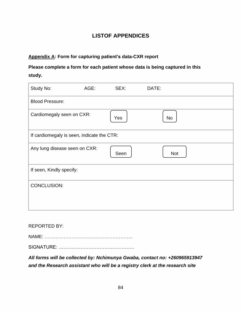
84
LISTOF APPENDICES
Appendix A: Form for capturing patient’s data-CXR report
Please complete a form for each patient whose data is being captured in this
study.
Study No: AGE: SEX: DATE:
Blood Pressure:
Cardiomegaly seen on CXR:
If cardiomegaly is seen, indicate the CTR:
Any lung disease seen on CXR:
If seen, Kindly specify:
CONCLUSION:
REPORTED BY:
NAME: ………………………………………………..
SIGNATURE: …………………………………………
All forms will be collected by: Nchimunya Gwaba, contact no: +260965913947
and the Research assistant who will be a registry clerk at the research site
Yes
s
No
Seen Not
Seen

85
Appendix B: Form for capturing patient’s data- ECHO report
Please complete a form for each patient whose data is being captured in this study.
Study No: AGE: SEX: DATE:
Blood Pressure:
Cardiomegaly seen on CXR:
ECHOCARDIOGRAM REPORT:
AORTIC VALVE:
AORTIC ROOT: RIGHT VENTRICULAR OUTFLOW TRACK:
LEFT ATRIUM:
LEFT VENTRICLE:
FRACTIONAL SHORTENING: EJECTION FRACTION:
MITRAL VALVE:
INTER VENTRICULAR SEPTUM:
POSTERIOR WALL (LV):
PERICARDIAL EFFUSION:
VEGETATION: THROMBUS:
OTHER FINDINGS: DOPPLER:
CONCLUSION:
REPORTED BY:
NAME: ……………………………………………………………………..…….
SIGNATURE: ……………………………………………………………..……
All forms will be collected by: Nchimunya Gwaba, contact no: +260965913947
Yes No

86
Appendix C: Participant Information Leaflet - for Research Assistants of the study.
Date:
Participant Information Leaflet by Nchimunya Gwaba; Cell no: +260 965913947
As Diagnostic Radiographer it is part of our goal to contribute and continuously offer
quality diagnostic services to our clients, as well as continuously strive into research
ventures that will bring new knowledge, enabling us to improve our patient management
and care.
Please take some time to read the information presented here which will explain the
details of this project. Please ask the principal investigator about any part of the project
that you do not fully understand. It is very important that you are fully satisfied as to what
the research entails. Your participation is entirely voluntary and you are free to withdraw
at any stage if you say so, this will not affect you negatively in any way whatsoever.
What is the Research Study all about?
This research study aims to determine the association between cardiomegaly identified
on CXR and ECHO results of the same patient at LMGH in Lusaka, Zambia.
Files of participants who have undergone an echocardiography examination after
cardiomegaly has been identified on a CXR, using the standard imaging procedures for
these examinations, will be analysed-in order to determine the common conditions found
in cardiomegaly patients.
Being a retrospective study, particular attention will be paid to issues of confidentiality by
ensuring that all information that come into the possession of the researcher and his
assistants during the study is not shared with any unauthorized person.
Why have you been invited to participate?
The researcher’s main interests are patients with an enlarged heart on CXR. The study
will be conducted retrospectively and thus patients will not be required to sign this consent
form. However, the research assistant, the head of the radiology department, and
research supervisor will be required to read this form as they will play a role in this
research and will thus be requested to keep patient details confidential. Refer to HWS-
REC 4.1 form.

87
What will your responsibility be?
The study will be conducted retrospectively and thus patients will not be required to sign
this consent form. However, the research assistants will be required to retrieve patient’s
files that have been identified with an enlarged heart on CXR and have undergone ECHO.
The research assistant, together with the research supervisor and head of the radiology
department will be required to keep patient confidentiality by not discussing patient’s
details with those that are not directly related to this study.
Will you benefit from taking part in this research?
Are there any benefits (financial/nonfinancial) to your taking part in this study?
There are no financial benefits, to those whose files will be used for this study, related to
this study. Using the patient’s participant’s file in this study will also not lead patients to
spend any extra money as is related to this research.
This study will help us determine the association between cardiomegaly identified on CXR
and ECHO findings of the same patient at LMGH in Lusaka, Zambia. The research may
thus benefit patients and their families or community in future as the research may
enhance more knowledge on the need for more consorted efforts in combating
cardiomegaly in particular and cardiovascular diseases in general. In addition, this
research may also benefit consulting cardiologist, physicians and clinical educators in the
management of cardiovascular diseases in adults above the age of 18 years with
cardiomegaly that is related to IHD and increased rate of morbidity and mortality.
It should also be brought to your attention that the researcher will not benefit financially
from the study as well, although the researcher will continue drawing his normal salary as
a radiographer.
Are there any risks involved in your taking part in this research?
There will be no known risks attributed to this research study as the research will be
conducted retrospectively.
Who will have access to your medical records?
All the data being collected in this research will be coded, and the patients name will not
be used to ensure confidentiality. The data will be stored in the department safe, and only

88
the principal investigator, research supervisor and the head of the radiology department
will have access to this information.
Thank you.

89
Appendix D: Permission to Carry out research

90
Appendix E: Ethics Certificate


![arXiv:1705.09850v3 [cs.CV] 27 Sep 2017 · the cardiomegaly localization is that the heart and its sur-rounding region is most responsible for cardiomegaly detec-tion, in contrast](https://static.fdocuments.net/doc/165x107/5e34802d595379010310239f/arxiv170509850v3-cscv-27-sep-2017-the-cardiomegaly-localization-is-that-the.jpg)