ECG Interpretation Program - Frank's Hospital Workshop
128
For Bedside Monitors User’s Guide ECG Interpretation Program ECAPS 12C 0614-006465A
Transcript of ECG Interpretation Program - Frank's Hospital Workshop

For Bedside Monitors
User’s Guide
ECG InterpretationProgram
ECAPS 12C
0614-006465A

If you have any comments or suggestions on this manual, please contact us at: www.nihonkohden.com
Copyright NoticeThe entire contents of this manual are copyrighted by Nihon Kohden. All rights are reserved. No part of this document may be reproduced, stored, or transmitted in any form or by any means (electronic, mechanical, photocopied, recorded, or otherwise) without the prior written permission of Nihon Kohden.

1
2
3
4
5
6
User’s Guide ECAPS 12C C.1
6-1
6-2
6-3
6-4
6-5
6-6
A
ContentsSection 1 Introduction ........................................................................... 1.1
Section 2 Precautions (Discrepancies between Physician’s and ECAPS 12C’s Findings) ........................................................ 2.1
Section 3 Outline of ECG Automatic Recording ................................. 3.1Entering Patient Data .......................................................................................................3.3
Recording ECG Data .......................................................................................................3.4
Improving Waveform Quality ............................................................................................3.5
Measuring ECG Waveform ..............................................................................................3.6
General Measurement Values to Be Printed .............................................3.10
Classification and Printing ECG Data ............................................................................3.11
Section 4 Recorded Analysis Results .................................................. 4.1
Section 5 How to Read Analysis Results ............................................. 5.1ECG Findings ..................................................................................................................5.3
Priority of ECG Findings ..................................................................................................5.3
Criteria .............................................................................................................................5.4
Overall Judgement ...........................................................................................................5.4
Analysis Result When ECG Cannot Be Analyzed ...........................................................5.5
Section 6 Criteria of Findings ............................................................ 6.0.1Indication of Analysis Criteria .......................................................................................6.0.2
Section 6-1 Arrhythmias ......................................................................................... 6.1.1Rhythm analysis ............................................................................................................6.1.2
1002 Marked rhythm irregularity, possible non-conducted PAC, SA block, AV block,
or sinus pause ................................................................................................6.1.13
1100 Sinus rhythm .....................................................................................................6.1.3
1102 Sinus arrhythmia .............................................................................................6.1.12
1108 Marked sinus arrhythmia ................................................................................6.1.13
1120 Sinus tachycardia .............................................................................................6.1.3
1130 Sinus bradycardia .............................................................................................6.1.3
1200 Atrial rhythm .....................................................................................................6.1.4
1210 Atrial fibrillation .................................................................................................6.1.7
12101 Atrial fibrillation with rapid ventricular response ...............................................6.1.7
12102 Atrial fibrillation with slow ventricular response ................................................6.1.7
12103 Atrial fibrillation with aberrant conduction, or ventricular premature
complexes .......................................................................................................6.1.18

C.2 User’s Guide ECAPS 12C
CONTENTS
12108 Atrial fibrillation with rapid ventricular response with aberrant conduction, or
ventricular premature complexes ....................................................................6.1.18
12109 Atrial fibrillation with slow ventricular response with aberrant conduction, or
ventricular premature complexes ....................................................................6.1.18
1220 Rapid atrial rhythm ...........................................................................................6.1.4
1250 Atrial flutter .......................................................................................................6.1.8
12503 Atrial flutter with aberrant conduction, or ventricular premature complexes ...6.1.19
12505 Cannot rule out atrial flutter ..............................................................................6.1.8
1300 Junctional rhythm ..............................................................................................6.1.5
1320 Rapid junctional rhythm ....................................................................................6.1.5
1400 Undetermined rhythm (Possible supraventricular rhythm) ................................6.1.6
1420 Undetermined rhythm (Possible supraventricular tachycardia) .........................6.1.6
1430 Undetermined rhythm (Possible supraventricular bradycardia) ........................6.1.6
1470 with occasional supraventricular premature complexes .................................6.1.14
1474 with frequent supraventricular premature complexes .....................................6.1.14
1475 with frequent supraventricular premature complexes in a pattern of
bigeminy .........................................................................................................6.1.14
1570 with occasional ventricular premature complexes ..........................................6.1.15
15708 with occasional ventricular premature complexes (Unreliable analysis due
to noise) ..........................................................................................................6.1.15
1574 with frequent ventricular premature complexes ..............................................6.1.15
15748 with frequent ventricular premature complexes (Unreliable analysis due to
noise) ..............................................................................................................6.1.15
1575 with frequent ventricular premature complexes in a pattern of bigeminy ........6.1.15
15758 with frequent ventricular premature complexes in a pattern of bigeminy
(Unreliable analysis due to noise)...................................................................6.1.15
1577 with couplet ventricular premature complexes ................................................6.1.15
15778 with couplet ventricular premature complexes (Unreliable analysis due to
noise) ..............................................................................................................6.1.15
16006 Electronic atrial pacemaker ..............................................................................6.1.9
16007 Electronic ventricular pacemaker ......................................................................6.1.9
16008 Electronic atrial pacemaker (Unreliable analysis due to noise) ........................6.1.9
16009 Electronic ventricular pacemaker (Unreliable analysis due to noise) ...............6.1.9
1901 Undetermined regular rhythm .........................................................................6.1.10
1902 Undetermined rhythm .....................................................................................6.1.10
1921 Undetermined regular rhythm (tachycardia) ...................................................6.1.10
1922 Undetermined rhythm (tachycardia) ...............................................................6.1.10
1931 Undetermined regular rhythm (bradycardia) ...................................................6.1.10
1932 Undetermined rhythm (bradycardia) ...............................................................6.1.10
1938 Extreme bradycardia ......................................................................................6.1.11
1970 with occasional ectopic premature complexes ...............................................6.1.17
19708 with occasional ectopic premature complexes (Unreliable analysis due to
noise) ..............................................................................................................6.1.17
1974 with frequent ectopic premature complexes ...................................................6.1.17
19748 with frequent ectopic premature complexes (Unreliable analysis due to
noise) ..............................................................................................................6.1.17
1975 with frequent ectopic premature complexes in a pattern of bigeminy .............6.1.17

User’s Guide ECAPS 12C C.3
CONTENTS
1
2
3
4
5
6
6-1
6-2
6-3
6-4
6-5
6-6
A
Section 6-2 Conductive Defect .............................................................................. 6.2.12210 Short PR interval ..............................................................................................6.2.2
2216 Type-A Wolff-Parkinson-White syndrome .........................................................6.2.3
2217 Type-B Wolff-Parkinson-White syndrome .........................................................6.2.3
2218 Atypical Wolff-Parkinson-White syndrome .......................................................6.2.3
2219 Intermittent Wolff-Parkinson-White syndrome ..................................................6.2.3
2231 First degree AV block .......................................................................................6.2.5
2232 2nd degree AV block, Mobitz type I ..................................................................6.2.5
2233 2nd degree AV block, Mobitz type II .................................................................6.2.5
2234 Possible 3rd degree AV block ...........................................................................6.2.5
2320 Nonspecific intraventricular conduction delay ................................................6.2.10
2330 Nonspecific intraventricular conduction block ................................................6.2.10
2420 RSR (QR) in lead V1/V2, consistent with right ventricular conduction delay ...6.2.6
2440 Incomplete right bundle branch block...............................................................6.2.6
2450 Right bundle branch block ................................................................................6.2.6
24501 Right bundle branch block, plus possible RVH .................................................6.2.6
2540 Incomplete left bundle branch block .................................................................6.2.8
2550 Left bundle branch block ..................................................................................6.2.8
2630 Left anterior fascicular block.............................................................................6.2.9
2730 Left posterior fascicular block ...........................................................................6.2.9
Section 6-3 Myocardial Infarction .......................................................................... 6.3.1Analysis Criteria ..................................................................................................6.3.2
Anterior Myocardial Infarction .............................................................................6.3.5
Septal Myocardial Infarction ...............................................................................6.3.8
Lateral Myocardial Infarction .............................................................................6.3.10
Inferior Myocardial Infarction.............................................................................6.3.12
Children ............................................................................................................6.3.14
Section 6-4 ST-T Abnormality ................................................................................ 6.4.1ST Depression ....................................................................................................6.4.2
Injury ...................................................................................................................6.4.4
Subendocardial Ischemia ...................................................................................6.4.8
Early Repolarization .........................................................................................6.4.11
Pericarditis ........................................................................................................6.4.12
T Wave Abnormality ..........................................................................................6.4.13
Nonspecific ST Elevation ..................................................................................6.4.15
Section 6-5 Ventricular Hypertrophy ..................................................................... 6.5.1Point Score System ......................................................................................................6.5.2
Analysis Criteria for RVH ..............................................................................................6.5.2
5120 Possible right ventricular hypertrophy .......................................................6.5.4
5130 Right ventricular hypertrophy ....................................................................6.5.4
5134 Right ventricular hypertrophy, probably repolarization abnormality .........6.5.5
Analysis Criteria for LVH ...............................................................................................6.5.6
5211 Minimal voltage criteria for LVH, may be normal variant ..........................6.5.7
5220 Possible left ventricular hypertrophy .........................................................6.5.7
5222 Moderate voltage criteria for LVH, may be normal variant ........................6.5.7
5233 Voltage criteria for LVH .............................................................................6.5.7

C.4 User’s Guide ECAPS 12C
CONTENTS
5234 Left ventricular hypertrophy with repolarization abnormality ....................6.5.7
Section 6-6 Atrial Enlargement, Abnormal Axis Deviation and Others.............. 6.6.16120 Possible right atrial enlargement ......................................................................6.6.2
6130 Right atrial enlargement ...................................................................................6.6.2
6220 Possible left atrial enlargement .........................................................................6.6.3
6230 Left atrial enlargement ......................................................................................6.6.3
7100 Abnormal right axis deviation ...........................................................................6.6.4
7102 Moderate right axis deviation ............................................................................6.6.4
7200 Abnormal left axis deviation ..............................................................................6.6.4
7202 Moderate left axis deviation ..............................................................................6.6.4
7300 Indeterminate axis ............................................................................................6.6.4
7400 S1-S2-S3 pattern, consistent with pulmonary disease, RVH, or normal
variant ...............................................................................................................6.6.6
7500 Abnormal QRS-T angle ....................................................................................6.6.7
8003 Consistent with pulmonary disease ..................................................................6.6.8
8100 Low QRS voltage ..............................................................................................6.6.9
8101 Low QRS voltage in limb leads .........................................................................6.6.9
8102 Low QRS voltage in chest leads .......................................................................6.6.9
8200 Dextrocardia ...................................................................................................6.6.10
8304 Long QTc interval ...........................................................................................6.6.11
8305 Short QTc interval ..........................................................................................6.6.11
0101 Possible arm leads reversed, check lead requested ......................................6.6.12
0102 ARTIFACT PRESENT .....................................................................................6.6.13
0103 CANNOT ANALYZE ECG ...............................................................................6.6.13
0104 ELECTRODE(S) FAILURE...Repeat ECG is required. ...................................6.6.13
0201 ...Analysis based on intrinsic rhythm ..............................................................6.6.13
Appendix Modified Minnesota Code ................................................... A.1General ............................................................................................................................A.2
Code List ..........................................................................................................................A.3
Priority of Code Printing ...................................................................................................A.7
Detailed Criteria ...............................................................................................................A.8

User’s Guide ECAPS 12C 1.1
1Section 1 Introduction

1.2 User’s Guide ECAPS 12C
1. INTRODUCTION
ECAPS 12C is the ECG analysis program for the Nihon Kohden’s instruments, such as electrocardiographs. A computer analysis program is merely a collection of ECG evaluation criteria created by physicians. It is not possible for a computer program to correctly judge every unique ECG, so sometimes it makes wrong interpretations where a physician could very easily read and interpret the waveforms. The final decision can only be made by the qualified physicians. Use this system only as a diagnostic aid, based on proper understanding of its features and limitations.
This manual describes the criteria of the analysis results of the ECAPS 12C output data.
For the detailed operation procedure of the system, refer to the operator’s manual of the instrument.
NOTE
The contents of this manual are subject to change without prior notice for
improvement of analysis precision.

1
User’s Guide ECAPS 12C 1.3
1. INTRODUCTION
ADVISORYThe ECAPS 12C analysis program is applicable to ages 3 and older.
Ages below 3 years are treated according to the criteria for the age of
3 years.
Final determination of overall interpretation judgement, diagnosis and
treatment must be made by a qualified physican.
Patient Age and the Age Used for Analysis
• When classifying as adult or child:
Age Age which is used for analysisChild (3-15) 12Adult (>=16) 35
• When classifying in age range:
Age Age which is used for analysis<=5 36-9 7
10-14 1215-34 25>=34 35
• When patient’s actual age is entered: The patient’s actual age is used for analysis.

User’s Guide ECAPS 12C 2.1
2Section 2 Precautions (Discrepancies
between Physician’s and ECAPS 12C’s Findings)

2.2 User’s Guide ECAPS 12C
2. PRECAUTIONS (DISCREPANCIES BETWEEN PHYSICIAN’S AND ECAPS 12C’S FINDINGS)
The causes for discrepancy between computer analysis and physician’s findings and the countermeasures to be taken for these causes are given below.
Causes CountermeasuresDifference in judgement criteria between physician and computer program, or lack of applicable findings by computer program.
Refer to Section 6 “Criteria of Findings” which list all the judgement criteria used by ECAPS 12C.
The ECG waveform is on the borderline of the judgement criteria.
Compare the measured data with the data in Section 6 “Criteria of Findings”.This may be the limit of computer analysis.Patient data other than ECG should be taken into consideration.
Artifact (EMG, AC interference, baseline wandering, etc.) not recognized, leading to wrong interpretation.
Try to record ECG with as little artifact as possible.
Arrhythmia, etc. which are intrinsically difficult for the computer to analyze.
This is a limit for computer analysis. The advice of a physician should be obtained.

2
User’s Guide ECAPS 12C 2.3
2. PRECAUTIONS (DISCREPANCIES BETWEEN PHYSICIAN’S AND ECAPS 12C’S FINDINGS)
Although various means of eliminating errors and discrepancies between computer analysis and physician’s findings are employed in the ECAPS 12C, not all ECG diagnostic cases are incorporated, therefore, consider the following points when using the computer’s analysis results.
(1) ECAPS 12C is not programmed to compensate for the influence of medicine.Check the dosing record when reading the ECG. However, the influence of digitalis is noted on the recording paper according to the presence of atrial fibrillation.
(2) ECAPS 12C is not programmed to compensate for fluid balance such as abnormal electrolytes. When interpreting ECG, read the QTc interval, T waveform and U waveform, along with other relevant test results.
(3) ECAPS 12C does not further classify premature complex into trigeminy, short-run, etc.
(4) ECAPS 12C is not programmed to identify escaped beats and pararrhythmia.These may be interpreted as “undetermined rhythms”.
(5) ECAPS 12C is not programmed to identify LGL syndrome. Judge this syndrome from “short PR interval”.
(6) ECAPS 12C does not identify wandering pacemakers.

User’s Guide ECAPS 12C 3.1
3
Section 3 Outline of ECG Automatic Recording
Entering Patient Data ..........................................................................................................................................3.3
Recording ECG Data ...........................................................................................................................................3.4
Improving Waveform Quality ................................................................................................................................3.5
Measuring ECG Waveform ..................................................................................................................................3.6
General Measurement Values to Be Printed ...................................................................................3.9
Classification and Printing ECG Data ................................................................................................................3.10

3.2 User’s Guide ECAPS 12C
3. OUTLINE OF ECG AUTOMATIC RECORDING
ECG Measurement Flowchart
START
Enter patient information. Refer to p. 3.3.
Record ECG. Refer to p. 3.4.
Remove AC interference and base line
wandering from ECG. Refer to p. 3.4.
Measure P, QRS and T waveforms. Refer to p. 3.6.
Analyze ECG. Refer to p. 3.7.
Print out analysis results (finding). Refer to p. 3.10.
END

3
User’s Guide ECAPS 12C 3.3
3. OUTLINE OF ECG AUTOMATIC RECORDING
Entering Patient Data
Enter the following data before acquiring ECG data. With some instruments, some of these items cannot be entered. Refer to the operator’s manual of the instrument for details of entering the patient data.
• Patient identification number (ID)• Name• Gender• Birth Date• Age• Height• Weight• Blood pressure• Medication• Date*• Hour* * Date and hour are automatically set.
NOTE
• Amongtheseitems,onlyageandgenderaffecttheanalysis.Other
items have no effect on the analysis.
• Ifnoageisinput,thefactorydefaultsettingof35isused.Ifnogender
is specified, the factory default setting of male is used.
• Foraccurateanalysisresults,inputgenderandage.

3.4 User’s Guide ECAPS 12C
3. OUTLINE OF ECG AUTOMATIC RECORDING
Recording ECG Data
The ECGs of all 12 standard leads are acquired simultaneously for 10 seconds at an accuracy of 1.25 µV/bit and 500 samples/s.
Refer to the operator’s manual of the instrument for details of recording ECG.

3
User’s Guide ECAPS 12C 3.5
3. OUTLINE OF ECG AUTOMATIC RECORDING
Improving Waveform Quality
During ECG data acquisition, the quality of the ECG waveforms is improved with digital filters and the adverse influence of baseline wandering due to electrode potential drifting and AC interference is minimized.
• AC interference A digital filter is used in the system to reduce the AC frequency components.
A digital filter eliminates adverse influence on the ECG waveform more than a conventional analog filter.
Conventional analog filter Digital filter
• Baseline wandering
Shortening the time constant distorts the ST segments which reduces diagnostic accuracy. A digital filter is used to remove the components which cause baseline wandering.
• High frequency noise High cut filters of different frequencies are incorporated in the instruments.
The high cut filter cuts the high frequency components of ECG but reduces the effect of EMG. The high cut filter attenuates the QRS amplitude although the influence on judgement of LVH (left ventricular hypertrophy) is reduced as much as possible. However, the ECAPS 12C program always analyzes the ECG waveform acquired by 150 Hz filter. Therefore, some differences may occur between the recorded waveform and the analysis result.

3.6 User’s Guide ECAPS 12C
3. OUTLINE OF ECG AUTOMATIC RECORDING
Measuring ECG Waveform
ECG waveforms are measured as shown below.
QRSs are classified by patterns and only the typical waveforms are extracted and averaged for measurement.
The P waves for each heartbeat are searched and classified by pattern.
The most characteristic P wave pattern among the classified patterns is taken as the typical P wave.
P wave, QRS wave, T wave and ST segment are measured.For the detailed measurement method, refer to the next page.
Rhythm is analyzed based on QRS wave and P wave waveform measurement data.
Waveforms are measured as shown on the next page. The reference point in measuring waveforms is the starting point of the QRS wave.

3
User’s Guide ECAPS 12C 3.7
3. OUTLINE OF ECG AUTOMATIC RECORDING
(a) Waveform measurement parameters
PR interval QRS duration
V.A.T.
R amp.
P duration
P amp.
P' amp.
Q amp.
S amp.
T amp.
Q duration S durationR duration
QT interval
STM STE STJ
1/16 R-R interval
QTc interval = QT interval +7
(1000-R-R interval)
QTc interval: QT interval changes with heart rate. QTc interval is the converted value when the heart rate is 60/min.
QRS duration
P amp. P' amp.
T amp.
S duration
R duration
S amp.
R amp.
R max amp.
S max amp.
R' amp.
T' amp.S' amp.
S' duration
R' duration
Total QRS amplitude = R max. amp. + S max. amp.Net QRS amplitude = R max. amp. − S max. amp.

3.8 User’s Guide ECAPS 12C
3. OUTLINE OF ECG AUTOMATIC RECORDING
(b) QRS area
(c) Upward oriented T
T is upward oriented: C > 0.05 mV + (1.5 × STJ)
(d) Modified T amplitude (T amp. (mod)) To simplify the treatment of biphasic T wave, and to explain the T amplitude
when ST and T differ from the QRS starting point, T amplitude is modified as below.
When T’ is present: T amp. (mod) = (T amp. or T’ amp., whichever is smaller) − (STE or T end,
whichever is larger) When T’ is not present: T amp. (mod) = T amp. − (STE or T end, whichever is larger)

3
User’s Guide ECAPS 12C 3.9
3. OUTLINE OF ECG AUTOMATIC RECORDING
General Measurement Values to Be Printed
Printout MeaningHR (Vent. rate) -- bpm Heart ratePR int. -- ms PR intervalQRS dur. -- ms QRS durationQT int. -- ms QT intervalQTc int. -- ms QTc interval (See p. 3-7(a))P axis -- ° P axis deviationQRS axis -- ° QRS axis deviationT axis -- ° T axis deviationRV5 amp -- mV R amplitude in V5SV1 amp -- mV S amplitude in V1
When a measurement value cannot be measured, “***” is displayed.
Patient data
General measurement values
Rhythm lead

3.10 User’s Guide ECAPS 12C
3. OUTLINE OF ECG AUTOMATIC RECORDING
Classification and Printing ECG Data
The ECGs are classified into the findings. For the details of the classification method, refer to Section 6 “Criteria of Findings”.
The findings are printed out as “Analysis Result”. Refer to the next section.

User’s Guide ECAPS 12C 4.1
4
Section 4 Recorded Analysis Results

4.2 User’s Guide ECAPS 12C
4. RECORDED ANALYSIS RESULTS
The analysis results of the system are printed out in the format selected by the operator. The outline of the formats and the operation are described in the operator’s manual of the instrument. The explanation of the common features of the printout with typical examples are given below. These are factory default settings.
Not all of the following items are printed with some instruments. For details, refer to the operator’s manual of the instrument.
(1) Patient’s data and recording conditions Data and Time Name ID No. Gender Birth date and Age (in some formats, birth date is omitted): years Vent. rate (Heart Rate) Paper speed Lead name
NOTE
, , and are automatically entered. Items to are printed
as entered by the operator.
(2) ECG waveform printout (In some formats, these are omitted.) Dominant ECG waveform (averaged) Rhythm lead Calibration wave
(3) Analysis result Overall judgement ECG findings name [criteria] General measurement values Physician’s signature (Reviewed)
NOTE
• Thefindings[criteria]areprintedasasupplementarywhichisoneof
the judgement criteria. For details, refer to Section 5 “How to Read
Analysis Results”.
• Insomefindings,[criteria]arenotprinted.

4
User’s Guide ECAPS 12C 4.3
4. RECORDED ANALYSIS RESULTS

User’s Guide ECAPS 12C 5.1
5
Section 5 How to Read Analysis Results
ECG Findings ......................................................................................................................................................5.3
Priority of ECG Findings ......................................................................................................................................5.3
Criteria .................................................................................................................................................................5.4
Overall Judgement ...............................................................................................................................................5.4
Analysis Result When ECG Cannot Be Analyzed ...............................................................................................5.5

5.2 User’s Guide ECAPS 12C
5. HOW TO READ ANALYSIS RESULTS
The analysis result printout, as shown below, contains the following data.
1. ECG findings [Criteria]2. Overall judgement3. General measurement values
General measurement values
CodeFindings
Overall judgement code Overall judgement

5
User’s Guide ECAPS 12C 5.3
5. HOW TO READ ANALYSIS RESULTS
ECG Findings
The ECAPS 12C classifies the ECGs into about 200 findings by comparing the features of the ECGs with the analysis criteria specified for each finding. The analysis criteria are all taken from the judgement criteria used by the physicians and arranged for computer processing. For details, refer to Section 6 “Criteria of Findings”.
For the analysis process, refer to Section 3 “Outline of ECG Automatic Recording”.
When the obtained ECG exactly conforms to the analysis criteria, the finding name is classified as “determined”. However, there are findings which cannot be determined definitely and findings which are on the borderline between conformance and nonconformance, liable to be influenced by slight noises. Those findings which are close to “determined” but not definite are classified as “possible”, and those findings which may or might not conform but cannot be completely ruled out are classified as “Cannot rule out”.Some arrhythmia ECGs are difficult to classify into finding names; these are classified as “undetermined rhythm”.
NOTE
When the ECG is classified as “undetermined rhythm” (1901, 1902, 1921,
1922, 1931, 1932), there is no further rhythm analysis.
Priority of ECG Findings
When the obtained ECG conforms to two or more findings criteria, only the most important finding name is printed. For example, if “ischemia” is found to apply, less important findings such as “nonspecific ST & T wave abnormality” or “ST elevation” are not printed.
Further details of analysis are given as NOTE under the respective findings in Section 6 “Criteria of Findings”.

5.4 User’s Guide ECAPS 12C
5. HOW TO READ ANALYSIS RESULTS
Criteria
The analysis criteria can be printed out on the recording paper after the findings, as shown on p. 5.2. They should serve as an aid in observing the ECG.
For example, when the instrument judges the ECG as abnormal junctional ST depression because there is junctional ST depression of more than 0.1 mV in V5 and V6, the criteria is printed as “4023 Abnormal junctional ST depression [0.1 + mV junctional ST depression (V5, V6)].
For reading convenience, the analysis findings and criteria are printed in simplified form.
For further details of the analysis criteria, refer to Section 6 “Criteria of Findings”.
Overall Judgement
The findings are classified into one of five overall judgements. The list of all findings for each overall judgement is shown in Section 6 “Criteria of Findings”.
Overall Judgement Code12345
abnormal ECGabnormal rhythm ECGborderline ECGnormal ECGatypical ECG
91509140913091109120
Where two or more findings are output, only the highest priority finding is printed.
Although only the highest priority finding is selected for judgement, the findings requiring immediate treatment, such as “Myocardial infarction, possible acute” are printed as “abnormal ECG” instead of “borderline ECG”. This is to raise an alarm in case of emergency although there is actually little possibility of danger. When an unusual ECG pattern is recognized as neither “normal ECG” nor “abnormal ECG”, it is judged as “atypical ECG”. For example, “Low QRS voltage”, “undetermined axis”, and “undetermined rhythm” are judged as “atypical ECG”.

5
User’s Guide ECAPS 12C 5.5
5. HOW TO READ ANALYSIS RESULTS
Analysis Result When ECG Cannot Be Analyzed
When a measurement value cannot be measured, “***” is displayed. When no measurement values can be measured, the ECG cannot be analyzed and a “0103 CANNOT ANALYZE ECG” message is displayed. For recording example, refer to “General Measurement Values to Be Printed” in Section 3.

User’s Guide ECAPS 12C 6.0.1
6
Section 6 Criteria of Findings
Section 6-1 Arrhythmias .................................................................................................................................6.1.1
Section 6-2 Conductive Defect .......................................................................................................................6.2.1
Section 6-3 Myocardial Infarction ...................................................................................................................6.3.1
Section 6-4 ST-T Abnormality .........................................................................................................................6.4.1
Section 6-5 Ventricular Hypertrophy ...............................................................................................................6.5.1
Section 6-6 Atrial Enlargement, Abnormal Axis Deviation and Others ...........................................................6.6.1

6.0.2 User’s Guide ECAPS 12C
6. CRITERIA OF FINDINGS
Indication of Analysis Criteria
1. How to read analysis criteria The analysis criteria are marked with (1), (2) and , .
(1) ...indicates AND ...indicates OR
[ Example 1 ](1) condition A(2) condition B(3) condition C condition D
[ Example 2 ](1) condition A(2) • condition B • condition C
2. Age reference tables The following tables show the values used for certain analysis parameters
which vary according to age. The applicable items are marked with “*” in the criteria column.
NOTE
• Whennoageisinputinthepatientinformation,thefactorydefault
setting of age 35 is used.
• Whentheageis2oryounger,thecomputer’sresultsmaynotbe
accurate.
[ PR interval ] (%)
Age PR ratio<=5<=9<=11<=14<=18>18
7683858891
100
Conditions A and B are satisfied, and either condition C or condition D are satisfied.
Either condition A is satisfied, or both conditions B and C are satisfied.
(Example)
PR interval = 170 msAge = 6 years oldPR criteria = PR int × PR ratio/100 = 170 × 83/100 = 141 ms

6
User’s Guide ECAPS 12C 6.0.3
6. CRITERIA OF FINDINGS
[ QRS duration ] (%)
Age QRS duration<=5<=9<=11<=14<=18>18
8285859295
100
[ S duration: Lateral lead (I, aVL, V4, V5, V6) ] (s)
Age S duration S duration0.040 (s) 0.060 (s)
<=5<=9<=11<=14<=18>18
0.0310.0340.0370.0390.0390.039
0.0470.0510.0560.0590.0590.059
shows the parameters used in criteria for each finding.
[ R duration (V1, V2) ] (s)
Age R duration R duration0.020 (s) 0.030 (s)
<=5<=9<=11<=14<=18>18
0.0160.0170.0190.0200.0200.020
0.0240.0260.0280.0300.0300.030
shows the parameters used in criteria for each finding.
(Example)
QRS duration: 0.12 sAge = 10 years oldQRS criteria = QRS dur. × QRS dur (%)/100 = 0.12 × 85/100 = 0.102 s

6.0.4 User’s Guide ECAPS 12C
6. CRITERIA OF FINDINGS
[ R duration (I, aVL, V4, V5, V6) ] (s)
Age R duration R duration R duration0.060 (s) 0.100 (s) 0.250 (s)
<=5<=9<=11<=14<=18>18
0.0490.0510.0540.0570.0590.059
0.0820.0860.0900.0950.0990.099
0.2070.2180.2280.2400.2500.250
shows the parameters used in criteria for each finding.
[ Electrical axis (left axis) ] (degree)
AgeLAXD1 LAXD2
Male Female Male Female<=5<=9<=11<=14<=18>18
15°9°4°5°
−14°−20°
19°9°
23°20°13°
−20°
5°−1°−6°−5°
−24°−30°
9°−1°13°10°3°
−30°
[ Electrical axis (right axis) ] (degree)
AgeRAXD1 RAXD2
Male Female Male Female<=5<=9<=11<=14<=18>18
97°97°92°97°99°90°
101°97°
100°97°
100°90°
107°107°102°107°109°100°
111°107°110°107°110°100°
6
User’s Guide ECAPS 12C 6.0.5
6. CRITERIA OF FINDINGS
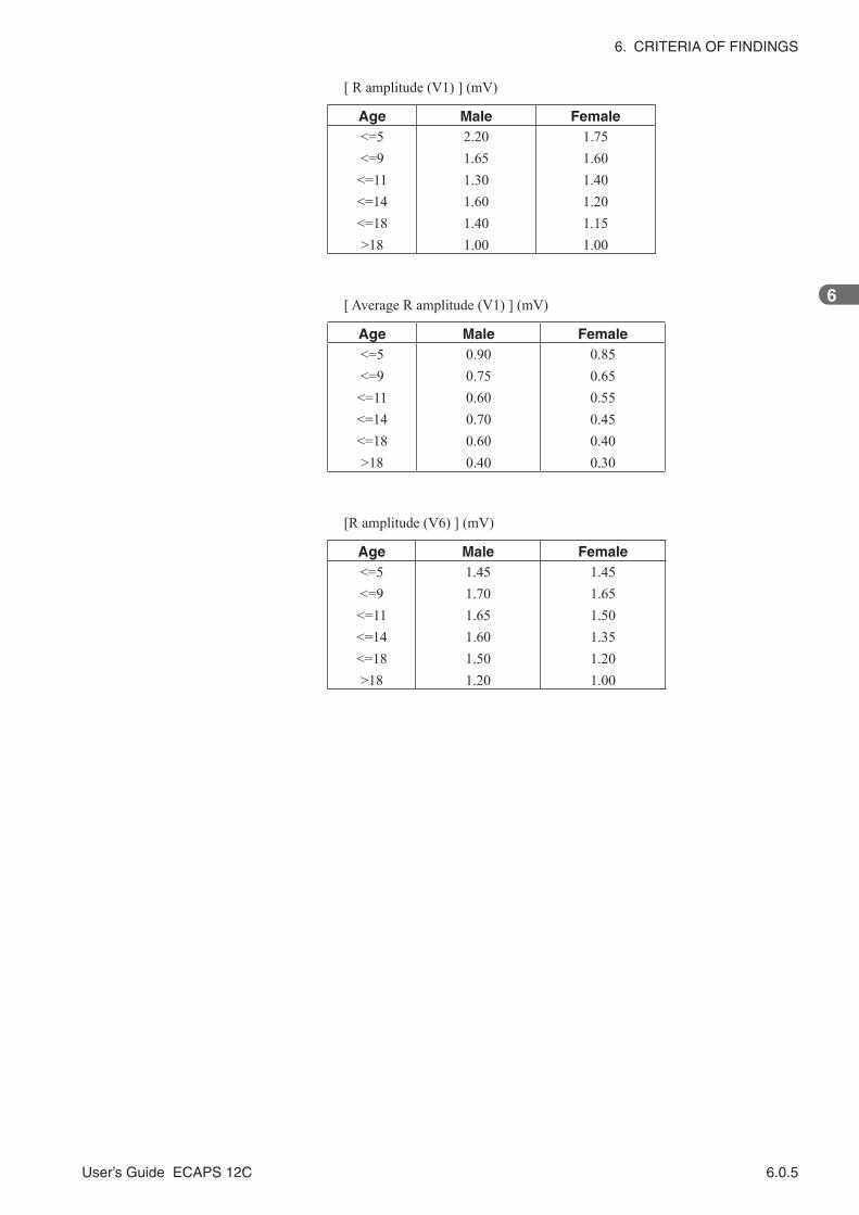
[ R amplitude (V1) ] (mV)
Age Male Female<=5<=9<=11<=14<=18>18
2.201.651.301.601.401.00
1.751.601.401.201.151.00
[ Average R amplitude (V1) ] (mV)
Age Male Female<=5<=9<=11<=14<=18>18
0.900.750.600.700.600.40
0.850.650.550.450.400.30
[R amplitude (V6) ] (mV)
Age Male Female<=5<=9<=11<=14<=18>18
1.451.701.651.601.501.20
1.451.651.501.351.201.00

6.0.6 User’s Guide ECAPS 12C
6. CRITERIA OF FINDINGS
[ S depth (V1) ] (mV)
Age Male Female<=5<=9<=11<=14<=18>18
1.201.201.351.501.651.20
1.251.251.301.151.150.95
[ S depth (V6) ] (mV)
Age Male Female<=5<=9<=11<=14<=18>18
0.540.650.610.470.500.40
0.540.600.610.350.360.30
[R/S (V1) ratio]
Age Male Female<=5<=9<=11<=14<=18>18
2.71.82.32.21.31.0
2.01.81.31.61.71.0

6
User’s Guide ECAPS 12C 6.0.7
6. CRITERIA OF FINDINGS
[ VAT (V1) ] (s)
Age Male Female<=5<=9<=11<=14<=18>18
0.0370.0330.0450.0410.0390.050
0.0390.0290.0280.0320.0340.050
[ Heart Rate ] (BPM) - For arrhythmia analysis -
Age Bradycardia base Tachycardia base<=5<=8<=12<=16>16
6560555050
140135130120100
BPM : Beats per minute

User’s Guide ECAPS 12C 6.1.1
6
6-1
Rhythm analysis ...............................................................................................................................................6.1.2
1002 Marked rhythm irregularity, possible non-conducted PAC, SA block, AV block, or sinus pause .........6.1.13
1100 Sinus rhythm ........................................................................................................................................6.1.3
1102 Sinus arrhythmia ................................................................................................................................6.1.12
1108 Marked sinus arrhythmia ....................................................................................................................6.1.13
1120 Sinus tachycardia .................................................................................................................................6.1.3
1130 Sinus bradycardia .................................................................................................................................6.1.3
1200 Atrial rhythm .........................................................................................................................................6.1.4
1210 Atrial fibrillation .....................................................................................................................................6.1.7
12101 Atrial fibrillation with rapid ventricular response ...................................................................................6.1.7
12102 Atrial fibrillation with slow ventricular response ....................................................................................6.1.7
12103 Atrial fibrillation with aberrant conduction, or ventricular premature complexes .................................6.1.18
12108 Atrial fibrillation with rapid ventricular response with aberrant conduction, or ventricular premature
complexes ..........................................................................................................................................6.1.18
12109 Atrial fibrillation with slow ventricular response with aberrant conduction, or ventricular premature
complexes ..........................................................................................................................................6.1.18
1220 Rapid atrial rhythm ...............................................................................................................................6.1.4
1250 Atrial flutter ...........................................................................................................................................6.1.8
12503 Atrial flutter with aberrant conduction, or ventricular premature complexes .......................................6.1.19
12505 Cannot rule out atrial flutter ..................................................................................................................6.1.8
1300 Junctional rhythm .................................................................................................................................6.1.5
1320 Rapid junctional rhythm ........................................................................................................................6.1.5
1400 Undetermined rhythm (Possible supraventricular rhythm) ....................................................................6.1.6
1420 Undetermined rhythm (Possible supraventricular tachycardia) ............................................................6.1.6
1430 Undetermined rhythm (Possible supraventricular bradycardia) ............................................................6.1.6
1470 with occasional supraventricular premature complexes .....................................................................6.1.14
1474 with frequent supraventricular premature complexes .........................................................................6.1.14
1475 with frequent supraventricular premature complexes in a pattern of bigeminy ..................................6.1.14
1570 with occasional ventricular premature complexes ..............................................................................6.1.15
15708 with occasional ventricular premature complexes (Unreliable analysis due to noise) ........................6.1.15
1574 with frequent ventricular premature complexes ..................................................................................6.1.15
15748 with frequent ventricular premature complexes (Unreliable analysis due to noise) ............................6.1.15
1575 with frequent ventricular premature complexes in a pattern of bigeminy ...........................................6.1.15
15758 with frequent ventricular premature complexes in a pattern of bigeminy (Unreliable analysis due
to noise)..............................................................................................................................................6.1.15
1577 with couplet ventricular premature complexes ...................................................................................6.1.15
15778 with couplet ventricular premature complexes (Unreliable analysis due to noise) .............................6.1.15
16006 Electronic atrial pacemaker ..................................................................................................................6.1.9
16007 Electronic ventricular pacemaker .........................................................................................................6.1.9
16008 Electronic atrial pacemaker (Unreliable analysis due to noise) ............................................................6.1.9
16009 Electronic ventricular pacemaker (Unreliable analysis due to noise) ...................................................6.1.9
1901 Undetermined regular rhythm .............................................................................................................6.1.10
1902 Undetermined rhythm .........................................................................................................................6.1.10
Section 6-1 Arrhythmias

6.1.2 User’s Guide ECAPS 12C
6. CRITERIA OF FINDINGS
1921 Undetermined regular rhythm (tachycardia) .......................................................................................6.1.10
1922 Undetermined rhythm (tachycardia) ...................................................................................................6.1.10
1931 Undetermined regular rhythm (bradycardia) ......................................................................................6.1.10
1932 Undetermined rhythm (bradycardia) ...................................................................................................6.1.10
1938 Extreme bradycardia ..........................................................................................................................6.1.11
1970 with occasional ectopic premature complexes ...................................................................................6.1.17
19708 with occasional ectopic premature complexes (Unreliable analysis due to noise) .............................6.1.17
1974 with frequent ectopic premature complexes .......................................................................................6.1.17
19748 with frequent ectopic premature complexes (Unreliable analysis due to noise) .................................6.1.17
1975 with frequent ectopic premature complexes in a pattern of bigeminy.................................................6.1.17
Rhythm analysisArrhythmia analysis is divided into three major categories; “basic rhythm analysis”, “basic rhythm fluctuation analysis”, and “premature complex analysis”. In the basic rhythm analysis, the presence of P wave and P wave axis are important factors. The connection between P waves and QRS waves is an important factor in classifying the waveform into basic rhythm, basic rhythm fluctuation and premature complex.

6
User’s Guide ECAPS 12C 6.1.3
6-1. ARRHYTHMIAS
6-1
1. Basic rhythm analysis(1) With P wave • Sinus
Code Findings [Criteria] Judgement1100 Sinus rhythm normal ECG1120 Sinus tachycardia abnormal rhythm ECG1130 Sinus bradycardia abnormal ECG
Analysis criteria
Findings Criteria
Sinus rhythm
(1) Electronic atrial pacemaker is not used.(2) P waveform; constant, PR interval; regular(3) −30° <= P axis < 120°(4) 50 <= heart rate < 100*
Sinus tachycardia
(1) Electronic atrial pacemaker is not used.(2) P waveform; constant, PR interval; regular(3) −30° <= P axis < 120°(4) 100 <= heart rate*
Sinus bradycardia
(1) Electronic atrial pacemaker is not used.(2) P waveform; constant, PR interval; regular(3) −30° <= P axis < 120°(4) 50 > heart rate*
NOTE
The values marked with “*” vary with age. For details, refer to p.6.0.7.
50/minute 100/minuteBradycardia Sinus rhythm Tachycardia

6.1.4 User’s Guide ECAPS 12C
6. CRITERIA OF FINDINGS
• Atrial
Code Findings [Criteria] Judgement1200 Atrial rhythm abnormal rhythm ECG1220 Rapid atrial rhythm abnormal rhythm ECG
Analysis criteria
Findings Criteria
Atrial rhythm
(1) Electronic atrial pacemaker is not used.(2) P waveform; constant, PR interval; regular(3) 120° <= P axis <= 240° −30° > P axis >= −60° (1) PR interval > 0.14s (2) 120° <= P axis <= 270° −30° > P axis >= −90°(4) Heart rate <= 70
Rapid atrial rhythm
(1) Electronic atrial pacemaker is not used.(2) P waveform; constant, PR interval; regular(3) 120° <= P axis <= 240° −30° > P axis >= −60° (1) PR interval > 0.14 s (2) 120° <= P axis <= 270° −30° > P axis >= −90°(4) 70 < heart rate
70/minuteAtrial rhythm Rapid

6
User’s Guide ECAPS 12C 6.1.5
6-1. ARRHYTHMIAS
6-1
• AV junction
Code Findings [Criteria] Judgement1300 Junctional rhythm abnormal rhythm ECG1320 Rapid junctional rhythm abnormal rhythm ECG
Analysis criteria
Findings Criteria
Junctional rhythm
(1) Electronic atrial pacemaker is not used.(2) P waveform; constant, PR interval; regular PR <= 0.14 s(3) −60° > P axis > −90° 240° < P axis <= 270°(4) Heart rate <= 70 (age > 3 years) Heart rate <= 80 (age <= 3 years)
Rapid junctional rhythm
(1) Electronic atrial pacemaker is not used.(2) P waveform; constant, PR interval; regular PR <= 0.14 s(3) −60° > P axis > −90° 240° < P axis <= 270°(4) 70 < heart rate (age > 3 years) 80 < heart rate (age <= 3 years)
70/minuteJunctional rhythm Rapid

6.1.6 User’s Guide ECAPS 12C
6. CRITERIA OF FINDINGS
(2) Without P wave• Without atrial fibrillation or without atrial flutter
Code Findings [Criteria] Judgement
1400Undetermined rhythm (Possible supraventricular rhythm)
abnormal rhythm ECG
1420Undetermined rhythm (Possible supraventricular tachycardia)
abnormal rhythm ECG
1430Undetermined rhythm (Possible supraventricular bradycardia)
abnormal rhythm ECG
Analysis criteria
When the P wave is not joined to the QRS, the following criteria are used for classification.
Findings Criteria
Undetermined rhythm (Possible supraventricular rhythm)
(1) Electronic atrial pacemaker is not used.(2) Atrial fibrillation and atrial flutter are not present.(3) QRS duration < 120 ms*(4) 50 <= heart rate < 100*
Undetermined rhythm (Possible supraventricular tachycardia)
(1) Electronic atrial pacemaker is not used.(2) Atrial fibrillation and atrial flutter are not present.(3) QRS duration < 120 ms*(4) heart rate >= 100*
Undetermined rhythm (Possible supraventricular bradycardia)
(1) Electronic atrial pacemaker is not used.(2) Atrial fibrillation and atrial flutter are not present.(3) QRS duration < 120 ms*(4) heart rate < 50*
NOTE
• Thevaluesmarkedwith“*”varywithage.Fordetails,refertop.6.0.3
and 6.0.7.
• Whenthesefindingsarerecognized,otherlessimportantfindingsare
not printed on the recording paper.
50/minute 100/minuteBradycardia Supraventricular rhythm Tachycardia

6
User’s Guide ECAPS 12C 6.1.7
6-1. ARRHYTHMIAS
6-1
• With atrial fibrillation or with atrial flutter
Code Findings [Criteria] Judgement1210 Atrial fibrillation abnormal rhythm ECG12101 Atrial fibrillation with rapid ventricular response abnormal rhythm ECG12102 Atrial fibrillation with slow ventricular response abnormal rhythm ECG
Analysis criteria
Findings Criteria
Atrial fibrillation
(1) (1) P wave is not present (2) RR interval deviation > 0.125 × mean RR interval (3) Random RR interval Fibrillation wave (f wave) is detected.(2) 50 <= heart rate < 100*
Atrial fibrillation with rapid ventricular response
(1) (1) P wave is not present (2) RR interval deviation > 0.125 × mean RR interval (3) Random RR interval Fibrillation wave (f wave) is detected(2) 100 <= heart rate*
Atrial fibrillation with slow ventricular response
(1) (1) P wave is not present (2) RR interval deviation > 0.125 × mean RR interval (3) Random RR interval Fibrillation wave (f wave) is detected(2) 50 > heart rate*
NOTE
The values marked with “*” vary with age. For details, refer to p.6.0.7.
50/minute 100/minuteBradycardia Atrial fibrillation Tachycardia

6.1.8 User’s Guide ECAPS 12C
6. CRITERIA OF FINDINGS
Code Findings [Criteria] Judgement1250 Atrial flutter abnormal rhythm ECG12505 Cannot rule out atrial flutter abnormal rhythm ECG
Analysis criteria
Findings CriteriaAtrial flutter (1) Flutter wave (including fibrillation or flutter wave) is present.
Atrial flutter with aberrant conduction, or venticular premature complexes
(1) Atrial flutter is present.(2) Ectopic QRS is not pacemaker waveform.(3) Ectopic QRS with duration > 0.12 s*
NOTE
The values marked with “*” vary with age. For details refer to p. 6.0.3.

6
User’s Guide ECAPS 12C 6.1.9
6-1. ARRHYTHMIAS
6-1
(3) With pacemaker
Code Findings [Criteria] Judgement16006 Electronic atrial pacemaker atypical ECG16007 Electronic ventricular pacemaker atypical ECG
16008Electronic atrial pacemaker (Unreliable analysis due to noise)
atypical ECG
16009Electronic ventricular pacemaker (Unreliable analysis due to noise)
atypical ECG
Analysis criteria
Findings Criteria
Electronic atrial pacemaker rhythm
(1) QRS is dominant waveform.(2) Pacemaker pulse recognized within 0.08 s before and after
the beginning of the P wave(3) Three or more heartbeats satisfying above two conditions
are present.Electronic ventricular pacemaker rhythm
Pacemaker pulse and dominant QRS are joined.
NOTE
• Whenthereisanydetachedelectrodeornoiseduringanalysis,“16008”
or “16009” appear in the findings column even if the above conditions
are satisfied.
• IfspontaneoussystolesoccurduringECGobservation,theECG
system analyzes the spontaneous systoles. In this case, “0201 Analysis
based on intrinsic rhythm” is additionally printed out in the findings
column.

6.1.10 User’s Guide ECAPS 12C
6. CRITERIA OF FINDINGS
2. Basic rhythm fluctuation analysis
Code Findings [Criteria] Judgement1901 Undetermined regular rhythm atypical ECG1902 Undetermined rhythm atypical ECG1921 Undetermined regular rhythm (tachycardia) atypical ECG1922 Undetermined rhythm (tachycardia) atypical ECG1931 Undetermined regular rhythm (bradycardia) atypical ECG1932 Undetermined rhythm (bradycardia) atypical ECG
Analysis criteria
Findings Criteria
Undetermined regular rhythm
(1) Normal P wave is not present.(2) QRS duration >= 0.12 s*(3) Atrial fibrillation is not present.(4) Atrial flutter is not present.(5) Electronic pacemaker is not used.(6) All QRSs are dominant type.(7) Maximum RR interval - Minimum RR interval < 1/8 mean
RR interval
Undetermined rhythm
(1) Normal P wave is not present.(2) QRS duration >= 0.12 s*(3) Atrial fibrillation is not present.(4) Atrial flutter is not present.(5) Electronic pacemaker is not used.(6) Regular rhythm is not present.
Undetermined regular rhythm (tachycardia)
(1) Cannot determine the rhythm (PR interval; regular)(2) 100* <= Heart rate
Undetermined rhythm (tachycardia)
(1) Cannot determine the rhythm(2) 100* <= Heart rate
Undetermined regular rhythm (bradycardia)
(1) Cannot determine the rhythm (PR interval; regular)(2) 50* > Heart rate
Undetermined rhythm (bradycardia)
(1) Cannot determine the rhythm(2) 50* > Heart rate
NOTE
The values marked with “*” vary with age. For details refer to p. 6.0.3 and
6.0.7.

6
User’s Guide ECAPS 12C 6.1.11
6-1. ARRHYTHMIAS
6-1
Code Findings [Criteria] Judgement1938 Extreme bradycardia abnormal rhythm ECG
Analysis criteria
Findings Criteria
Extreme bradycardia(1) 40 > Heart rate(2) 2° AV block (Mobitz type II) is not present.

6.1.12 User’s Guide ECAPS 12C
6. CRITERIA OF FINDINGS
• With abnormal rhythm
Code Findings [Criteria] Judgement
1002Marked rhythm irregularity, possible non-conducted PAC, SA block, AV block, or sinus pause
abnormal rhythm ECG
Analysis criteria
Findings CriteriaMarked rhythm irregularity, possible non-conducted PAC, SA block, AV block or sinus pause
(1) 2° AV block is not present.(2) Heart rate < 100(3) Random RR interval

6
User’s Guide ECAPS 12C 6.1.13
6-1. ARRHYTHMIAS
6-1
Code Findings [Criteria] Judgement1102 Sinus arrhythmia normal ECG1108 Marked sinus arrhythmia normal ECG
Analysis criteria
When sinus rhythm, sinus tachycardia and sinus bradycardia are judged and satisfy the below conditions, then the classification is as below.
Findings Criteria
Sinus arrhythmia(1) Premature complexes are not present.(2) Marked rhythm irregularity is not present.(3) RR interval deviation > 0.2 × mean RR interval
Marked sinus arrhythmia(1) Premature complexes are not present.(2) Marked rhythm irregularity is not present.(3) RR interval deviation > 0.4 × mean RR interval
NOTE
“Premature complexes: absent” and “Marked rhythm irregularity: absent”
are analyzed according to their analysis criteria on p. 6.1.13 to 6.1.16.

6.1.14 User’s Guide ECAPS 12C
6. CRITERIA OF FINDINGS
3. Premature complex analysis
(1) Supraventricular
Code Findings [Criteria] Judgement1470 with occasional supraventricular premature complexes abnormal rhythm ECG1474 with frequent supraventricular premature complexes abnormal rhythm ECG
1475 with frequent supraventricular premature complexes in a pattern of bigeminy abnormal rhythm ECG
Analysis criteria
Findings Criteria
with occasional supraventricular premature complexes
(1) Intermittent WPW is not present.(2) 2° AV block is not present.(3) Marked rhythm irregularity is not present.(4) RR interval < mean RR interval × 3/4 • P wave is not sinus-induced • RR interval < mean RR interval − mean RR interval
× 1/10 (maximum 100 ms) • The second heartbeat on the recording paper.
Or, when the heartbeat is the third or later on the recording paper; the RR interval of the previous heartbeat ≥ RR interval + 10 ms. Or, when the heartbeat is the third or later on the recording paper; the previous heartbeat is premature complexes.
with frequent supraventricular premature complexes
Three or more supraventricular premature complexes given above (code 1470) and/or ectopic premature complexes are present.
with frequent supraventricular premature complexes in a pattern of bigeminy
Supraventricular premature complexes given above (code 1470) and dominant waveform appear alternately.

6
User’s Guide ECAPS 12C 6.1.15
6-1. ARRHYTHMIAS
6-1
(2) Ventricular
Code Findings [Criteria] Judgement1570 with occasional ventricular premature complexes abnormal rhythm ECG1574 with frequent ventricular premature complexes abnormal rhythm ECG
1575with frequent ventricular premature complexes in a pattern of bigeminy
abnormal rhythm ECG
1577 with couplet ventricular premature complexes abnormal rhythm ECG
15708with occasional ventricular premature complexes (Unreliable analysis due to noise)
abnormal rhythm ECG
15748with frequent ventricular premature complexes (Unreliable analysis due to noise)
abnormal rhythm ECG
15758with frequent ventricular premature complexes in a pattern of bigeminy (Unreliable analysis due to noise)
abnormal rhythm ECG
15778with couplet ventricular premature complexes (Unreliable analysis due to noise)
abnormal rhythm ECG

6.1.16 User’s Guide ECAPS 12C
6. CRITERIA OF FINDINGS
Analysis criteria
Findings Criteria
with occasional ventricular premature complexes
(1) Intermittent WPW is not present.(2) Ectopic QRS duration > 0.12 s*(3) For the first heartbeat on the recording paper: • P wave does not precede QRS-complexes • next RR interval − 40 ms > mean RR interval For the first heartbeat on the recording paper: • P wave precedes QRS-complex • next RR interval − 100 ms > mean RR interval When the heartbeat is the second or later: • P wave does not precede QRS-complexes • RR interval + 40 ms < mean RR interval When the heartbeat is the second or later: • P wave precedes QRS-complexes • RR interval < mean RR interval − mean RR interval × 1/10 (maximum
100 ms) • The second heartbeat on the recording paper. Or, when the heartbeat is
the third or later on the recording paper; the RR interval of the previous heartbeat ≥ RR interval + 10 ms. Or, when the heartbeat is the third or later on the recording paper; the previous heartbeat is premature complexes.
with frequent ventricular premature complexes
Three or more ventricular premature complexes given above (code 1570) are present.
with frequent ventricular premature complexes in a pattern of bigeminy
The ventricular premature complexes given above (code 1570) and dominant waveform appear alternately.
with couplet ventricular premature complexes
More than two ventricular premature complexes given above (code 1570) appear consecutively.
with occasional ventricular premature complexes (Unreliable analysis due to noise)
Analysis criteria for code 1570 is satisfied and there is electrode detachment or noise during analysis.
with frequent ventricular premature complexes (Unreliable analysis due to noise)
Analysis criteria for code 1574 is satisfied and there is electrode detachment or noise during analysis.
with frequent ventricular premature complexes in a pattern of bigeminy (Unreliable analysis due to noise)
Analysis criteria for code 1575 is satisfied and there is electrode detachment or noise during analysis.
with couplet ventricular premature complexes (Unreliable analysis due to noise)
Analysis criteria for code 1577 is satisfied and there is electrode detachment or noise during analysis.
NOTE
• Whenanyofaboveconditionsissatisfiedandthereisanyelectrode
detached or noise exists during analysis, “15708”, “15748”, “15758”, or
“15778” appears in the finding column.
• Thevaluemarkedwith“*”varieswithage.Fordetails,refertop.6.0.3.

6
User’s Guide ECAPS 12C 6.1.17
6-1. ARRHYTHMIAS
6-1
(3) Ectopic
Code Findings [Criteria] Judgement1970 with occasional ectopic premature complexes abnormal rhythm ECG1974 with frequent ectopic premature complexes abnormal rhythm ECG
1975with frequent ectopic premature complexes in a pattern of bigeminy
abnormal rhythm ECG
19708with occasional ectopic premature complexes (Unreliable analysis due to noise)
abnormal rhythm ECG
19748with frequent ectopic premature complexes (Unreliable analysis due to noise)
abnormal rhythm ECG
Analysis criteria
Findings Criteria
with occasional ectopic premature complexes
(1) Satisfies all criteria for ventricular premature complexes other than ectopic QRS
(2) 0.06 s < ectopic QRS duration <= 0.12 swith frequent ectopic premature complexes
Three or more ectopic premature complexes given above are present.
with frequent ectopic premature complexes in a pattern of bigeminy
Above occasional ectopic premature complexes and dominant waveform appear alternately.
NOTE
• Wheneithertheconditions1970or1974issatisfiedandthereisany
electrode detached or noise exists during analysis, “19708” or “19748”
appears in the finding column.
• Ectopicprematurecomplexesdonotappearinthefindingcolumnwhen
it is judged together with the occasional supraventricular premature
complexes. This is because the instrument judges it to be occasional
supraventricular premature complexes with abberant conduction.

6.1.18 User’s Guide ECAPS 12C
6. CRITERIA OF FINDINGS
(4) With atrial fibrillation or with atrial flutter
Code Findings [Criteria] Judgement
12103Atrial fibrillation with aberrant conduction, or ventricular premature complexes
abnormal rhythm ECG
12108Atrial fibrillation with rapid ventricular response with aberrant conduction, or ventricular premature complexes
abnormal rhythm ECG
12109Atrial fibrillation with slow ventricular response with aberrant conduction, or ventricular premature complexes
abnormal rhythm ECG
Analysis criteria
Findings CriteriaAtrial fibrillation with aberrant conduction, or ventricular premature complexes
(1) Atrial fibrillation is present.(2) Ectopic QRS is not pacemaker waveform(3) Ectopic QRS with interval >= 0.12 s*
Atrial fibrillation with rapid ventricular response with aberrant conduction, or ventricular premature complexes
(1) Atrial fibrillation with rapid ventricular response is present.
(2) Ectopic QRS is not pacemaker waveform(3) Ectopic QRS with interval >= 0.12 s*
Atrial fibrillation with slow ventricular response with aberrant conduction, or ventricular premature complexes
(1) Atrial fibrillation with slow ventricular response is present.
(2) Ectopic QRS is not pacemaker waveform(3) Ectopic QRS with interval >= 0.12 s*
NOTE
• For“Atrialfibrillation”,“Atrialfibrillationwithrapidventricularresponse”
and “Atrial fibrillation with slow ventricular response”, the same analysis
criteria given on p. 6.1.7 are used.
• Thevaluesmarkedwith“*”varywithage.Fordetails,refertop.6.0.3.

6
User’s Guide ECAPS 12C 6.1.19
6-1. ARRHYTHMIAS
6-1
Code Findings [Criteria] Judgement
12503Atrial flutter with aberrant conduction, or ventricular premature complexes
abnormal rhythm ECG
Analysis criteria
Findings CriteriaAtrial flutter with aberrant conduction, or ventricular premature complexes
(1) Atrial flutter is present.(2) Ectopic QRS is not pacemaker waveform(3) Ectopic QRS with interval >= 0.12 s*
NOTE
• For“Atrialflutter”,thesameanalysiscriteriagivenonp.6.1.8isused.
• Thevaluesmarkedwith“*”varieswithage.Fordetails,refertop.6.0.3.

User’s Guide ECAPS 12C 6.2.1
6
6-2
2210 Short PR interval ..................................................................................................................................6.2.2
2216 Type-A Wolff-Parkinson-White syndrome .............................................................................................6.2.3
2217 Type-B Wolff-Parkinson-White syndrome .............................................................................................6.2.3
2218 Atypical Wolff-Parkinson-White syndrome ...........................................................................................6.2.3
2219 Intermittent Wolff-Parkinson-White syndrome ......................................................................................6.2.3
2231 First degree AV block ...........................................................................................................................6.2.5
2232 2nd degree AV block, Mobitz type I ......................................................................................................6.2.5
2233 2nd degree AV block, Mobitz type II .....................................................................................................6.2.5
2234 Possible 3rd degree AV block ...............................................................................................................6.2.5
2320 Nonspecific intraventricular conduction delay ....................................................................................6.2.10
2330 Nonspecific intraventricular conduction block .....................................................................................6.2.10
2420 RSR (QR) in lead V1/V2, consistent with right ventricular conduction delay ........................................6.2.6
2440 Incomplete right bundle branch block ...................................................................................................6.2.6
2450 Right bundle branch block ....................................................................................................................6.2.6
24501 Right bundle branch block, plus possible RVH .....................................................................................6.2.6
2540 Incomplete left bundle branch block .....................................................................................................6.2.8
2550 Left bundle branch block ......................................................................................................................6.2.8
2630 Left anterior fascicular block .................................................................................................................6.2.9
2730 Left posterior fascicular block ...............................................................................................................6.2.9
Section 6-2 Conductive Defect

6.2.2 User’s Guide ECAPS 12C
6. CRITERIA OF FINDINGS
1. A-V conductive defect
Code Findings [Criteria] Judgement2210 Short PR interval atypical ECG
Analysis criteria
Findings Criteria
Short PR interval
(1) Pacemaker is not used.(2) P waveform and PR interval are both constant.(3) −60° <= P axis <= 240°(4) PR interval < 0.12 s*
NOTE
The value marked with “*” varies with age. Refer to p. 6.0.2.

6
6-2
User’s Guide ECAPS 12C 6.2.3
6-2. CONDUCTIVE DEFECT
• Wolff-Parkinson-White syndrome
Code Findings [Criteria] Judgement2216 Type-A Wolff-Parkinson-White syndrome abnormal ECG2217 Type-B Wolff-Parkinson-White syndrome abnormal ECG2218 Atypical Wolff-Parkinson-White syndrome abnormal ECG2219 Intermittent Wolff-Parkinson-White syndrome abnormal ECG
Analysis criteria
Findings Criteria
Type-A WPW syndrome
(1) • PR interval <= 0.12 s* • Delta waves are recognized in at least two leads. • PR interval <= 0.14 s* • Delta waves are recognized in at least three leads. Delta waves are recognized in at least five leads. • PR interval <= 0.12 s* • Q wave is not present and VAT > 0.08 s in at least two leads(2) Maximum R amplitude > Maximum S amplitude in V1
Type-B WPW syndrome
(1) 1 • QRS area ratio > 0.4 in at least two leads among I, V5 and V6 • R duration > 0.03 s in V2 • PR interval <= 0.14 s* • PR interval <= 0.12 s* • Delta waves are recognized in at least two leads. • PR interval <= 0.14 s* • Delta waves are recognized in at least three leads. Delta waves are recognized in at least five leads. • PR interval <= 0.12 s* • Q wave is not present and VAT > 0.08 s in at least two leads(2) Maximum R amplitude <= Maximum S amplitude in V1
Atypical WPW syndrome
• PR interval <= 0.12 s* • Delta waves are recognized in at least two leads.
• PR interval <= 0.14 s* • Delta waves are recognized in at least three leads.
Delta waves are recognized in at least five leads. • PR interval <= 0.12 s*
• Q wave is not present and VAT > 0.08 s in at least two leads
Intermittent WPW syndrome
(1) Four or more ectopic QRS without pacemaker pulses are present.(2) Heart rate < 120(3) RR interval of the ectopic beat + 0.16 s > RR interval of dominant beat(4) Delta waves are recognized in at least two leads(5) PR interval of the ectopic beat < 0.14 s*(6) PR interval of the ectopic beat < Mean PR interval of the dominant QRS-
0.02 s(7) PJ interval of the ectopic beat > PJ interval of the dominant QRS-0.02 s

6.2.4 User’s Guide ECAPS 12C
6. CRITERIA OF FINDINGS
NOTE
(1) If any of the following is satisfied, WPW check is not done.
P wave is not the same type as the dominant beat’s.
PR interval > 0.17 s*
QRS duration < 0.10 s*
QRS duration > 0.20 s*
Heart rate > 120*
(2) If WPW is determined by the above analysis criteria, other waveform
analysis will be omitted.
(3) The values marked with “*” vary with age. Refer to p. 6.0.2, 6.0.3 and
6.0.7.
(4) PJ interval is the time between the starting point of P wave to the end
point of QRS wave (STJ point).

6
6-2
User’s Guide ECAPS 12C 6.2.5
6-2. CONDUCTIVE DEFECT
• AV block
Code Findings [Criteria] Judgement2231 First degree AV block abnormal ECG2232 2nd degree AV block, Mobitz type I abnormal ECG2233 2nd degree AV block, Mobitz type II abnormal ECG2234 Possible 3rd degree AV block abnormal ECG
Analysis criteria
Findings CriteriaFirst degree AV block (1) No pacemaker is used.
(2) P waveform and PR interval are both constant.(3) −60° <= P axis <= 240°(4) PR interval >= 0.21 s*
2nd degree AV block, Mobitz type I (1) 2nd degree AV block (Mobitz type II) is not present.(2) Both the preceding and current heartbeats are in
dominant waveform.(3) QRS dropout (which is characteristic of Mobitz type I)
exists which is calculated from RR interval.2nd degree AV block, Mobitz type II QRS dropout (which is characteristic of Mobitz type II)
exists which is calculated from RR interval.Possible 3rd degree AV block (1) P wave is not present.
(2) Heart rate < 50(3) Differences between RR intervals are less than 2% of
the mean RR interval.
NOTE
The values marked with “*” vary with age. Refer to p. 6.0.2.

6.2.6 User’s Guide ECAPS 12C
6. CRITERIA OF FINDINGS
2. Intra-ventricular conductive defect• Right bundle branch block
Code Findings [Criteria] Judgement
2420RSR (QR) in lead V1/V2 consistent with right ventricular conduction delay
borderline ECG
2440 Incomplete right bundle branch block borderline ECG2450 Right bundle branch block abnormal ECG24501 Right bundle branch block, plus possible RVH abnormal ECG
Analysis criteria
Findings Criteria
RSR (QR) in lead V1/V2 consistent with right ventricular conduction delay
In V1 or V2 • R amplitude > 0.1 mV
• R duration > 0.02 s* • S wave is not present.
• R’ amplitude > 0.1 mV • R’ duration > 0.02 s*
Incomplete right bundle branch block
(1) 0.09 s < QRS duration < 0.12 s*(2) In two leads among I, a VL, V4, V5 and V6, S duration >= 0.04 s*(3) Right ventricular conduction delay is present.
Right bundle branch block
(1) QRS duration >= 0.12 s*(2) QRS area > 0 in V1(3) S duration > 0.04 s* in 2 or more leads among I, aVL, V4, V5, V6(4) R duration < 0.10 s* in 4 or more leads among I, aVL, V4, V5, V6(5) Does not end with S or S’ wave in V1Or(1) QRS duration >= 0.105 s*(2) S duration > 0.06 s* in 3 or more leads among I, aVL, V4, V5, V6(3) R or R’ duration > 0.06 s* in V1(4) QRS area > 0 in V1

6
6-2
User’s Guide ECAPS 12C 6.2.7
6-2. CONDUCTIVE DEFECT
Findings Criteria
Right bundle branch block, plus possible RVH
(1) RBBB is present.(2) • Age >= 1 year old • R or R’ amplitude > 1.5 mV in V1 R or R’ amplitude > 2.0 mV in V1(3) 110° < QRS axis <= 270° (> 14 years old) 120° < QRS axis <= 270° (<= 14 years old)
NOTE
• Thevaluesmarkedwith“*”varywithage.Refertop.6.0.3and6.0.4.
• WhenRBBBisrecognized,norightaxisdeviationisjudged.
• Withrightbundlebranchconductiondefects,theterminatingvectoris
directed towards the anterior and right and it is extended. In the ECAPS
12C criteria, not only QRS duration but also the presence of R at V1,
and of wide S in at least two lateral leads are required.
• Previously,QRSdurationover0.12secondwasacriterionforbundle
branch blocks.
However, since QRS duration over 0.105 second with wide S in the
lateral leads and wide R in V1 appearing is also judged as RBBB, this
case has been included in the ECAPS 12C program.

6.2.8 User’s Guide ECAPS 12C
6. CRITERIA OF FINDINGS
• Left bundle branch block
Code Findings [Criteria] Judgement2540 Incomplete left bundle branch block abnormal ECG2550 Left bundle branch block abnormal ECG
Analysis criteria
Findings Criteria
Incomplete left bundle branch block
(1) QRS duration > 0.105 s*(2) Net QRS amplitude < 0 in V1, V2(3) Q/S duration >= 0.080 s* in V1, V2(4) Q wave is not present in at least 2 leads among I, V5, V6(5) R duration >= 0.060 s* in at least 2 leads among I, aVL, V5, V6
Left bundle branch block
(1) Incomplete LBBB is present.(2) R + R’ duration >= 0.1 s* in any leads among I, aVL, V6(3) QRS area ratio > 0.25 in I or V6(4) QRS duration >= 0.16 s* • QRS duration >= 0.14 s* • total of each R + R’ duration >= 0.25 s* in I, aVL, V6 QRS duration >= 0.12 s* • total of each R + R’ duration >= 0.25 s* in I, aVL, V6 • QRS area ratio > 0.4 in at least 2 leads among I, aVL, V6
NOTE
• Thevaluesmarkedwith“*”varywithage.Refertop.6.0.3and6.0.4.
• When“incompleteLBBB”isjudged,“Moderatelevelleftaxisdeviation”
is not printed out.
• When“LBBB”isjudged,“Moderatelevelleftaxisdeviation”,“Left
anterior fascicular block”, and “Left posterior fascicular block” are not
printed out.
• QRSareaintheanalysiscriteriaforLBBBmeanstheareafromthe
start to the end of QRS. Refer to p. 3.8 (b). This area increases through
R type, expansion, notch, etc. Therefore, the boundary values are
determined on the basis of typical LBBB cases. This value is used
instead of R wave to classify between the true LBBB and R pattern
where R wave is expanded by nonspecific slur at the end of the wave.
• ThereisnospecificdefinitionforincompleteLBBBinECGanalysis.In
the ECAPS 12C program, incomplete LBBB is defined very narrowly,
and whenever other findings are applicable, such as left posterior
fascicular block, then one of those findings is taken.

6
6-2
User’s Guide ECAPS 12C 6.2.9
6-2. CONDUCTIVE DEFECT
• Fascicular block
Code Findings [Criteria] Judgement2630 Left anterior fascicular block abnormal ECG2730 Left posterior fascicular block abnormal ECG
Analysis criteria
Findings Criteria
Left anterior fascicular block
(1) −90° < QRS axis <= −45°(2) R amplitude > Q amplitude in I and aVL(3) Q wave is present in I(4) S or S’ amplitude > maximum R amplitude in II
Left posterior fascicular block
(1) Age >= 1 year old(2) S pattern is not present.(3) Right atrial enlargement is not present.(4) Lung disease is not recognized.(5) 110° <= QRS axis <= 270° (> 14 years old) 120° <= QRS axis <= 270° (<= 14 years old)(6) R amplitude > Q amplitude (III and aVF)(7) Q wave is present (III and aVF).
NOTE
• When“Leftanteriorfascicularblock”isjudged,“Moderateleftaxis
deviation” and “Left axis deviation” are not printed out.
• When“Leftposteriorfascicularblock”isjudged,“Moderaterightaxis
deviation” and “Right axis deviation” are not printed out.

6.2.10 User’s Guide ECAPS 12C
6. CRITERIA OF FINDINGS
• Nonspecific
Code Findings [Criteria] Judgement2320 Nonspecific intraventricular conduction delay borderline ECG2330 Nonspecific intraventricular conduction block abnormal ECG
Analysis criteria
Findings CriteriaNonspecific intraventricular conduction delay
(1) Block-related findings are not present.(2) QRS duration > 0.11 s*
Nonspecific intraventricular conduction block
(1) Criteria for RBBB and LBBB are not satisfied.(2) QRS duration > 0.13 s*
NOTE
• Thevaluesmarkedwith“*”varywithage.Refertop.6.0.3.
• Intraventricularconductiondelayisjudgedwhenthecriteriaonp.6.2.5
to 6.2.8 are not satisfied, and QRS duration is not large enough to be
judged as blocks.

User’s Guide ECAPS 12C 6.3.1
6
6-3
Section 6-3 Myocardial Infarction
Analysis Criteria .....................................................................................................................................6.3.2
Anterior Myocardial Infarction .................................................................................................................6.3.5
Septal Myocardial Infarction ...................................................................................................................6.3.8
Lateral Myocardial Infarction ................................................................................................................6.3.10
Inferior Myocardial Infarction ................................................................................................................6.3.12
Children ................................................................................................................................................6.3.14

6.3.2 User’s Guide ECAPS 12C
6. CRITERIA OF FINDINGS
Analysis Criteria
Myocardial infarction arises from the stricture and obstruction of coronary arteries and death of heart muscles. It is mainly caused by coronary arteriosclerosis. It is said that myocardial infarction shows a characteristic ECG waveform.
1. Features and analysis method of ECG of myocardial infarction The Q wave duration is commonly taken as the main factor for judging the
presence of myocardial infarction and likewise, ECAPS 12C also checks the Q wave duration. In addition to Q wave duration, repolarization abnormality that is sometimes reported to accompany “acute” or “recent” cases of myocardial infarction is also considered to be useful in detecting myocardial infarction. For example, elevated ST junctions and negative T waves are very clear signs of myocardial infarction, even when significant Q waves are absent.
When abnormal repolarization is taken into consideration, both the sensitivity and discrimination level of analysis are improved for acute and recent cases. For old cases, observing the QRS amplitude and duration improves the analysis precision.
With this program, these factors are integrated into “equivalent Q duration” for analysis processing.
The use of this new factor does not differ greatly from conventional analysis. It measures and processes age, gender, Q duration, Q amplitude QRS duration and QRS amplitude as a whole to improve the precision level of analysis. This “equivalent Q duration” is used in the analysis criteria unless stated otherwise.

6
6-3
User’s Guide ECAPS 12C 6.3.3
6-3. MYOCARDIAL INFARCTION
The ECAPS 12C analysis program classifies ECGs for myocardial infarction diagnostic purposes as follows:
Item Division Criteria Description
Age classification
Acute (?)ST elevation
Varies with the extent of ST elevation
Recent (?)Old (?) No ST elevation
Age undeterminedAge cannot be determined.
Level classificationDetermined
Equivalent Q duration:40 ms ~
Classified by eq. Q duration (Eq. Q duration is one corrected for Q amp., R amp. and QRS duration.)
Possible 35 ~ 39 msCannot rule out 30 ~ 34 ms
(1) Age classification It is said that after the occurrence of myocardial infarction, the T wave
increases height and ST elevates within several hours, and then, with some delay, abnormal Q waves appear. It is also said that the coronary T wave starts to appear while the ST elevation is improving (2 days to 1 week) and remains for a long period of time. Although the time division, “acute (?)”, “recent (?)”, “age undetermined” and “old (?)”, is classified according to the ST elevation level, this is a classification of ECG diagnosis, not a clinical diagnosis.
In atypical instances, ST elevation may be observed for more than a few months.
(2) Level classification Abnormal Q wave is the most important factor in identifying myocardial
infarction. The divisions “determined”, “possible” and “cannot rule out” given here according to the durations of abnormal Q waves are the terms expressing the level of definiteness of the ECG features or the level of computer analysis, and they are not directly related to the level of seriousness in clinical diagnosis.

6.3.4 User’s Guide ECAPS 12C
6. CRITERIA OF FINDINGS
2. Diagnosis of infarct portion by abnormal Q wave The locations of infarction are classified according to the leads showing
abnormal Q waves:
I II III aVR aVL aVF V1 V2 V3 V4 V5 V6
Anterior V V V V
Septal ∆1 V
Lateral V V V V
Inferior V V
Posterior inferior V V ∆2 ∆2
Anterolateral V V V V V V
Anteroseptal ∆1 V V V V
1: Q wave is present 2: By R duration
The program analyzes the infarct portions in the following sequence:
3. Other features(1) Abnormal Q waves may appear in cases of “LBBB”, “WPW syndrome”,
“pulmonary embolism”, “LVH”, and “RVH”.(2) Sometimes the characteristic ECG of infarction does not appear at all
depending on the time after the nosogenesis, the size and portion of the infarction. Clinical reviews including chemical blood tests (GOT, GPT, LDH, CPK) are recommended.
(3) An intermixed EMG or AC noise is sometimes mistaken for small R waves. This program is designed to use the typical heartbeat waves that contain Q waves with priority among all typical heartbeat waves, but record favorable waveforms by eliminating artifact as much as possible.

6
6-3
User’s Guide ECAPS 12C 6.3.5
6-3. MYOCARDIAL INFARCTION
Anterior Myocardial Infarction
When the following criteria are satisfied, anterior myocardial infarction is not analyzed.• LBBB• QRS duration > 140 ms, negative net QRS amplitude at V1
Analysis criteria
Age classification Criteria Other
Acute (?) STM > 0.2 mV in V3 and V4and STE > 0.2 mV in V3 and V4and Modified T amplitude >= 0* in V3 and V4
Recent (?) STM > 0.05 mV in V3 or V4and STE > 0.05 mV in same leads as aboveand Modified T amplitude < 0* in V3 or V4
Old (?) STM < 0.03 mV in V3 and V4and Modified T amplitude >= 0* in V3 and V4
Acute (?) Recent (?) are not satisfied.
Age undetermined Acute (?), recent (?) and Old (?) are not satisfied.
* Refer to p. 3.8 (d) for measurement method.

6.3.6 User’s Guide ECAPS 12C
6. CRITERIA OF FINDINGS
Level
classificationCriteria Other
Cannot rule out
Q duration >= 30 ms in V2 and V3, or V3and V4, or V4 and V5or R amplitude < 0.2 mV in V4
Possible
Q duration >= 30 ms in V2 or V4and Q duration >= 35 ms in V3or Q duration >= 30 ms in V3 or V5and Q duration >= 35 ms in V4
LVH is not present.
(Determined)
Q duration >= 30 ms in V2 or V4and Q duration >= 40 ms in V3
or Q duration >= 30 ms in V3 or V5and Q duration >= 40 ms in V4or “Cannot rule out” is satisfied
LVH is not present.Chest lead low voltage is not present.Nonspecific intraventricular conduction block is not present.
Acute (?) or Recent (?) is satisfied.
Code and findings
Code Findings [Criteria] Judgement3113 Cannot rule out anterior myocardial infarction, probably old abnormal ECG3114 Cannot rule out anterior myocardial infarction, age undetermined abnormal ECG3121 Possible anterior myocardial infarction, possibly acute abnormal ECG3122 Possible anterior myocardial infarction, probably recent abnormal ECG3123 Possible anterior myocardial infarction, probably old abnormal ECG3124 Possible anterior myocardial infarction, age undetermined abnormal ECG3131 Anterior myocardial infarction, possibly acute abnormal ECG3132 Anterior myocardial infarction, probably recent abnormal ECG3133 Anterior myocardial infarction, probably old abnormal ECG3134 Anterior myocardial infarction, age undetermined abnormal ECG3213 Cannot rule out anteroseptal myocardial infarction, probably old abnormal ECG3214 Cannot rule out anteroseptal myocardial infarction, age undetermined abnormal ECG3221 Possible anteroseptal myocardial infarction, possibly acute abnormal ECG3222 Possible anteroseptal myocardial infarction, probably recent abnormal ECG3223 Possible anteroseptal myocardial infarction, probably old abnormal ECG3224 Possible anteroseptal myocardial infarction, age undetermined abnormal ECG3231 Anteroseptal myocardial infarction, possibly acute abnormal ECG3232 Anteroseptal myocardial infarction, probably recent abnormal ECG3233 Anteroseptal myocardial infarction, probably old abnormal ECG3234 Anteroseptal myocardial infarction, age undetermined abnormal ECG3313 Cannot rule out anterolateral myocardial infarction, probably old abnormal ECG
3314 Cannot rule out anterolateral myocardial infarction, age undetermined abnormal ECG
3321 Possible anterolateral myocardial infarction, possibly acute abnormal ECG3322 Possible anterolateral myocardial infarction, probably recent abnormal ECG3323 Possible anterolateral myocardial infarction, probably old abnormal ECG

6
6-3
User’s Guide ECAPS 12C 6.3.7
6-3. MYOCARDIAL INFARCTION
Code Findings [Criteria] Judgement3324 Possible anterolateral myocardial infarction, age undetermined abnormal ECG3331 Anterolateral myocardial infarction, possibly acute abnormal ECG3332 Anterolateral myocardial infarction, probably recent abnormal ECG3333 Anterolateral myocardial infarction, probably old abnormal ECG3334 Anterolateral myocardial infarction, age undetermined abnormal ECG

6.3.8 User’s Guide ECAPS 12C
6. CRITERIA OF FINDINGS
Septal Myocardial Infarction
When the following criteria are satisfied, septal myocardial infarction is not analyzed.• LBBB• QRS duration > 140 ms and net QRS amplitude is negative in V1.• Cannot rule out anterior infarction and Q wave is not present in V1.
Analysis criteria
Age classification Criteria Other
Acute (?) STM and STE > 0.2 mV in V2and Modified T amplitude >= 0* in V2
Recent (?) STM and STE > 0.05 mV in V2and Modified T amplitude < 0* in V2
Old (?) STM < 0.05 mV in V2and Modified T amplitude >= 0* in V2
Acute (?) and Recent (?) is satisfied.
Age undeterminedAcute (?), Recent (?) and Old (?) are not satisfied.
Level
classificationCriteria Other
Cannot rule out Q duration >= 30 ms in V2or Q duration > 20 ms in V2 RBBB is present.
Possible Q duration >= 35 ms in V2 LVH is not present.(Determined) Q duration >= 40 ms in V2 LVH is not present.
* Refer to p. 3.8 (d) for measurement method.

6
6-3
User’s Guide ECAPS 12C 6.3.9
6-3. MYOCARDIAL INFARCTION
Code and findings
Code Findings [Criteria] Judgement3411 Cannot rule out septal myocardial infarction, possibly acute abnormal ECG3412 Cannot rule out septal myocardial infarction, probably recent abnormal ECG3413 Cannot rule out septal myocardial infarction, probably old abnormal ECG3414 Cannot rule out septal myocardial infarction, age undetermined abnormal ECG3421 Possible septal myocardial infarction, possibly acute abnormal ECG3422 Possible septal myocardial infarction, probably recent abnormal ECG3423 Possible septal myocardial infarction, probably old abnormal ECG3424 Possible septal myocardial infarction, age undetermined abnormal ECG3431 Septal myocardial infarction, possibly acute abnormal ECG3432 Septal myocardial infarction, probably recent abnormal ECG3433 Septal myocardial infarction, probably old abnormal ECG3434 Septal myocardial infarction, age undetermined abnormal ECG

6.3.10 User’s Guide ECAPS 12C
6. CRITERIA OF FINDINGS
Lateral Myocardial Infarction
Analysis criteria
Age classification Criteria Other
Acute (?) STM and STE > 0.2 mV in V5 and V6and STM and STE > 0.1 mV in I and aVLand Modified T amplitude >= 0* in I, aVL, V5 and V6
Recent (?)
STM and STE > 0.05 mV in one lead among I, aVL, V5 and V6
and Modified T amplitude < 0* in one lead among I, aVL, V5 and V6
Old (?) STM < 0.03 mV in I, aV1, V5 and V6and Modified T amplitude >= 0* in I, aVL, V5 and V6
Acute (?) and Recent (?) are not satisfied.
Age undetermined Acute (?), Recent (?) and Old (?) are not satisfied.
Level
classificationCriteria Other
Cannot rule out Q duration >= 30 ms in 2 leads among I, aVL, V5 and V6
PossibleQ duration >= 35 ms in one lead among I, V5 and V6 Cannot rule out is
satisfied.
(Determined)
Q duration >= 40 ms in one lead among I, V5 and V6or Cannot rule out
Cannot rule out is satisfied.Acute (?) and Recent (?) are satisfied.
* Refer to p. 3.8 (d) for measurement method.

6
6-3
User’s Guide ECAPS 12C 6.3.11
6-3. MYOCARDIAL INFARCTION
Code and findings
Code Findings [Criteria] Judgement3513 Cannot rule out lateral myocardial infarction, probably old abnormal ECG3514 Cannot rule out lateral myocardial infarction, age undetermined abnormal ECG3521 Possible lateral myocardial infarction, possibly acute abnormal ECG3522 Possible lateral myocardial infarction, probably recent abnormal ECG3523 Possible lateral myocardial infarction, probably old abnormal ECG3524 Possible lateral myocardial infarction, age undetermined abnormal ECG3531 Lateral myocardial infarction, possibly acute abnormal ECG3532 Lateral myocardial infarction, probably recent abnormal ECG3533 Lateral myocardial infarction, probably old abnormal ECG3534 Lateral myocardial infarction, age undetermined abnormal ECG

6.3.12 User’s Guide ECAPS 12C
6. CRITERIA OF FINDINGS
Inferior Myocardial Infarction
Analysis criteria
Age classification Criteria Other
Acute (?) STM and STE > 0.1 mV in II and aVFand Modified T amplitude >= 0* in II and aVF
Recent (?) STM and STE > 0.05 mV in II or aVFand Modified T amplitude < 0* in II or aVF
Old (?) STM < 0.03 mV in II and aVFand Modified T amplitude >= 0* in II and aVF
Acute (?) and Recent (?) are not satisfied.
Age undetermined Acute (?), Recent (?) and Old (?) are not satisfied.
Level classification Criteria Other
Cannot rule out
Q duration >= 30 ms in II or aVFand Q amplitude (I) < Q amplitude (II)or Q duration >= 30 ms in II or aVFand Q amplitude (I) < Q amplitude (aVF)
Possible Q duration >= 35 ms in II or aVFCannot rule out is satisfied.
(Determined)
Cannot rule out is satisfiedand Q duration >= 40 ms in II or aVFor Cannot rule out is satisfiedand Recent (?) is satisfiedor Cannot rule out is satisfiedand Acute (?) is satisfied.
Findings Criteria
Posterior Extension
Any of the inferior myocardial infarction is satisfied.and complete RBBB is not present.and Q amplitude = 0 mV (in V1 and V2)and R duration >= 40 ms (in V1 and V2) or R duration >= 35 ms and Net QRS amplitude > 0 mV (in V1 or V2) or R duration >= 30 ms and Net QRS > 0 (in V1 and V2)
* Refer to p. 3.8 (c) and (d) for measurement method.

6
6-3
User’s Guide ECAPS 12C 6.3.13
6-3. MYOCARDIAL INFARCTION
Code and findings
Code Findings [Criteria] Judgement3613 Cannot rule out inferior myocardial infarction, probably old abnormal ECG
36132Cannot rule out inferior myocardial infarction with posterior extension, probably old
abnormal ECG
3614 Cannot rule out inferior myocardial infarction, age undetermined abnormal ECG
36142Cannot rule out inferior myocardial infarction with posterior extension, age undetermined
abnormal ECG
3621 Possible inferior myocardial infarction, possibly acute abnormal ECG
36212Possible inferior myocardial infarction with posterior extension, possibly acute
abnormal ECG
3622 Possible inferior myocardial infarction, probably recent abnormal ECG
36222Possible inferior myocardial infarction with posterior extension, probably recent
abnormal ECG
3623 Possible inferior myocardial infarction, probably old abnormal ECG
36232Possible inferior myocardial infarction with posterior extension, probably old
abnormal ECG
3624 Possible inferior myocardial infarction, age undetermined abnormal ECG
36242Possible inferior myocardial infarction with posterior extension, age undetermined
abnormal ECG
3631 Inferior myocardial infarction, possibly acute abnormal ECG
36312Inferior myocardial infarction with posterior extension, possibly acute
abnormal ECG
3632 Inferior myocardial infarction, probably recent abnormal ECG
36322Inferior myocardial infarction with posterior extension, probably recent
abnormal ECG
3633 Inferior myocardial infarction, probably old abnormal ECG36332 Inferior myocardial infarction with posterior extension, probably old abnormal ECG3634 Inferior myocardial infarction, age undetermined abnormal ECG
36342Inferior myocardial infarction with posterior extension, age undetermined
abnormal ECG

6.3.14 User’s Guide ECAPS 12C
6. CRITERIA OF FINDINGS
Children
With children under 18 years old, the following analysis are executed. The criteria are the same as the myocardial infarction. When the last number of the code (right) is 1, see the criteria for the code with the same first 4 numbers. When the last number is 3, see the criteria for the code with the same first 4 numbers but with the last (fifth) number 2.
For example, for the criteria of 31211 see the code 3121. For 36213, see the code 36212.
Code and findings
Code Findings [Criteria] Judgement31211 Abnormal Q wave in lead V3/V4, cannot rule out cardiomyopathy abnormal ECG31221 Abnormal Q wave in lead V3/V4, cannot rule out cardiomyopathy abnormal ECG31311 Abnormal Q wave in lead V3/V4, cannot rule out cardiomyopathy abnormal ECG31321 Abnormal Q wave in lead V3/V4, cannot rule out cardiomyopathy abnormal ECG32211 Abnormal Q wave in lead V2 + V3/V4, cannot rule out cardiomyopathy abnormal ECG32221 Abnormal Q wave in lead V2 + V3/V4, cannot rule out cardiomyopathy abnormal ECG32311 Abnormal Q wave in lead V2 + V3/V4, cannot rule out cardiomyopathy abnormal ECG32321 Abnormal Q wave in lead V2 + V3/V4, cannot rule out cardiomyopathy abnormal ECG33211 Abnormal Q wave in lead I/aVL/V3-V6, cannot rule out cardiomyopathy abnormal ECG33221 Abnormal Q wave in lead I/aVL/V3-V6, cannot rule out cardiomyopathy abnormal ECG33311 Abnormal Q wave in lead I/aVL/V3-V6, cannot rule out cardiomyopathy abnormal ECG33321 Abnormal Q wave in lead I/aVL/V3-V6, cannot rule out cardiomyopathy abnormal ECG34111 Abnormal Q wave in lead V2, cannot rule out cardiomyopathy abnormal ECG34121 Abnormal Q wave in lead V2, cannot rule out cardiomyopathy abnormal ECG34211 Abnormal Q wave in lead V2, cannot rule out cardiomyopathy abnormal ECG34221 Abnormal Q wave in lead V2, cannot rule out cardiomyopathy abnormal ECG34311 Abnormal Q wave in lead V2, cannot rule out cardiomyopathy abnormal ECG34321 Abnormal Q wave in lead V2, cannot rule out cardiomyopathy abnormal ECG35211 Abnormal Q wave in lead I/aVL/V5/V6, cannot rule out cardiomyopathy abnormal ECG35221 Abnormal Q wave in lead I/aVL/V5/V6, cannot rule out cardiomyopathy abnormal ECG35311 Abnormal Q wave in lead I/aVL/V5/V6, cannot rule out cardiomyopathy abnormal ECG35321 Abnormal Q wave in lead I/aVL/V5/V6, cannot rule out cardiomyopathy abnormal ECG36211 Abnormal Q wave in lead II/aVF, cannot rule out cardiomyopathy abnormal ECG36221 Abnormal Q wave in lead II/aVF, cannot rule out cardiomyopathy abnormal ECG36311 Abnormal Q wave in lead II/aVF, cannot rule out cardiomyopathy abnormal ECG36321 Abnormal Q wave in lead II/aVF, cannot rule out cardiomyopathy abnormal ECG36213 Abnormal Q wave in lead II/aVF, cannot rule out cardiomyopathy abnormal ECG36223 Abnormal Q wave in lead II/aVF, cannot rule out cardiomyopathy abnormal ECG36313 Abnormal Q wave in lead II/aVF, cannot rule out cardiomyopathy abnormal ECG36323 Abnormal Q wave in lead II/aVF, cannot rule out cardiomyopathy abnormal ECG

User’s Guide ECAPS 12C 6.4.1
6
6-4
Section 6-4 ST-T Abnormality
ST Depression ........................................................................................................................................6.4.2
Injury ......................................................................................................................................................6.4.4
Subendocardial Ischemia .......................................................................................................................6.4.8
Early Repolarization .............................................................................................................................6.4.11
Pericarditis ............................................................................................................................................6.4.12
T Wave Abnormality .............................................................................................................................6.4.13
Nonspecific ST Elevation ......................................................................................................................6.4.15

6.4.2 User’s Guide ECAPS 12C
6. CRITERIA OF FINDINGS
ST Depression
Analysis criteria
(1) • Nonspecific intraventricular conduction block • RBBB* • LBBB • ST elevation* • Possible acute pericarditis • RVH (with repolarization abnormality)* • LVH (with repolarization abnormality)
(2) ST depression classification Processing the data of all the leads except for a VR and III, the system
adopts the finding uniformly judged by the data of two or more leads. When the age is less than 16, V1 and V2 leads are not considered for “ST
depression, possible digitalis effect” and “Minimal ST depression”.
Classification STJ STM STEJunctional ST depression, probably normal
< −0.1 mV >= 0 mV
Abnormal junctional ST depression < −0.1 mV< 0 mV and
>= STJ/2
ST depression, possible digitalis effect
< STJ and< −0.05 mV
< STJ and< −0.05 mV
Minimal ST depression < −0.025 mV < −0.025 mV < −0.025 mV
Moderate ST depression
< −0.05 mV < 0 mV< STJ and < STM
and< −0.05 mV
*Marked ST depression, possible subendocardial injury
< −0.1 mV < −0.1 mV < −0.1 mV
Marked ST depression consistent with subendocardial injury
< −0.2 mV < −0.2 mV < −0.2 mV
NOTE
• Whenthefindingmarkedwith“*”,i.e.“markedSTdepression,possible
subendocardial injury” is judged, and at the same time “atrial fibrillation”
is found, “possible digitalis effect” is added to the findings.
• WhenanyoftheSTdepressionfindingsisrecognized,“nonspecificST
elevation” is not analyzed.
When any of these findings is recognized, the process of “(2) ST depression classification” is omitted.However, when the findings marked with “*” are recognized, ST depression classification is performed, but for RBBB and RVH (repolarization abnormality), leads V1 and V2 are not analyzed.

6
6-4
User’s Guide ECAPS 12C 6.4.3
6-4. ST-T ABNORMALITY
Code and findings
Code Findings [Criteria] Judgement40106 ST depression, possible digitalis effect abnormal ECG4011 Minimal ST depression borderline ECG40116 Minimal ST depression, probably digitalis effect borderline ECG4012 Moderate ST depression abnormal ECG40126 Moderate ST depression, probably digitalis effect abnormal ECG4016 Marked ST depression, possible subendocardial injury abnormal ECG40166 Marked ST depression, possible subendocardial injury or digitalis effect abnormal ECG4017 Marked ST depression, consistent with subendocardial injury abnormal ECG4021 Junctional ST depression, probably normal borderline ECG4023 Abnormal junctional ST depression borderline ECG

6.4.4 User’s Guide ECAPS 12C
6. CRITERIA OF FINDINGS
Injury
Analysis criteria
(1) • Left bundle branch block • Right bundle branch block • Nonspecific ventricular conductive defect • Possible acute percarditis
(2) Injury Classification
Portion Finding Judgement Criteria Lead
Anterior
Possible injury
All of the following conditions are satisfied:(1) STJ ≥ J threshold value. Refer to the table 1.(2) STE – STJ ≤ JE threshold value. Refer to the table 2.(3) Inclination from STJ to STM is –1.0 μV/ms to 3.5 μV/ms.(4) The difference between the inclination from STJ to STM
and the inclination from STE to Tpeak is –0.8 μV/ms to 2.5 μV/ms.
(5) T wave is not upward oriented.(6) The findings of possible left ventricular hypertrophy or
left ventricular hypertrophy are not present.(7) The findings of anterior myocardial infarction are not
present.
2 leads consecutive in V2, V3, V4, and V5
All of the following conditions are satisfied:(1) STJ ≥ 450 μV and inclination from STJ to STM is
positive.(2) R amplitude × 1.8 < S amplitude, or S amplitude = 0
InjuryAll of the following conditions are satisfied:(1) The criteria of possible anterior injury is met.(2) STJ > Total QRS amplitude/4
2 leads or more among V2, V3, V4, V5
Anteroseptal
Possible injury
All of the following conditions are satisfied:(1) The criteria of possible anterior injury is met.(2) The criteria of possible septal injury is met.(3) The criteria of possible anterolateral injury is not met.
–
Injury
All of the following conditions are satisfied:(1) The criteria of possible anteroseptal injury is met.(2) The criteria of septal injury is met.All of the following conditions are satisfied:(1) The criteria of possible anteroseptal injury is met.(2) The criteria of anterior injury is met.
Anterolateral
Possible injuryAll of the following conditions are satisfied:(1) The criteria of possible anterior injury is met.(2) The criteria of possible lateral injury is met.
–
Injury
All of the following conditions are satisfied:(1) The criteria of possible anteroseptal injury is met.(2) The criteria of anterior injury is met.All of the following conditions are satisfied:(1) The criteria of possible anteroseptal injury is met.(2) The criteria of lateral injury is met.
When any of these findings is recognized, subendocardial injury is not analyzed.

6
6-4
User’s Guide ECAPS 12C 6.4.5
6-4. ST-T ABNORMALITY
Portion Finding Judgement Criteria Lead
Septal
Possible injury
All of the following conditions are satisfied:(1) STJ ≥ J threshold value. Refer to the table 1.(2) STE – STJ ≤ JE threshold value. Refer to the table 2.(3) Inclination from STJ to STM is –1.0 μV/ms to 3.5 μV/ms.(4) The difference between the inclination from STJ to STM
and the inclination from STE to Tpeak is –0.8 μV/ms to 2.5 μV/ms.
(5) T wave is not upward oriented.(6) The findings of possible left ventricular hypertrophy or
left ventricular hypertrophy are not present.(7) The findings of septal myocardial infarction are not
present.
V1 and V2
All of the following conditions are satisfied:(1) STJ ≥ 450 μV and inclination from STJ to STM is
positive.(2) R amplitude × 1.8 < S amplitude, or S amplitude = 0
InjuryAll of the following conditions are satisfied:(1) The criteria of possible septal injury is met.(2) STJ > Total QRS amplitude/4
Lateral
Possible injury
All of the following conditions are satisfied:(1) STJ ≥ J threshold value. Refer to the table 1.(2) STE – STJ ≤ JE threshold value. Refer to the table 2.(3) Inclination from STJ to STM is –1.0 μV/ms to 3.5 μV/ms.(4) The difference between the inclination from STJ to STM
and the inclination from STE to Tpeak is –0.8 μV/ms to 2.5 μV/ms.
(5) T wave is not upward oriented.(6) The findings of lateral myocardial infarction are not
present.
V5 and V6
All of the following conditions are satisfied:(1) STJ ≥ 450 μV and inclination from STJ to STM is
positive.(2) R amplitude × 1.8 < S amplitude, or S amplitude = 0All of the following conditions are satisfied:(1) STJ > STLMT/2(2) T wave is not upward oriented.(3) The findings of lateral myocardial infarction are not
present. I and aVLAll of the following conditions are satisfied:(1) STJ > Total QRS amplitude/6(2) The findings of lateral myocardial infarction are not
present.
InjuryAll of the following conditions are satisfied:(1) The criteria of possible lateral injury is met.(2) STJ > Total QRS amplitude/4
2 leads or more among I, aVL, V5, V6
Inferior
Possible injury
All of the following conditions are satisfied:(1) STJ > STLMT/2(2) T wave is not upward oriented.(3) The findings of inferior myocardial infarction are not
present.
II and aVFAll of the following conditions are satisfied:(1) STJ > Total QRS amplitude/6(2) The findings of inferior myocardial infarction are not
present.
InjuryAll of the following conditions are satisfied:(1) The criteria of possible inferior injury is met.(2) STJ > Total QRS amplitude/4

6.4.6 User’s Guide ECAPS 12C
6. CRITERIA OF FINDINGS
The threshold values of STJ and the threshold of potential difference between STJ and STM are as follows.
Table 1: J threshold values
Age Lead Threshold value (μV)18 years or less No Judgement19 to 29 years V1 195
V2 208V3, V4, V5 and V6 221
30 to 39 years V1 180V2 192
V3, V4, V5 and V6 20440 years or more V1 150
V2 160V3, V4, V5 and V6 170
Table 2: JE threshold values
Lead Threshold value (μV)V1 200V2 300
V3, V4, V5 and V6 320
STLMT is used for comparison of STJ. The values are below limb leads: STLMT = 0.3 mV
NOTE
(1) When injury or possible injury is found, the following findings are not
printed out:
• STelevation,probablyearlyrepolarization
• Earlyrepolarization
• STelevationconsistentwithepicardialinjury,pericarditis,orearly
repolarization
• NonspecificSTelevation
(2) Relationship between injury portion and leads
The portion of the injury is judged by the relationship between the
standard 12 leads and heart portions as shown in the table below.
(3) For the method of measuring upward oriented T, refer to p. 3.8 (c) of
the ECAPS12C user’s guide.
(4) For the method of measuring total QRS amplitude and net QRS
amplitude, refer to p. 3.7 of the ECAPS12C user’s guide.

6
6-4
User’s Guide ECAPS 12C 6.4.7
6-4. ST-T ABNORMALITY
Portion I II III aVR aVL aVF V1 V2 V3 V4 V5 V6
Anterior * * * *
Anteroseptal * * * * *
Anterolateral * * * * * * *
Septal * *
Lateral * * * *
Inferior * *
Code and findings
Code Findings [Criteria] Judgement4136 Possible anterior injury or acute infarct abnormal ECG4137 Anterior injury or acute infarct abnormal ECG4236 Possible anteroseptal injury or acute infarct abnormal ECG4237 Anteroseptal injury or acute infarct abnormal ECG4336 Possible anterolateral injury or acute infarct abnormal ECG4337 Anterolateral injury or acute infarct abnormal ECG4436 Possible septal injury or acute infarct abnormal ECG4437 Septal injury or acute infarct abnormal ECG4536 Possible lateral injury or acute infarct abnormal ECG4537 Lateral injury or acute infarct abnormal ECG4636 Possible inferior injury or acute infarct abnormal ECG4637 Inferior injury or acute infarct abnormal ECG

6.4.8 User’s Guide ECAPS 12C
6. CRITERIA OF FINDINGS
Subendocardial Ischemia
Analysis criteria
(1) The new version has some changes in the criteria to withhold judgement of T wave abnormality (possible subendocardial ischemia).
The criteria changes are underlined.
When any of the following findings is present, T wave abnormality (possible subendocardial ischemia) is not judged.• Nonspecific intraventricular conduction block• LBBB• Myocardia infarction in relevant portion• Subendocardial injury in relevant portion• Possible subendocardial injury in relevant portion• Possible acute pericarditis• Marked ST depression consistent with subendocardial injury• RVH (repolarization abnormality)• LVH (repolarization abnormality)
(2) T wave abnormality and ST-T wave abnormality classification
Portion Finding Criteria Lead Judgement criteria
Anterior
T wave abnormality (Possible subendocardial ischemia)
modified T amplitude< −0.1 mV
V3 and V4Cannot rule out anterior myocardial infarction is not satisfied.
two or more leads among V2, V3 and V4
Cannot rule out anterior myocardial infarction is not satisfied.Complete RBBB is not satisfied.Age >= 16 years old
T wave abnormality (subendocardial ischemia)
modified T amplitude< −0.5 mV
V3 or V4Possible anterior subendocardial ischemia is satisfied
and of V2, V3 and V4
Possible anterior subendocardial ischemia is satisfiedComplete RBBB is not satisfied.Age >= 16 years old
Lateral
T wave abnormality (Possible subendocardial ischemia)
modified T amplitude< −0.1 mV
two or more leads among I, V4, V5 and V6
Cannot rule out lateral myocardial infarction is not satisfied.
maximum R amplitude> 0.5 mV
modified T amplitude < −0.1 mV
aVL
at least two in I, aVL, V4, V5 and V6
Cannot rule out lateral myocardial infarction is not satisfied.

6
6-4
User’s Guide ECAPS 12C 6.4.9
6-4. ST-T ABNORMALITY
Portion Finding Criteria Lead Judgement criteria
Lateral
T wave abnormality (subendocardial ischemia)
modified T amplitude< −0.5 mV
any of I, V5 and V6
Possible lateral subendocardial ischemia is satisfied
maximum R amplitude > 0.5 mV
modified T amplitude < −0.5 mV
aVL
any of I, aVL, V5 and V6
Possible lateral subendocardial ischemia is satisfied
Inferior
T wave abnormality (Possible subendocardial ischemia)
modified T amplitude< 0 mV
modified T amplitude< −0.1 mV
II and aVF
II
Cannot rule out inferior myocardial infarction is not satisfied.
modified T amplitude< 0 mV
modified T amplitude< −0.1 mV
Net QRS amplitude > 0 mV
II and aVF
aVF
aVF
Cannot rule out inferior myocardial infarction is not satisfied.
T wave abnormality (subendocardial ischemia)
modified T amplitude< −0.5 mV
II or aVFPossible inferior subendocardial ischemia is satisfied
Net QRS amplitude > 0 mV
modified T amplitude < −0.5 mV
aVF
aVF
Possible inferior subendocardial ischemia is satisfied
NOTE
• Whenatrialfibrillationisjudgedtogetherwithsubendocardialischemia,
“possible digitalis effect” is added to the findings.
• Whenbothanteriorischemiaandlateralischemiaarejudged,
anterolateral ischemia is printed out.

6.4.10 User’s Guide ECAPS 12C
6. CRITERIA OF FINDINGS
Code and findings
Code Findings [Criteria] Judgement4164 T wave abnormality, possible anterior ischemia abnormal ECG41646 T wave abnormality, possible anterior ischemia or digitalis effect abnormal ECG4165 T wave abnormality, consistent with anterior ischemia abnormal ECG4364 T wave abnormality, possible anterolateral ischemia abnormal ECG43646 T wave abnormality, possible anterolateral ischemia or digitalis effect abnormal ECG4365 T wave abnormality, consistent with anterolateral ischemia abnormal ECG4564 T wave abnormality, possible lateral ischemia abnormal ECG45646 T wave abnormality, possible lateral ischemia or digitalis effect abnormal ECG4565 T wave abnormality, consistent with lateral ischemia abnormal ECG4664 T wave abnormality, possible inferior ischemia abnormal ECG46646 T wave abnormality, possible inferior ischemia or digitalis effect abnormal ECG4665 T wave abnormality, consistent with inferior ischemia abnormal ECG

6
6-4
User’s Guide ECAPS 12C 6.4.11
6-4. ST-T ABNORMALITY
Early Repolarization
Analysis criteria
(1) • QTc interval > 450 ms • Nonspecific intraventricular conduction block • RBBB • LBBB • Myocardial infarction • LVH
(2) When following two conditions are satisfied, the total number of the leads and the total amplitude of STJ are used to classify the ECG data as shown in (3).• Chest lead: STJ and STM amplitude > 0.075 mV (age >= 20 years old) STJ and STM amplitude > 0.150 mV (age < 20 years old)• Limb lead: STJ and STM amplitude > 0.049 mV (age >= 20 years old) STJ and STM amplitude > 0.099 mV (age < 20 years old)
(3) Classification
Findings No. of leads Total STJ amplitude T waveform*
ST elevation consistent with subepicardial, pericarditis, or early repolarization
3 or more
>= 0.45 mV (Age >= 20 years old)>= 0.90 mV (Age < 20 years old)
not upward
ST elevation, probably early repolarization
3 or more
>= 0.45 mV (Age >= 20 years old)>= 0.90 mV (Age < 20 years old)
upward in more than half the number of leads satisfied (2)
Early repolarization 6 or more >= 0.45 mV
* For the method of measuring upward T waveform, refer to p. 3.8 (c).
Code and findings
Code Findings [Criteria] Judgement40302 ST elevation, probably early repolarization borderline ECG40303 Early repolarization normal ECG
40371ST elevation, consistent with subepicardial injury, pericarditis, or early repolarization
abnormal ECG
When any of these findings is recognized, the analysis processes of (2) and (3) are not executed.

6.4.12 User’s Guide ECAPS 12C
6. CRITERIA OF FINDINGS
Pericarditis
Analysis criteria
(1) • Nonspecific intraventricular conduction block • RBBB • LBBB • Myocardial infarction • LVH
(2) The number of leads that satisfy the following conditions is counted. • STJ and STM amplitude > 0.075 mV (in I, II, aVF) • STJ and STM amplitude > 0.09 mV (in V2 ~ V6)
(3) The number of leads that satisfy the following conditions is counted. • STJ and STM amplitude > 0.09 mV (in I, II, aVF) • STJ and STM amplitude > 0.11 mV (in V2 ~ V6)
(4) Classification
Findings STJ amplitude × 4 STJ and STM amplitude No. of leads
Possible acute pericarditis (40304)
> T amplitude > 0 mV 4 or more leads among I, II, V4, V5, V6
> -0.1 mVAll leads other than aVR
5 or more leads in (2)
Possible acute pericarditis (40305)
> T amplitude > 0 mV 4 or more leads among I, II, V4 V5, V6
> -0.1 mVAll leads other than aVR
5 or more leads in (3)
NOTE
When “possible acute pericarditis” is found, the following findings are not
printed out.
• STelevation,probablyearlyrepolarization
• Earlyrepolarization
• STelevation,consistentwithsubendocardialinjury,pericarditis,orearly
repolarization
• NonspecificSTelevation
Code and findings
Code Findings [Criteria] Judgement40304 Possible acute pericarditis abnormal ECG40305 Possible acute pericarditis abnormal ECG
When any of these findings is recognized, the analysis processes of (2), (3) and (4) are not executed.

6
6-4
User’s Guide ECAPS 12C 6.4.13
6-4. ST-T ABNORMALITY
T Wave Abnormality
Analysis criteria
(1) • Nonspecific intraventricular conduction block • RBBB • LBBB • Myocardial infarction • Possible acute pericarditis • ST elevation • Subendocardial injury • Subendocardial ischemia • RVH (repolarization abnormality) • LVH (repolarization abnormality)
(2) Classification 1
FindingsModified T
amplitude*
Maximum R
amplitude
ST
abnormalityTall T wave
Nonspecific T wave abnormality
< T min 2 or more leads among I, II, aVL, aVF, V3 to V6
> 0.5 mVIn same leads as left
Nonspecific ST&T abnormality
< T min 2 or more leads among I, II, aVL, aVF, V3 to V6
> 0.5 mVIn same leads as left
Present Absent
Here, T min is defined as follows:1: Net QRS amplitude > 0 mV: T min = 0.025 mV + Net QRS amplitude/202: Net QRS amplitude < 0 mV: T min = 0.025 mV
* For the method of measuring modified T amplitude, refer to p. 3.8 (d).
When any of these findings is recognized, the analysis processes of (2) and (3) are not executed.

6.4.14 User’s Guide ECAPS 12C
6. CRITERIA OF FINDINGS
(3) Classification 2 When QTc <= 0.45 s, the following analysis is made.
Findings T amplitude
Tall T wave, possible hyperkalemia
(1) > 1.0 mV > R amplitude/2 3 or more leads among I, II, V1 to V6(2) > 1.5 mV > R amplitude/2 any lead among I, II, V1 to V6
NOTE
When “nonspecific T wave abnormality” or “nonspecific ST&T wave
abnormality” is recognized with “atrial fibrillation”, “probably digitalis effect”
is added to the findings.
Code and findings
Code Findings [Criteria] Judgement4048 Nonspecific ST&T wave abnormality normal ECG40486 Nonspecific ST&T wave abnormality, probably digitalis effect borderline ECG4050 Tall T waves, possible hyperkalemia abnormal ECG4068 Nonspecific T wave abnormality borderline ECG40686 Nonspecific T wave abnormality, probably digitalis effect borderline ECG

6
6-4
User’s Guide ECAPS 12C 6.4.15
6-4. ST-T ABNORMALITY
Nonspecific ST Elevation
Analysis criteria
(1) • Nonspecific intraventricular conduction block • RBBB • LBBB • Myocardial infarction • LVH (repolarization abnormality)
(2) STJ >= 0.049 mV (Age >= 20 years old):
STJ >= 0.099 mV (Age < 20 years old): STM >= 0.049 mV (Age >= 20 years old):
STM >= 0.099 mV (Age < 20 years old):
STE >= 0.049 mV (Age >= 20 years old):
STE >= 0.099 mV (Age < 20 years old): T wave is not upward*: In same leads as above. * For measurement method, refer to p. 3.8 (c).
Code and findings
Code Findings [Criteria] Judgement4038 Nonspecific ST elevation normal ECG
When any of these findings is recognized, the analysis processes for (2), is not executed.
2 or more leads in I, II, III, aVF, V3, V4, V5 and V6
In same leads as above.
In same leads as above.

User’s Guide ECAPS 12C 6.5.1
6
6-5
Section 6-5 Ventricular Hypertrophy
Point Score System ..........................................................................................................................................6.5.2
Analysis Criteria for RVH ..................................................................................................................................6.5.2
5120 Possible right ventricular hypertrophy .............................................................................................6.5.4
5130 Right ventricular hypertrophy ..........................................................................................................6.5.4
5134 Right ventricular hypertrophy, probably repolarization abnormality ................................................6.5.5
Analysis Criteria for LVH ...................................................................................................................................6.5.6
5211 Minimal voltage criteria for LVH, may be normal variant .................................................................6.5.7
5220 Possible left ventricular hypertrophy ................................................................................................6.5.7
5222 Moderate voltage criteria for LVH, may be normal variant ..............................................................6.5.7
5233 Voltage criteria for LVH ....................................................................................................................6.5.7
5234 Left ventricular hypertrophy with repolarization abnormality ...........................................................6.5.7

6.5.2 User’s Guide ECAPS 12C
6. CRITERIA OF FINDINGS
Point Score System
For judging cardiac hypertrophy, the major identifying features such as amplitude, QRS duration and repolarization abnormality are given points that vary with age and gender, and their total is used as the criteria.
Analysis Criteria for RVH
Analysis criteria
Complete RBBB Complete LBBB QRS duration > 140 ms, net QRS duration < 0 mV (V1)
• Judgement criteria for ages 17 years old and below
No. Judgement Points
1In V1 : Q amplitude > 0.04 mV R amplitude > 0.7 mV S amplitude < 0.5 mV
3
2 In V1 : R amplitude > 1.0 mV* 33 In V6 : S amplitude > 0.4 mV* 3
4
Right ventricular conduction delay is present and S amplitude (V1) < 0.5 mV,and R’ amplitude > 1.0 mV (V1) (Age >= 1 year old)or R’ amplitude > 1.5 mV (V1)
2
5 T amplitude (V1) >= 0, Age <= 9 years old 26 (RV1 + SV5) / (SV1 + RV5) > 1.0 (Age >= 3 years old) 37 Right axis deviation is present 28 Moderate right axis deviation is present 1
9Moderate right axis deviation is present or Right axis deviation is present, and Incomplete right bundle branch block is present.
1
NOTE
When the below condition is satisfied, right ventricular hypertrophy is not
analyzed:
maximum S depth > 2 × maximum R height (V1)
The values marked with “*” vary with age. Refer to p. 6.0.5 and 6.0.6.
When any of these findings is recognized, the analysis of RVH is not executed.

6
6-5
User’s Guide ECAPS 12C 6.5.3
6-5. VENTRICULAR HYPERTROPHY
• Judgement criteria for 18 years and over
No. Judgement Point1 R or R’ amplitude > 0.5 mV in V1 12 Net QRS amplitude > 0 mV in V1 13 Net QRS amplitude > 0.5 mV in V1 1
4Net QRS amplitude < 0 mV in V5 or V6
1and S amplitude > 0.5 mV in V5 or V6
5 QRS axis >= 90° (no unidentified axis) 16 QRS axis >= 100° (no unidentified axis) 17 QRS axis >= 110° (no unidentified axis) 18 Possible Right atrial enlargement is present 19 S pattern is present 110 Age > 30 years old 111 Unidentified axis is present 1
However, when the following conditions are satisfied, right ventricular hypertrophy is not analyzed. Q, S, S’ amplitude < 0.25 mV in I QRS axis < 60° Maximum S amplitude > 1 mV (in V1)

6.5.4 User’s Guide ECAPS 12C
6. CRITERIA OF FINDINGS
Code Findings [Criteria] Judgement
5120Possible right ventricular hypertrophy [Some/all of; prominent R in V1, late transition, RAD, RAE, SSS]
borderline ECG
5130Right ventricular hypertrophy [Some/all of; prominent R in V1, late transition, RAD, RAE, SSS]
abnormal ECG
Analysis criteria
Findings CriteriaPossible RVH Points >= 4RVH Points >= 6
NOTE
When possible RVH is found, the following findings are not analyzed.
• Nonspecificintraventricularconductiondelay
• RSR(QR)inleadV1/V2consistentwithrightventricularconduction
delay
• Leftposteriorfascicularblock
• Moderaterightaxisdeviation
• Abnormalrightaxisdeviation
• S1-S2-S3pattern,consistentwithpulmonarydisease,RVH,ornormal
variant
• LowQRSvoltage
• LowQRSvoltageinlimbleads
• LowQRSvoltageinchestleads

6
6-5
User’s Guide ECAPS 12C 6.5.5
6-5. VENTRICULAR HYPERTROPHY
Code Findings [Criteria] Judgement
5134Right ventricular hypertrophy with repolarization abnormality [Some/all of; prominent R in V1, late transition, RAD, RAE, SSS, ST depression in lead V1, V2, V3]
abnormal ECG
Analysis criteria
Findings Criteria
Right ventricular hypertrophy with repolarization abnormality
(1) RVH or possible RVH : present(2) STJ > STM > STE : in V1, V2, V3 • STM < −0.1 mV : in V1, V2, V3 • STE < −0.1 mV T amplitude < –0.1 mV : in V1, V2, V3(3) QRS duration < 120 ms*
NOTE
The value marked with “*” varies with age. Refer to p. 6.0.3.

6.5.6 User’s Guide ECAPS 12C
6. CRITERIA OF FINDINGS
Analysis Criteria for LVH
(1) LBBB is present QRS duration > 0.14 s* and Net QRS amplitude < 0 mV in V1 *The value marked with * varies with age. Refer to p. 6.0.3.
(2) Point score calculation and classification The following four items are analyzed and the points are calculated. Obtained point scores and the respective finding analysis criteria are used to
identify the findings given on p. 6.5.7.
Item Lead Judgement Points
1. R or R’ amplitude aVL > 1.1 mV2 points, 1 point added at every + 0.1 mV
2. S or S’ amplitude V1 > Threshold value***2 points, 1 point added at every + 0.2 mV*
3. R or R’ amplitude V5 > Threshold value***2 points, 1 point added at every + 0.2 mV*
4. R or R’ amplitude + S or S’ amplitude
V5 or V6V1
> Threshold value***2 points, 1 point added at every + 0.3 mV**
* The values marked with “*” are used for 17 years old and over. 1 point is added at every another 0.3 mV when 16 years old and under.** The values marked with “**” is used for 17 years old and over. 1 point is added at every another 0.45 mV when 16 years old and under.*** Threshold values for age groups are given below.
R (V5) S (V1) R (V5 / V6) +S (V1)YEARS MALE FEMALE MALE FEMALE MALE FEMALE
3 to 5 6 to 8 9 to 1213 to 1617 to 1920 to 2324 to 29>= 30
3.50 mV3.50 mV3.75 mV3.75 mV3.75 mV3.75 mV3.50 mV3.00 mV
3.50 mV3.50 mV3.50 mV3.00 mV3.00 mV3.00 mV3.00 mV3.00 mV
2.50 mV2.75 mV3.00 mV3.75 mV3.75 mV3.75 mV3.50 mV3.00 mV
2.50 mV2.75 mV3.00 mV3.00 mV3.00 mV3.00 mV3.00 mV3.00 mV
6.00 mV6.00 mV6.00 mV6.00 mV6.00 mV5.55 mV5.00 mV4.50 mV
6.00 mV6.00 mV5.30 mV4.80 mV4.50 mV4.50 mV4.50 mV4.50 mV
When either of these findings is recognized, LVH is not analyzed.

6
6-5
User’s Guide ECAPS 12C 6.5.7
6-5. VENTRICULAR HYPERTROPHY
Code Findings [Criteria] Judgement5211 Minimal voltage criteria for LVH, may be normal variant borderline ECG5220 Possible left ventricular hypertrophy abnormal ECG5222 Moderate voltage criteria for LVH, may be normal variant borderline ECG5233 Voltage criteria for LVH abnormal ECG5234 Left ventricular hypertrophy with repolarization abnormality abnormal ECG
Analysis criteria
Findings CriteriaMinimal voltage criteria for LVH
Point >= 2
Moderate voltage criteria for LVH
Point >= 3
Voltage criteria for LVH Point >= 5
Possible left ventricular hypertrophy
(1) Point >= 2(2) Maximum change point of inclination – QRS start point > 68 ms (V5)* Left atrial enlargement or Possible right atrial enlargement.
Left ventricular hypertrophy with repolarization abnormality
(1) Point >= 2(2) Atrial fibrillation is not present(3) T amplitude (V1) > T amplitude (V6) + 0.2 mV • STE height < STJ height in any lead among I, aVL, V4, V5, V6 • STE height < −0.05 mV in same lead as above • R amplitude > 1.1 mV in same lead as above
NOTE
• Whenleftventricularhypertrophywithrepolarizationabnormalityis
found, “Nonspecific intraventricular conduction delay”, “Incomplete left
bundle branch block” and “ST-T Abnormality” are not analyzed.
• Thevaluemarkedwith“*”varieswithage.Refertop.6.0.3.

User’s Guide ECAPS 12C 6.6.1
6
6-6
Section 6-6 Atrial Enlargement, Abnormal Axis Deviation and Others
6120 Possible right atrial enlargement ..........................................................................................................6.6.2
6130 Right atrial enlargement .......................................................................................................................6.6.2
6220 Possible left atrial enlargement ............................................................................................................6.6.3
6230 Left atrial enlargement .........................................................................................................................6.6.3
7100 Abnormal right axis deviation ...............................................................................................................6.6.4
7102 Moderate right axis deviation ...............................................................................................................6.6.4
7200 Abnormal left axis deviation .................................................................................................................6.6.4
7202 Moderate left axis deviation ..................................................................................................................6.6.4
7300 Indeterminate axis ................................................................................................................................6.6.4
7400 S1-S2-S3 pattern, consistent with pulmonary disease, RVH, or normal variant ..................................6.6.6
7500 Abnormal QRS-T angle ........................................................................................................................6.6.7
8003 Consistent with pulmonary disease ......................................................................................................6.6.8
8100 Low QRS voltage .................................................................................................................................6.6.9
8101 Low QRS voltage in limb leads ............................................................................................................6.6.9
8102 Low QRS voltage in chest leads ..........................................................................................................6.6.9
8200 Dextrocardia .......................................................................................................................................6.6.10
8304 Long QTc interval ...............................................................................................................................6.6.11
8305 Short QTc interval ..............................................................................................................................6.6.11
0101 Possible arm leads reversed, check lead requested ..........................................................................6.6.12
0102 ARTIFACT PRESENT ........................................................................................................................6.6.13
0103 CANNOT ANALYZE ECG ...................................................................................................................6.6.13
0104 ELECTRODE(S) FAILURE...Repeat ECG is required. .......................................................................6.6.13
0201 ...Analysis based on intrinsic rhythm ..................................................................................................6.6.13

6.6.2 User’s Guide ECAPS 12C
6. CRITERIA OF FINDINGS
Atrial Enlargement
Code Findings [Criteria] Judgement6120 Possible right atrial enlargement borderline ECG6130 Right atrial enlargement abnormal ECG
Analysis criteria
Findings Criteria
Possible right atrial enlargement(1) Heart rate < 120/minute(2) P amplitude > 0.25 mV in any lead among II, III, aVF,
V1, V2
Right atrial enlargement(1) Heart rate < 120/minute(2) P amplitude > 0.3 mV in any lead among II, III, aVF,
V1, V2
NOTE
• Thereasonfornotanalyzingwhenheartrateisover120/minuteis
that at such a high heart rate, the increased P wave height may not
definitely be caused by atrial enlargement.
• Asshownin(2)inthetable,whenthePwaveamplitudeis0.25to0.3
mV, the finding is marked “possible”.

6
6-6
User’s Guide ECAPS 12C 6.6.3
6-6. ATRIAL ENLARGEMENT, ABNORMAL AXIS DEVIATION AND OTHERS
Code Findings [Criteria] Judgement6220 Possible left atrial enlargement borderline ECG6230 Left atrial enlargement abnormal ECG
Analysis criteria
Findings Criteria
Possible left atrial enlargement(1) Negative P amplitude < −0.1 mV in V1(2) Negative P area >= 4.0 mV × ms in same lead as above
Left atrial enlargement(1) Negative P amplitude < −0.15 mV in V1(2) Negative P area >= 6.0 mV × ms in same lead as above
NOTE
As shown in (1) in the table, when the negative P amplitude is between
−0.15 and −0.1 mV, the finding is marked “possible”.

6.6.4 User’s Guide ECAPS 12C
6. CRITERIA OF FINDINGS
Abnormal Axis Deviation
Code Findings [Criteria] Judgement7100 Abnormal right axis deviation borderline ECG7102 Moderate right axis deviation normal ECG7200 Abnormal left axis deviation borderline ECG7202 Moderate left axis deviation normal ECG7300 Indeterminate axis atypical ECG
Analysis criteria
Findings CriteriaModerate right axis deviation 90° < QRS axis <= 100° (RAXD1)*Abnormal right axis deviation 100° < QRS axis <= 270° (RAXD2)*Moderate left axis deviation −30° <= QRS axis < −20° (LAXD1)*Abnormal left axis deviation −90° <= QRS axis < −30° (LAXD2)*Indeterminate axis Net QRS amplitude < 33% of total QRS amplitude in I, II, III
NOTE
• Thevaluesmarkedwith“*”varywithage.Refertop.6.0.4.

6
6-6
User’s Guide ECAPS 12C 6.6.5
6-6. ATRIAL ENLARGEMENT, ABNORMAL AXIS DEVIATION AND OTHERS
• When the measured values are at the boundary, the expression “moderate” is added to the findings.
• Since measuring the axis is irrelevant when the net QRS amplitude in I, II and III is smaller than 1/3 of the total QRS amplitude, the expression “indeterminate” is used.
(For net QRS amplitude and total QRS amplitude, refer to p. 3.7.)

6.6.6 User’s Guide ECAPS 12C
6. CRITERIA OF FINDINGS
Code Findings [Criteria] Judgement
7400S1-S2-S3 pattern, consistent with pulmonary disease, RVH, or normal variant
borderline ECG
Analysis criteria
When QRS duration >= 120 ms*, S pattern is not analyzed.The value marked with * varies with age. Refer to p. 6.0.3.
Findings Criteria
S1-S2-S3 pattern
(1) R < S (amplitude) in I, II, III • S > 0.3 mV in I • S > 0.4 mV in II • S > 0.7 mV in III(2) R’ wave is not present in I, II, III(3) S > 0.2 mV in I, II, III(4) Age >= 16 years old

6
6-6
User’s Guide ECAPS 12C 6.6.7
6-6. ATRIAL ENLARGEMENT, ABNORMAL AXIS DEVIATION AND OTHERS
Code Findings [Criteria] Judgement7500 Abnormal QRS-T angle borderline ECG
Analysis criteria
• Nonspecific intraventricular conduction block • RBBB • LBBB • Myocardial infarction • ST elevation • Subendocardial injury • Acute pericarditis • RVH (with repolarization abnormality) • LVH (with repolarization abnormality) • Possible marked ST depression consistent with subendocardial injury • Age < 1 year old
FindingsCriteria
QRS axis − T axis T axisAbnormal QRS-T angle 1
2> 60° < 0°
< −60° > 90°
When any of these findings are recognized, the abnormal QRS-T angle is not analyzed.

6.6.8 User’s Guide ECAPS 12C
6. CRITERIA OF FINDINGS
Others
Code Findings [Criteria] Judgement8003 Consistent with pulmonary disease abnormal ECG
Analysis criteria
Findings Criteria
Consistent with pulmonary disease(1) QRS duration < 120 ms*(2) Total points >= point 4
NOTE
Pulmonary disease is judged from the points representing the features
of pulmonary diseases. The point scores are calculated as shown in the
table below. This logic is not enough to identify pulmonary diseases, but
if 4 or more of these features in the table are present in the ECGs, the
probability of pulmonary diseases can be said to be fairly high.
Features Points1. Right atrial enlargement or possible right atrial enlargement 12. −90° <= QRS axis < LAXD2* 13. RAXD1* < QRS axis <= 270° 14. Indeterminate axis 15. S1-S2-S3 pattern 16. Low QRS voltage in limb leads 17. Low QRS voltage in chest leads 18. (1) Net QRS amplitude < 0 mV in V5 (2) R (and R’) amplitude < 0.5 mV in V6
3
• The value marked with “*” varies with age. Refer to p. 6.0.4.

6
6-6
User’s Guide ECAPS 12C 6.6.9
6-6. ATRIAL ENLARGEMENT, ABNORMAL AXIS DEVIATION AND OTHERS
Code Findings [Criteria] Judgement8100 Low QRS voltage abnormal ECG8101 Low QRS voltage in limb leads atypical ECG8102 Low QRS voltage in chest leads atypical ECG
Analysis criteria
Findings Criteria
Low QRS voltage(1) QRS duration < 0.12 s*(2) Total QRS amplitude < 0.5 mV in all limb leads(3) Total QRS amplitude < 1.0 mV in all chest leads
Low QRS voltage in limb leads(1) QRS duration < 0.12 s*(2) Total QRS amplitude < 0.5 mV in all limb leads
Low QRS voltage in chest leads(1) QRS duration < 0.12 s*(2) Total QRS amplitude < 1.0 mV in all chest leads
NOTE
The values marked with “*” vary with age. Refer to p. 6.0.3.

6.6.10 User’s Guide ECAPS 12C
6. CRITERIA OF FINDINGS
Code Findings [Criteria] Judgement8200 Dextrocardia atypical ECG
Analysis criteria
Findings Criteria
Dextrocardia
(1) 90° < QRS axis <= 270°(2) 90° < P axis <= 270°(3) PR interval >= 110 ms*(4) Q wave is present in I • Q wave is not present in I • R amplitude < 0.15 mV in I(5) R amplitude < 0.5 mV in V6(6) Net QRS amplitude <= 0 mV in V6(7) P amplitude < 0.02 mV in V6(8) Negative P amplitude < −0.02 mV in V6
NOTE
• Referto“0101Possiblearmleadsreversed,checkleadrequested”.
• Thevaluesmarkedwith“*”varieswithage.Refertop.6.0.2.

6
6-6
User’s Guide ECAPS 12C 6.6.11
6-6. ATRIAL ENLARGEMENT, ABNORMAL AXIS DEVIATION AND OTHERS
Code Findings [Criteria] Judgement8304 Long QTc interval abnormal ECG8305 Short QTc interval abnormal ECG
Analysis criteria
• Nonspecific intraventricular conduction block • RBBB • LBBB • Myocardial infarction • ST elevation (Subendocardial injury) • Subendocardial ischemia • Possible acute pericarditis • Marked ST depression (Subendocardial injury) • RVH (with repolarization abnormality) • LVH (with repolarization abnormality) • Age < 1 year old
Findings CriteriaLong QTc interval QTc interval > 0.45 s
Short QTc interval (1) QTc interval < 0.36 s(2) heart rate < 140/minute
When any of these findings are recognized, long QTc interval is not analyzed.

6.6.12 User’s Guide ECAPS 12C
6. CRITERIA OF FINDINGS
Code Findings [Criteria] Judgement0101 Possible arm leads reversed, check lead requested ---
Analysis criteria
Findings Criteria
ARM LEADS REVERSED
(1) 90° < QRS axis <= 270°(2) 90° < P axis <= 270°(3) PR interval >= 110 ms*(4) Q wave is present in I • Q wave is not present in I • R amplitude < 0.15 mV in I(5) R amplitude >= 0.5 mV at V6 R amplitude > S amplitude in V6 P amplitude >= 0.02 mV in V6 Negative P amplitude >= −0.02 mV in V6
NOTE
• InproperlyrecordedECGs,thePandQRSwavesofLeadIarenot
expected to appear in negative simultaneously. If the type of QRS
wave is Qr (or rSr’), the most probable cause is either dextrocardia or
reversed arm lead electrodes. If the V6 lead shows the typical upward-
oriented waveform, the probability of reversed electrode is high,
otherwise, the probability for dextrocardia is high.
• PulmonarydiseasetendstoshowrightaxisdeviationinbothPwave
and QRS wave, and the rS type occurs. The same is true with other
diseases which show right axis deviation. A Qr type rarely appears in
myocardial infarction, but an inverted P is not expected to appear at the
same time.
• Both“Armleadsreversed”and“Dextrocardia”have“invertedPand
QRS wave” as criteria, but in the actual decision, the Qr or rSr type of
QRS wave is taken as a significant factor.
• Thevaluemarkedwith“*”varieswithage.Refertop.6.0.2.

6
6-6
User’s Guide ECAPS 12C 6.6.13
6-6. ATRIAL ENLARGEMENT, ABNORMAL AXIS DEVIATION AND OTHERS
Code Findings [Criteria] Judgement0102 ARTIFACT PRESENT ---
Artifact on waveform.Artifact may cause incorrect analysis. Remove artifacts and record ECG again.
Code Findings [Criteria] Judgement0103 CANNOT ANALYZE ECG ---
ECG could not be analyzed because• Artifact on waveform, or• Heart beats are too small, or• The ECG has less than 3 heart beats which the instrument can measure.
Code Findings [Criteria] Judgement
0104ELECTRODE(S) DETACHED…Repeat ECG is required.
---
Electrode detachmentAttach electrodes correctly and record ECG again.
Code Findings [Criteria] Judgement0201 …Analysis based on intrinsic rhythm ---
Both artificial pacemaker rhythm and intrinsic heart beats are in ECG recording and the instrument analyzed intrinsic heart beats.Refer to p. 6.1.9 “(3) With pacemaker”.

User’s Guide ECAPS 12C A.1
A
Appendix Modified Minnesota Code
General ................................................................................................................................................................A.2
Code List .............................................................................................................................................................A.3
Priority of Code Printing ......................................................................................................................................A.7
Detailed Criteria ...................................................................................................................................................A.8

A.2 User’s Guide ECAPS 12C
APPENDIX MODIFIED MINNESOTA CODE
General
The Minnesota code is a classification of adult ECG waveforms according to a certain criteria for the purpose of disease research. This criteria is adopted by WHO.
In Japan, the Japanese Association for Cerebro-Cardiovascular Disease Control arranged classification criteria of Minnesota code under consideration for the Japanese body size (modified Minnesota code).
The electrocardiograph uses a modified Minnesota code which has been adopted by the Japanese Association for Cerebro-Cardiovascular Disease Control.
The modified Minnesota code after exercise is coded by comparing the code for rest ECG with the code for After Exercise ECG. Therefore, to print out the modified Minnesota code after exercise, be sure to analyze the rest ECG before attempting exercise tests.
Up to 8 codes can be printed at the same time. Modified Minnesota code for rest ECG is 1-n to 9-n, modified Minnesota code after exercise is 11-n to 16-n. For classification criteria, refer to page 3.2 and later.
Some instruments do not print out modified Minnesota codes. For the procedure to print out modified Minnesota code, refer to the electrocardiograph operator’s manual.
NOTE
• ThemodifiedMinnesotacodeclassifiesECGwaveformaccordingto
a criteria different from that of the ECAPS12C. Therefore, the analysis
result of ECAPS12C and the classification by the modified Minnesota
code may differ.
• TheECAPS12Cclassifiesaveragedwaveformsaccordingtothe
modified Minnesota code.

A
User’s Guide ECAPS 12C A.3
APPENDIX MODIFIED MINNESOTA CODE
Code List
1-0 Normal1. Q and QS patterns
1-1 Class1 1-1-1 1-1-5 1-1-2 1-1-6 1-1-3 1-1-7 1-1-41-2 Class2 1-2-1 1-2-5 1-2-2 1-2-6 1-2-3 1-2-7 1-2-4 1-2-81-3 Class3 1-3-1 1-3-4 1-3-2 1-3-5 1-3-3 1-3-6
2. QRS Axis Deviation 2-1 Left axis deviation 2-2 Right axis deviation 2-3 Right axis deviation 2-4 Extreme axis deviation 2-5 Indetermination axis
3. High Amplitude R Waves 3-1 Left: High amplitude R waves 3-2 Right: High amplitude R waves* 3-3 Left: Moderate high amplitude R waves*** S amplitude > R amplitude in either V2, V3, V4, V5 or V6 R amplitude > 0.5 mV and R amplitude > S amplitude in V1 (If criteria for 3-2 is met, 7-3 is not coded.)** For easy understanding, 3-3 is divided into 3-3-1, 3-3-2 and 3-3-3.
4. ST Junction (J) and Segment Depression 4-1* 4-3 4-2 4-4* For easy understanding, 4-1 is divided into 4-1-1 and 4-1-2 according to
degree of phenomenon.
5. T-Wave Items 5-1 5-4 5-2 5-5 5-3
6. A-V Conduction Defect 6-1 Complete (third degree) A-V block 6-2* Partial (second degree) A-V block 6-3 First degree A-V block

A.4 User’s Guide ECAPS 12C
APPENDIX MODIFIED MINNESOTA CODE
6-4* Wolff-Parkinson-White Pattern (WPW) 6-5 Short P-R interval 6-8 Artificial pacemaker* 6-2 is divided into 6-2-1 (Mobitz type II) and 6-2-3 (Wenckebach’s
phenomenon)* 6-4 is divided into 6-4-1 (WPW pattern, persistent) and 6-4-2 (WPW
pattern, intermittent)
7. Ventricular Conduction Defect 7-1 Complete left bundle branch block 7-2 Complete right bundle branch block 7-3 Incomplete right bundle branch block 7-4 Intraventricular block 7-5 R-R’ pattern 7-6 Incomplete left bundle branch block
8. Arrhythmias 8-1* with frequent premature complexes 8-2 Ventricular tachycardia 8-3* Atrial fibrillation or atrial flutter 8-4 Supraventricular tachycardia 8-5 Ventricular rhythm 8-6 Atrioventricular (A-V) nodal rhythm 8-7 Sinus tachycardia 8-8 Sinus bradycardia 8-9 other arrhythmias* 8-1 is divided into 8-1-1 (supraventricular) and 8-1-2 (ventricular)* 8-3 is divided into 8-3-1 (Atrial fibrillation) and 8-3-2 (Atrial flutter).
9. Miscellaneous Items 9-1* Low QRS amplitude 9-2 ST elevation 9-3-1 Tall P waves 9-3-2 Widened P waves 9-4-1 Transition zone 9-4-2 Transition zone 9-5 Tall T waves 9-6 Dextrocardia
9-8 Measurement failure because of technical problems (electrode detached, arm leads reversed)
* 9-1 is divided into 9-1-1 (limb leads and chest leads) , 9-1-2 (limb leads), 9-1-3 (chest leads)
10. ST Items after Exercise11-1 No ST Junction (J) and Segment Depression code (4-x) is present in
rest ECG and 4-1 appears after exercise.11-2 No ST Junction (J) and Segment Depression code (4-x) is present in
rest ECG and 4-2 appears after exercise.

A
User’s Guide ECAPS 12C A.5
APPENDIX MODIFIED MINNESOTA CODE
11-3 No ST Junction (J) and Segment Depression code (4-x) is present in rest ECG and 4-3 appears after exercise.
11-4 No ST Junction (J) and Segment Depression code (4-x) is present in rest ECG and 4-4 appears after exercise.
11-5 Any ST Junction (J) and Segment Depression code present in rest ECG changes to a lower ST Junction (J) and Segment Depression code after exercise.
11-6 Any ST Junction (J) and Segment Depression code present in rest ECG changes to a higher ST Junction (J) and Segment Depression code after exercise.
11-7 Any ST Junction (J) and Segment Depression code is present in rest ECG and the same ST Junction (J) and Segment Depression code appears after exercise.
11-8 Any ST Junction (J) and Segment Depression code is present in rest ECG and no ST Junction (J) and Segment Depression code appears after exercise.
11-9 both 11-x and 9-8
11. T Items after Exercise12-1 No T-wave Items code (5-x) is present in rest ECG and 5-1 appears
after exercise.12-2 No T-wave Items code (5-x) is present in rest ECG and 5-2 appears
after exercise.12-3 No T-wave Items code (5-x) is present in rest ECG and 5-3 appears
after exercise.12-4 Any T-wave Items code present in rest ECG changes to a lower T-
wave Items code after exercise.12-5 Any T-wave Items code present in rest ECG changes to a higher T-
wave Items code after exercise.12-6 Any T-wave Items code is present in rest ECG and the same T-wave
Items code appears after exercise.12-7 Any T-wave Items code is present in rest ECG and no T-wave Items
code appears after exercise.12-8 both 12-x and 9-8
12. A-V Conduction after Exercise13-1 No A-V Conduction Defect code (6-x) is present in rest ECG and
Complete (third degree) A-V block appears after exercise.13-2 No A-V Conduction Defect code (6-x) is present in rest ECG and
Partial (second degree) A-V block appears after exercise.13-3 No A-V Conduction Defect code (6-x) is present in rest ECG and First
degree A-V block (P-R interval >= 0.22 sec) appears after exercise.13-4 No A-V Conduction Defect code (6-x) is present in rest ECG and 6-4-
1 (WPW pattern, persistent) appears after exercise.13-5 6-3 or 6-2 present in rest ECG changes to any other A-V Conduction
Defect code after exercise.13-6 Any A-V Conduction Defect code is present in rest ECG and the same
code appears after exercise.

A.6 User’s Guide ECAPS 12C
APPENDIX MODIFIED MINNESOTA CODE
13-7 Any A-V Conduction Defect code is present in rest ECG and no A-V Conduction Defect code appears after exercise.
13. Ventricular Conduction after Exercise14-1 No Ventricular Conduction Defect code (7-x) is present in rest ECG
and complete left bundle branch blocks (7-1) appears after exercise.14-2 No Ventricular Conduction Defect code (7-x) is present in rest ECG
and complete right bundle branch block (7-2) appears after exercise.14-3 No Ventricular Conduction Defect code (7-x) is present in rest ECG
and incomplete right bundle branch block (7-3) appears after exercise.14-4 No Ventricular Conduction Defect code (7-x) is present in rest ECG
and intraventricular block appears after exercise.14-5 7-1, 7-2, 7-3, 7-4, 7-5 or 7-6 present in rest ECG changes to another
Ventricular Conduction Defect code after exercise.14-6 Any Ventricular Conduction Defect code is present in rest ECG and
the same code appears after exercise.14-7 7-1, 7-2, 7-3, 7-4, 7-5 or 7-6 is present in rest ECG and no Ventricular
Conduction Defect code appears after exercise.
14. Arrhythmia after Exercise Sinus arrhythmia, sinus tachycardia (8-7) and sinus bradycardia (8-8) are
excluded from judgement, but when sinus bradycardia (8-8) in rest ECG changes to atrioventricular (A-V) nodal rhythm (8-6) after exercise, this is included regardless transient or persistent.15-1 No Arrhythmias code (8-x) in rest ECG and any of Arrhythmias code
appears after exercise.15-2 Any Arrhythmias code (8-x) present in rest ECG changes to another
Arrhythmias code after exercise.15-3 Any Arrhythmias code (8-x) is present in rest ECG and the same code
appears after exercise.15-4 Any Arrhythmias code (8-x) is present in rest ECG and no
Arrhythmias code appears after exercise.
15. Miscellaneous Items after Exercise16-1 ST elevation code (9-2) is not present in rest ECG and 9-2 (ST
elevation) appears after exercise.16-2 ST elevation code (9-2) is present in rest ECG and it appears after
exercise too.16-3 ST elevation code (9-2) is present in rest ECG and it does not appear
after exercise.
NOTE
The judgement criteria in this section are extracted from the handbook
by the Japan Association for Cerebro-Cardiovascular Disease Control.
These criteria were not written for computer analysis. Where the
computer needs more detailed criteria for analysis, Nihon Kohden has
added some criteria.

A
User’s Guide ECAPS 12C A.7
APPENDIX MODIFIED MINNESOTA CODE
Priority of Code Printing
1) For each top level group 1 to 5, only the highest priority item is coded.Example (1) 1-1-1 suppresses 1-2-4 and 1-3-2 In this case only 1-1-1 is
coded and 1-2-4 and 1-3-2 are ignored.Example (2) When 1-1-1, 1-2-4, 2-1 and 3-1 are present in analysis, only
1-1-1, 2-1 and 3-1 are coded.
2) For 6 to 9, two or more codes are coded together among each 6 to 9. All the present codes in analysis result are coded.Example (1) When 8-1 and 8-3 are present, both 8-1 and 8-3 are coded. For
6, 7, 8 and 9, each group is coded separately.
3) Q and QS patterns (1-x) Do not code if 6-4 or 7-1 is present. Q wave < 0.1 mV is not coded.
4) QRS axis deviation (2-x) Do not code if 6-4, 7-1, 7-2, 7-4 or 9-1 is present.
5) High amplitude R waves (3-x) Do not code if 6-4, 7-1, 7-2 or 7-4 is present.
6) ST Junction (J) and Segment Depression (4-x) Do not code if 6-4, 7-1, 7-2 or 7-4 is present.
7) T-wave Items (5-x) Do not code if 6-4, 7-1, 7-2 or 7-4 is present.

A.8 User’s Guide ECAPS 12C
APPENDIX MODIFIED MINNESOTA CODE
Detailed Criteria
1. Q and QS patterns1-1 Class1
1-1-1 Q/R amplitude ratio >= 1/3, plus Q duration >=0.03 sec in any of leads I, II, V2, V3 ,V4, V5, V6.
1-1-2 Q duration >= 0.04 sec in any of lead I, II, V1, V2, V3 ,V4, V5, V6.1-1-3 Q duration >= 0.04 sec, plus R amplitude >= 0.3 mV in lead aVL.1-1-4 Q duration >= 0.05 sec in lead III, plus Q amplitude >= 0.1 mV in
lead aVF.1-1-5 Q duration >= 0.05 sec in lead aVF.1-1-6 QS pattern when initial R-wave is present in adjacent lead to the right
on the chest wall, in any of leads V2, V3 ,V4, V5, V6.1-1-7 QS pattern in all of leads V1-V4 ,V1-V5 or V1-V6.
1-2 Class21-2-1 Q/R amplitude ratio >= 1/3, plus Q duration >=0.02 sec and < 0.03
sec in any of leads I, II, V2, V3 ,V4, V5, V6.1-2-2 Q duration >= 0.03 sec and < 0.04 sec in any leads of I, II, V2, V3,
V4, V5, V6.1-2-3 QS pattern in lead II.1-2-4 Q duration >= 0.04 sec and < 0.05 sec in lead III, plus a Q-wave >=
0.1 mV in lead aVF.1-2-5 Q duration >= 0.04 sec and < 0.05 sec in lead aVF.1-2-6 Q amplitude >= 0.5 mV in leads III or aVF.1-2-7 QS pattern in all of leads V1, V2, and V3.1-2-8 Initial R amplitude decreasing to 0.2 mV or less in every beat (and
absence of codes 3-2, 7-2, 7-3) between any of leads V2 and V3, V3 and V4, V4 and V5, V5 and V6. All beats in the lead immediately to the right on the chest must have an initial R > 0.2 mV.
1-3 Class31-3-1 Q/R amplitude ratio >= 1/5 and < 1/3 plus Q duration >=0.02 sec and
< 0.03 sec in any of leads I, II, V2, V3 ,V4, V5, V6.1-3-2 QS pattern in lead V1 and V2. (Do not code in the presence of 3-1,
6-4, 7-1.)1-3-3 Q duration >= 0.03 sec and < 0.04 sec, plus R amplitude >= 0.3 mV
in lead aVL.1-3-4 Q duration >= 0.03 sec and < 0.04 sec in lead III, plus a Q-wave >=
0.1 mV in lead aVF.1-3-5 Q duration >= 0.03 sec and < 0.04 sec in lead aVF.1-3-6 QS pattern in each of leads III and aVF.

A
User’s Guide ECAPS 12C A.9
APPENDIX MODIFIED MINNESOTA CODE
2. QRS Axis Deviation2-1 Left axis deviation: QRS axis from −30° through −90° in leads I, II, III. (The algebraic
sum of major positive and major negative QRS waves must be positive in I, zero or negative in II and negative in III.)
2-2 Right axis deviation: QRS axis from +120° through -150° . (the algebraic sum of major
positive and major negative QRS waves must be negative in I, zero or positive in III, and the sum in I must be one-half or more of the sum in III.)
2-3 Right axis deviation: QRS axis from + 90° through +119° . (The algebraic sum of major
positive and major negative QRS waves must be zero or negative in I and positive in II and III.)
2-4 Extreme axis deviation (usually S1-S2-S3 pattern) QRS axis from −91° through −149° . (The algebraic sum of major
positive and major negative QRS waves must be negative in each of leads I, II and III.)
2-5 Indeterminate axis: (The algebraic sum of major positive and major negative QRS waves
is zero in each of leads I, II and III.)
3. High amplitude R waves3-1 Left: High amplitude R waves R amplitude > 2.6 mV in either V5 or V6, or R amplitude > 2.0 mV
in either I, II, III, aVF, or R amplitude > 1.2 mV in lead aVL.3-2 Right: High amplitude R waves R amplitude >= 0.5 mV and R amplitude >= S amplitude in lead
V1, when S amplitude is > R amplitude somewhere to the left on the chest of V1 (Code as 3-2 if criteria for 3-2 and 7-3 are met. Do not code 1-2-8 in the presence of 3-2)
3-3-1 Left: Moderate high amplitude R waves R amplitude >= 1.5 mV but <= 2.0 mV in lead I.3-3-2 Left: Moderate high amplitude R waves R amplitude in lead V5 plus S amplitude in lead V1 >= 3.5 mV3-3-3 Left: Moderate high amplitude R waves R amplitude in lead V6 plus S amplitude in lead V1 >= 3.5 mV.
4. ST Junction (J) and Segment Depression4-1-1 STJ depression >= 0.2 mV and ST segment is horizontal or
downward sloping in any of leads I, II, aVL, aVF, V1, V2, V3, V4, V5, V6.
4-1-2 STJ depression >= 0.1 mV but < 0.2 mV, and ST segment is horizontal or downward sloping in any of leads I, II, aVL, V1, V2, V3, V4, V5, V6.
4-2 STJ depression >= 0.05 mV and < 0.1 mV and ST segment is horizontal or downward sloping in any of leads I, II, aVL, V2, V3, V4, V5, V6.

A.10 User’s Guide ECAPS 12C
APPENDIX MODIFIED MINNESOTA CODE
4-3 No STJ depression as much as 0.05 mV but ST segment downward sloping and segment or T-wave nadir >= 0.05 mV below P-R baseline, in any of leads I, II, aVL, V2, V3, V4, V5, V6.
4-4 STJ depression >= 0.1 mV, and ST segment upward sloping or U-shaped, in any of leads I, II, aVL, aVF, V1, V2, V3, V4, V5, V6.
5. T Wave Items5-1 T amplitude negative 0.5 mV or more in any of leads I, II, V2, V3,
V4, V5, V6, or in lead aVL when R amplitude is >= 0.5 mV, or in lead aVF when QRS is mainly upright.
5-2 T amplitude negative or diphasic (negative-positive or positive-negative type) with negative phase at least 0.1 mV but not as deep as 0.5 mV in any of leads I, II, V2, V3, V4, V5, V6, or in lead aVL when R amplitude is >= 0.5 mV, or in lead aVF when QRS is mainly upright.
5-3 T amplitude zero (flat) or negative or diphasic (negative-positive type) with less than 0.1 mV negative phase, in any leads of I, II, V3, V4, V5, V6, or in lead aVL when R amplitude is >= 0.5 mV. (Do not code in lead aVF.)
5-4 T amplitude positive and T/R amplitude ratio < 1/20 in any of leads I, II, aVL, V3, V4, V5, V6: R wave amplitude must be >= 1.0 mV.
5-5 T amplitude positive and T/R amplitude ratio < 1/10 and >= 1/20 in any of leads I, II, aVL, V3, V4, V5, V6: R wave amplitude must be >= 1.0 mV.
6. A-V Conduction Defect6-1 Complete (third degree) A-V block (permanent or intermittent) in any
lead.6-2 Partial (second degree) A-V block (permanent or intermittent, 2:1 or
3:1 block, Wenckebach’s phenomenon) in any lead.6-2-1 Mobitz Type II6-2-3 Wenckebach’s phenomenon6-3 P-R (P-Q) interval >= 0.22 sec in any of leads I, II, III, aVL, aVF.6-4-1 Wolff-Parkinson-White Pattern (WPW) P-R interval < 0.12 sec, plus QRS duration >= 0.12 sec, plus R peak
duration >= 0.06 sec, coexisting in the same beat and present in all beats in any of leads I, II, aVL, V4, V5, V6.
6-4-2 Wolff-Parkinson-White Pattern (WPW), intermittent P-R interval < 0.12 sec, plus QRS duration >= 0.12 sec, plus R peak
duration >= 0.06 sec, coexisting in the same beat and present in some beats in any of leads I, II, aVL, V4, V5, V6.
6-5 Short P-R interval: P-R (P-Q) interval < 0.12 sec in all beats of any two of leads I, II, III,
aVL, aVF. (in the absence of 8-6, 8-7)6-8 Artificial pacemaker: Artificial pacemaker pulse is present.

A
User’s Guide ECAPS 12C A.11
APPENDIX MODIFIED MINNESOTA CODE
7. Ventricular Conduction Defect7-1 Complete left bundle branch block (Do not code in presence of 6-4)
QRS duration >= 0.12 sec in any of leads I, II, III, aVL, aVF, plus R peak duration >= 0.06 sec and a codable Q-wave is not present in any of leads I, II, aVL,V5, V6.
7-2 Complete right bundle branch block (Do not code in the presence of 6-4)
QRS duration >= 0.12 sec in any of leads I, II, III, aVL, aVF, plus: R’ > R or R peak duration >= 0.06 sec in V1 or V2.
7-3 Incomplete right bundle branch block: QRS duration < 0.12 sec in each of leads I, II, III, aVL, aVF, and R’
> R in either of leads V1, V2. (Code as 3-2 if criteria for 3-2 is met.)7-4 Intraventricular block (Do not code in presence of 6-4, 7-1, 7-2) QRS duration >= 0.12 sec.7-5 R-R’ pattern: R-R’ pattern which do not meet the criteria of 7-2 and 7-3 in V1 or
V27-6 Incomplete left bundle branch block: QRS duration >= 0.10 sec but < 0.12 sec and codable Q-wave is not
present in leads I, aVL and either V5 or V6.
8. Arrhythmias8-1 with frequent atrial, junctional or ventricular premature complexes
(10% or more of recorded complexes)8-1-1 with frequent supraventricular premature complexes (10% or more of
recorded complexes)8-1-2 with frequent ventricular premature complexes (10% or more of
recorded complexes) If supraventricular or ventricular is undetermined, code as 8-1.8-2 Ventricular tachycardia (>= 100/min)8-3-1 Atrial fibrillation8-3-2 Atrial flutter8-4 Supraventricular tachycardia (>= 100/min)8-5 Ventricular rhythm (<= 100/min)8-6 atrioventricular (A-V) nodal rhythm (<= 100/min) Negative P in aVF, and P-R interval <= 0.12 sec in any of leads I, II,
III, aVL, aVF.8-7 Sinus tachycardia (>= 100/min)8-8 Sinus bradycardia (<= 50/min)8-9 other arrhythmias
9. Miscellaneous Items9-1 Low QRS amplitude:9-1-1 Low QRS amplitude: QRS peak-to-peak amplitude < 0.5 mV in all
beats in each of leads I, II, III, and < 1.0mV in all beats in each of leads V1, V2, V3, V4, V5, V6.
9-1-2 Low QRS amplitude: QRS peak-to-peak amplitude < 0.5 mV in all beats in each of leads I, II, III.

A.12 User’s Guide ECAPS 12C
APPENDIX MODIFIED MINNESOTA CODE
9-1-3 Low QRS amplitude: QRS peak-to-peak amplitude < 1.0 mV in all beats in each of leads V1, V2, V3, V4, V5, V6.
9-2 ST segment elevation >= 0.1 mV in any of leads I, II, III, aVL, aVF, V5, V6, or >= 0.2 mV in any of leads V1, V2, V3, V4. (Do not code in the presence of 6-4, 7-1, 7-2 or 7-4)
9-3-1 P-wave amplitude >= 0.25 mV in any of leads II, III, aVF.9-3-2 P-wave duration >= 0.10 sec in any of leads I, II, aVL.9-4-1 QRS Transition zone to the right of V3 on the chest wall. (Do not
code in the presence of 6-4, 7-1, 7-2 or 7-4)9-4-2 QRS Transition zone at V4 or to the left of V4 on the chest wall. (Do
not code in the presence of 6-4, 7-1, 7-2 or 7-4)9-5 T wave amplitude > 1.2 mV in any of leads I, II, III, aVL, aVF, V1,
V2, V3, V4, V5, V6. (Do not code in the presence of 6-4, 7-1, 7-2 or 7-4)
9-6 Dextrocardia9-8 Measurement failure because of electrode detachment or reversed
arm leads.

Serial numberModel
Your Representative
Contact information is accurate as of May 2013. Visit www.nihonkohden.com for the latest information.
The model and serial number of your instrument are identified on the rear or bottom of the unit.Write the model and serial number in the spaces provided below. Whenever you call your representative concerningthis instrument, mention these two pieces of information for quick and accurate service.
Manufacturer
NIHON KOHDEN CORPORATION1-31-4 Nishiochiai, Shinjuku-kuTokyo 161-8560, JapanPhone +81 (3) 5996-8036Fax +81 (3) 5996-8100
Sales
USA
NIHON KOHDEN AMERICA, INC.90 Icon Street, Foothill Ranch, CA 92610, USAPhone +1 (949) 580-1555Fax +1 (949) 580-1550
Europe
European Representative
NIHON KOHDEN EUROPE GmbHRaiffeisenstrasse 10D-61191 Rosbach v.d.H., GermanyPhone +49 6003 827-0 Fax +49 6003 827-599
NIHON KOHDEN ITALIA S.r.l.Via Fratelli Bronzetti 28I-24124 Bergamo, ItalyPhone +39 035 219 543Fax +39 035 232 546
NIHON KOHDEN FRANCE SARL8, rue Francois Delage, 94230 Cachan, FrancePhone +33 1 49 08 05 50 Fax +33 1 49 08 93 32
NIHON KOHDEN IBERICA S.L.C/Ulises 75AE-28043 Madrid, SpainPhone +34 917 161 080Fax +34 913 004 676
NIHON KOHDEN UK LTDTolworth Tower, Ewell Road, SurbitonSurrey KT6 7EL, UKPhone +44 20-8390-8622 Fax +44 20-8390-4675
Asia
SHANGHAI KOHDEN MEDICAL ELECTRONIC INSTRUMENT CORPORATION 7th Floor, Dawning Centre Tower A No.500Hongbaoshi Road, Changning District Shanghai 201103, ChinaPhone +86 (21) 6270-0909Fax +86 (21) 6270-9700 Beijing Branch Room 1701, East Ocean Centre No. 24A JianGuoMenWai Street, Beijing, 100004 Phone 010-6515-5750 Fax 010-6515-5758 Guangzhou Branch Room 2514, Yian Plaza No. 33 Jian She Liu Ma Road, Guangzhou, 510060 Phone 020-8363-3737 Fax 020-8363-3807
NIHON KOHDEN SINGAPORE PTE LTD1 Maritime Square, #10-34 (Lobby C), Harbour Front CentreSingapore 099253Phone +65 6376-2210Fax +65 6376-2264
NIHON KOHDEN KOREA, INC.Hannam Tower Annex Bldg. Suite 203 730 Hannam-dong, Yongsan-gu, Seoul, Korea 141-210Phone +82 (2) 3273-2310Fax +82 (2) 3273-2352
Production Reagent Production
NIHON KOHDEN FIRENZE S.r.l.Via Torta 72/74I-50019 Sesto Fiorentino Firenze, ItalyPhone +39 055 3045 1 Fax +39 055 308548