e-learning Pregnancy-related myocardial infarction - … · Pregnancy-related myocardial infarction...
5
ORIGINAL ARTICLE - E-LEARNING DOI 10.1007/s12471-017-0989-9 Neth Heart J (2017) 25:365–369 e-learning Pregnancy-related myocardial infarction H. Lameijer 1,2 · M. C. Lont 3 · H. Buter 3 · A. J. van Boven 4 · P. W. Boonstra 4 · P. G. Pieper 1 Published online: 19 April 2017 © The Author(s) 2017. This article is an open access publication. Abstract Introduction The risk of acute myocardial infarction in young women is low, but increases during pregnancy due to the physiological changes in pregnancy, including hy- percoagulability. Ischaemic heart disease during pregnancy is not only associated with increased maternal morbidity and mortality, but also with high neonatal complications. Advancing maternal age and other risk factors for cardio- vascular diseases may further increase the risk of ischaemic heart disease in young women. Methods We searched the coronary angiography database of a Dutch teaching hospital to identify women with acute myocardial infarction who presented during pregnancy or postpartum between 2011 and 2013. Results We found two cases. Both women were in their early thirties and both suffered from myocardial infarction in the postpartum period. Acute myocardial infarction was due to coronary stenotic occlusion in one patient and due to coronary artery dissection in the other patient. Coronary artery dissection is a relatively frequent cause of myocardial infarction during pregnancy. Both women were treated by percutaneous coronary intervention and survived. H. Lameijer [email protected] 1 Department of Cardiology, University Medical Center Groningen, University of Groningen, Groningen, The Netherlands 2 Department of Emergency Medicine, University of Groningen, UMCG Groningen, Groningen, The Netherlands 3 Department of Intensive Care, Medical centre Leeuwarden, Leeuwarden, The Netherlands 4 Department of Cardiology and Cardiothoracic Surgery, Medical Centre Leeuwarden, Leeuwarden, The Netherlands Conclusion Physicians should be aware of the increased risk of myocardial infarction when encountering pregnant or postpartum women presenting with chest pain. Keywords Pregnancy · Myocardial infarction · Ischaemic heart disease · Gender Introduction Ischaemic heart disease (IHD) and acute myocardial in- farction in fertile women are rare [1]. However, pregnancy greatly increases the risk for IHD in these women [2, 3]. This can be explained by the physiological changes in preg- nancy, including a hyperdynamic circulation and hyperco- agulability. Advancing maternal age and other risk factors for cardiovascular diseases may further increase the risk of IHD in young women [4, 5]. IHD during pregnancy is not only associated with increased maternal morbidity and mortality, but also with high neonatal complications [2, 6, 7]. Information about IHD during pregnancy or the postpar- tum period is scarcely available and mainly consists of case reports, two studies, and few reviews [2, 6–9]. While the treatment of IHD advances, contemporary cases of preg- nancy-related IHD are scarce [7]. We therefore present two recent cases of acute myocardial infarction presenting dur- ing pregnancy or in the postpartum period. Methods We searched the coronary angiography database of the De- partment of Cardiology of the Medical Centre Leeuwar- den, a teaching hospital in Leeuwarden, the Netherlands. We selected fertile women (defined as <45 years) who un-
Transcript of e-learning Pregnancy-related myocardial infarction - … · Pregnancy-related myocardial infarction...
ORIGINAL ARTICLE - E-LEARNING
DOI 10.1007/s12471-017-0989-9Neth Heart J (2017) 25:365–369
e-learning
Pregnancy-related myocardial infarction
H. Lameijer1,2 · M. C. Lont3 · H. Buter3 · A. J. van Boven4 · P. W. Boonstra4 · P. G. Pieper1
Published online: 19 April 2017© The Author(s) 2017. This article is an open access publication.
AbstractIntroduction The risk of acute myocardial infarction inyoung women is low, but increases during pregnancy dueto the physiological changes in pregnancy, including hy-percoagulability. Ischaemic heart disease during pregnancyis not only associated with increased maternal morbidityand mortality, but also with high neonatal complications.Advancing maternal age and other risk factors for cardio-vascular diseases may further increase the risk of ischaemicheart disease in young women.Methods We searched the coronary angiography databaseof a Dutch teaching hospital to identify women with acutemyocardial infarction who presented during pregnancy orpostpartum between 2011 and 2013.Results We found two cases. Both women were in theirearly thirties and both suffered from myocardial infarctionin the postpartum period. Acute myocardial infarction wasdue to coronary stenotic occlusion in one patient and dueto coronary artery dissection in the other patient. Coronaryartery dissection is a relatively frequent cause of myocardialinfarction during pregnancy. Both women were treated bypercutaneous coronary intervention and survived.
� H. [email protected]
1 Department of Cardiology, University Medical CenterGroningen, University of Groningen, Groningen, TheNetherlands
2 Department of Emergency Medicine, University ofGroningen, UMCG Groningen, Groningen, The Netherlands
3 Department of Intensive Care, Medical centre Leeuwarden,Leeuwarden, The Netherlands
4 Department of Cardiology and Cardiothoracic Surgery,Medical Centre Leeuwarden, Leeuwarden, The Netherlands
Conclusion Physicians should be aware of the increasedrisk of myocardial infarction when encountering pregnantor postpartum women presenting with chest pain.
Keywords Pregnancy · Myocardial infarction · Ischaemicheart disease · Gender
Introduction
Ischaemic heart disease (IHD) and acute myocardial in-farction in fertile women are rare [1]. However, pregnancygreatly increases the risk for IHD in these women [2, 3].This can be explained by the physiological changes in preg-nancy, including a hyperdynamic circulation and hyperco-agulability. Advancing maternal age and other risk factorsfor cardiovascular diseases may further increase the riskof IHD in young women [4, 5]. IHD during pregnancy isnot only associated with increased maternal morbidity andmortality, but also with high neonatal complications [2, 6,7]. Information about IHD during pregnancy or the postpar-tum period is scarcely available and mainly consists of casereports, two studies, and few reviews [2, 6–9]. While thetreatment of IHD advances, contemporary cases of preg-nancy-related IHD are scarce [7]. We therefore present tworecent cases of acute myocardial infarction presenting dur-ing pregnancy or in the postpartum period.
Methods
We searched the coronary angiography database of the De-partment of Cardiology of the Medical Centre Leeuwar-den, a teaching hospital in Leeuwarden, the Netherlands.We selected fertile women (defined as <45 years) who un-
366 Neth Heart J (2017) 25:365–369
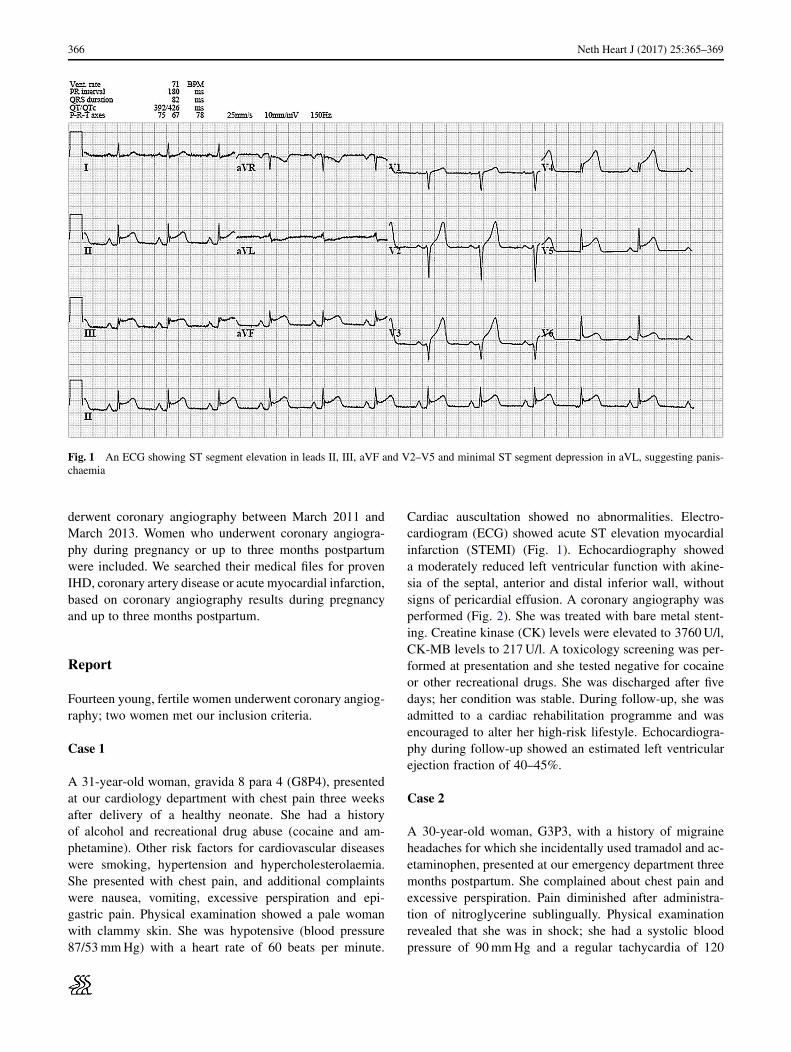
Fig. 1 An ECG showing ST segment elevation in leads II, III, aVF and V2–V5 and minimal ST segment depression in aVL, suggesting panis-chaemia
derwent coronary angiography between March 2011 andMarch 2013. Women who underwent coronary angiogra-phy during pregnancy or up to three months postpartumwere included. We searched their medical files for provenIHD, coronary artery disease or acute myocardial infarction,based on coronary angiography results during pregnancyand up to three months postpartum.
Report
Fourteen young, fertile women underwent coronary angiog-raphy; two women met our inclusion criteria.
Case 1
A 31-year-old woman, gravida 8 para 4 (G8P4), presentedat our cardiology department with chest pain three weeksafter delivery of a healthy neonate. She had a historyof alcohol and recreational drug abuse (cocaine and am-phetamine). Other risk factors for cardiovascular diseaseswere smoking, hypertension and hypercholesterolaemia.She presented with chest pain, and additional complaintswere nausea, vomiting, excessive perspiration and epi-gastric pain. Physical examination showed a pale womanwith clammy skin. She was hypotensive (blood pressure87/53mmHg) with a heart rate of 60 beats per minute.
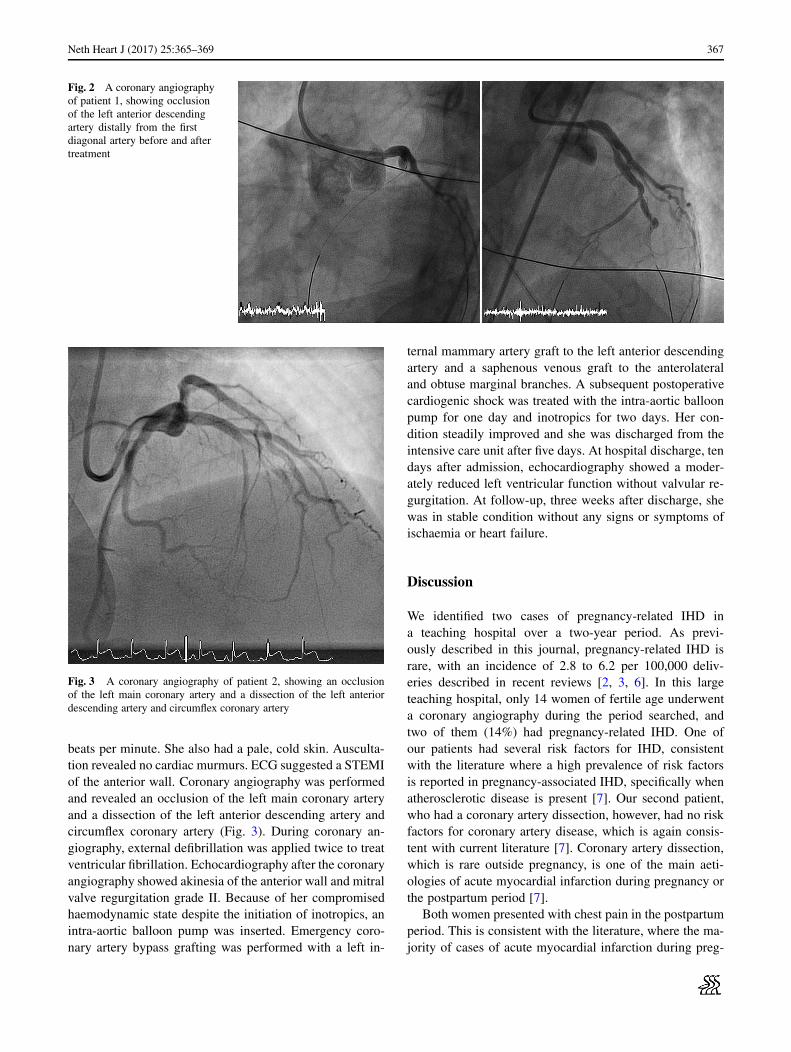
Cardiac auscultation showed no abnormalities. Electro-cardiogram (ECG) showed acute ST elevation myocardialinfarction (STEMI) (Fig. 1). Echocardiography showeda moderately reduced left ventricular function with akine-sia of the septal, anterior and distal inferior wall, withoutsigns of pericardial effusion. A coronary angiography wasperformed (Fig. 2). She was treated with bare metal stent-ing. Creatine kinase (CK) levels were elevated to 3760U/l,CK-MB levels to 217U/l. A toxicology screening was per-formed at presentation and she tested negative for cocaineor other recreational drugs. She was discharged after fivedays; her condition was stable. During follow-up, she wasadmitted to a cardiac rehabilitation programme and wasencouraged to alter her high-risk lifestyle. Echocardiogra-phy during follow-up showed an estimated left ventricularejection fraction of 40–45%.
Case 2
A 30-year-old woman, G3P3, with a history of migraineheadaches for which she incidentally used tramadol and ac-etaminophen, presented at our emergency department threemonths postpartum. She complained about chest pain andexcessive perspiration. Pain diminished after administra-tion of nitroglycerine sublingually. Physical examinationrevealed that she was in shock; she had a systolic bloodpressure of 90mmHg and a regular tachycardia of 120
Neth Heart J (2017) 25:365–369 367
Fig. 2 A coronary angiographyof patient 1, showing occlusionof the left anterior descendingartery distally from the firstdiagonal artery before and aftertreatment
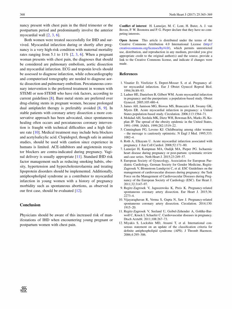
Fig. 3 A coronary angiography of patient 2, showing an occlusionof the left main coronary artery and a dissection of the left anteriordescending artery and circumflex coronary artery
beats per minute. She also had a pale, cold skin. Ausculta-tion revealed no cardiac murmurs. ECG suggested a STEMIof the anterior wall. Coronary angiography was performedand revealed an occlusion of the left main coronary arteryand a dissection of the left anterior descending artery andcircumflex coronary artery (Fig. 3). During coronary an-giography, external defibrillation was applied twice to treatventricular fibrillation. Echocardiography after the coronaryangiography showed akinesia of the anterior wall and mitralvalve regurgitation grade II. Because of her compromisedhaemodynamic state despite the initiation of inotropics, anintra-aortic balloon pump was inserted. Emergency coro-nary artery bypass grafting was performed with a left in-
ternal mammary artery graft to the left anterior descendingartery and a saphenous venous graft to the anterolateraland obtuse marginal branches. A subsequent postoperativecardiogenic shock was treated with the intra-aortic balloonpump for one day and inotropics for two days. Her con-dition steadily improved and she was discharged from theintensive care unit after five days. At hospital discharge, tendays after admission, echocardiography showed a moder-ately reduced left ventricular function without valvular re-gurgitation. At follow-up, three weeks after discharge, shewas in stable condition without any signs or symptoms ofischaemia or heart failure.
Discussion
We identified two cases of pregnancy-related IHD ina teaching hospital over a two-year period. As previ-ously described in this journal, pregnancy-related IHD israre, with an incidence of 2.8 to 6.2 per 100,000 deliv-eries described in recent reviews [2, 3, 6]. In this largeteaching hospital, only 14 women of fertile age underwenta coronary angiography during the period searched, andtwo of them (14%) had pregnancy-related IHD. One ofour patients had several risk factors for IHD, consistentwith the literature where a high prevalence of risk factorsis reported in pregnancy-associated IHD, specifically whenatherosclerotic disease is present [7]. Our second patient,who had a coronary artery dissection, however, had no riskfactors for coronary artery disease, which is again consis-tent with current literature [7]. Coronary artery dissection,which is rare outside pregnancy, is one of the main aeti-ologies of acute myocardial infarction during pregnancy orthe postpartum period [7].
Both women presented with chest pain in the postpartumperiod. This is consistent with the literature, where the ma-jority of cases of acute myocardial infarction during preg-
368 Neth Heart J (2017) 25:365–369
nancy present with chest pain in the third trimester or thepostpartum period and predominantly involve the anteriormyocardial wall [2, 3, 6].
Both women were treated successfully for IHD and sur-vived. Myocardial infarction during or shortly after preg-nancy is a very high-risk condition with maternal mortalityrates ranging from 5.1 to 11% [2, 3, 6]. When a pregnantwoman presents with chest pain, the diagnoses that shouldbe considered are pulmonary embolism, aortic dissectionand myocardial infarction. ECG and troponin levels shouldbe assessed to diagnose infarction, while echocardiographyand computerised tomography are needed to diagnose aor-tic dissection and pulmonary embolism. Percutaneous coro-nary intervention is the preferred treatment in women withSTEMI or non-STEMI who have risk factors, according tocurrent guidelines [8]. Bare metal stents are preferred overdrug-eluting stents in pregnant women, because prolongeddual antiplatelet therapy is preferably avoided [8, 9]. Instable patients with coronary artery dissection a more con-servative approach has been advocated, since spontaneoushealing often occurs and percutaneous coronary interven-tion is fraught with technical difficulties and a high fail-ure rate [10]. Medical treatment may include beta blockersand acetylsalicylic acid. Clopidogrel, though safe in animalstudies, should be used with caution since experience inhumans is limited. ACE-inhibitors and angiotensin recep-tor blockers are contra-indicated during pregnancy. Vagi-nal delivery is usually appropriate [11]. Standard IHD riskfactor management such as reducing smoking habits, obe-sity, hypertension and hypercholesterolaemia and treatinglipoprotein disorders should be implemented. Additionally,antiphospholipid syndrome as a contributor to myocardialinfarction in young women with a history of pregnancymorbidity such as spontaneous abortions, as observed inour first case, should be evaluated [12].
Conclusion
Physicians should be aware of this increased risk of man-ifestations of IHD when encountering young pregnant orpostpartum women with chest pain.
Conflict of interest H. Lameijer, M. C. Lont, H. Buter, A. J. vanBoven, P. W. Boonstra and P. G. Pieper declare that they have no com-peting interests.
Open Access This article is distributed under the terms of theCreative Commons Attribution 4.0 International License (http://creativecommons.org/licenses/by/4.0/), which permits unrestricteduse, distribution, and reproduction in any medium, provided you giveappropriate credit to the original author(s) and the source, provide alink to the Creative Commons license, and indicate if changes weremade.
References
1. Vinatier D, Virelizier S, Depret-Mosser S, et al. Pregnancy af-ter myocardial infarction. Eur J Obstet Gynecol Reprod Biol.1994;56:89–93.
2. Ladner HE, Danielsen B, Gilbert WM. Acute myocardial infarctionin pregnancy and the puerperium: a population-based study. ObstetGynecol. 2005;105:480–4.
3. James AH, Jamison MG, Biswas MS, Brancazio LR, Swamy GK,Myers ER. Acute myocardial infarction in pregnancy: a UnitedStates population-based study. Circulation. 2006;113:1564–71.
4. Mokdad AH, Serdula MK, Dietz WH, Bowman BA, Marks JS, Ko-plan JP. The spread of the obesity epidemic in the United States,1991–1998. JAMA. 1999;282:1519–22.
5. Cunningham FG, Leveno KJ. Childbearing among older women– the message is cautiously optimistic. N Engl J Med. 1995;333:1002–4.
6. Roth A, Elkayam U. Acute myocardial infarction associated withpregnancy. J Am Coll Cardiol. 2008;52:171–80.
7. Lameijer H, Kampman MA, Oudijk MA, Pieper PG. Ischaemicheart disease during pregnancy or post-partum: systematic reviewand case series. Neth Heart J. 2015;23:249–57.
8. European Society of Gynecology, Association for European Pae-diatric Cardiology, German Society for Gender Medicine, Regitz-Zagrosek V, Blomstrom Lundqvist C, et al. ESC Guidelines on themanagement of cardiovascular diseases during pregnancy: the TaskForce on the Management of Cardiovascular Diseases during Preg-nancy of the European Society of Cardiology (ESC). Eur Heart J.2011;32:3147–97.
9. Regitz-Zagrosek V, Jaguszewska K, Preis K. Pregnancy-relatedspontaneous coronary artery dissection. Eur Heart J. 2015;36:2273–4.
10. Vijayaraghavan R, Verma S, Gupta N, Saw J. Pregnancy-relatedspontaneous coronary artery dissection. Circulation. 2014;130:1915–20.
11. Regitz-Zagrosek V, Seeland U, Geibel-Zehender A, Gohlke-Bar-wolf C, Kruck I, Schaefer C. Cardiovascular diseases in pregnancy.Dtsch Arztebl. 2011;108:267–73.
12. Miyakis S, Lockshin MD, Atsumi T, et al. International con-sensus statement on an update of the classification criteria fordefinite antiphospholipid syndrome (APS). J Thromb Haemost.2006;4:295–306.
Neth Heart J (2017) 25:365–369 369