Dysphagia In the Aerodigestive · PDF fileDysphagia in the Aerodigestive Patient ... liquids...
85
Dysphagia In the Aerodigestive Patient Gwen Provo‐Bell, SLP‐CCC Christopher T. Wootten, MD
Transcript of Dysphagia In the Aerodigestive · PDF fileDysphagia in the Aerodigestive Patient ... liquids...

Dysphagia In the AerodigestivePatient
Gwen Provo‐Bell, SLP‐CCCChristopher T. Wootten, MD

Overview• Changes in Normal Anatomy Over Time
• Normal and Abnormal Physiology of Swallowing in Children
• The Evolution of Etiologies of Dysphagia After the NICU
• The Evaluation and Treatment of Dysphagia in the AerodigestivePatient
• Dysphagia Through the Airway Reconstruction Process

Changes in Normal Anatomy Over Time
AdultAdult 2 y/o2 y/o 2 d/o2 d/o


Changes in Normal Anatomy Over Time

Normal and Abnormal Physiology of Swallowing in Children
Dysphagia in the Aerodigestive Patient
Definition of Dysphagia‐difficulty with any step of the feeding process—from accepting foods and liquids into the mouth to the entry of food into the
stomach and intestines.
Dysphagia Encompasses both feeding and Swallowing‐‐ Oral Stage‐ Oropharyngeal Phase‐ Pharyngeal Phase‐ Esophageal Phase

Efficient Feeding and Swallowing Requires:
• Oral Preparation Stage—preparing the food or liquid in the oral cavity to form a bolus‐including sucking liquids, manipulating soft boluses, and chewing solid food.
• Oropharyngeal Phase—moving or propelling the bolus posteriorly through the oral cavity.
• Pharyngeal Phase—initiating the swallow; moving the bolus through the pharynx.
• Esophageal Phase—moving the bolus through the cervical and thoracic esophagus and into the stomach via esophageal peristalsis (Logemann, 1998).

Phases of the Swallow
‐

Coordination of Respiration/Swallowing
• Pharynx is a channel for both airflow and the passage of ingested material, breathing and swallowing cannot occur simultaneously without risk to the airway
• Respiratory Patterns and Swallowing are highly coordinated, ( sucking/breathing may occur simultaneously, but at moment of swallowing, airway is closed
The Evolution of Etiologies of Dysphagia After the NICU
• Oral Aversion/Textural Concerns• Neurocognitive Delay• Structural Problems• Laryngeal Inflammation
Source: IP THA Data via Strategic Marketing Office

Dysphagia: Structural Problems• Choanal stenosis• Cleft lip/palate• Laryngotracheal cleft• TEF/EA• Vascular ring• Tumors• Craniofacial anomalies• Glottic insufficiency
Source: Capacity Management
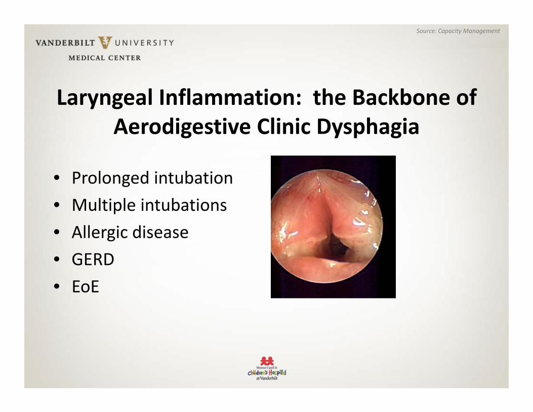
Laryngeal Inflammation: the Backbone of Aerodigestive Clinic Dysphagia
• Prolonged intubation• Multiple intubations• Allergic disease• GERD• EoE
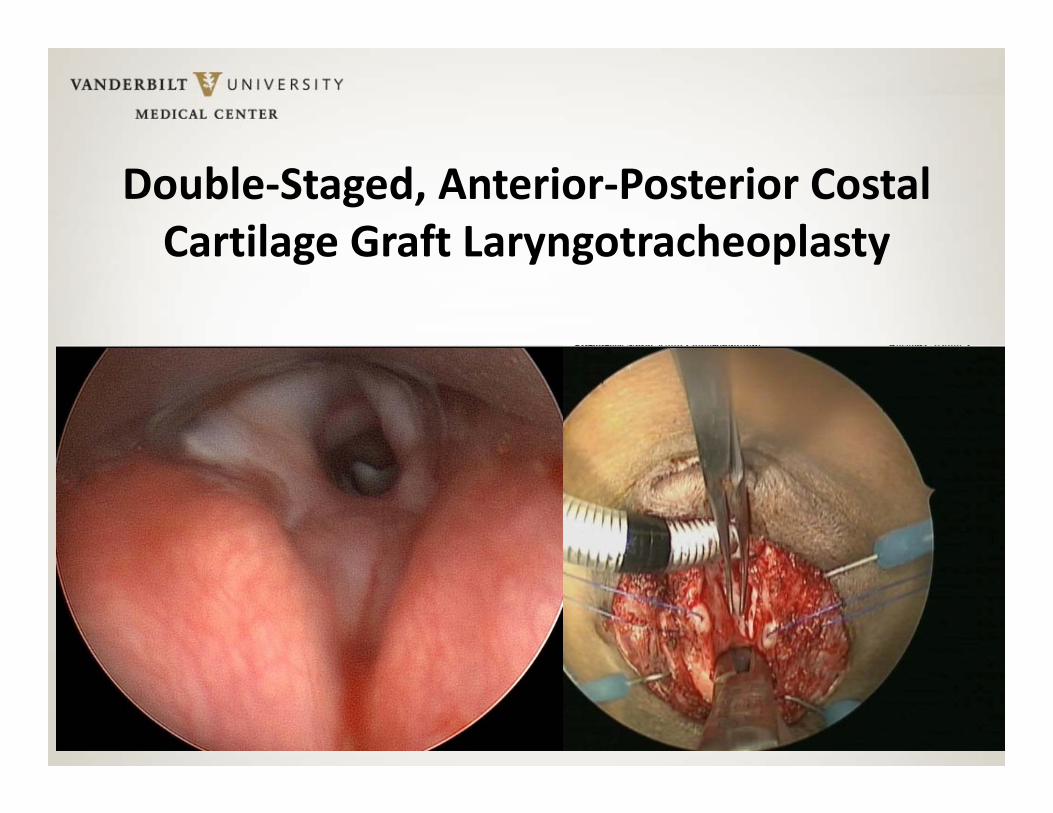
Double‐Staged, Anterior‐Posterior Costal Cartilage Graft Laryngotracheoplasty

Three Weeks Later

The Evaluation and Treatment of Dysphagia in the Aerodigestive Patient

Evaluation• Starting point: baseline assessment of feeding status, oral stage problems, related medical problems, identify clinical signs & symptoms of dysphagia
• Limitations: cannot assess beyond the oral phase• Outcome: provides basis for clinical pathway to determine further assessments, instrumental & other

Common feeding Presentations in the Aerodigestive Patient
• Compromised nutritional status• Poor growth/Failure to thrive due to poor
volume/mechanics• Food refusal/Limited food repertoire/Food Sensitivities• Decreased volume• Delayed oral motor/feeding skills• Choking/coughing/gagging/vomiting while eating/drinking• Difficulty transitioning from enteral to oral feeds• Maladaptive/Behavioral feeding problems
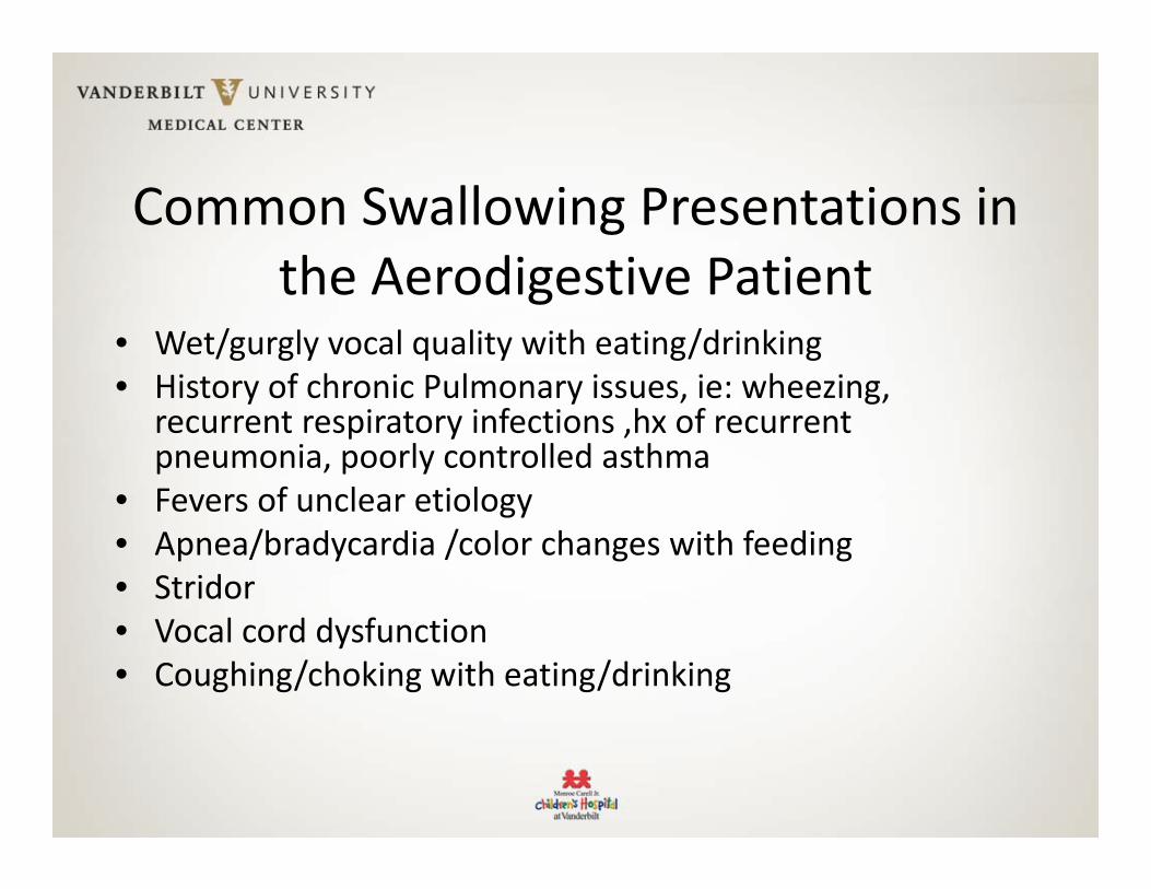
Common Swallowing Presentations in the Aerodigestive Patient
• Wet/gurgly vocal quality with eating/drinking• History of chronic Pulmonary issues, ie: wheezing,
recurrent respiratory infections ,hx of recurrent pneumonia, poorly controlled asthma
• Fevers of unclear etiology• Apnea/bradycardia /color changes with feeding• Stridor• Vocal cord dysfunction• Coughing/choking with eating/drinking

Videofluoroscopic Swallow Study• Indications: : patient demonstrates signs or symptoms of swallowing dysfunction
• Collaborative examination via radiologist & speech pathologist• SLP: positioning, use of feeding equipment & food/liquid to which
patient is accustomed– Implementation of compensatory strategies– Involvement of family– Dual interpretation with radiologist, decision‐making following
study in collaboration with others on team

VFSS : Advantages
• Advantages: – View of all phases of swallow, no discomfort, ability to try compensatory strategies
– Provides ongoing view of airway protection during rapid chain swallowing sequences i.e. bottle‐feeds
– Provides opportunity to exclude structural problem in esophagus as source of dysphagia

VFSS: Disadvantages• Radiation exposure• Child may resist barium – may not get sufficient sample for meaningful interpretation
• Not feasible if child has negligible oral intake• Operator dependent• Poor interrator reliability – implications for accuracy of interpretation and appropriateness of subsequent recommendations re: feeds

FEES Assessment
• Pediatric FEES – A definition– Transnasal passage of endoscope to view pharyngeal and laryngeal structures
– Assessment of ability to protect airway during swallowing
– Collaborative exam – Speech Pathology & Otolaryngology
Non‐Surgical Treatment of Dysphagia: Infant Treatment

Goals of Treatment
Safely support adequate nutrition/hydrationDetermine feeding techniques to maximize feeding safety and efficiencyAttain age/developmentally appropriate feeding skills

Treatment• Positioning• Diet Modifications• Equipment/Utensils• Feeding Techniques• Oral Motor Skills• Pacing/Feeding Strategies• Sensory• Behavioral Interventions• Parent Education

The Treatment of Dysphagia: Surgical
Anatomical Problem = Anatomical Solution*
*Not universally true

*Not universally true
Anatomical Problem = Anatomical Solution
• Specific Anatomies• Dysphagia in Airway Reconstruction

Suck
Swallow
Breathe

Suck
Swallow
Breathe
FrenulectomyPalatoplasty
Part. glossectomy

Suck
Swallow
Breathe
Cricopharyngeal myotomyTx. Vallecular cystPalatoplastyRepair laryngeal cleft

Suck
Swallow
Breathe
Repair CNPASTx. Choanal atresiaTx. Vallecular cystSupraglottoplastyAirway Recon.

More About Breathing: Dysphagia and Airway Reconstruction
I. Preoperative Causes of DysphagiaII. Assessing Dysphagia in the Preoperative
PatientIII. Postoperative Dysphagia: Early PhaseIV. Postoperative Dysphagia: Late Phase
Dysphagia and Airway Reconstruction
I. Preoperative Causes of DysphagiaII. Assessing Dysphagia in the Preoperative
PatientIII. Postoperative Dysphagia: Early PhaseIV. Postoperative Dysphagia: Late Phase

Preoperative Causes of DysphagiaAnatomical Considerations• Laryngomalacia• Subglottic stenosis• Tracheal stenosis• Vocal fold paralysis• Cricoarytenoid fixation• Laryngeal cleft• Tracheoesophageal fistula• Laryngeal scarring

Preoperative Causes of DysphagiaAnatomical Considerations• Laryngomalacia• Subglottic stenosis• Tracheal stenosis• Vocal fold paralysis• Cricoarytenoid fixation• Laryngeal cleft• Tracheoesophageal fistula• Laryngeal scarring

Preoperative Causes of DysphagiaAnatomical Considerations• Laryngomalacia• Subglottic stenosis• Tracheal stenosis• Vocal fold paralysis• Cricoarytenoid fixation• Laryngeal cleft• Tracheoesophageal fistula• Laryngeal scarring

Preoperative Causes of DysphagiaAnatomical Considerations• Laryngomalacia• Subglottic stenosis• Tracheal stenosis• Vocal fold paralysis• Cricoarytenoid fixation• Laryngeal cleft• Tracheoesophageal fistula• Laryngeal scarring

Preoperative Causes of DysphagiaAnatomical Considerations• Laryngomalacia• Subglottic stenosis• Tracheal stenosis• Vocal fold paralysis• Cricoarytenoid fixation• Laryngeal cleft• Tracheoesophageal fistula• Laryngeal scarring

Preoperative Causes of DysphagiaAnatomical Considerations• Laryngomalacia• Subglottic stenosis• Tracheal stenosis• Vocal fold paralysis• Cricoarytenoid fixation• Laryngeal cleft• Tracheoesophageal fistula• Laryngeal scarring

Preoperative Causes of DysphagiaAnatomical Considerations• Laryngomalacia• Subglottic stenosis• Tracheal stenosis• Vocal fold paralysis• Cricoarytenoid fixation• Laryngeal cleft• Tracheoesophageal fistula• Laryngeal scarring

Preoperative Causes of DysphagiaAnatomical Considerations• Laryngomalacia• Subglottic stenosis• Tracheal stenosis• Vocal fold paralysis• Cricoarytenoid fixation• Laryngeal cleft• Tracheoesophageal fistula• Laryngeal scarring

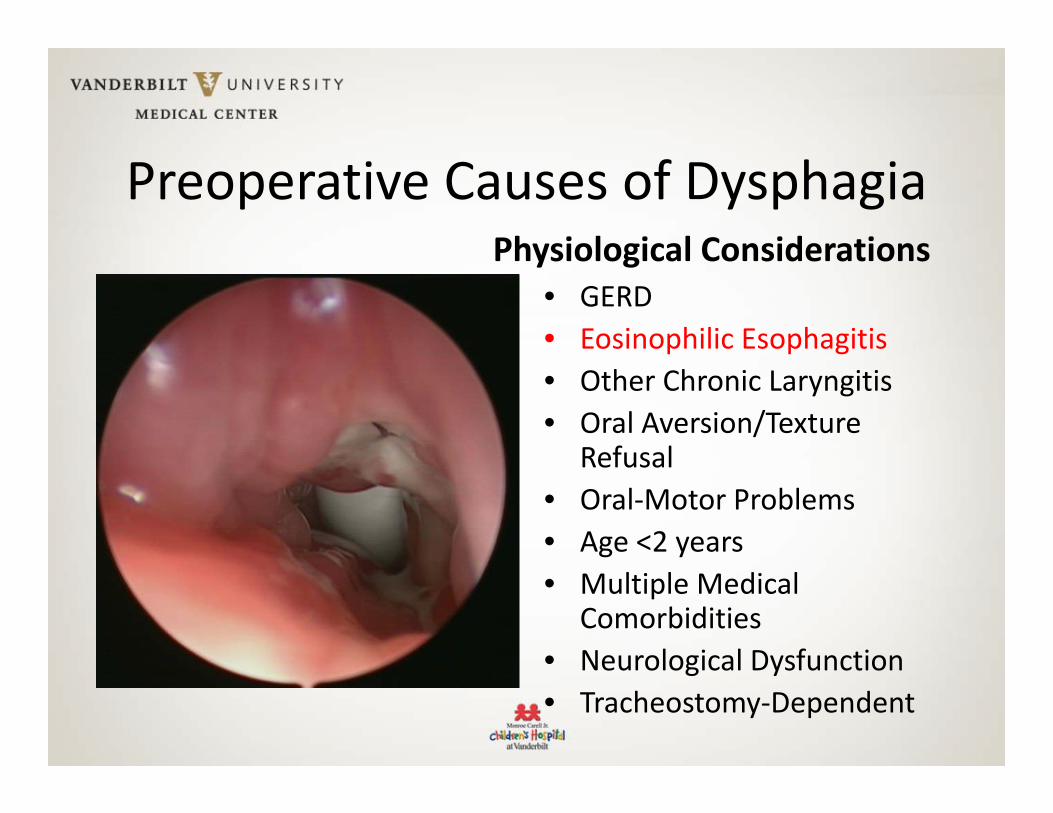
Preoperative Causes of DysphagiaPhysiological Considerations
• GERD• Eosinophilic Esophagitis• Other Chronic Laryngitis• Oral Aversion/Texture
Refusal• Oral‐Motor Problems• Age <2 years• Multiple Medical
Comorbidities• Neurological Dysfunction• Tracheostomy‐Dependent

Preoperative Causes of DysphagiaPhysiological Considerations
• GERD• Eosinophilic Esophagitis• Other Chronic Laryngitis• Oral Aversion/Texture
Refusal• Oral‐Motor Problems• Age <2 years• Multiple Medical
Comorbidities• Neurological Dysfunction• Tracheostomy‐Dependent
Preoperative Causes of DysphagiaPhysiological Considerations
• GERD• Eosinophilic Esophagitis• Other Chronic Laryngitis• Oral Aversion/Texture
Refusal• Oral‐Motor Problems• Age <2 years• Multiple Medical
Comorbidities• Neurological Dysfunction• Tracheostomy‐Dependent

Preoperative Causes of DysphagiaPhysiological Considerations
• GERD• Eosinophilic Esophagitis• Other Chronic Laryngitis• Oral Aversion/Texture
Refusal• Oral‐Motor Problems• Age <2 years• Multiple Medical
Comorbidities• Neurological Dysfunction• Tracheostomy‐Dependent

Preoperative Causes of DysphagiaPhysiological Considerations
• GERD• Eosinophilic Esophagitis• Other Chronic Laryngitis• Oral Aversion/Texture
Refusal• Oral‐Motor Problems• Age <2 years• Multiple Medical
Comorbidities• Neurological Dysfunction• Tracheostomy‐Dependent

Preoperative Causes of DysphagiaPhysiological Considerations
• GERD• Eosinophilic Esophagitis• Other Chronic Laryngitis• Oral Aversion/Texture
Refusal• Oral‐Motor Problems• Age <2 years• Multiple Medical
Comorbidities• Neurological Dysfunction• Tracheostomy‐Dependent

Dysphagia and Airway Reconstruction
I. Preoperative Causes of DysphagiaII. Assessing Dysphagia in the Preoperative
PatientIII. Postoperative Dysphagia: Early PhaseIV. Postoperative Dysphagia: Late Phase

Assessing Dysphagia in the Preoperative Patient
• Specifically, the sphincteric functions of the larynx may be temporarily or permanently altered by airway reconstruction.

Assessing Dysphagia in the Preoperative Patient
• History• Physical• Flexible Nasopharyngoscopy• FEES/FEEST• Video Fluoroscopic Swallow Study

Fiberoptic Endoscopic Evaluation of Swallowing (FEES)
Assess For:• Pooling of secretions• Poor oral motor skills with premature spillage• Laryngeal penetration• Aspiration• Hypopharyngeal residue

Fiberoptic Endoscopic Evaluation of Swallowing (FEES)

Videofluoroscopic Swallow Study (VFSS)

WHAT IS AIRWAY RECONSTRUCTION?

Interlude: What is “Airway Reconstruction?”
Resection
Single Stage = Leave the OR without a tracheostomy
Double Stage = Leave the OR with a tracheostomy

Interlude: What is “Airway Reconstruction?”
Augmentation (Grafting)

Interlude: What is “Airway Reconstruction?”
Slide Tracheoplasty

Dysphagia and Airway Reconstruction
I. Preoperative Causes of DysphagiaII. Assessing Dysphagia in the Preoperative
PatientIII. Postoperative Dysphagia: Early PhaseIV. Postoperative Dysphagia: Late Phase

Postoperative Dysphagia: Early Phase
0%
20%
40%
60%
80%
100%
OralPharyngeal
EsophagealNo
Dysphagia
Tracheostomies and the Etiologies of Dysphagia

Postoperative Dysphagia: Early Phase
0%
20%
40%
60%
80%
100%
OralPharyngeal
EsophagealNo
Dysphagia
Tracheostomies and the Etiologies of Dysphagia
Postoperative Dysphagia: Early Phase
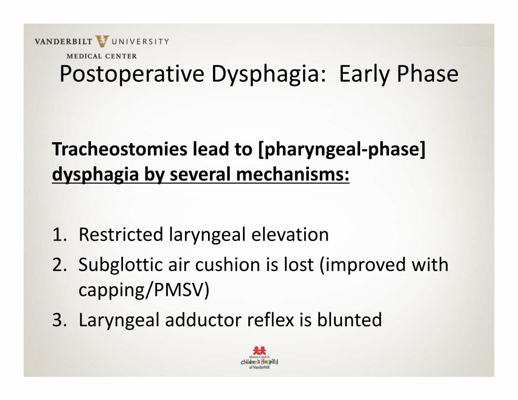
Tracheostomies lead to [pharyngeal‐phase] dysphagia by several mechanisms:
1. Restricted laryngeal elevation2. Subglottic air cushion is lost (improved with
capping/PMSV) 3. Laryngeal adductor reflex is blunted

Postoperative Dysphagia: Early Phase
In airway reconstruction, the patient is getting more than simply a tracheostomy (or its removal). What is the nature and timeline of dysphagia in this unique population?

Postoperative Dysphagia: Early Phase
Types of Stents Used in Airway Reconstruction:1. Endotracheal tube2. Suprastomal stent3. Montgomery laryngeal stent4. T‐tube5. Tracheal [bronchial] stent6. None
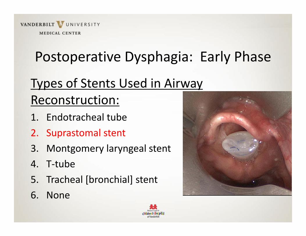
Postoperative Dysphagia: Early Phase
Types of Stents Used in Airway Reconstruction:1. Endotracheal tube2. Suprastomal stent3. Montgomery laryngeal stent4. T‐tube5. Tracheal [bronchial] stent6. None

Postoperative Dysphagia: Early Phase
Types of Stents Used in Airway Reconstruction:1. Endotracheal tube2. Suprastomal stent3. Montgomery laryngeal stent4. T‐tube5. Tracheal [bronchial] stent6. None

Postoperative Dysphagia: Early Phase
Types of Stents Used in Airway Reconstruction:1. Endotracheal tube2. Suprastomal stent3. Montgomery laryngeal stent4. T‐tube5. Tracheal [bronchial] stent6. None
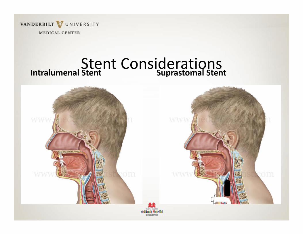
Stent ConsiderationsIntralumenal Stent Suprastomal Stent

What Is a “Tracheal Stent?”

POD 4 ssCTR with a tracheal stent

What is a Sutured (vs Non‐Sutured) SuprastomalStent?

VFSS Example of Dysphagia To Thins With a Suprastomal Stent

• 18 ss cases, 22 ds cases –oral feeders preop
• 7/16 patients had successful feeding (stent in)– 4 had stent sutured, 12 had non‐sutured stents– 4/4 with sutured stent tolerated feeds
• 34/40 (85%) resumed preop diet within 0‐8 days of stent removal/extubation, average 1.9 days 6/40 (15%) had significant postop dysphagia– No difference in graft sizes, age, previous LTR, ss, ds…

Early Postoperative Dysphagia• Improving Voice and
Swallow– Passy Muir Valve usage
• Dysphagia of a greater duration is associated with: – DS anterior+posterior grafts
with a T‐tube stent – DS CTR with a T‐tube stent
and – DS VF lateralization – DS petiole repositioning– DS open arytenoidectomy
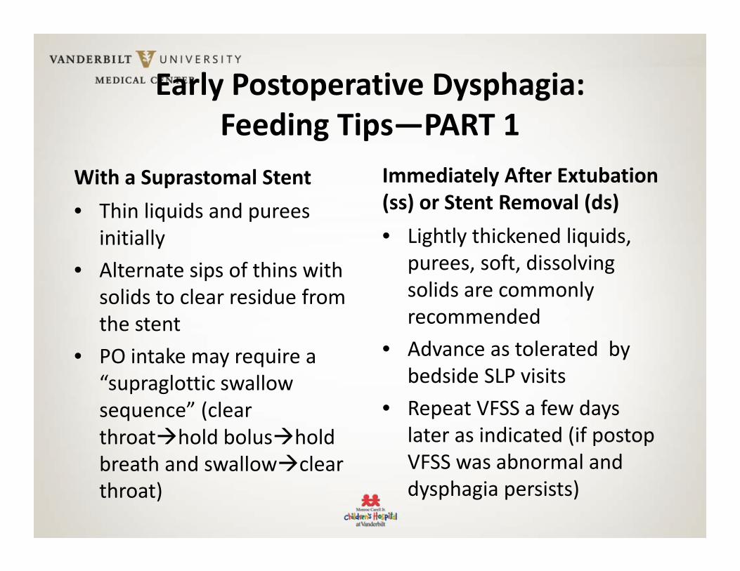
Early Postoperative Dysphagia: Feeding Tips—PART 1
With a Suprastomal Stent• Thin liquids and purees
initially• Alternate sips of thins with
solids to clear residue from the stent
• PO intake may require a “supraglottic swallow sequence” (clear throathold bolusholdbreath and swallowclearthroat)
Immediately After Extubation(ss) or Stent Removal (ds)• Lightly thickened liquids,
purees, soft, dissolving solids are commonly recommended
• Advance as tolerated by bedside SLP visits
• Repeat VFSS a few days later as indicated (if postop VFSS was abnormal and dysphagia persists)

Early Postoperative Dysphagia• A success story: VFSS examples from a 67 y/o child with end‐stage rheumatoid arthritis affecting the larynx requiring urgent tracheotomy and ultimately repaired with an endoscopic posterior graft LTP
Her capped suprastomal stent

POD 3 ds Posterior LTP (endoscopic) with a capped suprastomal stent

Same patient, POD 1 after stent removal
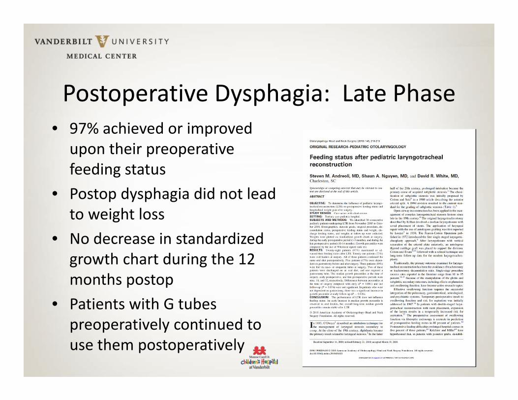
Postoperative Dysphagia: Late Phase• 97% achieved or improved
upon their preoperative feeding status
• Postop dysphagia did not lead to weight loss
• No decrease in standardized growth chart during the 12 months postop
• Patients with G tubes preoperatively continued to use them postoperatively

Distilling the Data From Three Studies: Postoperative Oral Feeding Prognosis For Patients Who Were Oral Feeders
Preoperatively
# Back to Preop Oral Diet after dsLTP/CTR (stent out)
28/29 Pts@ CCHMC; 39/40 Pts@ CHOP; 28/29 Pts@ MUSC
Time to PO after ssLTP/CTR in oral feeders (extubated)
1‐5 days (13/13 Pts@ CCHMC); 0‐8 days (16/18 Pts@ CHOP)
Time to PO after dsLTP/CTR in pts who took SOME PO (stent in)
4‐28 days (16/18 Pts@ CCHMC); “by POD 2”(7/16 Pts@ CHOP)
CW

Postoperative Dysphagia: Late Phase

Conclusions
• Instrumental measures to assess dysphagia preoperatively (FEES and/or VFSS) are useful in predicting long‐term postoperative feeding status after airway reconstruction
• Respect the young (2 y/o and younger), tracheostomy‐dependent, medically complex

Conclusions
• Transient dysphagia is common following airway reconstruction
• Nearly universally, airway patients, in time, return to or improve upon their preoperative swallowing abilities

THANK YOUDysphagia in the Aerodigestive Patient