Dynamics of cork mycobiota throughout stopper ... phylotypes. The possible production of...
154
Maria do Carmo Barreto Baptista Dissertation presented to obtain the Ph.D degree in Biology Instituto de Tecnologia Química e Biológica | Universidade Nova de Lisboa Oeiras, December, 2011 Dynamics of cork mycobiota throughout stopper manufacturing process: from diversity to metabolite !"#$ &'()*) +,-
Transcript of Dynamics of cork mycobiota throughout stopper ... phylotypes. The possible production of...
Maria do Carmo Barreto Baptista
Dissertation presented to obtain the Ph.D degree in Biology Instituto de Tecnologia Química e Biológica | Universidade Nova de Lisboa
Oeiras, December, 2011
Dynamics of cork mycobiota throughout stopper manufacturing process: from diversity to metabolite
!"#$%&'()*)%+,-%


T o J o ão , S o f i a an d m y p ar e n ts
“ T i m e i s l i f e i ts e lf , a n d l i fe re s i d e s i n t he hu m an h e ar t . ”
M i c h a e l E n d e

T a b l e o f c o n t e n t s
Acknowledgements 7
Summary 11
Sumário 14
Chapter 1 Introduction 19
Chapter 2 Taxonomic studies of the fungal mycobiota
presented in cork samples collected
throughout cork manufacturing discs
55
Unveiling the fungal mycobiota present
throughout cork stopper manufacturing
process
57
Taxonomic studies of the Penicillium
glabrum complex and the description of a
new species P. subericola
89
Chapter 3 Exo-metabolites produced by some fungal
isolates in several media cultures
101
Exo-metabolome of some fungal isolates
growing on cork-based medium
103
Chapter 4 Volatile compounds produced by cork
mycobiota
111
Volatile Compounds in Samples of Cork
and also Produced by Selected Fungi
113
Supporting information 120
Chapter 5 Discussion 123
Chapter 6 Bibliography 133


A c k n o w l e d g m e n t s
I thank my supervisor Doutora Vitória San Romão for all her
support and confidence, which were necessary for the good
conclusion of this PhD thesis. Her friendship and encouragement
were also extremely important. I also want to thank my co-
supervisor Doutora Teresa Barreto Crespo for her collaboration,
support and enthusiasm showed in several occasions during the
course of this work.
Collaborate with Professor Luis Vilas Boas gave me the
opportunity to learn more about chemistry and volatile
compounds. The conversations (scientific or not), studies and the
revisions of either the manuscript or the thesis were important
and inspiring steps for my learning process. I will always be
grateful to him.
Professor Jens Frisvad with whom I learned many things about
exo-metabolites in fungi and had the privilege to work with him at
DTU, Denmark. Our many scientific discussions and work
resulted in a manuscript already published. I am also thankful to
the other co-authors Professor Thomas Larsen and Jesper
Mogensen for their collaboration and support. I am thankful to all
the persons that worked at DTU that made my stay more
pleasant, especially Marina Venturini for her help and friendship.
Cristina Silva Pereira for her continuous support, friendship and
the opportunity to learn with her.
7
I am grateful to Professor Rogério Tenreiro for sharing his
experience and scientific knowledge with me that gave me the
opportunity to learn. To Mário Gadanho whose collaboration
resulted in a publication.
I am grateful to the group Applied and Industrial Mycology from
CBS-KNAW Fungal Biodiversity Centre where I stayed during
some time to work in the identification of the fungal isolates. I am
gratefully to Professor Rob Samson that gave me the opportunity
to learn and to benefit with the experience of his work group in
fungal taxonomy. Bedankt daarvoor.
To work with Jos Houbraken was not only an excellent
experience but also a funny one. I learned many things especially
at microscopic level. He was always available to any question
and we had some interesting scientific (and non-scientific)
discussions. Janos Vargas helped to identify the Aspergillus
group and shared his experience in molecular taxonomy
techniques.
Richard van Leeuwen I want to thank you for all the good
moments that we share at CBS and on the conferences that we
went. Always keep your sense of humor and good mood.
Bedankt daarvoor (kleine snoeperd).
I want to thank Tineke van Doorn for not only her technical
support but also for her friendship and good times that we had at
CBS. Bedankt daarvoor.
Martin Meijer who was also present in the few times that I’ve
been at CBS but always gave technical support and watch out for
all of us. Bedankt.
Ferry Hagen who helped me to identify some yeasts. His sense
of humor and support helped me to make my staying at CBS
8

more pleasant one. Also for the very good moments spend at
conferences and in Lisbon.
Jan Dijksterhuis with whom we shared good moments at CBS
and at the conferences that we attend.
I am also grateful to Paramee Noonim (Tao) for the great times
that we spend at CBS and at Key West.
To Professors J. J. Baptista Ferreira and Margarida Barata that
initiated me in fungal taxonomic studies and with whom I learned
very much about the fungi world. I also thank them their
encouragement.
To Doctor Ian Smith for his revision and corrections of the thesis
title and Summary and also for his suggestions that helped to
improve the quality of the manuscript.
To Drª Teresa Melo that revised part of the manuscript and gave
very useful suggestions.
To my sister Rita Baptista for her support and a set of photos
taken during a visit to the cork factory that helped to improve this
work. Also to Doutora Patrícia Noronha for the photos taken to
the fungi isolated and identified in the course of this work.
To Professor Julian Mitchell for his advises, suggestions,
interesting conversations and emails that helped me to overcome
some practical problems.
I want to acknowledge Vanessa for her support and the
opportunity to work in her group that resulted in a publication.
9

To Andreia Santos, Susana Marcelino, Liliana Pinto, Rita Bento
and Mário Gil Dias for their help processing the cork samples and
with the molecular work. To my lab colleagues Paula Alves,
Catarina Dourado, Beatriz, Ana Margarida, Filipa, Sandra,
Doudou, Teresa, Neuza, Paulo Marujo, Gilda, Patrícia and all my
former colleagues for their help and support in several occasions.
To Cristina Leitão, Maria joão Fernandes and Fernanda Spínola
for their continuous support and friendship that helped me to
complete this task.
To my friend and colleague Dra. Dulce Brito for her
encouragement, support and motivation that helped me in many
occasions. Especially, the many conversations shared with tea.
To my friend Bárbara (Ayahua) that helped me greatly and whom
we shared many special moments either in the lab or outside. I
will always be grateful to her. David, thank you for all your
support and friendship that helped me all the way. Especially for
always being present. Ana Paula A., thank you for everything.
“May the force be with you all”.
This thesis is dedicated to my parents for strength and
confidence that helped me to cross this path and to my kids,
Sofia and João, they were the motivation that I needed to finish
this goal.
To Mani, Mariana, Catarina and my family for their
encouragement and support that helped me to look further.
Thanks to all my friends and many persons that in any way
helped me to accomplished this task.
FCT Fundação para a Ciência e Tecnologia for financing my PhD
with the grant BD/19264/ 2004.
10

S u m m a r y
Cork, the continuous layer of outer bark of the Quercus suber L.
tree, has physical and chemical properties that are unique.
Portugal possesses 33 % of the world’s cork oak forests and
accounts for approximately half of total global cork production.
The manufacture of cork discs (or stoppers) comprises several
stages, including two boiling stages, during which slabs of cork
are steeped in boiling water. In days following the boiling the
humidity of the slabs decreases and they become completely
covered in a white mycelium of Chrysonilia sitophila until the cork
achieves a certain water activity level (ca 0.9 aw). Below this
level other fungal species (e.g. Penicillium, Aspergillus or
Trichoderma) can germinate and shift the fungal colonization of
the cork slabs.
The two main objectives of the research described in the
presented PhD thesis are (1) a taxonomic identification of the
mycobiota present in cork slabs throughout the manufacture of
cork discs, and (2) an investigation into the chemical compounds,
which can give unfavourable properties to the cork, produced by
these fungi.
To perform the identification of the fungi present in the cork
samples, one culture-dependent (isolation) and two culture-
independent methods (denaturing gradient gel electrophoresis
and cloning technique) were employed. Results show that most
of the isolated fungi belong to the Penicillium, Eurotium,
Chrysonilia, Cladosporium and Mucor genera with the most
commonly encountered isolated fungal species being Penicillium
glabrum which was detected in 70 % of the samples.
11

Consequently, a detailed taxonomic study of Penicillium glabrum
complex was carried out. One isolate with unique phenotypical
and molecular characteristics has been classified as a new
species (Penicillium subericola).
All employed methods indicate that the mycobiota occurring in
the samples taken prior to the first boiling stage appear to be
distinct from the population present in subsequent manufacturing
stages. Furthermore, the cloning technique confirmed the
presence of uncultivable fungi, Ascomycota and endophytes in
the raw cork and uncultivable fungi in the samples taken after the
first boiling. In the remaining stages the samples were mostly
composed of Penicillium glabrum, Penicillium sp. and Chrysonilia
sitophila phylotypes.
The possible production of exo-metabolites by some fungal
isolates that colonize cork slabs in the resting stage after the first
boiling was assessed in one cork-based and two semi-synthetic
media cultures. The studied fungi in the cork-based medium
culture produced few metabolites with some isolates not
producing any metabolite. However, the addition of Chrysonilia
sitophila remains to the cork-based medium enhanced the exo-
metabolome profiles of almost all studied fungi. Deleterious exo-
metabolites or mycotoxins were not produced by the studied
fungal species in either cork media culture employed.
The study of the chemical compounds produced by the fungi
focused on the volatile compounds released by microbial
communities during the cork manufacturing process. Results
show that the majority of volatiles was produced during two
stages: resting stage after the first boiling and cork discs
(nontreated) The volatile profiles produced during both stages are
similar.
12

The releasable volatile compounds produced by five isolated
fungi either in pure or mixed cultures were analysed using gas
chromatography coupled with mass-spectroscopy. Results show
that 1-octen-3-ol and esters of fatty acids (medium chain length
C8–C20) were the main volatile compounds produced either in
pure or mixed culture. Penicillium glabrum seems to be the
fungal species that contributed most to the global volatile
composition obtained by the fungal mixture.
Preliminary results in the analysis of releasable 2,4,6-
trichloroanisole (TCA) and eventually produced by these fungi in
cork-based media were studied. Results show that the production
of releasable TCA cannot be attributed to any of the assayed
fungal isolates.
Results show the necessity to control the humidity levels of the
cork slabs after the boiling stage to avoid the colonization by
fungal species that could impart any unpleasant sensory
properties to the final cork product.
13

S u m á r i o
A cortiça é a camada externa e contínua do tronco da árvore de
Quercus suber L. e tem propriedades físicas e químicas únicas.
Portugal possui 33% das florestas de cortiça mundiais e contribui
com aproximadamente metade da produção de cortiça global.
A manufactura de discos de cortiça ( ou rolhas) compreende
várias etapas, incluindo duas cozeduras, durante as quais as
pranchas estão mergulhadas em água em ebulição. Nos dias
seguintes à cozedura, o nível de humidade das pranchas
diminuiu e estas ficaram completamente cobertas pelo micélio
branco de Chrysonilia sitophila até a cortiça atinjir um
determinado nível de actividade de água (ca 0.9 aw). Abaixo
desse nível outras espécies de fungos (exemplo: Penicillium,
Aspergillus ou Trichoderma) podem germinar mudando assim as
colonizações fúngicas existentes nas pranchas.
Os dois principais objectivos da investigação efectuada durante
este Doutoramento foram: (1) identificação taxonómica do
mycobiota presente nas pranchas de cortiça durante toda a
manufactura dos discos de cortiça (2) investigar os compostos
químicos produzidos por esses fungos, que podem transmitir à
cortiça propriedades desfavoráveis.

Para identificar os fungos presentes nas amostras de cortiça,
usou-se um método dependente de cultura (isolamento) e dois
métodos independentes (electroforese em gel de gradiente e a
técnica de clonagem). Os resultados mostraram que a maioria
dos fungos isolados pertenciam aos géneros Penicillium,
Eurotium, Chrysonilia, Cladosporium e Mucor, sendo Penicillium
glabrum a espécie predominante detectada em 70% das
amostras. Como consequência, foi efectuado um estudo
taxonómico detalhado no grupo ao qual o Penicillium glabrum
pertence. Um dos isolados apresentou características fenotípicas
e moleculares únicas, sendo por isso classificado como espécie
nova (Penicillium subericola).
Todos os métodos utilizados indicaram que o mycobiota
presente em amostras colhidas após a primeira cozedura
aparentou ser distinto da população fúngica presente nos
subsequentes estádios de manufactura. Para além disso, a
técnica de clonagem confirmou a presença de fungos não
cultiváveis, Ascomycota e endófitos, na cortiça crua, e de fungos
não cultiváveis em amostras colhidas após a primeira cozedura.
As amostras colhidas nos restantes estádios continham filótipos
pertencentes maioritariamente a Penicillium glabrum, Penicillium
sp. e Chrysonilia sitophila.
Foi estudada a possível produção de exo-metabolitos por alguns
dos isolados fúngicos, que colonizam as pranchas na fase de
descanso após a primeira cozedura, nos seguintes meios de
cultura: um de cortiça e dois meios semi-sintéticos. Os fungos
estudados produziram poucos metabolitos no meio de cortiça,
havendo mesmo alguns isolados que sem produção detectável
quando crescidos nesse meio de cultura. No entanto, a adição
de restos de micélio de Chrysonilia sitophila ao mesmo meio de
cortiça aumentou os perfis exo-metabolómicos da maioria dos
fungos estudados. As espécies fúngicas analisadas, quando
crescidas em qualquer um dos meios de cortiça, não produziu
qualquer exo-metabolito prejudicial ou micotoxina.
O estudo de compostos químicos gerados pelos fungos focou-se
nos compostos voláteis libertados pelas comunidades
microbianas presentes em amostras de cortiça colhidas durante
o processo de manufactura. Os resultados mostram que a
maioria dos compostos voláteis foi detectada durante dois
estádios de manufactura: fase de repouso após a primeira
cozedura e discos de cortiça (não tratados). Os perfis dos
voláteis produzidos nestas duas fases é semelhante.
Foram analisados os compostos voláteis produzidos por cinco
fungos isolados, tanto em cultura pura como mista, usando

cromatografia gasosa acoplada com espectrofotometria de
massa. Os resultados mostraram que os principais compostos
voláteis detectados, em ambas as culturas, foram o octen-3-ol e
ésteres de ácidos gordos (de cadeia média C8-C20). A espécie
fúngica que contribuiu mais para a composição volátil global
obtida pela mistura fúngica foi, segundo os resultados obtidos, o
Penicillium glabrum.
Estudou-se também a eventual produção de 2,4,6-tricloroanisole
(TCA) por estes fungos em meio de cortiça. Os resultados
indicam que a produção de TCA não pode ser atribuída a
qualquer uma das espécies fúngicas estudadas.
Os resultados mostram ainda a necessidade de controlar os
níveis de humidade das pranchas de cortiça após a fase de
cozedura, para evitar a colonização destas por espécies fúngicas
que podem produzir alterações sensoriais desagradáveis no
produto final da cortiça (rolha).


1
Introduction


I n t r o d u c t i o n
1. Cork oak forests – Montado
The Quercus suber L. forests are spread along the western
Mediterranian basin occupying 2 million hectares across Portugal,
Spain, Algeria, Italy, Morocco, Tunisia and France (Pereira,
2007b). Portugal owns the world largest area of cork oak forest
with 730 thousands hectares (WWF data; http://www.wwf.org.uk).
These forests (montado - Portugal) are located in the South part
of the country and are well adapted to dry summers (Gourlay,
1998). This unique ecosystem reduces the soil erosion and
prevents soil desertification since these trees possess deep root
systems that capture water from deep soil depths. These forests
are biodiversity hotspots that serve as habitat for a number of
animals, like the endangered Iberian lynx and the Spanish
Imperial eagle and also for certain plants used in culinary (e.g.
aromatic) and medicine (WWF, 2006).
Quercus suber trees are typical national species and constitute
the basis of several economic activities with national interests;
law protects it since 1927. In 1988 a new decree (Decree-law nº
172/88) was written to assure an efficient protection of this
national species. It is a very strict law that forbids the cutting of
cork oak trees, dead or alive (Oliveira, 2000). Quercus suber
forests are also protected by the European Union (Habitats
Directive 92/43/EEC).
1.2 Cork structure
Cork is the continuous layer of bark produced on the outer layer
of the Quercus suber L. tree. It is the suberized parenchyma
21

C h a p t e r 1
originated by the suber-felodermic meristem and constitutes the
outer layer of trunk and branches. The meristem grows
continuously to the exterior that is an important characteristic of
cork. The cork structure can be seen spatially according to three axes
in relation to its original position in the tree, radial (horizontal),
axial (vertical) and tangential (horizontal angle) (fig.1).
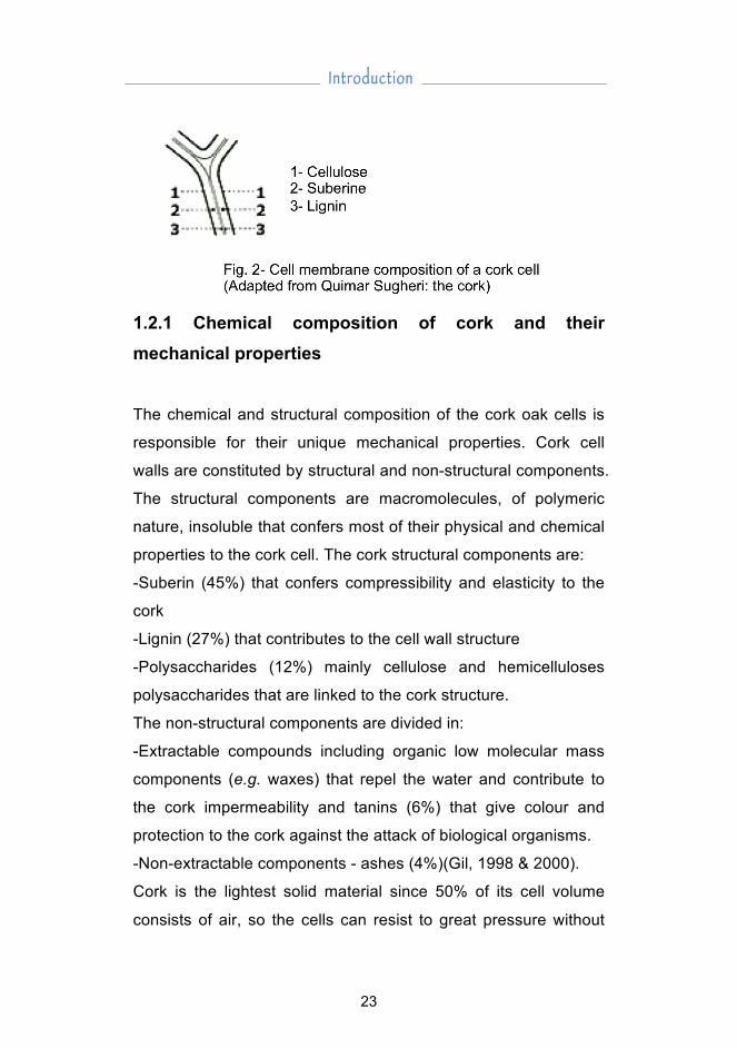
Structurally, cork is constituted by several cells in tangential
section having a polygonal shape, disposed in a regular and
compact manner without any empty spaces. Images from
scanning electron microscope show that cork cells have a
structure similar to a honeycomb. During its growth their cellular
content disappears and latter a suberization process
(impermeability) of its cellular membranes occurs (Gil, 1998).
These micro-cells are filled with a gas similar to air around 60 –
85% of the total volume (Maga, 2005). Their cell walls have five
layers: two formed by cellulose that surrounds the cellular
cavities; followed by two middle layers suberized (with suberin
and waxes) and lately one more internal constituted by lignin
(that confers the rigidity and structure) (fig 2).
22
I n t r o d u c t i o n
1.2.1 Chemical composition of cork and their mechanical properties The chemical and structural composition of the cork oak cells is
responsible for their unique mechanical properties. Cork cell
walls are constituted by structural and non-structural components.
The structural components are macromolecules, of polymeric
nature, insoluble that confers most of their physical and chemical
properties to the cork cell. The cork structural components are:
-Suberin (45%) that confers compressibility and elasticity to the
cork
-Lignin (27%) that contributes to the cell wall structure
-Polysaccharides (12%) mainly cellulose and hemicelluloses
polysaccharides that are linked to the cork structure.
The non-structural components are divided in:
-Extractable compounds including organic low molecular mass
components (e.g. waxes) that repel the water and contribute to
the cork impermeability and tanins (6%) that give colour and
protection to the cork against the attack of biological organisms.
-Non-extractable components - ashes (4%)(Gil, 1998 & 2000).
Cork is the lightest solid material since 50% of its cell volume
consists of air, so the cells can resist to great pressure without
23

C h a p t e r 1
breaking and retain 90% of its original form after the pressure is
released, so maintaining their dimensions in one direction even
when the pressure is applied in another one (Fortes, 2004; Gil,
2007).
1.3 Cork forest sustainability The cork oaks trees can live up to 350 years and produce cork
continuously during its life (Fortes, 2004).
The first harvest (virgin cork) is made when the tree is
approximately 25 years old. The first two harvests produce poor
quality cork, which are not used to produce stoppers. The
following harvests are done every 9-12 years, when the cork is
able to produce a layer of cork with the adequate thickness to
produce stoppers.
Cork harvesting is done entirely by hand, and it is not a harmful
operation. It is done at the end of the spring and during summer,
so the cork cells can regenerate and continue to divide (Fortes,
2004).
Cork is a biodegradable product and has a positive impact in
carbon fixing thus reduction the greenhouse gas emissions that
cause the climate change
(htttp://www.corkcomposites.amorim.com/client/skins/english/sim
ples.asp?produto=23).
The needs to provide an adequate management service of the
cork oak forests thus assuring its sustainability lead to the
certification of the montados. In Portugal the Forest Stewardship
Council (FSG) attributed around 28 thousand hectares of certified
cork forest.
24

I n t r o d u c t i o n
1.4 Cork applications The unique physical properties of cork make it a material suitable
for many applications. Traditionally, cork has been used since the
early antiquity as floating device, sealant and insulator. Several
cork devices were found (e.g. fishing items, cork lids, sandals
soles) in China, Egypt and in the Mediterranean area. However,
the most important application of cork is to seal wines, drinks and
sparkling wines as stoppers, which started in the 17th century
(http://www.corkfacts.com/nchoice1.htm).
The manufacturing of cork stoppers originates many remains
however all of them used. The by-products from the cork
stoppers manufacturing are used in other purposes.
Some of these products can be used in vibration, thermal and
acoustic insulators in roofs, walls and floors (Gil, 1996). The
lipophilic extractives of cork and cork by-products are promising
sources of bioactive chemicals or chemical intermediates for the
synthesis of value added compounds (Sousa, 2006).
Some of these wastes can be have several uses like hockey and
baseball balls, golf cubs and rackets. They can also be used in
computer printers, handbags, fishing rods, floats, carpets and
helmets (APCOR) and in electrical and automobile Industries
(Silva, 2005)
In recent years, the interest in cork increased and it is a material
used by architects, designers and decorators due to the fact that
it as natural resource extremely easy to use, renewable and
environmental friendly. This noble material also serves to make
clothes (http://www.trendhunter.com/trends/cork-clothing-gets-
25

C h a p t e r 1
fashionable), little pieces of furniture (e.g. cushions, sofas and
pillows) (http://www.squidoo.com/decorate-with-cork) and even
an entire house made from cork
(http://www.tvleak.com/overflow/house-made-of-cork/).
Recently, the cosmetic investigations discovered the anti-aging
effect of the cork extract. This fact is mainly due to the suberin
content present in the cork cell wall, specially its hydroxycarbolic
acids that provides special mechanical and chemical properties
originating the possible smoothing effect on the surface skin
(Coquet, 2005).
1.5 Manufacturing of cork stoppers On the early times of cork stopper industry, all the procedures
were quite rudimentary and mainly based on empiric knowledge.
Although, the cork stopper is still punched with its axis
synchronized to the plank axial position and the noblest part of
the cork plank is used.
The Industry introduced some new technological innovations to
guarantee the consumers the manufacture of high quality
products. Moreover, their production processes have been
checked and objectively analysed so the European cork
federation (CELIEGE) elaborated the International Code of Cork
Stopper Manufacturing Practice (SYSTECODE), which consists
of several rules to assure the quality system and accreditation of
the cork Industry (Celiège, 2006).
According to the SYSTECODE, the manufacture of cork stoppers
includes several manufacturing stages starting with the stripping
of the cork trees in the forest. After stripping, the cork is pilled
26
I n t r o d u c t i o n
and stacked in the forest or in the factory outdoors, exposed to
rainfall, for 6 months to one year, in order to become flattened
and to enable the elimination of most of their dirt, also to oxidize
most of their polyphenols and to partially stabilize their structure
(Gourlay, 1998). In a second stage, cork slabs are prepared for
the industrial processing. The cork slabs showing visible defects
are immediately separated and the other ones are arranged in
pallets with 3 levels.
The next stage in the Industrial processing is the boiling of cork
slabs in water (95 – 100ºC) for about 1 hour. This is a very
important stage as humidity allows the raw slabs to flat, and
expand the cork tissue and to stabilize it dimensionally, mainly in
the radial direction. Also cork becomes softer and more elastic
(Fortes, 2004; Silva, 2005; Pereira, 2007b). Some tannins and
minerals salts of the cork are partially extracted during this stage.
To prevent the cork contamination by chlorinated compounds, no
chlorine treated water is used and the boiling water is periodically
renewed as some cork phenolic compounds, soluble solids or
volatile compounds can be extracted from it. Their accumulation
can be a contamination source for cork slabs during subsequent
boiling since cork can absorb many volatiles and phenolic
compounds (Pollnitz, 1996).
The boiling stage does not promote the microbial sterilization of
cork. Although the water temperature reaches near 100º C, the
specific physical-mechanical and chemical properties of cork
namely its waterproof properties do not allow the water to reach
some deeper layers of cork slabs. Moreover, the particular
structure of cork with its lenticels filled with air leads to a specific
27
C h a p t e r 1
niche where water does not normally enter so the fungal spores
are not destroyed.
After boiling, the cork slabs are left to dry in the factory
atmosphere for some days (3-4 days). During this period the
slabs will become more flat and straight. The humidity levels
starts to decrease immediately after boiling and after two days
attain a humidity level ranging from 14–18% (0.9 of water activity
(aw) (Pires, 2007), which is the adequate level for cork
transformation (slabs slicing and stoppers or discs punching)
(Pereira, 2007b). After this period the cork planks are selected
once again, and separated according to their thickness and
quality. Then they are sliced and punched according to its
thickness in stoppers or for cork discs.
Regarding the manufacturing of cork discs, the slabs are first cut
into strips, where the inner and outer-backs are removed. Then
the slabs are punched into discs using an automated process.
Furthermore, the discs and stoppers are washed and bleached
with hydrogen peroxide aqueous solution; dried until they reach a
humidity level that ranges between 5–8%, and dimensionally
rectified. Both, cork stoppers and discs are then carefully chosen
using optometric and manual choice. Therefore they are
classified according to different quality classes mainly based on
their visual appearance.
Lately, the cork stoppers are washed, dried and their surface is
treated with a lubricant film to reduce friction and to allow a better
introduction and extraction into the bottleneck. Additionally,
regarding the technical stoppers there is the assembly of the
different parts of their constituents to form the stopper (e.g.
28
I n t r o d u c t i o n
champagne cork stopper an agglomerated body that is glued to
two natural cork discs in one side).
2. Fungi on cork Several different types of fungi co-exist in the same habitat as
Quercus suber trees. Some of them are important to the tree
adaptation to the habitat, establishing a mutual relation with the
tree roots, as mycorriza. Others colonize the tree itself in the
trunk, leaves or branches. When the cork is harvested several
fungi colonizing it constitute a specific mycobiota as seen in non-
boiled cork planks.
Fungi are eukaryotic organisms possessing absorptive
heterotrophic nutrition. Their vegetative body is constituted by
million of threads like structures, with or without septa, called
hyphae constituting a multinucleate structure that exhibit apical
growth. They form spores, as reproductive and/or resistance
structures (Bennet, 2001). Their walls are constituted mainly by
chitin, a polymer of N-acetylglucosamine.
The number of known fungi is estimated to be around 1.5 million
members (Hawksworth, 1991). Most of their members colonize
the terrestrial habitat, although some of them can be marine or
aquatic. Fungi possess a remarkable ability to utilize almost any
carbon source as food. Moreover, they colonize most habitats,
surviving some extreme physiological conditions (e.g.
temperature, pH, water activity and oxygen) (Alexopoulos, 1996)
Fungi own an important role in the decomposition and recycling
of the ecosystem (Pitt, 1997) because they produce a number of
29

C h a p t e r 1
extracellular enzymes, able to decompose recalcitrant substrates
into more easily metabolised ones.
Fungi are members of the kingdom Fungi constituted by five
phyla: Chytridiomycota, Gloemeromycota, Zygomycota,
Ascomycota and Basidiomycota (Carlile, 2001). The latter three
phyla belong to the Eumycota Division. Eumycota members
reproduce by spores and can have an asexual stage (anamorph)
or sexual stage (teleomorph) or both (holomorph) phases. When
the teleomorph of some fungi are not known, they are named
mitosporic fungi (Hawksworth, 1995). The most important fungi
involved in food and airborne contamination are included in this
group. Recently the holomorph of more fungal species was
known using heat shock treatment of the anamorph species
(Houbraken, 2008).
The fungal genera Aspergillus and Penicillium are the most well
known to be common air and foodborne habitants and may also
be involved in the spoilage of food products. They are taxonomic
placed in the Eurotiales, within the phylum Ascomycota (Frisvad,
2004).
2.1 Fungi present in cork and cork environment
The presence of fungi colonizing the cork was described in
earlier works by Mathieu (1900), Bordas (1904) and Sharf & Lyon
(1958) according to Riboulet (Riboulet, 1982).
Several studies concerning the identification and characterization
of the cork mycoflora along several stages of cork stoppers
manufacturing process were made along the time.
30

I n t r o d u c t i o n
The quality of the air environment of a cork factory was also
assessed to characterize the fungal spores present in there
(Lacey, 1973; Ávila, 1974). Additionally, since cork is produced
mainly in a country and then exported elsewhere, the cork planks
that were transported in the ships containers and the cork bales
were investigated to see if any fungi were present in them (Lacey,
1973; Ávila, 1974). Additionally, cork stoppers exported to
Australia were studied to detect the eventual presence of natural
mycoflora and to see if the ship conditions could originate the
fungal contamination (Davis, 1981; Lee, 1993).
The whole cork manufacturing process involves the shift of
humidity levels along the time. This factor is of the outmost
importance as it allows the germination and development of
fungal mycelia in cork. All the cultivable fungi that colonize cork
grow at 0.97 aw, some of them continue to grow at 0.85 aw,
however very few are able to grow at 0.80 aw (Simpson, 1990).
On the other had, the critical point for C. sitophila development is
0.9 wa (Pires, 2007), which is the fungal species that covers the
cork slabs after boiling with its white to pink abundant mycelium.
In the beginnings of cork stoppers industry it was assumed that
there presence indicate that the slabs had the adequate humidity
to be sliced and punched. When the humidity levels decrease
below 0.9 other fungi mycelium develop on the cork slabs, like
Penicillium, Aspergillus and Trichoderma. So it is important that
the maturating stage should last a maximum of 3 to 4 days. It
was recommended that the cork Industry respected that period of
time. In the case that they could not continue the process during
that time, a new boiling step should be done to maintain the
humidity levels to stop the growing of other fungi.
31
C h a p t e r 1
The cork mycoflora has been characterized over the past
decades. The predominant fungal genera found were:
Chrysonilia, Penicillium, Aspergillus, Trichoderma, Mucor (Davis,
1981; Daly, 1984; Simpson, 1990; Lee, 1993; Danesh, 1997;
Silva Pereira, 2000b; Alvarez-Rodriguez, 2002; Oliveira, 2003).
However, some studies refer genera like Rhizoctonia (Daly,
1984), Aphanocladium (Simpson, 1990), Fusarium, Acremonium,
Paecylomyces (Alvarez-Rodriguez, 2002).
The yeast community was also studied and the predominant
genera are Rhodotorula, Candida and Streptomyces (Lefebvre,
1983). Although, Candida famata, Sporodiobolus johnsonii and
Rhodotorula glutinis were species also isolated from cork
(Danesh, 1997). Recently, a polyphasic approach used on the
identification of some yeasts isolated from cork resulted in the
characterization of new genera like Rhodosporium,
Debaryomyces and Trichosporon (Villa-Carvajal, 2004) and the
characterization of Rhodotorula subericula, a new species
isolated from cork oak (Belloch, 2007).
2.2 Fungal communities
Fungal communities have been traditionally studied using
culture-based methods, where the fungal species were isolated
through the use of several media cultures. These methods are
not only time consuming but can only detect a limited number of
microorganisms. Some previous studies have calculated that
only 1 – 10% of the soil microflora is known by these methods.
The low biodiversity encountered when using this approach is
maybe due to the interdependence of the different
32
I n t r o d u c t i o n
microorganisms upon each other. Some species depend on other
ones and are not able to grow on artificial media cultures, also
the culture conditions that are normally used are not suited to
isolate most of the species that exist in a certain habitat (Muyzer,
1998).
To overcome this fact, some culture independent methods, which
are DNA or RNA based, have been developed and their use has
increased our knowledge of the biodiversity and functioning of
more mycobiota present in some habitats (Simon, 1993; Vainio,
2000; Vandenkoornhuyse, 2002; Anderson, 2003b;
Vandenkoornhuyse, 2003).
Most of these techniques have an initial step that consists in the
extraction of nucleic acids, directly or indirectly from the sample,
followed by amplification using polymerase chain reaction (PCR)
of a specific part of the genomic DNA/RNA. The method used to
extract DNA or RNA is vital for the success of the subsequent
steps, since the quality and the purity of the extracted nucleic
acids is important for the subsequent PCR amplification.
Another aspect to take into account is the primers choice for
doing the PCR reaction. Some primer pairs amplify preferentially
some fungal species, usually the ones that are present in higher
concentrations at the expenses of the ones that are in lower
concentrations in the template (Dickie, 2002). The excessive
specificity of the primers can create this bias in the fungal
biodiversity of the sample.
Another factor to consider when dealing with the fungal genome
is the multicopy nature of some target region in the fungal
genome. The rRNA operon, which is widely used in these studies,
is assumed to have equal number of copies for each fungal
33
C h a p t e r 1
species. However, the number of copies in the genome of
different fungal species is different and the exact number of the
copies it is unknown for most of the species (Hibbet, 1992).
The techniques used to investigate the fungal communities are
able to identify different fungal species, genera, families or higher
taxonomic groups depending on their sensitivity (Nannipieri,
2003). Most of the molecular methods have intermediate
resolution and have been used to detect mycota groups instead
of mycota species such as denaturing gradient gel
electrophoresis (DGGE).
DGGE technique have been based in the electrophoretic mobility
of DNA fragments of the same size, previously amplified by PCR,
in polyacrylamide gels that possess a linear gradient denaturant
made with urea and formamide. The DNA double-stranded that is
subjected to increasing concentrations of denaturant starts to
partially melts in the so-called “melting domains”. The DNA
mobility depends on its base composition and this technique
allows the differentiation of two DNA molecules differing in only
one single base (Muyzer, 1999). Moreover, an optimal resolution
is obtained when the molecules do not completely denature this
is assured by the addition of a 30 to 40 GC clamp to one of the
PCR primers. This technique is simple and rapid to perform and
allows the identification of shifts or changes that occur in the
fungal community composition through a period of time (van
Elsas, 2000; Vainio, 2000; Anderson, 2003b). Furthermore, the
bands obtained after DGGE can be excised, re-amplified and
sequenced. The obtained DNA sequences can be compared with
known sequences present in databases. The objective is to
identify the fungal species corresponding to each amplicon
34

I n t r o d u c t i o n
(Nielsen, 2005). The primers choice is crucial for the technique
sucess. Most of the used primers correspond to some parts of
the rDNA region, which is a multicopy gene present in each
fungal cell. However, the use of one copy gene primer is starting
to be taken into consideration to be used.
The rapid analysis and comparisons between different samples
in one gel, and also amongst several gels, is quickly made.
However, that comparison depends on the use of suitable
internal standards that allows an accurate analysis amongst all
gels. The reproducibility between gels has been mentioned as
one of the main pitfalls of the technique that can be overcome
using standardized equipment to prepare each gel.
One disadvantages of this technique are the use of shorter DNA
amplicons (<500 bp), thus limiting the taxonomic information
obtained from excised band gels, although some larger PCR
products have been used successfully (Ranjard, 2000;
Landeweert, 2004). However, the obtained biodiversity profiles
could not correspond to the real variety of the sample.
Another unfavourable condition is the staining method that often
is not too sensitivity to detect the less dominant members of the
fungal community. Besides, in some cases, a single band on the
gel does not correspond to a fungal isolate, because some DNA
amplicons with different base compositions co-migrate together.
To overcome this problem the excised band is cloned and then
sequenced.
Another technique use to study fungal communities is the cloning
of the amplified PCR amplicons from specific samples to be
studied (Chen, 2002; Anderson, 2003a). The obtained clones
can be screened using restriction fragment length polymorphism
35
C h a p t e r 1
technique. (RFLP). This reduces the number of clones to be
sequenced since the use of restriction enzymes groups the
clones into different OTUs. So the choice of how many and which
restriction enzymes are going to be used is an important step to
have in consideration, since previous studies using Pisolithus
isolates and using two restriction enzymes showed that the same
species of Pisolithus can have different RFLP profiles (Hitchcock,
2003). The advantages of this technique are that it provides a
diversity profile of the fungal community existing in a specific
habitat. The disadvantages are that it takes a lot of time to obtain
clones with different operational taxonomic units (OTUs) and to
sequence them being a laborious and potentially costly technique.
The presence of chimeras (DNA sequence originated from two
different organisms) is another factor to take into account.
Moreover, the taxonomic identification of the clones depends on
the correct identification of the sequences deposited in the known
public sequence databases.
3. Secondary metabolites produced by fungi Secondary metabolites were defined in opposition to primary
metabolites (or central metabolites), which are common to all
fungi and serves for its survival in the producing of carbon flux
and energy.
Exo-metabolites (or secondary metabolites) are natural chemical
compounds that are synthesized by fungi in response to an
interaction with the surrounding environment (Thrane, 2007).
They are energetic costly chemical products usually produced
late in the cell differentiation or development processes and
36

I n t r o d u c t i o n
normally produced with sporulation (Calvo, 2002). The production
of exo-metabolites is not consistent for all fungi; instead it is
specific to certain genera and species (Frisvad, 1998). Although,
some researchers claim that it can be strain specific (Engel,
1982).
Some of the exo-metabolites produced by fungi are considered to
be mycotoxins. According to the definition proposed by Frisvad,
Thrane and Samson (2007), mycotoxins are chemical
compounds that when present in low concentrations, are toxic to
any vertebrate animal. Some of them can be neurotoxins, or
carcinogenic, while others can lead to the kidney or liver
deterioration. Some of them can also interfere with the protein
synthesis producing several effects like, skin sensitivity to
extreme immunodeficiency (Sweeney, 1998). The fungal genera
that produce most of the fungal exo-metabolites and mycotoxins
are, Penicillium, Aspergillus and Fusarium genera and their
teleomorphs.
In the last decades, some exo-metabolites showed potential as
source of new pharmaceutical compounds like, antibiotics,
immunosuppressant’s and antiviral compounds (Larsen, 2005).
These data added interest in the demand to search new
molecular active molecules.
The exo-metabolism specificity encouraged the mycologists to
use the exo-metabolite profile as a taxonomic tool (Karlovsky,
2008). The Penicillium, Aspergillus and Fusarium genera have
had good results. The first two are known to be present in food
and also in environment and the former genus is constituted
mostly by plant panthogens (Sweeney, 1998).
37

C h a p t e r 1
The production of exo-metabolites by fungi is studied in the
laboratory using a solid culture media, since it is quite similar to
fungal natural substrata (e.g. food, plants, decaying wood). The
exo-metabolites production depends on several factors. The
substrate ingredients of the culture media are carefully chosen to
obtain the production of metabolites. Some of them are produced
under certain environmental conditions and only when certain
trace metals are present (e.g. iron or cupper).
The physiological conditions of the strains are a very important
factor and several transfers can deteriorate them, as well as the
accumulation of carbon dioxide can inhibit the metabolite
production. The investigation done by some research groups
(Filtenborg, 1990), (Frisvad, 1989) showed that the substrates
that have easily assimilable nutrients originates the production of
most exo-metabolites (Thrane, 2007). Most of the synthetic
media cultures, like yeast extract sucrose agar (YES), malt
extract agar (MEA), potato dextrose agar (PDA) and oatmeal
agar (OA) are the most used and give the best results.
Furthermore, the study of the mycobiota composition of food or
products indirectly used in the food chain is necessary to assess
the possible existence of mycotoxins to assure the quality and
safety of those products, for instance in wheat and white wheat
(Weindenboerner, 2000) and in maize (Soriano, 2004).
3.2 Volatile compounds produced by fungi Volatile organic compounds (VOCs) produced by the fungi
normally contribute its intense and characteristic odours. Their
production has been shown to be consistent and related to
38

I n t r o d u c t i o n
cultural conditions and abiotic environment (Larsen, 1998). This
characteristic can be used taxonomically to distinct different
species at least in some fungi groups, e.g. Trichophyton (Sahgal,
2006) or Penicillium (Larsen, 1995a), (Larsen, 1995b), as
dissimilarity among them is a difficult task.
Moreover, volatile metabolites can have some allergic effects on
humans (Fischer et al, 2000). Their presence in foodstuff
(Schnurer, Olsson & Borjesson, 1999) and inside buildings has
been widely studied (Girman et al, 1999; Hodgson et al, 2003).
VOC-mediated positive, negative or neutral interactions can
occur between a very wide range of soil bacteria and fungi.
These effects include both stimulation and inhibition of growth,
the enzyme production being only an example of the different
surviving strategies used by the biological communities. Many
organisms are known to modify the environment in order to
construct an adequate niche where natural selection can take
place (Brown, 2009).
3.3 Cork taint and off-flavours Taint is by definition a foreign taste or odor imparted to a product
(ISO, 1992), which gives disagreeable taste and most often
results in consumer rejection of the product.
Cork is referred in most studies as the main source for the cork
taint in wines. The main reason lies in the fact that several
microorganisms colonize the cork tree and most of them are
lodged in their cork lenticels and structural fissures (Macku,
2009). Latter the microorganisms can also colonize cork along
the stopper manufacturing process. These microorganisms can
39
C h a p t e r 1
metabolize chemical compounds causing the cork taint in the
presence of some humidity from: a) some compounds belonging
to the cork lignin or suberin b) some chemical compounds that
does not exist in the nature but were used for many decades as
pesticides, wood preservatives, fungicides. However, some
investigations also showed that the taint can be originated from
a) contaminated oaks barrels b) contaminated winery machinery
used for bottling the wines c) airborne fungus eventually present
in the winery environment and d) molds present in wooden
barrels and wine making devices (Haas, 2010).
The first presence of cork taint in bottled wine was described in
1904 (Lefèbvre et al, 1983). The report of several types of cork
taint was well described by Duncan (1995) although, the real
definition of “cork taint” refers to a very unpleasant odour still of
unknown origin extremely rare to occur (Ribéreau-Gayon et al.,
1998). The term taint can include several odors like earthy, musty,
medicinal, moldy, mushroom-like, earthy (Margalit, 1997),
depending on what compound originated the taint.
The proportion of wines contaminated with cork taint has been
the subject of some divergence depending on the informative
source. According to the Industry, the problem affects 0.7% of
the bottled wine (Hall, 2002). On the other hand, the studies done
so far indicate that the real value should be around 1-5% (Soleas,
2002). These values will depend which chemical compounds
originated the taint as well as the origin of the taint, since the
compounds detection threshold can vary.
Several chemical compounds have been identified as
responsible for the taint in wines. Some of them possess different
flavors (Pena-Neira et al, 2000; Alvarez-Rodriguez et al, 2003).
40

I n t r o d u c t i o n
However, in most cases not only one but also a mixture of
compounds can be detected in tainted wines (Silva Pereira,
2000). These compounds can act alone or synergistically; a
phenomenon by which the effect is produced by all the
components that is greater than the sum of their individual
contribution. This fact makes it difficult to assess the contribution
that each components gives to the taint. The main chemical contaminants identified as responsible for
taints are haloanisoles.
3.3.1 Haloanisoles Haloanisoles are the most common volatile compounds detected
in tainted wines, mainly chloro- and bromoanisoles (Amon, 1989;
Pollnitz, 1996; Chatonnet, 2004; Coque, 2006; AFGC, 2007).
Chloro- and bromoanisoles are derivatives of the anisole (or
methoxybenzene) that possesses at least one substituted
chlorine (or bromide) in the phenolic ring. These chemical
compounds are volatiles produced by fungal methylation, when
environmental humidity level and the corresponding halophenols
precursors are present (Alvarez-Rodriguez, 2002). This chemical
reaction occurs as a detoxification way, to transform the toxic
halophenols in non-toxic haloanisoles. S-adenosyl-L-methyonine
(SAM)-dependent methyltransferase is the enzyme that catalyses
this chemical reaction resulting that the halophenol is converted
into the respective anisole (Coque, 2003).
Chloro and bromoanisoles have been detected in most cases of
tainted wine (Amon, 1989; Chatonnet, 2004; Coque, 2006; AFGC,
2007) as being responsible for that defect. According to Coque et
41

C h a p t e r 1
al. (2006) the most important chloroanisoles implicated in the
contamination of wine are: 2,4,6-trichloroanisole (TCA), 2,3,4,6-
tetrachloroanisole (TeCA), pentachloroanisole (PCA) and 2,4,6-
tribromoanisole (TBA). However, 2,4- and 2,6- dichloroanisole
were also found in some tainted wines by some researchers
(Pollnitz, 1996). Moreover, in most cases not one but several
compounds were detected in tainted wines (Pollnitz, 1996).
Apart from the wine (Pollnitz, 1996; Chatonnet, 2004) several
cases of food tainted with haloanisoles have been reported, e.g.
eggs (Curtis, 1974), sake ({MIki, 2005 #1265) and coffee
(Spadone, 1990). Some studies indicate that these chemical
compounds are probably the main contamination source of
tainted food (AFGC, 2007). Some studies indicated that the main
source contamination comes from the use of chlorophenols
mainly in the last decades as fungicides for wood protection and
preservatives (AFGC, 2007). However, now their use is forbidden
specially in Europe, United States as it has been classified as
“highly hazardous pesticides” by the International pesticide action
network (PAN International) and is listed in the highly hazardous
pesticides (International, 2010). Around the 1980s, other
compounds were detected in tainted products, namely
tribromoanisoles. Bromophenols are now used instead of the
banned chlorophenols. They are produced by the industry and
used as antifungal agents and flame-retardants, on wood, plastic
and paintings (Chatonnet, 2004). Moreover, the TBA can be
naturally found in the marine environment since it is synthesized
by the red algae Polysiphonia sphaerocarpato to remove
excessive bromine (Flodin, 2000). Although these compounds
are less toxic to the environment, they are present in many cases
42

I n t r o d u c t i o n
of food taint, like contaminated oysters (Watanabe, 1983) and in
several Australian Food products (AFGC, 2007), especially in
wine (Coque, 2006). The main problem concerning the chloro- and bromoanisoles
contamination is that these compounds possess a low threshold
detection levels, in ng/L range. This results in the product
rejection when these chemical contaminants are present in low
amounts (Saxby, 1996). The necessity to detect their presence in
very low quantities, at least sensitive to the human nose, as a
standard quality, has lead to the improvement of methods used
to detect the chemical compounds responsible for the off-flavours.
Such methods are: gas chromatography-mass spectrometry
(GC/MS) (Soleas, 2002; Chatonnet, 2004; Dias, 2008) or stir bar
sorptive extraction (SBSE) and thermal desorption GC/MS
(Hoffman, 2000) or even gas chromatography-olfactometry (GC-
O) (Plutowska, 2008).
3.3.1.1 2,4,6 -Trichloroanisole (TCA) Tanner (1981) published the first work relating the cork taint with
the presence of 2,4,6-trichloroanisole (TCA) but Amon (1989)
determined its content by gas chromatography-m ass
spectrometry (GC-MS) and found its major contribution on cases
of cork taint.
The most studied compound by the Cork Industry involved in the
cork taint is TCA (Riboulet, 1982; Daly, 1984; Rigaud, 1984;
Simpson, 1990, Lee, 1993; Pollnitz, 1996; Duncan, 1997; Sefton,
2005; Coque, 2006; Prak, 2007). This compound was detected in
80-90% of the taints (Maga, 2005) and its concern resides on the
43

C h a p t e r 1
low detection threshold, which lies between 1.4 ng/L and 10 ng/L
(Buser, 1982; Silva Pereira, 2000a; Alvarez-Rodriguez, 2002).
The detection of low quantities of TCA is difficult in a more
complex matrix than water (Teixeira, 2006). When this compound
is present in wines the rejection odor threshold is 10 ng/L (white
wine) and 16 ng/L (red wines) according to Teixeira et al. ( 2006).
Most of the studies performed until now focused on the ability of
the cork isolated fungus to methylate TCP into TCA (Lee, 1993;
Pollnitz, 1996; Silva Pereira, 2000a; Howland, 2008; Maggi,
2008). Other studies refer the mycobiota diversity present in
tainted corks and related them to the TCA presence (Daly, 1984;
Caldentey, 1998; Alvarez-Rodriguez, 2002; Prak, 2007).
Although the fungal species content in cork is different for each
research studies, some fungal genera isolated are common to
most of the studies, like: Penicillium sp., Chrysonilia sp.,
Cladosporium sp., Trichoderma sp., Aspergillus sp. (Davis, 1981;
Lee, 1993; Hill, 1995; Silva Pereira, 2000b; Alvarez-Rodriguez,
2002).
Silva-Pereira (2000) incubated some cork fungi in a medium
culture containing TCP and analysed their capacity to convert the
chlorophenol into TCA. The work showed that the isolated fungi
have low potentially to convert TCP into TCA. C. sitophila was
able to metabolize TCP but had a conversion rate of 0.03 %,
appearing to be a non-TCA producer (Silva Pereira, 2000b).
Alvarez-Rodriguez (2002) also studied the capacity of 14 cork
isolates belonging to several fungal genera to convert TCP into
TCA. The results showed that Fusarium sp. and Trichoderma sp.
were strong producers transforming 25% of TCP into TCA; the
moderate producers were two Penicillium strains, Acremonium
44

I n t r o d u c t i o n
strictum, C. sitophila and Cladosporium oxysporium that
converted between 10-25% of the TCA into TCA, P. viridis, P.
chrysogenum and V. psalliotae were low producers and M. alpine,
M. plumbeus and P. decumbens non-producers (Alvarez-
Rodriguez, 2002).
Other researchers showed that Mucor sp., Paecylomyces sp.,
Penicillium sp., Trichoderma sp. were able to convert TCP into
TCA. However, the best yields were obtained by Paecylomyces
sp. and P. chrysogenum. Additionally, in this study C. sitophila
and Penicillium sp. did not produce TCA (Prak, 2007).
Another research work reported the capacity of the fungi
Cryptococcus sp., Rhodotorula sp., P. glabrum and P. variable to
produced TCA to a great extent (Prat., 2009).
The results obtained concerning the capacity of some fungal
isolates to produce TCA from TCP leads to the hypothesis that
this feature could be a strain dependent effect, thus explaining in
different works by the different methylation capacities obtained
for different isolates of the same species, e.g. C. sitophila and P.
glabrum. Moreover, no direct relation between the methylation of
TCP to TCA and the amount of TCP on the cork could be
established, except for the work of Simpson and Lee that
reported that the tested fungus had 74-100% of conversion to
TCA, however, only 0-23% of them was converted to TCA
(Simpson, 2007). All the other works reported lower conversion
rates suggesting that TCP metabolites can be incorporated in the
cellular material (Silva Pereira, 2000b; Alvarez-Rodriguez, 2002).
The quantity and diversity of moulds present in cork stoppers,
some of them with TCA while others with none were studied. The
results showed that the number of moulds is identical in both
45

C h a p t e r 1
cases, although the biodiversity is higher in TCA-containing
stoppers (Prak, 2007).
It was observed that the number of corks that possess TCA or
TCP is higher compared to the percentage of the wines that
became tainted (Juanola, 2005). This fact was the starting point
to to look for the factors that affect the TCA transference into the
wine (Capone, 2002; Soleas, 2002; Sefton, 2005; Juanola, 2005;
Alvarez-Rodriguez, 2009).
Additionally, in case of contamination of tainted wines present in
the winery environment and absorbed by the cork, the main
question is the TCA capacity to migrate through the cork to
contaminate the wine.
Research showed that TCA has more affinity to the cork than to
the hydro-alcoholic solution or wine (Juanola, 2005; Sefton,
2005; Alvarez-Rodriguez, 2009), so if this compound is present in
the wine the cork will absorb at least most of it. However, if the
cork is the source of contamination depending on the cork’s
surface of contact with the wine the percentage of the TCA that
passes into the wine depends on the time, temperature as well
as the compound amount that is present. In cases of bottled wine
could range between 0.7 to 2.7% (Soleas, 2002) or less than
0.1% (Juanola, 2005).
The amount of TCA is not uniformly distributed in the cork
stopper. Research done so far indicates that TCA migrates
poorly inside the cork and stays mainly at its surface (Capone,
2002), (Sefton, 2005; Alvarez-Rodriguez, 2009). Moreover, after
24h of cork exposure to d5-TCA the compound was restricted to
the 2 mm of the outer layer, although 15 to 25% penetrated
beyond this layer, possibly via lenticels (Capone, 2002).
46

I n t r o d u c t i o n
However, in long term experiments that tries to mimic the long
storage of wines, the studies done so far indicate that the surface
equilibrium attained by the TCA-cork stopper is disturbed and
some level of TCA can be released into the wine (Capone, 2002),
(Juanola, 2005). It seems that TCA located in the cork matrix
reaches more accessible layers and is slowly released into the
wine. This could happen after 12 months storage and migration
ranges from 0.7 to 2.7% (Soleas, 2002). Additionally, the
releasable pollutants analysed (TCA included) could not be
completely extracted by soaking into a hydro-alcoholic solution
(Alvarez-Rodriguez, 2009). The study concluded that the TCA
does not present a problem to wine closed with natural stoppers.
3.4 Other chemical compounds capable of producing taint
Some other chemical compounds were detected in tainted wines,
which are capable to contribute or cause the off-flavor. Several
compounds can impart a musty earthy off-flavor to the wines e.g.
geosmin, 2-methylisoborneol (Karahadian, 1985; Darriet, 2000),
2-methoxy-3-isopropylpyrazine (IPMP), 2-methoxy-3,5-
dimethylpyrazine (MDMP), guaiacol (Álvarez-Rodríguez, 2003),
1-octen-3-ol and 1-octen-3-one (Karahadian, 1985; Darriet, 2000).
The contribution that each compound gives to the taint is not
equal; it depends on the perception threshold and the chemical
stability of each one of them in the wine, as well as the wine
composition. The detection thresholds of these compounds are in
the ngL-1 range (µgL-1 for guaiacol and 1-octen-3-ol) (Prat, 2008).
47

C h a p t e r 1
Among these compounds geosmim appears to cause less
problems since it is chemically unstable in wine (Sefton, 2005).
4 Economic consequences of wine cork taint
Portugal is the world leader of the production and manufacturing
of cork stoppers, responsible for 70 % of the global exporting
(WWF, 2006). According to the Portuguese National Institute of
Statistics (INE) cork exportation registered an increase of 0.6 %,
export value in 2007 compared with 2006. In 2007, natural cork
stoppers are the major Portuguese exported product ranging
values of 415 million euros followed by champagne cork stoppers
with 88 million euros and agglomerates with 86 million euros. The
main destination countries of cork exportation in 2007 were:
France (20.6 %), USA (15.7 %), Spain (13 %), Germany (8 %)
and Italy (7,6 %).
It is reported that cork taints occur in 1 % to 8 % of the
commercialised wines (Pereira, 2006) and its occurrence is
responsible for economic losses suffered by either the cork and
wine industry (Álvarez-Rodríguez, 2009). Even if the responsible
for the wines contamination cannot always be imputed to the cork,
the Industry is ensuring a high standard product to decrease its
trustworthy.
However, the cork taint occurrence even with a low incidence,
can origin high losses for both the cork stopper and wine
Industries. As a consequence, in the nineties the synthetic wine
stoppers to seal wine bottles developed and acquired an
increased importance mainly due to economic interests of non-
cork producing countries. The main drawback of these devices is
48
I n t r o d u c t i o n
their negative environmental impact. Furthermore, to our
knowledge, they possess low quality, at least, to seal high quality
wines that usually require long storage periods to ensure the
proper aging of the wine bouquet. Several studies were done to
investigate the impact of the closure in wine aroma and aging,
using wines sealed with both synthetic closures and natural cork
stoppers (Karbowiak, 2010; Skouroumounis, 2005). The results
showed that wines with synthetic closures exhibit a relatively
oxidised aroma, a brown colour and low levels of sulphur dioxide.
Natural cork stoppers, on the other hand, showed negligible
reduced aroma characters. The closures act primarily as a barrier
to oxygen, since if this gas is present in low or high levels can
impart an oxidized or reduced flavour, respectively. Moreover,
micro-oxygenation enabled by the cork porosity is a very
important process to allow wines age correctly inside the bottles
(Mills, 2006; Toit, 2006).
Currently, new cork products manufactured according the
Industry technological research knowledge are available. These
new products were developed to eradicate all TCA precursors
and to be well adapted to the demands of the wine market.
i) natural cork stoppers are 100% natural product and can be
made with a) single piece of cork and it is recommended for seal
reserve wines or wines that need to age in the bottle or b) a
multipiece natural stoppers which comprises more than one
piece of cork glued together by FDA approved contact glues.
When the wines do not need to age in bottles the stoppers can
be made of:
49
C h a p t e r 1
ii) colmated cork stoppers which are natural cork stoppers that
contain pores or lenticels preserved with glued cork dust. They
are used for the same wines as the multipiece stopper
iii) champagne and sparkling wines stoppers that are constituted
by an agglomerated body with two cork discs glued to one side iv) Technical cork stoppers which are constituted by an
agglomerated body possessing a) one cork disc in each side or
b) two cork discs in one side. These closures are used to seal
wines that need to be consumed within shorter periods.
v) agglomerated cork stoppers formed from granulated corks
which are constituted by little pieces of corks glued together.
They are perfect for wines that require storage for no more than
12 months.
vi) capsulated cork closures. These stoppers derives from a)
natural cork or b) colmated cork stoppers which possess the
upper portion tied with glass, metal, porcelain, wooden or PVC.
They are used to seal fortified wines and spirits, and can be
reused (http://www.cork.pt/cork-stoppers.html).
Some studies done with “technical” cork stoppers (Neutrocork),
natural cork stoppers, and synthetic closures (Nomacorc)
showed that for natural and technical closures the oxygen
diffuses into the bottle during the first 12 and 24 months of
storage, respectively. Equally, under the study conditions,
“Nomacorc” closures showed to be permeable to the atmospheric
oxygen (Lopes, 2007). In conclusion, mimicking the heterogenic
cellular structure of the cork even if the material had the same
impermeability and mechanical properties can be a very difficult
task to achieve. Throughout recent years, the cork stopper
continues to be the noblest product used to seal wines.
50

I n t r o d u c t i o n
5 Fight against cork taint Several measures were implemented in the cork manufacturing
process in order to eliminate or reduce the TCA from the cork, as
a way to decrease the cork responsibility in the wine spoilage.
Research has been developed either in some industries or in
straight cooperation with public research institutions. Since the nineties, several cork stoppers producers in
conjunction with several investigation groups developed some
processes that aim to eliminate TCA. However the presence of
this compound remains to be not completely solved. Some of the
actually most important available strategies can be summarized: - Amorim & Irmãos Company developed the ROSA
(http://www.amorim.com/cor_ied_rolhas.php) method based on a
steam cleaning process, which efficiency in the TCA removal was
reported by several authors (Sefton, 2005). -The OENEO group, formerly known as Sabaté developed a
method called “Diamont” based on the use of supercritical carbon
dioxide extraction. The haloanisoles are selectively removed from
the raw cork material. Several independent studies showed the
good results obtained by this technique.
(http://www.beveragedaily.com/Financial/Oeneo-and-the-
Diamond-route-to-recovery). -A group of researchers and cork companies developed a
method called DELFIN (direct environmental load focused
inactivation) that use a technology based on microwaves to
remove TCA from the stoppers. They claim that reduces TCA
levels at least 90% and it kills microbes throughout the stopper.
51
C h a p t e r 1
(http://www.winespectator.com/webfeature/show/id/Research-
Group-Claims-to-Have-Eliminated-Wine-Cork-Taint_20322). - A technique based on the use of gamma radiation was
developed to remove TCA from cork. This technique transforms
the TCA existing on the cork in molecular residues that do not
possess the same organoleptic characteristics (Pereira, 2007a)
(Portuguese patent application PT 103006). - A CTCOR developed a technique called SYMBIOS, which
consists in the addition of an addictive to the boiling water. This
compound reduces the manufacturing process to one boiling that
takes one hour and a half. This technique reduces the
microbiological population on the post-boiling stage (e.g.
development of mainly C. sitophila and Mucor mycelium in the
cork surface) increasing also the extraction of polyphenolic
compounds from the cork
(http://www.ctcor.com/fotos/gca/SymbiosE.pdf).
II– Aims and layout
The main objective of this thesis was to isolate and identify fungal
species present along the main stages of the manufacturing of
cork discs, including raw cork, using either molecular or
phenotypical techniques. The uncultivable mycobiota present at
each stage of the manufacturing was also assessed by DGGE
(denaturing gradient gel electrophoresis), cloning and
subsequent sequencing. The elucidation of the relationship
between cork fungal community and the different cork niches
created along cork stoppers manufacturing process was also
envisaged.
52
I n t r o d u c t i o n
The most predominant fungal isolated fungal species or the most
susceptible of producing harmful exo-metabolites were
inoculated in several culture media: semi-synthetic and cork
based culture media. Their exo-metabolite profiles were analysed
and compared among them and the possible production of
mycotoxins was evaluated especially in cork-based culture
medium. Additionally, the production of volatiles and TCA by some
isolated fungal species inoculated in cork-based culture medium
was investigated. The main results obtained along this work were published or
submitted to publication and they constitute the following
chapters of this thesis: - Taxonomic studies of the fungal mycobiota presented in cork
samples collected throughout cork manufacturing discs
- Exo-metabolites produced by some fungal isolates in several
media cultures
- Volatile compounds produced by cork mycobiota
53


2
Taxonomic studies of the fungal mycobiota present in samples collected
throughout cork manufacture discs

C h a p t e r 2
This chapter focus the identification of cork mycobiota present in
some cork samples taken along the manufacturing of cork discs.
Two different approaches were used: one culture-dependent
(isolation) and two culture independent (DGGE and cloning
technique). In the course of this work it was found that about half
of the isolated species belong to Penicillium genus, mostly to
Glabra series and taxonomic studies of the Penicilllium glabrum
complex were done. Additionally, one isolate showed to have
phenotypical and molecular characteristics to be included in a
new species (Penicillium subericola).
This chapter consists of two scientific articles:
- Unveiling the fungal mycobiota throughout cork stopper
manufacturing process (submitted to FEMS Microbiology
journal).
- Taxonomic studies of the Penicillium glabrum complex and the
description of a new species P. subericola (Fungal Diversity,
2011, 49:23-33).
The experimental work presented in this chapter was done by the
author except the taxonomic studies that were made in
collaboration with the Applied and Industrial Mycology Laboratory
at Fungal Biodiversity Centre, CBS-KNAW, Utrecht, Netherlands.
Cork was ground with the help of Mário Gil Dias, Susana
Marcelino and Liliana Pinto. The cloning technique was done by
Biopremier Inovação e Serviços em biotecnologia, S. A., Lisbon.
Both manuscripts were written by the author and revised by the
other co-authors of the articles.
Unveiling the fungal mycobiota present throughout cork stopper 1
manufacturing process 2
M. C. Barreto *(1,2), J. Houbraken(3), R. A. Samson(3), D. Brito(1,24), M. Gadanho(5), M. V. San 3
Romão(1,2,6) 4
5
6
7
1Instituto de Tecnologia Química e Biológica - Universidade Nova de Lisboa (ITQB-UNL), 8
Av. da República. Estação Agronómica Nacional. 2780-157 Oeiras, Portugal 9
2Instituto de Biologia Experimental e Tecnológica (IBET), Apartado 12, 2781-901 Oeiras, 10
Portugal. 11
3CBS-KNAW Fungal Biodiversity Centre, Uppsalalaan 8, 3584 CT Utrecht, The Netherlands 12
4INRB I.P.-L-INIA, Oeiras, 2780-157, Portugal 13
5Bio-Fig, FC-UL, Lisboa, Portugal 14
6INRB I.P.-L-INIA, Dois Portos, 2565-191, Portugal 15
16
17
*Corresponding author. Phone: +351 214469555 Fax: +351 214 421 161. E-mail: 18
20
57

Abstract 21
The knowledge of the fungal population present in the main stages of the manufacturing 22
process of cork discs allow us to evaluate which species could produce any chemical 23
compounds that could spoil cork final product. 24
The fungal mycobiota using both culture dependent (isolation) and independent-methods 25
(denaturing gel gradient electrophoresis and cloning of the ITS1-5.8S-ITS2 region) was 26
studied. The mycobiota present in the samples taken in the stages before and after the first 27
boiling seems to be distinct from the population present in the subsequent manufacturing 28
stages. Most isolated fungi belong to the genera Penicillium, Eurotium and Cladosporium. 29
The presence of uncultivable fungi, Ascomycota and endophytes in raw cork was confirmed 30
by sequencing technique. The samples taken after the first boiling possessed uncultivable 31
fungi, still in few samples some isolated fungi were also detected. The main detected taxa 32
present in the following stages were Chrysonilia sitophila, Penicillium glabrum and 33
Penicillium spp. All applied techniques had complementary outcomes. 34
The main driven factors of shift in cork fungal colonization seems to be the high levels of 35
humidity and temperature, which the slabs are subjected during the boiling process. 36
37
Keywords: Mycobiota diversity, uncultivable fungal species, cloning, DGGE, cork 38
58

Introduction 39
Cork constitutes the outer layer of Quercus suber L. tree. It is a natural resource that is 40
continuously produced during the tree life span. Its unique chemical composition and 41
physical properties (compressibility, impermeability, resilience and chemical inertness) make 42
it a unique material suitable to be used in cork stoppers. Cork appears to be a specific 43
habitat due to its special chemical and physical constitution that harbours a unique 44
mycobiota population. 45
The European cork federation (CELIEGE) elaborated an International Code of Cork Stopper 46
Manufacturing Practice (SYSTECODE) that defines the necessary steps to acquire the 47
accreditation in the cork Industry. To assure the high standard of the final product the cork 48
industry follows this code. 49
The manufacturing of the cork discs (stoppers) comprises several stages and the most 50
important ones are the boiling of the cork during 1 hour and their subsequent resting stage. 51
These two stages allow the cork to become flat, softer and more elastic and thus being 52
workable. The high humidity levels of the boiling make cork a good habitat for the 53
development of the cork mycobiota. Few days after boiling the cork slabs become 54
completely covered by white/salmon mycelium of Chrysonilia sitophila. When the moisture 55
levels decrease below 0.9 aw, mycelia from other fungi (Penicillium, Trichoderma, 56
Cladosporium, Mucor and Aspergillus) start to germinate and replace C. sitophila mycelium 57
(Danesh et al. 1997; Alvarez-Rodriguez et al. 2003; Oliveira et al. 2003; Basilio et al. 2006; 58
Prat et al. 2009). 59
Fungi start to colonize the cork in the forest and different types of fungi can colonize the 60
root, trunk, leaves and even the surrounding soil. Some fungi will establish saprobyotic 61
relations with the tree, while others can be parasytic or mycorhizic. Most of these fungal 62
communities will also colonize the cork layer and could be actively growing on them. The 63
effect that the manufacturing process and mostly the boiling stage do in the original fungal 64
59

communities it is not known. However, several studies indicate that the manufacturing 65
process does not kill all the mycobiota already present in it (Alvarez-Rodriguez et al. 2002; 66
Basilio et al. 2006). However, the origin of the mycobiota present in each stage (tree forest 67
or the factory environment), as well as the contribution that each one can have to the 68
mycobiota that exists in the cork slabs at each stage is unknown. Some fungal species 69
described in several works, like C. sitophila and P. glabrum, appear to constitute the 70
predominant mycobiota and were also detected and mentioned in other works. However, 71
some fungal species are described in some works but are not constantly detected in all cork 72
and seems to be dependent on the geographic location of the cork tree. Some authors even 73
describe the existence of new fungal species in cork, either filamentous or yeasts, although 74
most of them were isolated in raw cork (Belloch, et al. 2007; Barreto et al. 2011b). 75
Most of the studies in the mycobiota of cork are made using cultivation-dependent methods 76
and show that cork is colonized by a particular mycobiota. However, our knowledge of the 77
uncultivable fungal species present is scarce. Prat et al (2009) studied the microbial 78
community structure of cork stoppers and discs with and without marked musty-earthy 79
aromas. Denaturing gradient gel electrophoresis (DGGE) was the culture-independent 80
method employed. The predominant fungi that were present in all samples were: P. glabrum 81
and Neurospora spp. However, the sole use of the final product (cork stoppers and discs) 82
does not provide the knowledge about the mycobiota constitution in each manufacturing 83
stage and also the microbial shifts occurring in each stage. A study covering more stages of 84
the manufacturing cork discs is necessary to understand the mycobiota composition and 85
their variance along the manufacturing process. Moreover, the presence of uncultivable 86
species to our knowledge was never studied, although their presence is known from the 87
studies done in the diversity of fungal communities present in other substrates and habitats 88
(Vandenkoornhuyse et al. 2002; Schat et al. 2003; Nagano et al. 2010). Furthermore, the 89
60

endophytes presence was also studied in cork oak seedlings (Linaldeddu et al. 2009), 90
however it was not known if they were present in the cork slabs. 91
In this work, both the cultivable and uncultivable mycobiota of several cork samples taken 92
along the manufacturing of cork discs were studied. The cultivable methods were used to 93
study cork sample originated from Portugal and Spain. From each geographic location two 94
different sampling were made. The cork samples were taken from five stages of the 95
manufacturing of cork discs. Two culture-independent methods were used: denaturing gel 96
gradient electrophoresis (DGGE) and cloning methods. DGGE was employed to investigate 97
the mycobiota shifts occurring in the cork samples taken along the manufacturing process. 98
Fungal diversity was accessed through the cloning technique and their respective relative 99
clone frequencies were calculated. 100
The objective was to study the mycobiota diversity that exists in the cork using both 101
cultivable dependent and independent methods, relating them with the respective 102
manufacturing stages. 103
Materials and Methods 104
Sample collection 105
The cork samples used in this work were originated from Portugal and Spain. All the 106
samples were taken along the main stages of the processing of cork discs of the same batch 107
of cork slabs, to assure that traceability is maintained. Two samplings of cork from each 108
origin were: Portuguese (20/11/2006–batch 4 (autumn) and 23/07/2006–batch 38 (summer)) 109
and Spanish (20/2/2006 –batch 59 (winter) and 20/11/2006 –batch17 (autumn)). All cork 110
was assembled and processed at a factory, localised in the centre of Portugal. The samples 111
were collected in the following manufacturing steps: before cork slabs boiling (BB), 112
immediately after the first boiling (1B), during the resting stage after the first boiling (PB), 113
immediately after the second boiling (2B) and non-treated cork discs (D). An alphanumeric 114
code was used to designate the cork samples. The criteria followed to designate the 115
61
samples name included the first two letters of the sampling stage added the respective batch 116
number (e.g. BB4 or 2B38). 117
The slabs are normally disposed in stacks inside the factory; each stack has three levels 118
being separated by a metal structure. A piece of cork with about 20 cm side was taken in 119
three points on the upper part of each level, in the slab’s diagonal. 120
The same amount of cork was collected for the non-boiled samples, which are stored in one 121
stack in the factory yard. 122
Samples of cork discs consisted of approximately 300 gr, a weight very similar to the cork 123
taken in the other stages. 124
After the samplings, all the cork samples were ground to a fine powder (60 mesh) using a 125
centrifuge mill (Retsch, Haan, Germany). 126
Fungal isolation and characterization 127
The dilution Plating Method was used to isolate the fungi. One g of ground cork was 128
weighed and added to 9 mL of 0.1% (w/v) peptone in water. Serial dilutions (10mL of final 129
volume) were used and 1 mL of each dilution was plated in three culture media (in 130
duplicate). In the case of the batch 59 Malt extract medium (MEA, Oxoid, Basingstoke, 131
Hampshire, United Kingdom) with chloramphenicol, Dichloran and Rose Bengal agar with 132
chloramphenicol (DRBC, Oxoid, Basingstoke, Hampshire, United Kingdom) and Dichloran 133
with 18% glycerol and chloramphenicol (DGC18, Oxoid, Basingstoke, Hampshire, United 134
Kingdom) were used. For the remaining samplings the culture medium used was DG18. 135
The plates were incubated for 5 days at 25 ° C and 99% humidity. After the first 24 h and 136
every subsequent 24 h, the presence of fungal colonies was registered. After 5 days of 137
incubation the total numbers of fungal colonies was counted and, for each sample all the 138
different morphological types of colonies were isolated. The isolated specimens were 139
transferred to MEA without antibiotic and purified by serial inoculations to obtain pure 140
62

cultures. As soon as the fungal isolates constitute single cultures, spore suspensions were 141
made for each isolate and maintained in 30% glycerol at -80 ° C (Samson et al, 2010). 142
Sequencing and data analysis of the fungal isolates 143
The fungal isolates obtained from all samples were grouped according to their morphological 144
characteristics on MEA. Among the groups that possessed similar morphological 145
characteristics up to two or three isolates were chosen. These isolates were grown for 2-3 146
days at 25ºC on malt peptone medium (Samson et al., 2010). Total DNA of the selected 147
strains was extracted using Ultraclean TM Microbial DNA Isolation Kit (Mobio, Solana Beach, 148
U.S.A.) according to the manufacturer’s instructions. A fragment of the β-tubulin gene was 149
amplified and later sequenced according to the procedure previously described by 150
Houbraken et al. (2007). Regarding the Penicillium glabra group, the partial sequence of the 151
calmodulin gene was also amplified and sequenced according to Houbraken et al. (2008). 152
The obtained DNA sequences were added to the bioloMICS database (CBS, Utrecht, the 153
Netherlands) and their identity was obtained by comparison with DNA sequences of the type 154
strains that exist in the database. 155
The fungal DNA sequences previously obtained were deposited on the Genbank nucleotide 156
sequence database under the following numbers (or designation) JN858112 to JN858147. 157
Phenotypic identification of the fungal isolates 158
The isolates that gave a homology equal or greater than 99% with a particular type strain 159
were only studied in MEA culture medium to confirm its pureness and identification. The 160
isolates that gave homologies smaller than 98% with the type strain were studied in more 161
detail. One, two or several isolates, depending on the number of isolates in each cluster that 162
were positioned in different clades on the dendrograms obtained from the alignment of the 163
partial sequences of β-tubulin gene were morphological characterized. The selected strains 164
were grown on MEA, Czapek Yeast autolysate agar (CYA), creatine agar (CREA) and Yeast 165
Extract Sucrose agar (YES) (Samson et al., 2010). The inoculation was done in three - 166
63
points and the plates incubated at 25ºC for 7 days. Moreover, the CYA plates were 167
incubated at 30ºC and 37ºC. Following incubation, the culture characteristics were recorded. 168
Microscopic features were obtained in MEA and CYA. 169
The obtained data was used to construct a qualitative table (presence/absence) of the fungal 170
biodiversity isolated in all samples. Unweight pair-group method using arithmetic averages 171
(UPGMA) was applied to analyze the results. A principal component analysis (PCA) was 172
computed and the minimum spanning tree was superimposed on the projections. 173
NTSYS-pc software (Rohlf, 2000) was used in the data analysis. 174
Fungal genomic DNA extraction and PCR amplification 175
The fungal DNA was extracted from the cork samples and amplified to be used in two 176
experiments: DGGE and cloning. The samples used in the DGGE experiment were 177
originated from batches 4 and 38. Contrary, to the ones used in the cloning process that 178
came only from batch 4. Both experiments included samples from all five sampling stages. 179
The same methods were used to extract DNA and amplify it in both experiments. 180
Around 5 g of the powdered cork samples was used to extract total fungal DNA according to 181
a protocol previously described (Gadanho & Sampaio, 2004). After the extraction, the DNA 182
was kept at -20ºC. 183
Amplification of the internal transcribed spacer (ITS) was used in both cloning and DGGE 184
techniques. A nested-PCR was used in the DGGE technique. In the first PCR round the 185
primers ITS1F (Gardes and Bruns, 1993) and ITS4 (White et al, 1990) were used. On the 186
second step the following primers were used: ITS 5 plus a 40bp GC clamp (Muyzer et al, 187
1993) and ITS4. The PCR conditions follow those previously described by Pereira et al. 188
(2010). 189
Regarding the cloning technique ITS1F and ITS4 were the used primers and the PCR 190
reactions and conditions were the same as previously described by Gadanho and Sampaio 191
(2006). 192
64
DGGE technique 193
The PCR amplicons were analysed using the DcodeTM Universal Mutation Detection System 194
(Bio-Rad Laboratories Inc., CA, USA) according to the manufacturer’s instructions. The 195
denaturing gradient made with formamide-urea (Bio-Rad Laboratories Inc., CA, USA) was 196
35%-42% in a gel of 6% Acrylamide/Bis 40% After 5h at 200V, each gel was embedded in a 197
solution of SYBR® Gold (1:10,000, Invitrogen™ Molecular Probes, OR, USA) and revealed 198
when exposed to the UV light and photographed. 199
Data analysis of DGGE 200
The photographed gels were analysed by the software BioNumerics (version 5.0; applied 201
Maths, Sint-Martens-Latem, Belgium). Lanes were manually established and normalization 202
of the gels was done using standards to assure a comparison between samples from 203
different DGGE gels. A binary matrix was constructed using DICE product- moment 204
correlations, which provide similarity values for the presence or absence of bands existing in 205
all fingerprint profiles. Dendrograms showing DGGE profile similarity were calculated by the 206
unweight pair group method with arithmetic mean (UPGMA) algorithm. 207
Cloning technique 208
Amplicons obtained with the primers ITS1F and ITS4 were cloned into the pGEM-T vector 209
(Promega, Madison, WI, USA) according to the manufacturing instructions. Competent cells 210
of Escherichia coli were transformed and the target gene amplified by PCR following the 211
protocol described by Gadanho & Sampaio (2006). Clone screening was performed using 212
restriction fragment length polymorphism (RFLP) using the following restriction enzymes: 213
AluI, DdeI, MseI and RsaI (New England Biolabs). All the restriction profiles were compared 214
and grouped according to their identity. For identification, one clone of each group was 215
selected and sequenced using primer ITS4 as previously described (Gadanho and Sampaio, 216
2006). The clones were sequentially named according to Table 2. 217
218
65

ITS rDNA sequencing 219
DNA sequences originated from the cloning technique were obtained using standard 220
protocols. The nucleic sequences were compared with those previously deposited at the 221
GenBank database and identified using the Basic Local Alignment Search Tool (BLAST) 222
available at http://blast.ncbi.nlm.nih.gov/Blast.cgi (Altschul et al., 1990). The sequences 223
obtained by cloning were checked for chimeric regions by dividing each sequence in three 224
parts and performing a BLAST search for each part to confirm the identification of the entire 225
sequence. 226
Phylogenetic analysis 227
Sequences of the ITS region obtained were aligned with reference sequences retrieved from 228
the GenBank online database. Alignments were made using ClustalX 1.83 (Thompson et al., 229
1997) and visually corrected. Phylogenetic trees were computed with the PAUP software 230
(Swofford, 2001), using the Neighbour-Joining method (Saitou & Nei, 1987). Distances 231
between sequences were calculated using Kimura’s two parameter model (Kimura, 1980) 232
and bootstrap analysis (Felsenstein, 1985) was based on 1000 random re-samplings. 233
Results 234
Identification and characterization of the cultivable fungal species 235
Fungi lodged in the cork samples taken along the main stages of cork manufacturing discs 236
were isolated. Regarding the cork sample originated from batch 59 three culture media were 237
employed: DRBC, MEA and DG18. In DG18, the number of fungal spores per g of cork and 238
the diversity of the species isolated in those samples were similar or even higher than the 239
ones present in the other two culture media (data not shown). However, cork is a dry 240
substrate and the culture medium recommended to enumerate fungal species in foods with 241
reduced water activity (i.e. less than 0.95 aw) is DG18, according to Pitt and Hocking (2009). 242
In the remaining samplings only DG18 culture medium was used. This procedure agrees 243
66

with previous studies using different dry substrates like dried fruits, dried fish, meat products 244
and cereals (Hocking and Pitt, 1980; Samson et al, 2010). 245
Filamentous fungi and yeasts were isolated from the cork slabs sampled along the main 246
stages of the manufacturing of cork discs using the dilution plate method. Total fungi isolated 247
in each sample onto DG18 culture medium were counted and their species diversity was 248
also assessed. The results are presented in Table 1. 249
All the isolates were studied by sequencing the partial region of β-tubulin gene and most of 250
the isolates were identified to species level, except three isolates from the Portuguese cork, 251
two isolates collected in the before boiling cork (BB4 and BB38) and another from the non-252
treated cork discs stage (D38) that shared no homology with any type strains deposited at 253
the CBS database (data not shown). 254
Most of the isolates belonged to Penicillium (52.5%), followed by Eurotium (18.9%), 255
Chrysonilia (6.7%), Cladosporium (5.0%) and Mucor (5.0%), respectively. Furthermore, most 256
of the Penicillium isolates belonged to the Glabra series (Pitt, 1979), being P. glabrum the 257
main isolated species. In fact, sixty-nine strains belonging to Glabra series were 258
taxonomically studied and their partial β-tubulin and calmodulin gene sequences were 259
amplified. The results showed that all the isolates accommodate in three groups: most of the 260
isolates presented high homology with P. glabrum type strain, a second group of isolates 261
share similarity with P. spinulosum type strain, a last group of isolates possess new 262
taxonomic characteristics that allowed the description of a new species named P. subericola 263
(Barreto et al., 2011a). 264
Most of the DNA sequences of the other isolates when compared with the type strain 265
sequences deposited in the CBS database gave high homologies, values equal and even 266
higher than 95%, being therefore classified in the respective species. However, some 267
isolates showed intraspecific variation namely those belonging to P. citrinum, P. 268
citreonigrum, P. janczewskii and P. miczynskii. Since their macromorphology shared many 269
67

characteristics with the type strains previously mentioned they were classified under the 270
respective species names. 271
Samplings occurred in several seasons: winter (batch 59), autumn (batches 17 and 4) and 272
summer (batch 38). In overall, batch 38 possessed more diversity and fungal quantity per g 273
of cork than any one of the other batches (Table 1). The fungal diversity present in cork 274
slabs was higher in raw cork independently of the cork geographic origin (Portugal or Spain). 275
In fact, fungal diversity collected before the boiling stage (BB) ranged between 9 (batch 17) 276
different species to 13 (batch 4) in opposition with the post-boiling stages. In almost all 277
cases the diversity and counted fungi decreased along the manufacturing process of cork 278
discs, mainly immediately after the boiling processes, except after the first boiling (batch 38) 279
where six different fungal species were isolated from 27 CFU/g of cork. Most of the samples 280
collected in the resting stage (PB) showed a slightly increase in both the number of isolated 281
fungus and their diversity (Table 1), in comparison with the samples collected immediately 282
after the first boiling stage. Still in some samplings, the fungal quantity showed to be similar 283
(1B4) or even 100 fold increased (1B59) in those two stages. Non-treated cork discs stage 284
possesses some fungal biodiversity although most of the isolated species belong to 285
Penicillium (table 1). Even though, the number of isolated fungus in this stage is slightly 286
higher in batch 17 having 2200 CFU/g of cork. Perhaps cork being a heterogeneous 287
substrate did not possess the same quantity of fungal spores in all its structure. 288
The results presented in Table 1 were used to compute a principal component analysis 289
(PCA). The samples were projected in the space defined by the first three coordinates as 290
shown in fig. 1. Although in the first three axis only 50.55% of the sample variance is 291
explained, the cophenetic correlation coefficient between the original distance and the 292
distances implied in the first three principal coordinates is high (r=0.944). Consequently, the 293
original distances were well preserved to allow this analysis tool to be applied in this study. 294
68
All the three dimensions showed that (Fig. 1) the BB samples collected in the before boiling 295
stage were dispersed especially in the second and third dimensions, while the other 296
manufacturing samples stages clustered together. The fungal species from group A that 297
contributed to the positive part of the first axis were collected in the BB stages, mainly in the 298
Portuguese (batches 4 and 38) and Spanish (batch 17) samples. Furthermore, the species 299
that contributed to the negative part of the second dimension were mostly collected in the 300
Spanish cork (batch 59), with exception of P. toxicarium (batches 4 and 17) and the black 301
yeast (batch 4). Likewise, the fungal isolates that contributed both positively and negatively 302
to the third dimension were only collected in the before boiling stage, except E. herbarium 303
that was also collected in the first boiling stage (batch 38). The species that belong to group 304
F were isolated only from batch 38 while the species from the group G were collected in the 305
batches 4 and 17. All the BB samples possessed high diversity, nevertheless samples BB4 306
and BB17 grouped more closely than any of the other samples from the other batches. 307
Total Fungal population diversity i) DGGE technique 308
The objective of DGGE technique was to study the fungal diversity between two Portuguese 309
batches collected in different seasons: autumn (batch 4) and summer (batch 38). 310
To assure the amplification of only fungal genomic DNA the forward primer used in the first 311
PCR round was a universal primer specific for fungal amplification (ITS1F) together with a 312
universal primer for eukaryotes (ITS4). 313
Fig. 2 shows the dendrogram and values using the Dice coefficient. The cophenetic 314
correlation for each branch was calculated thus expressing the dendrogram robustness. 315
The reproducibility of this technique was assured; a duplicate randomly selected and 316
represented 10% of the samples were used. This sample belongs to the 4BB stage and 317
clusters with its duplicate at 100% similarity, as seen in fig. 2 confirming the reproducibility of 318
this method. 319
69
The DGGE fingerprint profiles visualized in the gel possessed in each lane low number of 320
bands. This factor indicated the small fungal diversity observed on each sample. The 321
dendrogram analysis showed that all the samples from the batch 4 grouped together in a 322
separate cluster from the samples of batch 38. However, one cluster was constituted by 323
samples from both batches (4 and 38). This could indicate that the samples from the 324
different Portuguese batches shared some diversity, especially in the cork processing stages 325
after the first boiling. 326
The BB samples (batch 38) presents the most distinct fingerprinting profile since clusters 327
with the remaining samples at 28.5% similarity supported with a cophenetic value of 87. 328
Nevertheless, this was not verified for the samples collected in the before boiling stage 329
belonging to batch 4. In this case, sample BB4 clustered with 1B4 at high similarity (90.9%). 330
As a result, the fungal population present in these two samples was probably very similar 331
and distinct from the remaining samples. BB4 and 1B4 clusters with the remaining samples 332
from batch 4 and 38 at 37.1% of similarity, except with the PB38 and BB38 samples. 333
Total Fungal population diversity ii) cloning technique 334
Total fungal population profile dynamics of all manufacturing stages of one batch (batch 4) 335
was studied in more detail through cloning technique. This technique enables the 336
identification of the phylotypes present in each stage of the cork manufacturing process. 337
Fifty clones from each sample were analysed, with exception of samples from the first 338
boiling stage from which only 30 clones were obtained and sequenced. Clone screening 339
resulted in around 20 different restriction profiles for the BB sample and 3 and 8 for the 340
remaining samples. For each profile a clone was randomly choose to be sequenced and 341
analysed. The correspondent phylogenetic analysis is presented in Fig. 3 and the nearest 342
probable identification is given in the Table 2 as well as their homology percentage. 343
Most of the represented clones present in the BB samples belonged to uncultivable species 344
(40.6%), followed by Ascomycete (35.1%) fungi; some of them were present in the 345
70

mitosporic phase (e.g. Cladosporium sp.). Additionally, endophytes (24.3%) were also 346
present. The first boiling samples possessed only an uncultivable fungal phylotype. All the 347
remaining samples shared two restriction profiles: C. sitophila, and Penicillium glabrum . 348
Moreover, samples collected in the resting stage (PB) had one Pezicula sp. phylotype. Also, 349
in samples taken in the second boiling (2B) and nontreated cork discs (D) a 350
Sporobolomyces sp. phylotype was also present in both stages. The main diversity was 351
obtained in the BB samples. 352
These results were confirmed by the phylogeny (fig. 3) where the clones detected before the 353
boiling stage (BB) was in higher number and distributed along all the dendogram. Except 354
two clusters that grouped most of the clones from the cork resting stage (PB), the second 355
boiling (2B) and non-treated cork discs (D). 356
With exception of Cladosporium (clone 16), some cultivable phylotypes detected in small 357
frequencies like Helicoma (clone 19, 20), Acanthostigma (clone 5), Gloniopsis (clone 2) were 358
described as colonizing other substrates than cork. Most of them, being wood colonizers. 359
Clones 1, 4, 17, 18, 22 and 23 constituted one of those clusters and were related to a fungal 360
endophyte. Another group constituted by clone 6 was related to an uncultured Ascomycete. 361
Another phylotype that was probably an Helicoma vacinii is the cluster formed by clones 7, 362
19 and 20 which is near the cluster formed by the Acanthostigma perpusil related phylotype 363
detected in the 1B samples (clone 18). 364
The group constituted by clones 8, 14, 15 and 21 was related to a fungal endophyte and an 365
uncultivable Pleosporales, thus probably being uncultivable fungi. 366
A group constituted by clones 3 and 12 is probably related to a Cryptococcus sp. or an 367
uncultivable Basidiomycota, probably being Basidiomycota yeast. Clone 13 was also near 368
the uncultured fungus and Auricularia auricula judae and it is a Basidiomycete. 369
Discussion 370
71

To our knowledge, some fungal species reported in this work were not previously isolated on 371
cork substrate. Most of them were described as soil inhabitants (E. amstelodami, E. 372
herbariorum, P. brasilianum, P. miczynskii, P. rubefaciens, P. venetum and P. westlingii), 373
others colonized some foods (P. crustosum, P. paneum, P. venetum), while other were 374
present in indoor environments (P. sumatrense), or in water (Cl. herbarum). The remaining 375
isolated fungal species have been reported in other works to occur in cork substrate. Among 376
all species P. glabrum and C. sitophila were the most common fungi found in cork substrate 377
originated from several locations and collected in different seasons (Simpson and Lee, 1988, 378
Danesh et al., 1997, Álvarez-Rodriguez et al., 2002, Oliveira et al., 2003, Serra et al., 2008, 379
Prat et al., 2009a, Prat et al., 2009). Mostly they were reported to occur in the post-boiling 380
stages of the cork stoppers except P. glabrum that was also isolated in raw cork samples, as 381
well as in the factory environment (Lacey, 1973). Probably, both species are well adapted to 382
the cork substrate. 383
The combined phenotypic and molecular techniques employed to the isolates allowed us to 384
identify several different fungi to species level. Moreover, the use of specific PCR primers for 385
β-tubulin in all the Penicillium and Aspergillus isolates and calmodulin for the isolates belong 386
to the Penicillium series Glabra, as well as the identification of the fungal isolates using the 387
bioloMICS database resulted in a reliable taxonomic identification of the isolates. Probably if 388
the cloning technique was done using multi-primer sets the results could show several 389
subsets of the fungal community, as reported in other studies (Nagano et al., 2008). 390
The presence of Eurotium species isolated in some stages of the cork manufacturing discs 391
was not very surprising since most mitosporic species when subjected to high temperatures 392
become heat-resistant species (Houbraken et al, 2008). Eurotium species were previously 393
reported to be present in fungal DNA directly extracted from cork stoppers (E. repens) (Pratt 394
et al., 2009b). Probably these species originated in the tree and survived the boiling 395
72
processes of cork. They both appeared in Portuguese and Spanish corks, however in batch 396
59 they were only detected in raw cork (BB59). 397
Cork possessed high fungal diversity in the before boiling stages (BB), independently of the 398
cork geographic origin (Portuguese and Spanish), as confirmed by the culture-dependent 399
methods applied. The diversity decreases after the first boiling, which is supported by the 400
results included in Table 1 and Fig.1. Batches 4 and 17 shared similar fungal diversity, 401
especially in the BB stage, probably due to the fact that they were both sampled in the same 402
season (autumn) even if the geographical origin of the cork was different (Portugal and 403
Spain), according to PCA analysis (Fig. 1). Moreover, two fungal species (P. spinulosum and 404
P. janczewskii) were only isolated in those cork samplings (Table 1). 405
The projections of the cork samples in the first three dimensions (Fig. 1) showed that the BB 406
samples were dispersed mostly in the second and third dimensions, while the remaining 407
samples collected in other manufacturing stages clustered together. The phylogenetic tree 408
(Fig.3) also shows that three clusters are constituted by phylotypes isolated in the BB 409
samples. Moreover, one cluster that possessed the phylotypes isolated in the first boiling 410
samples (1B4) was closer to another cluster formed by the BB4 samples (Fig. 3). These 411
results were also supported by the dendrogram (Fig. 2) where BB4 and 1B4 samples 412
clustered together and shared 90.1% similarity. The mycobiota present in before boiling (BB) 413
samples is diverse and mainly constituted by uncultivable fungi, followed by Ascomycetes 414
and endophytes, respectively (Fig. 3). Their relative frequencies ranged from 2.7 % to 13.5 415
% (table 2). Additionally, when a culture-dependent method was employed several fungi that 416
belonged mainly to Penicillium, Eurotium and Chrysonilia were isolated. Probably, some 417
samples taken in the before boiling stage (BB) could possess low quantity of cultivable 418
species that only germinated in enriched culture medium. Maybe, the use of other PCR 419
primers in the cloning technique could detect other phylotypes present in the samples in low 420
frequencies. 421
73
During the boiling stage, the mycobiota present in the slabs were subjected to high 422
temperatures and humidity levels. Consequently, due to those conditions some fungi already 423
present in the cork can be reduced mostly to <10 with no spores detected or 2 CFU/g of 424
cork. However, sample 1B38 possessed 27 CFU/g of cork and their diversity consisted of six 425
different fungal species (Table 1). This shows that most of the cork contained few fungal 426
spores. However in some cases due to its heterogeneity or the capacity to create 427
microhabitats inside its structure, cork can lodge some mycobiota that could survive the 428
boiling process. 429
When the cloning technique was employed the detected phylotype in the 1B sample 430
belonged to an uncultivable fungi (cluster 1B4) and none cultivable species were detected, 431
in accordance with results expressed by the cultivable method (<10 CFU/g of cork). 432
Probably the fungal concentration present in these samples was reduced and constituted 433
mainly by uncultivable fungal species. However, some cultivable fungi could be present in 434
low numbers and their DNA templates were not amplified by PCR. Some of them could 435
germinate when the cork was placed in enriched culture medium (Table 1), or in some cases 436
these cultivable fungal species could be absent from most of the analysed cork samples. 437
The remaining cork manufacturing stages (PB, 2B and D) shared similar fungal diversity, as 438
supported by the cluster that contained all the phylotypes isolated in those stages (Fig. 3) 439
and also in the DGGE dendogram where they clustered together (Fig. 2), even in samples 440
collected in different seasons (batches 4 and 38). The origin of the mycobiota present in 441
samples collected in these three manufacturing stages (PB, 2B and D) was probably mainly 442
originated from the factory environment. In fact, a survey made in the factory air of three 443
cork factories showed that around 103 viable particles/m3 of air were present in most of the 444
Industrial facilities, increasing in the rooms where the resting stage occurred (Pires, 2000). 445
Furthermore, P. glabrum was also isolated in factory air (Lacey, 1973). 446
74

In the course of this work, environmental fungi present in each sampling place was 447
assessed. The obtained results showed the presence of four predominant fungi: A. flavus 448
and C. sitophila, especially during the resting stage and P. glabrum and Penicillium sp 449
(biverticillate) mostly in all the sampling stages (data not shown). 450
Two phylotypes amplified in the first boiling stage present in low relative frequency (1.14 %) 451
(Table 2) were detected. One phylotype (Pezicula sp.) was detected in the resting stage 452
(PB) and another one (Sporobolomyces) in the second boiling (2B) and non-treated cork 453
discs (D). Both of them probably originated in the cork tree since they were associated to 454
colonize plant material (Bai et al., 2002, Verkley et al., 2003) and could survived the whole 455
manufacturing process (fig. 3). 456
In conclusion, the combination of culture dependent and independent methods contributed 457
to complement the obtained results. In the manufacturing process of cork discs the humidity 458
levels play a central role in the shifts of the cork fungal communities. Before the boiling stage 459
mycobiota mainly dominated by tree colonization. When the cork is placed in the factory to 460
be processed into cork discs (stoppers), the mycobiota that dominates the following stages 461
is originated from the factory environment. Some fungal species seems to be well adapted to 462
this substrate (P. glabrum and C. sitophila) while others seems to colonize cork in low 463
frequencies (e.g. P. brasilianum and P. rubefaciens) in one or two stages, preferentially in 464
raw cork. 465
The knowledge of the mycobiota that colonizes cork during all the stages of the 466
manufacturing process of cork discs (stoppers) allow us to localize the phases in which the 467
colonization shifts occurs in order to eliminate mostly the presence of fungal isolates that 468
could endanger cork. However, studies done until now (Barreto et al, 2011a and b) showed 469
that the most common fungal species occurring in cork did not produce any chemical 470
compound susceptible to spoil the cork final product. 471
472
75

Acknowledgements: This research received support from the SYNTHESYS Project 473
http://www.synthesys.info/ which is financed by European Community Research 474
Infrastructure Action under the FP6 "Structuring the European Research Area" Programme. 475
M.C.Barreto thanks Fundação para a Ciência e a Tecnologia for Grant BD/19264/2004.We 476
thanks Amorim Natural Cork, S. A. (Coruche, Portugal) for cork samples. We also thank to 477
Liliana Pinto, Susana Marcelino, Mário Gil Dias and Ana Rita Bento, for helping to collect 478
and process the cork samples. Tineke van Doorn and Martin Meijer are greatly 479
acknowledged for their excellent technical support. 480
Bibliography 481
Álvarez- Rodríguez, M. L., López-Ocaña, L., López-Coronado, J. M., Rodríguez, E., 482
Martínez, M. J., Larriba, G., Coque, J.-J. R. (2002) Cork taint of wines: role of the 483
filamentous fungi isolated from cork in the formation of 2,4,6-trichloroanisole by O 484
methylation of the 2,4,6-trichlorophenol. App. Env. Microb. 68(12): 5860-5869. 485
486
Altschul, S. F., Gisher, W., Miller, W., Myers, E. W., Lipman, D. J. (2009) Basic local 487
aligment search tool. J. Mol. Biol. 215: 403-410. 488
489
Bai, F.-Y., Zhao, J.-H., Takashima, M., Jia, J.-H., Boekhout, T., Nakase, T. (2002) 490
Reclassification of the Sporobolomyces roseus and Sporidiobolus paraseus complexes, with 491
the description of Sporobolomyces phaffii sp. nov. Int. J. Syst. Evol. Microb. 52: 2309-2314. 492
493
Barreto, M. C., Houbraken, J., Samson, R., Frisvad, J. C., San-Romão, M. V. (2011a) 494
Taxonomic studies of the Penicillium glabrum complex and the description of a new species 495
P. subericola. Fungal Diversity 49: 23-33. 496
497
76

Barreto, M. C., Frisvad, J. C., Larsen, T. O., Mogensen, J., San Romão, M. V. (2011b) Exo-498
metabolome of some fungal isolates growing on cork based medium. Eur. Food Res. 499
Technol. 232: 575-582. 500
501
Barreto, M. C., Vilas Boas, L., Carneiro, L. C., San-Romão, M. V. (2011c) Volatile 502
compounds in samples of cork and produced by selected fungi. J. Agric. Food Chem. 503
59(12): 6568-6574. 504
505
Basílio, M. C., Gaspar, R., Silva Pereira, C., San Romão, M. V. (2006) Penicillium glabrum 506
cork colonizing isolates – preliminary analysis of their genomic similarity. Rev. Iberoam. 507
Micol. 23: 151-154. 508
509
Belloch, C., Villa-Carvajal, M., Álvaerz-Rodríguez, M. L., Coque, J. J. R. (2009) Rhodotorula 510
subericola sp. nov., an anamosphic basidiomycetous yeast species isolated from bark of 511
Quercus suber (cork oak). Inter J. Syst Evo. Microb. 57: 1668-1671. 512
513
Danesh, P., Velez Caldas, F. M., Figueiredo Marques, J. J., San Romão, M. V. (1997) 514
Mycobiota in Portuguese “normal” and “green” cork throughout the manufacturing process of 515
stoppers. J. App. Microb. 82: 689-694. 516
517
Felsenstein, J. (1985) Confidence limits on phylogenies: an approach using bootstrap. 518
Evolution 39: 783-791. 519
520
Gadanho, M., Sampaio, J. P. (2004) Application of temperature gradient gel electrophoresis 521
to the study of yeast diversity in the estuary of the Tagus River. FEMS Yeast Res. 5: 253-522
261. 523
77

524
Gadanho, M., Sampaio, J. P. (2006) Microeukaryotic diversity in extreme environments of 525
the Iberian Pyrite: a comparison between universal and fungi-specific primer sets, 526
temperature gradient gel electrophoresis and cloning. FEMS Microbiol Ecol. 57: 139-148. 527
528
Gardes, M., Bruns, T. D. (1993) ITS primer with enhanced specificity for Basidiomycetes – 529
application to the identification of mycorrhizae and rusts. Mol. Ecol. 2 (2): 113-118. 530
531
Glass, N. L., Donaldson, G.C. (1995) Development of primer sets designed for use with the 532
PCR to amplify conserved genes from filamentous ascomycetes. Applied and Environmental 533
Microbiology 61: 1323-1330. 534
535
Hocking, A. D., Pitt, J. I. (1980) Dichloran glycerol medium for enumeration of xerophilic 536
fungi from low- moisture foods. Appl. Environ. Microbiol. 39: 488-492. 537
538
Houbraken, J., Due, M., Varga, J., Meijer, M., Frisvad, J. C., Samon, R. A. (2007) 539
Polyphasic taxonomy of Aspergillus section Usti. Stud. Mycol. 59: 107-128. 540
541
Houbraken J., Varga J., Rico-Munoz E., Johnson S., Samson, R. A. (2008) Sexual 542
reproduction as the cause of heat resistance in the food spoilage fungus Byssochlamys 543
spectabilis (anamorph: Paecilomyces variotii). Appl. Environ. Microbiol. 74:1613-1619. 544
545
Lacey, J. (1973) The air spora of a Portuguese cork factory. Ann. Occup. Hyg. 16: 223-230. 546
547
78

Linaldeddu, B. T., Sirca, C., Spano, D., Franceschini, A. (2009) Physiological responses of 548
cork oak and holm oak to infection by fungal pathogens involved in oak decline. For. Path. 549
39: 232-238. 550
551
Muyzer, G., de Waal, E. C., Uitierlinden, A. G. (1993) Profiling of complex microbial 552
populations by denaturing gel gradient electrophoresis analysis of polymerase chain 553
reaction-amplified genes coding for 16S rRNA. Appl. Env. Microbiol. 59(3): 695-700. 554
555
Nagano, Y., Nagahama, T., Hatada, Y., Nunoura, T., Takami, H., Miyazaki, J., Takai, K., 556
Horikoshi, K. (2010) Fungal diversity in deep-sea sediments – the presence of novel fungal 557
groups. Fungal Ecology 3: 316-325. 558
559
Oliveira, A. C., Peres, C. M., Correia Pires, J. M., Silva Pereira, C., Figueiredo Marques, 560
Barreto Crespo, M. T., San Romão, M. V. (2003) Cork stoppers industry: defining 561
appropriate mould colonization. Microbiol. Res. 158: 117-124. 562
563
Kimura, M. (1980) A simple method for estimating evolutionary rates of base substitutions 564
through comparative studies of nucleotide sequences. J. Mol. Evol. 16: 111-120. 565
566
Pereira, V. J., Fernandes, D., Carvalho, G., Benoliel, M. J., San Romão, M. V., Barreto 567
Crespo, M. T. (2010) Assessment of the presence and dynamics of fungi in drinking water 568
sources using cultural and molecular methods. Water Research 44: 4850-4859. 569
570
Pereira, H. (2007) Cork biology, production and uses. Elsevier. Amsterdam. 571
572
79

Pitt, J. I. (1979) The genus Penicillium and its teleomorphic states Eupenicillium and 573
Talaromyces. Academic, London. 574
575
Pitt, J., Hocking, A. D. (2009) Fungi and food spoilage. Springer, New York. 3rd edition, pp 576
540. 577
578
Pires, J. M. (2000) Relatório final do projecto de apoio à inovação tecnológica no sector 579
rolha de cortiça. Final report of Agronomic engineer University degree, ISA, UTL, Lisboa, 580
Portugal. 581
582
Pires, J. M., Pereira, H., San-Romão, M. V. (2007) Study of humidity and water activity of 583
cork slabs during cork stopper manufacturing process - preliminary results. Ciên. Tec. Vit 584
22(1): 15-20. 585
586
Prat, C., Ruiz-Rueda, O., Trias, R., Anticó, E., Capone, D., Sefton, M., Bañeras, L. (2009) 587
Molecular fingerprinting by PCR-Denaturing gradient gel electrophoresis reveals differences 588
in the levels of microbial diversity for musty-earthy tainted corks. App. Env. Microb. 75(7): 589
1922-1931. 590
591
Rohlf, J. F. (2000) Numerical taxonomy and multivariate analysis system. Version 2.1 Exeter 592
software. Setauket NY. 593
594
Saitou, N., Nei, M. (1987). The neighbor joining method: a new method for reconstructing 595
phylogenetic trees. Mol Biol Evol 4: 406-42. 596
597
80

Samson, R. A., Houbraken, J., Thrane, U., Frisvad, J. C., Andersen, B. (2010) Food and 598
Indoor Fungi. CBS Manual Laboratory Manual Series 2. Centraalbureau voor 599
Schimmelcultures, Utrecht, The Netherlands. 600
601
Schadt, C. W., Martin, A. P., Lipson, D. A., Schmidt, S. K. (2003) Seasonal dynamics of 602
previously unknown fungal lineages in tundra soils. Science 301: 1359-1361. 603
604
Serra, R., Peterson, S., CTCOR, Venâncio, A. (2008) Multilocus sequence identification of 605
Penicillium species in cork bark during plank preparation for the manufacture of stoppers. 606
Res. Microb. 159: 178-186. 607
608
Silva Pereira, C., Pires, A., Valle, M. J., Vilas Boas, L., Figueiredo Marques, J. J., San 609
Romão, M. V. (2000) Role of Chrysonilia sitophila in the quality of cork stoppers for sealing 610
wine bottles. J. Ind. Microbiol. 24(4): 256-261. 611
612
Simpson, R. F., Lee, T. H. (1990) The microbiology and taints of cork and oak. In: 613
International Association for modern Winery Technology and Management E. V. 614
Proceedings of the Ninth International Oenological Symposium; 24-26 May 1990; Cascais, 615
Portugal. (Internationale Interessengemeinschaft fur modern Kellertechnik and 616
Betriebstuhrung e.v. Breisach) pp. 653-667. 617
618
Swofford, D. L. (2001). PAUP*. Phylogenetic. Analysis Using Parsimony (*and Other 619
Methods). Sinauer Associates, Sunderland, Massachusetts. 620
81
621
Thompson, J. D., Gibson, T. J., Plewniak, F., Jeanmougin, F. & Higgins, D. G. (1997). The 622
CLUSTAL_X windows interface: flexible strategies for multiple sequence alignment aided by 623
quality analysis tools. Nucleic Acids Res 25: 4876-4882. 624
625
Verkley, G. J. M., Zijstra, J. D., Summerbell, R. C., Berendese, F. (2003) Phylogeny and 626
taxonomy of root-inhabiting Cryptosporiopsis species and C. rhizophila sp. nov., a fungus 627
inhabiting roots of several Ericaceae. Mycol. Res. 107(6): 689-698. 628
629
Vandenkoornhuyse P., Husband R., Fitter A.H., Young J.P.W. (2002) Arbuscular 630
mycorrhizal community composition with co-occurring plant species from a grassland 631
ecosystem. Molecular Ecology, 11: 1555-1564. 632
633
White, T. J., Bruns, T., Lee, S., Taylor, J. W. (1990) Amplification and direct sequencing of 634
fungal ribosomal RNA genes for phylogenetics. In: Innis, M. A., Gelfand, D. H., Sninsky, J. 635
J., White, T. J. (Eds). PCR protocols: A guide to methods and applications. Academic Press, 636
Inc., New York, N. Y., pp.315-322 637
638
82

83
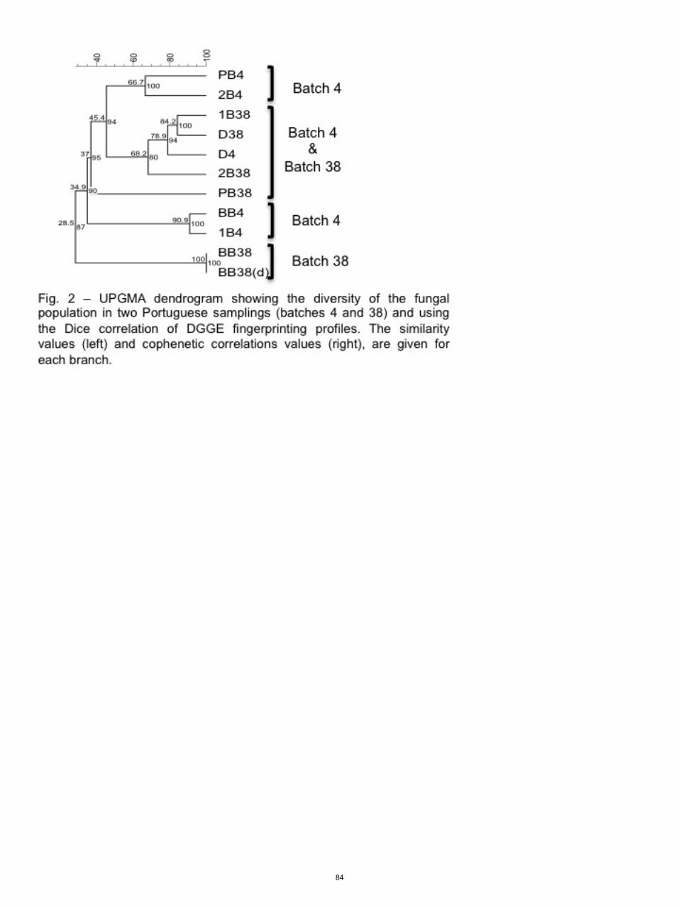
84
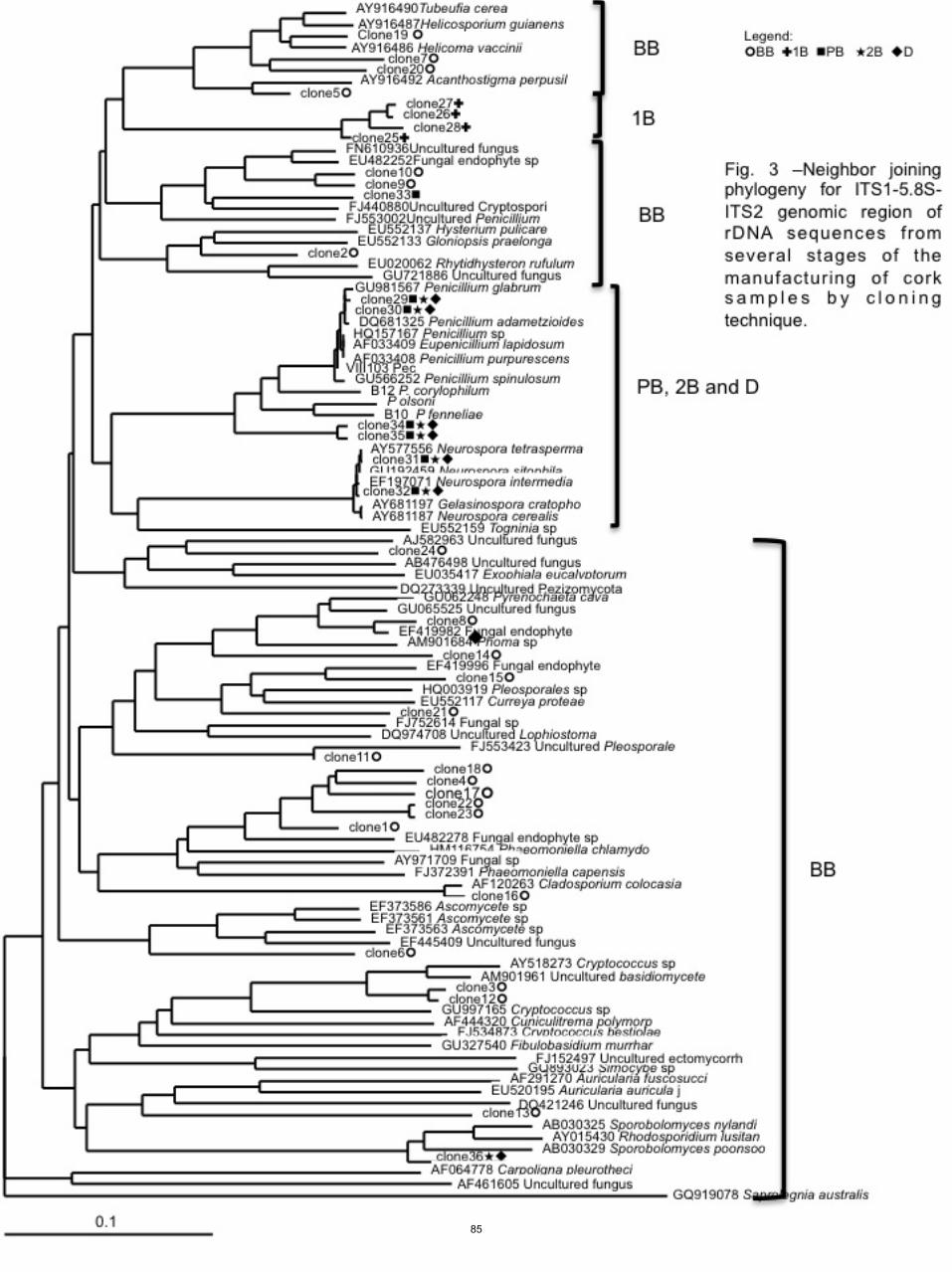
85

Tabl
e 1
– O
ccur
renc
e an
d qu
antif
icat
ion
of c
ultiv
able
fung
al s
peci
es fr
om tw
o sa
mpl
ings
of S
pani
sh (L
t 59
– 1st
sam
plin
g an
d Lt
17
– 2nd
sam
plin
g) c
ork
and
Por
tugu
ese
(Lt 4
– 1
st s
ampl
ing
and
Lt 3
8 –
2nd s
ampl
ing)
cor
k in
the
mai
n st
ages
of c
ork
man
ufac
turin
g di
scs
usin
g di
chlo
ran
glyc
erol
chl
oram
phen
icol
(D
GC
18) c
ultu
re m
ediu
m.
n.q.
= n
ot q
uant
ified
86

Tabl
e 2
- Per
cent
age
of s
eque
nce
iden
tity
to th
e ne
ares
t rel
ativ
e in
the
Gen
Ban
k da
taba
se, e
lativ
e fre
quen
cy a
nd th
eir g
enba
nk n
umbe
r for
the
clon
es s
eque
nced
in th
e pr
esen
t stu
dy.
R
elat
ive
frequ
ency
(%
) G
enba
nk n
º H
omol
ogy
Pro
babl
e id
entif
icat
ion
Clo
ne 1
13
.5
JN85
8112
96
U
ncul
tivab
le B
asid
iom
ycet
e cl
one
BF-
OTU
116
Clo
ne 2
8.
1 JN
8581
13
92
Glo
niop
sis
prae
long
a C
BS
119
332
Clo
ne 3
2.
7 JN
8581
14
96
Unc
ultiv
able
Bas
idio
myc
etes
C
lone
4
2.7
JN85
8115
93
Fu
ngal
end
ophy
te 5
T2.1
0 C
lone
5
2.7
JN85
8116
94
A
cant
host
igm
a pe
rpus
illum
C
lone
6
8.1
JN85
8117
90
U
ncul
tivab
le A
scom
ycet
e sp
. C
lone
7
5.4
JN85
8118
95
H
elic
oma
vaci
nii C
BS
216
.90
Clo
ne 8
2.
7 JN
8581
19
95
Fung
al e
ndop
hyte
isol
ate
CA
W 2
0 C
lone
9
2.7
JN85
8120
96
P
ezic
ula
sp. 3
ICM
P 1
8931
C
lone
10
2.7
JN85
8121
94
P
ezic
ula
sp. 3
ICM
P 1
8931
C
lone
11
2.7
JN85
8122
92
U
ncul
tivab
le P
leop
oral
es
Clo
ne 1
2 2.
7 JN
8581
23
94
Unc
ultiv
able
Bas
idio
myc
ete
Clo
ne 1
3 2.
7 JN
8581
24
85
Unc
ultiv
able
soi
l fun
gus
clon
e 13
8-33
C
lone
14
5.4
JN85
8125
98
U
ncul
tivab
le P
leur
opho
ma
clon
e K
L C
lone
15
2.7
JN85
8126
97
Fu
ngal
end
ophy
te is
olat
e 91
94
Clo
ne 1
6 5.
4 JN
8581
27
98
Cla
dosp
oriu
m c
oloc
asia
e C
lone
17
2.7
JN85
8128
93
Fu
ngal
end
ophy
te 5
T2.1
0 C
lone
18
2.7
JN85
8129
94
Fu
ngal
end
ophy
te 5
T2.1
0 C
lone
19
2.7
JN85
8130
94
H
elic
oma
vacc
inii
CB
S 2
16.9
0 C
lone
20
5.4
JN85
8131
96
H
elic
oma
vacc
inii
CB
S 2
16.9
0 C
lone
21
2.7
JN85
8132
96
Fu
ngal
end
ophy
te is
olat
e 91
94
Clo
ne 2
2 2.
7 JN
8581
33
94
Fung
al e
ndop
hyte
5T2
.10
Clo
ne 2
3 5.
4 JN
8581
34
94
Fung
al e
ndop
hyte
5T2
.10
Clo
ne 2
4 2.
7 JN
8581
35
91
Unc
ultiv
able
fung
us c
lone
TLF
34-
5 C
lone
25
56.3
JN
8581
36
86
Unc
ultiv
able
soi
l fun
gus
clon
e C
S2M
5c53
P
Clo
ne 2
6 3.
1 JN
8581
37
84
Unc
ultiv
able
soi
l fun
gus
clon
e C
S2M
5c53
P
Clo
ne 2
7 25
JN
8581
38
86
Unc
ultiv
able
soi
l fun
gus
clon
e C
S2M
5c53
P
Clo
ne 2
8 15
.6
JN85
8139
85
U
ncul
tivab
le s
oil f
ungu
s cl
one
CS
2M5c
53P
C
lone
29
21.2
JN
8581
40
99
P. g
labr
um
Clo
ne 3
0 1
JN85
8141
99
P
. gla
brum
C
lone
31
20.2
JN
8581
42
100
C. s
itoph
ila
Clo
ne 3
2 36
.5
JN85
81 4
3 99
C
. sito
phila
C
lone
33
1 JN
8581
44
88
Pez
icul
a sp
. 3 IC
MP
189
31
Clo
ne 3
4 1.
9 JN
8581
45
98
P. g
labr
um
Clo
ne 3
5 17
.3
JN85
8146
97
P
. gla
brum
C
lone
36
0.9
JN85
8147
99
S
poro
bolo
myc
es s
p.
87

Taxonomic studies of the Penicillium glabrum complexand the description of a new species P. subericola
M. C. Barreto & J. Houbraken & R. A. Samson &
J. C. Frisvad & M. V. San-Romão
Received: 22 October 2010 /Accepted: 6 January 2011 /Published online: 31 January 2011# The Author(s) 2011. This article is published with open access at Springerlink.com
Abstract A mycological survey of fungi, present in severalstages of the manufacturing of cork discs for champagnestoppers in Portugal, was made. Sixty-nine strains belong-ing to the Glabra series of the genus Penicillium wereisolated and subsequently grouped according to their partial!-tubulin gene sequences. Six groups with different partial!-tubulin gene sequences were observed, and a selection ofisolates of each group was made. These selected isolatesand various related ex-type strains were subjected to ataxonomical study using a polyphasic approach. Thisapproach included analysis of macro- and microscopicfeatures, the comparison of extrolite profiles and sequenc-
ing a part of the !-tubulin and calmodulin gene. The six !-tubulin types were reduced to three different species. Onegroup of isolates was centred on the ex-type strain of P.glabrum, a second group accommodated the type strain ofP. spinulosum and a third group contained isolates whichwere unique in their !-tubulin and calmodulin sequences,extrolite profiles and growth characteristics. This group ofisolates is described as the new species Penicilliumsubericola. The type strain of P. subericola CBS 125096T
was isolated from Portuguese raw cork, but additionalisolates were found from soil, air and lumen.
Keywords Taxonomy . Phylogeny . Tubulin . Cork
Introduction
Cork is the outer bark of the cork oak tree (Quercus suber).It is the most suitable material for cork stoppers, due to itsunique properties, such as elasticity, compressibility andimpermeability to gas or liquids (Lopes et al. 2001; Mano2002). During a survey of the colonizing mycobiota of corkslabs along the industrial manufacture of cork stoppers,numerous Penicillium isolates were isolated and identifiedusing morphological characters. More than half of theisolates belonged to the Glabra series, and were present inall production stages. However, identification of thedifferent isolates up to species level appeared to be difficultdue the high similarities in macro- and micromorphology.
Raper and Thom (1949) placed P. glabrum (as P.frequentans), P. spinulosum and P. purpurescens in the P.frequentans series, and later this series was synonymisedwith the Glabra series by Pitt (1979). The Glabra serieswas created to accommodate the fast growing Penicilliawith monoverticillate conidiophores and contains eightspecies (P. chermesinum, P. sclerotiorum, P. donkii, P.
M. C. Barreto :M. V. San-RomãoInstituto de Biologia Experimental e Tecnológica (IBET),Universidade Nova de Lisboa (UNL),Apartado 12,2781-901 Oeiras, Portugal
M. C. Barreto :M. V. San-RomãoInstituto de Tecnologia Química e Biológica (ITQB),Universidade Nova de Lisboa (UNL),2780-157 Oeiras, Portugal
J. Houbraken : R. A. Samson (*)CBS-KNAW Fungal Biodiversity Centre,Uppsalalaan 8,3584 CT Utrecht, The Netherlandse-mail: [email protected]
J. C. FrisvadCenter for Microbial Biotechnology, Biocentrum-DTU, TechnicalUniversity of Denmark,Søltofts Plads 221,DK-2800 Kgs. Lyngby, Denmark
M. C. Barreto :M. V. San-RomãoL-INIA, Ex Estação Vitivinícola Nacional,Quinta de Almoinha,2565-191 Dois Portos, Portugal
Fungal Diversity (2011) 49:23–33DOI 10.1007/s13225-011-0090-4
89
decumbens, P. thomii, P. glabrum, P. spinulosum and P.purpurescens). Among those species, P. glabrum and P.spinulosum were morphologically similar and could be bestdifferentiated based on conidial ornamentation. However,the morphological resemblance has caused much confusionand isolates are often misidentified or not differentiated bytaxonomists using morphological and physiological techni-ques (Pitt et al. 1990).
Sixty-nine strains originating from cork and belonging tothe Glabra series were grouped according to their partial !-tubulin gene sequences. A subset of these strains wasselected for macro- and microscopic analysis, extroliteprofiling and sequencing a part of the !-tubulin andcalmodulin gene. In addition, ex-type strains of variousrelated species were included in the analysis. Our polypha-sic taxonomic approach shows that a group of isolates sharepeculiar differences with other known species, and a newspecies is proposed for this group of isolates.
Materials and methods
Fungal strains
For our taxonomic study, a selection of these sixty-ninestrains isolated from cork, was made and supplementedwith related (ex-type) strains (Table 1). Spore suspen-sions of the cultures were maintained in 20% glycerol at!80°C.
Sequencing and data analysis
The strains were grown for 2–3 days at 25°C on maltpeptone medium. Genomic DNA was isolated using theUltraclean™ Microbial DNA Isolation Kit (MoBio, SolanaBeach, U.S.A.) according the manufacturer’s instructions.Fragments, containing a part of the !-tubulin or calmodulingene, were amplified and subsequently sequenced accord-ing the procedure previously described (Houbraken et al.2007). The alignments and analyses were preformed asdescribed by Samson et al. (2009). Newly obtainedsequences were deposited in Genbank nucleotide sequencedatabase under GQ367499-369547, GU372883-GU372894and GU991606-GU991609.
Phenotypic identification
All strains were grown on malt extract agar (MEA, Oxoid),Czapek Yeast autolysate agar (CYA), creatine agar (CREA)and Yeast Extract Sucrose agar (YES) (Samson et al. 2010).These media were inoculated in a three-point position andincubated at 25°C for 7 days. In addition, CYA plates wereincubated at 30°C and 37°C. After incubation, the culture
characteristics were recorded. Microscopic characters weredetermined on MEA and CYA.
Extrolite extraction and analysis
A selection of ten cork isolates was made based on the resultsof the !-tubulin analysis, and subjected to extrolite profiling.In addition, various related ex-type strains were examined.The extrolite extractions from the culture media werepreformed according to the methods described by Frisvadand Thrane (1987) and Smedsgaard (1997), using 500 "Lethylacetate/methanol/dichloromethane 3:2:1 (vol./vol./vol.)with 1% formic acid. The mixture was ultrasonicated in abath for 60 min. The organic solvent was transferred to anew vial and evaporated in a fume hood for 24 h. The extractwas re-dissolved in 400 "L methanol, analysed by HPLCwith diode array detection (DAD) and the extrolites wereidentified by their UV spectra and retention times.
Results
Grouping of members of the Glabra series isolatedfrom cork
The genetic variation within the strains isolated from cork wasinvestigated using the partial!-tubulin sequences. The strainsisolated from cork and four ex-type strains (P. glabrum, P.frequentans, P. paczoskii and P. spinulosum) were added tothe dataset, and subjected to an UPGMA analysis (Sneathand Sokal 1973). The sum of branch length of the optimaltree was 0.1301 and the dendrogram is shown in Fig. 1. Intotal, 422 positions were present in the final dataset. Sixgroups could be identified among the cork isolates belongingto the Glabra series. The largest group (50 isolates) sharedthe same partial !-tubulin sequence with the type of P.glabrum, CBS 125543 (Group 1). One cork isolate (CBS127703) appeared to have a unique partial !-tubulinsequence differing from other isolates in this clade (group2). Group 4 was the second largest group and consisted of 14isolates. This group was closely related with group 3 (3isolates) and these two groups only differed by one base pair.Group 5 and 6 were deviating from the other groups and the!-tubulin data shows that members of group 6 sharesequences with the type of P. spinulosum. Group 5 containedone isolate and this strain will be described here as a newspecies P. subericola. Each unique sequence type wascompared by a BLAST search in the NCBI database withthe P. glabrum strains identified by Serra et al. (2008). Intotal three P. glabrum sequences were deposited by Serra etal. (2008) and NRRL 35621 appeared to have identicalsequences as “group 2”, while the other two sequences(NRRL 35626 and NRRL 35684) were unique and not
24 Fungal Diversity (2011) 49:23–33
90

Table 1 List of isolates belonging to Series Glabra and related Penicillia
CBS no. Other no. Name Remarks
CBS 235.60 ATCC 18483=FRR 634 E. pinetorum Ex-type of P. silvaticum; forest soil, USSR
CBS 295.62 ATCC 14770=CCRC 31517=DSM 2438=IFO 7743=IMI 094209=MUCL 31196=NRRL 3008
E. pinetorum Ex-type; soil, conifer and hardwood forest,Wisconsin, USA
CBS 260.29 IMI 092242=NRRL 774=Thom4733.60 P. glabrum Ex-type of P. flavidorsum; unrecorded source
CBS 213.28 FRR 770=IMI 092265=IMI 092265ii=NRRL 770 P. glabrum Ex-type of P. oledzskii; soil under conifer,Poland
CBS 344.59 ATCC 18486=IFO 5359=IMI 068617=NRRL 3460 P. glabrum Ex-type P. spinuloramigenum; butter, Japan
CBS 228.28 FRR 752=IMI 092232=MUCL 29114=NRRL 752 P. glabrum Ex-type of P. terlikowskii; soil under conifer,Poland
CBS 229.28 FRR 751=IMI 092231=MUCL 29111=NRRL 751 P. glabrum Ex type of P. paczowskii; soil under conifer, Poland
CBS 105.11 P. glabrum Ex-type of P. frequentans; unknown substrate,Germany
CBS 127700 P. glabrum Non-boiled cork
CBS 127701 P. glabrum Cork, after the 1st boiling process
CBS 126333 P. glabrum Cork discs
CBS 127702 P. glabrum Non-boiled cork
CBS 127703 P. glabrum Non-boiled cork
CBS 127704 P. glabrum Non-boiled cork
CBS 127705 P. glabrum Non-boiled cork
CBS 126336 P. glabrum Non-boiled cork
CBS 125543 IBT 22658 P. glabrum Ex-type; unrecorded source
CBS 687.77 IJFM 3745=IMI 253783 P. grancanariae Ex-type of P. grancanariae; air, Gran Canaria, Spain
CBS 336.79 ATCC 38669=IJFM 3840=VKM F-2181 P. palmense Ex-type; air, Gran Canaria, Spain
CBS 126.64 P. purpurescens Soil, Erzurum, Turkey
CBS 366.48 ATCC 10485=IMI 039745=NRRL720=QM 1959
P. purpurescens Neotype; soil, Canada
CBS 328.48 ATCC 10444=IMI 040234=NRRL1915
P. spinulosum Ex-type of P. trzebinskii; forest soil, Poland
CBS 269.35 IMI 190574 P. spinulosum Ex-type of P. mucosum; soil, beech forest;Germany
CBS 268.35 IMI 189582 P. spinulosum Ex-type of P. mediocre; soil, pine forest; Germany
CBS 289.36 IMI 190573 P. spinulosum Ex-type of P. tannophagum; tannin solution,Germany
CBS 271.35 IMI 190675 P. spinulosum Ex-type of P. tannophilum; leaf litter,Germany
CBS 374.48 ATCC 10498=IMI 024316=MUCL13910=MUCL 13911=NRRL 1750
P. spinulosum Ex-type; culture contaminant, Germany
CBS 223.28 P. spinulosum Unknown source
CBS 127698 P. spinulosum Non-boiled cork
CBS 127699 P. spinulosum Non-boiled cork
CBS 125096 P. subericola Non-boiled cork, Portugal
CBS 127706 KAS 1289=IBT 22618 P. subericola Lumber, Vancouver, BC, Canada
CBS 125097 IBT 23009 P. subericola Air, margarine factory, Vejle, Denmark
CBS 125100 FRR 4914=IBT 30068 P. subericola From dried grapes (sultanas, Vitis vinifera),Mildura, Vic, Australia
CBS 125099 IBT 20217 P. subericola Acidified lake, Butte, Montana, USA
CBS 125098 IBT 20218 P. subericola Acidified lake, Butte, Montana, USA
CBS 347.59 FAT 340=IFO 6031=IMI 068221 P. thomii Ex-type of P. thomii var. flavescens; unrecordedsubstrate, Japan
CBS 350.59 ATCC 18333=FRR 3395=IFO 5362=IMI 068615
P. thomii Ex-type of P. yezoense; butter, Japan
Fungal Diversity (2011) 49:23–33 25
91
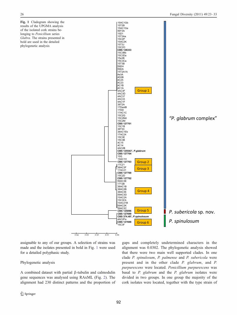
assignable to any of our groups. A selection of strains wasmade and the isolates presented in bold in Fig. 1 were usedfor a detailed polyphasic study.
Phylogenetic analysis
A combined dataset with partial !-tubulin and calmodulingene sequences was analysed using RAxML (Fig. 2). Thealignment had 230 distinct patterns and the proportion of
gaps and completely undetermined characters in thealignment was 0.0302. The phylogenetic analysis showedthat there were two main well supported clades. In oneclade P. spinulosum, P. palmense and P. subericola werepresent and in the other clade P. glabrum, and P.purpurescens were located. Penicillium purpurescens wasbasal to P. glabrum and the P. glabrum isolates weredivided in two groups. In one group the majority of thecork isolates were located, together with the type strain of
Fig. 1 Cladogram showing theresults of the UPGMA analysisof the isolated cork strains be-longing to Penicillium seriesGlabra. The strains presented inbold are used in the detailedphylogenetic analysis
26 Fungal Diversity (2011) 49:23–33
92

P. glabrum and the ex-type strains of P. flavidorsum, P.spinuloramigenum, P. terlikowskii, P. trzebinskii and P.oledzskii. The other group consisted of the type strains of P.frequentans and P. paczowskii. In the other clade, P.palmense was basal to P. spinulosum and P. subericola.The ex type of P. palmense clustered together with P.grancanariae CBS 687.77T.
Penicillium spinulosum and P. subericola were on abranch with a fair bootstrap support (72%). Three groups weredetected within this clade, but none of the phylogeneticrelations between those groups were well supported. Theisolates of P. subericola were on one branch. Interestingly, P.spinulosum was divided in two groups. One group com-passes the type culture of this species and the type strains of
P. mucosum CBS 269.35 and P. tannophilum CBS 271.35;the other group contained the type strains of P. mediocreCBS 268.35 and P. tannophagum CBS 289.36.
Phenotypic analysis
The strains isolated from cork were inoculated on the agarmedia MEA, CYA 25°C, CYA30°C, CYA 37°C, CREAand YES and were compared with the type strains of P.glabrum, P. spinulosum, P. frequentans and P. paczoskii.None of the examined strains were able to grow on CYAincubated at 37°C. In Fig. 3 an overview is shown ofgrowth patterns on various agar media. There was a largevariation in macromorphology among the Glabra strains.
Fig. 2 Phylogram based on thecombined dataset of partial !-tubulin and calmodulin genesequences and analysed usingRAxML. The strains in bold areisolated from cork
Fungal Diversity (2011) 49:23–33 27
93
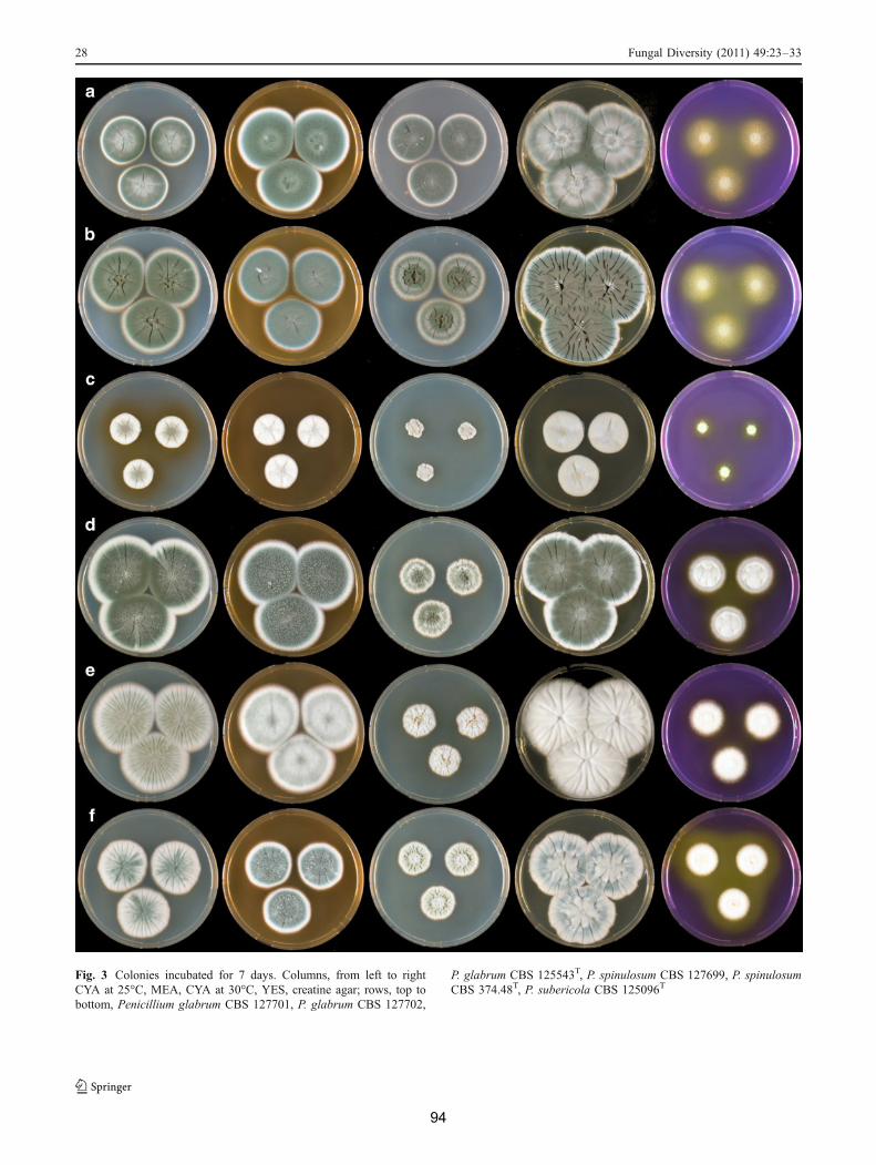
Fig. 3 Colonies incubated for 7 days. Columns, from left to rightCYA at 25°C, MEA, CYA at 30°C, YES, creatine agar; rows, top tobottom, Penicillium glabrum CBS 127701, P. glabrum CBS 127702,
P. glabrum CBS 125543T, P. spinulosum CBS 127699, P. spinulosumCBS 374.48T, P. subericola CBS 125096T
28 Fungal Diversity (2011) 49:23–33
94

The type strain of P. glabrum and P. spinulosum weredeviating and showed reduced growth rates and weaksporulation. The reverse colours on CYA of the Glabramembers were in shades of orange or orange brown, andoccasionally in crème colours. The intensity of thesecolours varied per isolate and ranged from pale orange-brown to vivid orange or red-orange (in P. spinulosum).The variation observed among the Glabra cork isolatescould not clearly be correlated to any of the six groupspreviously assigned with the partial !-tubulin data. Noclear distinctive characters to differentiate between P.glabrum, P. spinulosum and the new species could beobserved on CYA, MEA and YES. However, there was astriking difference on creatine agar. Isolates of P. spinulo-sum and the new species P. subericola grew moderate togood on this medium and the majority of both speciesproduced base compounds after prolonged incubation. Thecolony diameter was generally larger than 25 mm, while P.glabrum isolates grew more restricted (often less than25 mm) Fig. 3.
Microscopic analysis of the strains showed thatP. glabrum,P. spinulosum and P. subericola sp. nov. were very similar toeach other. All species were predominantly monoverticillate,with vesiculate conidiophores and 6–12 ampulliform phia-lides. The main microscopical difference was the conidiaornamentation, which was smooth to slightly rugose in P.glabrum and P. subericola sp. nov., and distinctly rugose inP. spinulosum. Moreover, the conidia of P. subericola tendedto be more rugose than in P. glabrum and the conidiophoresof this species occasionally were branched, a character notobserved in P. glabrum and P. spinulosum.
Extrolites analysis
The majority of the strains assigned to P. glabrum, P.spinulosum and P. subericola produced a pattern of extrolitestypical for each species (see Table 2). The P. glabrum isolateshad a typical extrolites profile containing asterric acid,bisdechlorogeodin, sulochrin or citromycetin, while isolatesof P. spinulosum produce asperfuran, palitantin and frequen-tin. Asperfuran, deoxybrevianamide E and unidentifiedcompounds which were tentatively named AMF were foundin the P. subericola. These AMF compounds are indols withan extended chromophore similar to penitremone. Two corkisolates which phylogenetically clearly belong to P. glabrum(CBS 126333 and 127701) were chemically weak and showno detectable extrolite production.
Discussion
The majority of cork isolates were identified as P. glabrumusing the current taxonomical schemes. Four different
sequence types of !-tubulin within P. glabrum could bedetected. BLAST searches on the NCBI database and localdatabases of the CBS-Fungal Biodiversity Centre showedthat many more sequence types are present in P. glabrum.This intra-species !-tubulin variation is in contrast withspecies in subgenus Penicillium, where various speciesshare the same tubulin sequence (Samson et al. 2004). Thelarge variability among P. glabrum isolates originating fromcork is also observed using microsatellite primers (Basílioet al. 2006). Our analysis show that P. flavidorsum, P.spinuloramigenum, P. terlikowskii, P. trzebinskii and P.oledzskii are synonyms of P. glabrum.
Raper and Thom (1949) placed P. glabrum (P. frequen-tans), P. spinulosum and P. purpurescens in the P.frequentans series. Our data show that these three speciesare phylogenetic related. Pitt (1979) named this the Glabraseries and expanded it with Penicillia, which have mono-verticillate penicilli and a colony diameter on CYA largerthan 30 mm after 7 days at 25°C. Penicillium chermesinum,P. sclerotiorum, P. donkii, P. decumbens, P. thomii, P.glabrum, P. spinulosum and P. purpurescens were included,but the phylogenetic analysis of the genus Penicillium byPeterson (2000) showed that the former four species werenot closely related to P. glabrum. Furthermore, Peterson(2000) named this monophyletic clade “Group 2”, andshowed that the species E. pinetorum, P. asperosporum, P.lividum and E. lapidosum were related to P. glabrum. Thesefindings in a large extent supported in our study, but thereare some differences. The taxonomic position of E.lapidosum warrants further attention. This species was notincluded in our phylogenetic study because the type strainof this species (CBS 343.48) is phylogenetically unrelatedto the Glabra group (J. Houbraken, unpublished data). Thisis in contrast with the observation made by Peterson (2000),which stated that E. lapidosum was conspecific with P.thomii.
Our data show that P. palmense and P. grancanariae,both isolated from air in Gran Canaria, Spain (Ramirez etal. 1978), are synonymous. The type strains of P.frequentans and P. paczowskii were considered to besynonyms of P. glabrum and P. spinulosum respectively(Pitt, 1979). However, based on calmodulin, tubulin andRPB2 data (data not shown) both type strains are placed ina separate clade related to P. glabrum, suggesting that P.frequentans/P. paczowskii and P. glabrum are two distinctspecies. This evidence is also supported by the extrolitesprofiles of these species (Frisvad, unpublished data).
Phenotypical differences were observed between the typestrains and the cultures isolated from the cork. This is probablydue to the fact that the type strains are maintained in culturescollections for a considerable period. Gradual degeneration ofvarious traits due to long-term maintenance and sub culturingare reported. Also degeneration could be due to the lyophili-
Fungal Diversity (2011) 49:23–33 29
95
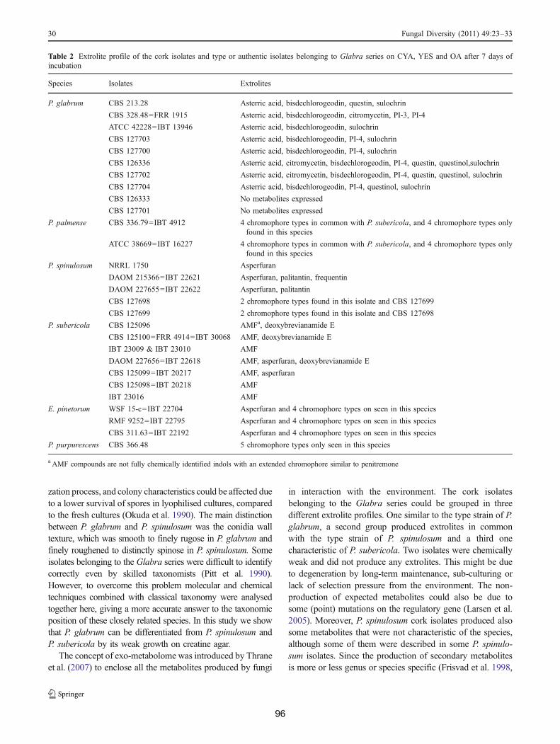
zation process, and colony characteristics could be affected dueto a lower survival of spores in lyophilised cultures, comparedto the fresh cultures (Okuda et al. 1990). The main distinctionbetween P. glabrum and P. spinulosum was the conidia walltexture, which was smooth to finely rugose in P. glabrum andfinely roughened to distinctly spinose in P. spinulosum. Someisolates belonging to the Glabra series were difficult to identifycorrectly even by skilled taxonomists (Pitt et al. 1990).However, to overcome this problem molecular and chemicaltechniques combined with classical taxonomy were analysedtogether here, giving a more accurate answer to the taxonomicposition of these closely related species. In this study we showthat P. glabrum can be differentiated from P. spinulosum andP. subericola by its weak growth on creatine agar.
The concept of exo-metabolome was introduced by Thraneet al. (2007) to enclose all the metabolites produced by fungi
in interaction with the environment. The cork isolatesbelonging to the Glabra series could be grouped in threedifferent extrolite profiles. One similar to the type strain of P.glabrum, a second group produced extrolites in commonwith the type strain of P. spinulosum and a third onecharacteristic of P. subericola. Two isolates were chemicallyweak and did not produce any extrolites. This might be dueto degeneration by long-term maintenance, sub-culturing orlack of selection pressure from the environment. The non-production of expected metabolites could also be due tosome (point) mutations on the regulatory gene (Larsen et al.2005). Moreover, P. spinulosum cork isolates produced alsosome metabolites that were not characteristic of the species,although some of them were described in some P. spinulo-sum isolates. Since the production of secondary metabolitesis more or less genus or species specific (Frisvad et al. 1998,
Table 2 Extrolite profile of the cork isolates and type or authentic isolates belonging to Glabra series on CYA, YES and OA after 7 days ofincubation
Species Isolates Extrolites
P. glabrum CBS 213.28 Asterric acid, bisdechlorogeodin, questin, sulochrin
CBS 328.48=FRR 1915 Asterric acid, bisdechlorogeodin, citromycetin, PI-3, PI-4
ATCC 42228=IBT 13946 Asterric acid, bisdechlorogeodin, sulochrin
CBS 127703 Asterric acid, bisdechlorogeodin, PI-4, sulochrin
CBS 127700 Asterric acid, bisdechlorogeodin, PI-4, sulochrin
CBS 126336 Asterric acid, citromycetin, bisdechlorogeodin, PI-4, questin, questinol,sulochrin
CBS 127702 Asterric acid, citromycetin, bisdechlorogeodin, PI-4, questin, questinol, sulochrin
CBS 127704 Asterric acid, bisdechlorogeodin, PI-4, questinol, sulochrin
CBS 126333 No metabolites expressed
CBS 127701 No metabolites expressed
P. palmense CBS 336.79=IBT 4912 4 chromophore types in common with P. subericola, and 4 chromophore types onlyfound in this species
ATCC 38669=IBT 16227 4 chromophore types in common with P. subericola, and 4 chromophore types onlyfound in this species
P. spinulosum NRRL 1750 Asperfuran
DAOM 215366=IBT 22621 Asperfuran, palitantin, frequentin
DAOM 227655=IBT 22622 Asperfuran, palitantin
CBS 127698 2 chromophore types found in this isolate and CBS 127699
CBS 127699 2 chromophore types found in this isolate and CBS 127698
P. subericola CBS 125096 AMFa, deoxybrevianamide E
CBS 125100=FRR 4914=IBT 30068 AMF, deoxybrevianamide E
IBT 23009 & IBT 23010 AMF
DAOM 227656=IBT 22618 AMF, asperfuran, deoxybrevianamide E
CBS 125099=IBT 20217 AMF, asperfuran
CBS 125098=IBT 20218 AMF
IBT 23016 AMF
E. pinetorum WSF 15-c=IBT 22704 Asperfuran and 4 chromophore types on seen in this species
RMF 9252=IBT 22795 Asperfuran and 4 chromophore types on seen in this species
CBS 311.63=IBT 22192 Asperfuran and 4 chromophore types on seen in this species
P. purpurescens CBS 366.48 5 chromophore types only seen in this species
a AMF compounds are not fully chemically identified indols with an extended chromophore similar to penitremone
30 Fungal Diversity (2011) 49:23–33
96

2008) the existence of P. glabrum cork isolates that producedtwo different extrolite profiles indicated the existence ofintraspecific variability.
The species concept, based not only on DNA sequences,but also in ecological, phenotypic characters and exo-metabolome profiles provide a more accurate and realclassification, as verified by studies on Penicillium subge-nus Penicillium (Samson and Frisvad 2004) and blackAspergilli (Samson et al. 2007). Applying this polyphasicapproach, P. spinulosum and P. subericola can be regardedas two separate species. Hoff et al. (2008) suggested in theirstudy of P. chrysogenum that closely related species couldbe mating types of the same biological species. However,no differences in extrolite patterns and phenotype could beobserved in isolates of different mating types of Paecilo-myces variotii (Houbraken et al. 2008, Samson et al. 2009).Furthermore, our studies showed that the two mating typesdiscovered in Aspergillus fumigatus (O’Gorman et al. 2009)
and Penicillium chrysogenum (Hoff et al. 2008) producedthe same pattern of extrolites and are identical in theirphenotype (Houbraken, Samson and Frisvad, unpublisheddata). In case of P. subericola we have observed differencesin both growth patterns and extrolite production and hencethe description of a new species is warranted.
The cork isolates now classified as P. glabrum speciesshowed a high intraspecific variability. The macro- andmicromorphologies, extrolites profiles and results of thesequencing of partial regions of the !-tubulin and calmod-ulin genes supported that variability. If the results wereanalyzed separately (e.g. the extrolite profile and !-tubulinsequencing) probably some of them could indicate theexistence of at least two different species. The analysis ofmore isolates of this species isolated from different sourcesand from different geographic locations is needed todetermine species boundaries in P. glabrum and relatedspecies.
Fig. 4 Penicillium subericola,cultures incubated for 7 days at25°C, A. MEA, B. CYA, C.YES. D-I. Conidiophores, phia-lides and conidia. Scale bar=10 "m
Fungal Diversity (2011) 49:23–33 31
97

Extrolites: asperfuran, deoxybrevianamide E and uniden-tified compounds which are indols with an extendedchromophore similar to penitremone.
Other isolates examined: CBS 127706 ex-lumber,Vancouver, BC, Canada; CBS 125100=IBT 30068, fromdried grapes (sultanas, Vitis vinifera), Mildura, Vic, Aus-tralia; CBS 125099=IBT 20218 and CBS 125098=IBT20217, both from acidified lake, Butte, Montana.
Acknowledgments This research received support from the SYN-THESYS Project http://www.synthesys.info/ which is financed byEuropean Community Research Infrastructure Action under the FP6"Structuring the European Research Area" Programme. Carmo Barretothanks Fundação para a Ciência e Tecnologia for the grant BD/19264/2004. Keith Seifert and John Pitt kindly provided strains and Tinekevan Doorn and Martin Meijer are greatly acknowledged for theirexcellent technical support.
Open Access This article is distributed under the terms of theCreative Commons Attribution Noncommercial License which per-mits any noncommercial use, distribution, and reproduction in anymedium, provided the original author(s) and source are credited.
References
Basílio MC, Gaspar R, Silva Pereira C, San Romão MV (2006)Penicillium glabrum cork colonising isolates—preliminary analysisof their genomic similarity. Rev Iberoam Micol 23:151–154
Okuda T, Klich MA, Seifert K, Ando K (1990) Media and incubationeffects on morphological characteristics of Penicillium andAspergillus. In: Samson R, Pitt JI (eds) Integration of moderntaxonomic methods for Penicillium and Aspergillus classification.Plenum Press, New York, pp 83–99
Peterson SW (2000) Phylogenetic analysis of Penicillium speciesbased on ITS and LSU-rDNA nucleotide sequences. In: SamsonR, Pitt J (eds) Integration of modern taxonomic methods forPenicillium and Aspergillus classification. Harwood, Reading, pp163–178
Pitt JI (1979) The genus Penicillium and its teleomorphic statesEupenicillium and Talaromyces. Academic, London
Pitt JI, Klich MA, Shaffer GP, Cruickshank RH, Frisvad JC, MullaneyEJ, Onions AHS, Samson RA, Williams AP (1990) Differentiationof Penicillium glabrum from Penicillium spinulosum and otherclosely related species: an integrated taxonomic approach. SystemAppl Microbiol 13:304–309
Ramirez C, Martinez AT, Ferrer S (1978) Three new species ofPenicillium. Mycopathol 66:77–82
Raper KB, Thom C (1949) Manual of the Penicillia. Williams andWilkins, Baltimore
Samson RA, Frisvad JC (2004) Penicillium subgenus Penicillium:new taxonomic schemes, mycotoxins and other extrolites. StudMycol 49:1–174
Samson RA, Seifert KA, Kuijpers AFA, Houbraken JAMP, Frisvad JC(2004) Phylogenetic analysis of Penicillium subgenus Penicilliumusing partial b-tubulin sequences. Stud Mycol 49:175–200
Samson RA, Noonim P, Meijer M, Houbraken J, Frisvad JC, Varga J(2007) Diagnostic tools to identify black Aspergilli. Stud Mycol59:129–145
Samson RA, Houbraken J, Varga J, Frisvad JC (2009) Polyphasictaxonomy of the heat resistant ascomycete genus Byssochlamysand its Paecilomyces anamorphs. Persoonia 22:14–27
32 Fungal Diversity (2011) 49:23–33
Penicillium subericola Baretto, Frisvad & Samson, sp.nov.—Mycobank MB 517383 - Fig. 4.
Penicillio glabro simile, sed bene crescenti in agarocreatino et formatione mixtionis chemicae obscurae (sed inP. glabro non producenti) distinguitur.
Culture ex type: CBS 125096, ex raw cork, PortugalColony diameters at 7 days in mm: CYA at 25º C: 37–
44; CYA at 30°C: 16–34; CYA at 37°C: no growth; MEA35–42; YES 39–46; CREA 14–26, moderate to goodgrowth with moderate to good acid production, baseproduction after prolonged incubation (14 days).
Good sporulation on CYA, grey-green, velvety andfloccose in centre, non sporulating margins 1–6 mm, fewsmall hyaline exudates droplets present, reverse colour creamto brownish. Colonies on MEA grey-green, good sporulation,floccose some isolates with velvety colonies and/or velvetywith floccose in the centre, exudate absent, reverse is orangebrown. Colonies on YES in various shades of green-grey,none or weak sporulation, mycelium inconspicuous, whitemargins with 1–2 mm, exudates absent, reverse orange-brownto yellow-brown, strongly sulcated (wrinkled).
Conidiophores strictly monoverticillate, stipes vesiculateup to 6 !m, smooth, occasionally short 40 !m, majoritylonger, width 3.0–4.0, vesicles 4.5–7.0 !m, phialides flaskshaped, 10–14!2.0–3.0 !m, conidia globose, finely rough-ened, 3–3.5 !m.
Frisvad JC, Thrane U (1987) Standardized high-performance liquidchromatography of 182 mycotoxins and other fungal metabolitesbased on alkylphenone retention indices and UV-VIS spectra(diode array detection). J Chromatogr 404:195–214
Frisvad JC, Thrane U, Filtenborg O (1998) Role and use of secondarymetabolites in fungal taxonomy. In: Frisvad JC, Bridge PD,Arora DK (eds) Chemical fungal taxonomy. Marcel Dekker, NewYork, pp 289–319
Frisvad JC, Andersen B, Thrane U (2008) The use of secondarymetabolite profiling in chemotaxonomy of filamentous fungi.Mycol Res 112:231–240
Hoff B, Pöggeler S, Kück U (2008) Eighty years after its discovery,Fleming’s Penicillium strain discloses the secret of its sex.Eukaryotic Cell 7:465–470
Houbraken J, Due M, Varga J, Meijer M, Frisvad JC, Samson RA(2007) Polyphasic taxonomy of Aspergillus section Usti. StudMycol 59:107–128
Houbraken J, Varga J, Rico-Munoz E, Johnson S, Samson RA (2008)Sexual reproduction as the cause of heat resistance in the foodspoilage fungus Byssochlamys spectabilis (anamorph: Paecilomycesvariotii). Appl Environ Microbiol 74:1613–1619
Larsen T, Smedsgaard J, Nielsen K, Hansen M, Frisvad J (2005)Phenotypic taxonomy and metabolite profiling in microbial drugdiscovery. Nat Prod Rep 22:672–695
Lopes M, Barros A, Neto C, Rutledge D, Delgadillo I, Gil A (2001)Variability of cork from Portuguese Quercus suber studied bysolid-state C-13-NMR and FTIR spectroscopies. Biopolymers62:268–277
Mano J (2002) The viscoelastic properties of cork. J Mat Sci 37:257–263
O'Gorman CM, Fuller HT, Dyer PS (2009) Discovery of a sexualcycle in the opportunistic fungal pathogen Aspergillus fumigatus.Nature 457:471–474
98

Samson RA, Houbraken J, Thrane U, Frisvad JC, Andersen B(2010) Food and Indoor Fungi. CBS Laboratory ManualSeries 2. Centraalbureau voor Schimmelcultures, Utrecht,The Netherlands
Serra R, Peterson S, CTCOR VA (2008) Multilocus sequenceidentification of Penicillium species in cork bark during plankpreparation for the manufacture of stoppers. Res Microbiol159:178–86
Smedsgaard J (1997) Micro-scale extraction procedure for standard-ized screening of fungal metabolite production in cultures. JChromatogr A 760:264–270
Sneath PHA, Sokal RR (1973) Numerical Taxonomy. Freeman, SanFrancisco
Thrane U, Andersen B, Frisvad J, Smedsgaard J (2007) The exo-metabolome in filamentous fungi in Topics in Current Genetics. Vol18. In: Nielsen J, Jewett MC (eds), Metabolomics. pp 235–252
Fungal Diversity (2011) 49:23–33 33
99


3
Exo-metabolites produced by some fungal isolates in several media cultures
C h a p t e r 3
This chapter includes the study of the possible production of exo-
metabolites by some isolated fungal species. The following
culture media were employed: two semi-synthetic media, a cork-
based medium and cork-based medium added with Chrysonilia
sitophila remains.
The following scientific article constitutes this chapter:
- Exo-metabolome of some fungal isolates growing on cork-
based medium (European Food Research Technology, 2011,
232:575-582)
The author performed the experimental work presented in this
chapter. The HPLC, GC-MS and data analysis were done in
collaboration with Center for Microbial biotechnology, Department
of Systems, Technical University of Denmark, Lyngby, Denmark.
The manuscript was written by the author and revised by the
other co-authors.

Eur Food Res Technol (2011) 232:575–582DOI 10.1007/s00217-011-1426-8
123
ORIGINAL PAPER
Exo-metabolome of some fungal isolates growing on cork-based medium
M. C. Barreto · J. C. Frisvad · T. O. Larsen · J. Mogensen · Maria Vitória San-Romão
Received: 28 October 2010 / Revised: 18 December 2010 / Accepted: 2 January 2011 / Published online: 20 January 2011! Springer-Verlag 2011
Abstract Fungal species colonize the cork slabs duringthe manufacturing of cork stoppers process. The mostimportant fungal species that colonizes cork slabs immedi-ately after boiling is Chrysonilia sitophila. Other fungalspecies may germinate replacing the C. sitophila myceliumon the cork slabs when the slabs’ water activity decreasesbelow 0.9. The possible production of exo-metabolites orvolatile compounds by some fungal species during the post-boiling stage was veriWed in pure cultures using threediVerent media compositions. The results suggest that nodeleterious exo-metabolites or mycotoxins are produced bythe studied fungal species, both in cork medium or in corkmedium added with C. sitophila extracts. However, theaddition of C. sitophila extract to the cork mediumenhanced the growth of the other studied fungal isolatesand altered the respective exo-metabolome proWle, leadingto the assumption that in their natural habitat, the late cork
colonizers like Penicillium spp. and Aspergilus spp. couldtake advantage from an earlier C. sitophila development asa result of its metabolism and/or mycelium remains. Fungalsuccessions may thus not only be a function of time andsubstrate, but also they can be dependent of the remains offormer colonizers. In fact, the production of the exo-metab-olites by the studied fungal isolates suggests that, under theused experimental conditions, they appear to play an impor-tant role in fungal interactions amongst the cork mycoXora.
Keywords Cork fungi · Extrolites · Volatile compounds · Cork niche
Introduction
The manufacturing of cork stoppers involves the boiling ofcork slabs. The boiling step increases the humidity in corkleading to fungal mycelium growth on the slabs surface.Chrysonilia sitophila (Mont.) Arx is the principal colonizerin slabs when the water activity is above 0.9 value [1, 2, Bar-reto & Gaspar, unpublished results]). When water activitydecreases below 0.9, cork fungal mycobiota shifts, whichis characterized by the growth of other fungi, for instancePenicillium spp., Aspergillus spp. and Trichoderma spp. [3–5].
Since C. sitophila is only visible during the Wrst days ofcork slab resting period, after the boiling step, its establish-ment implies that this species can metabolize the availablesubstrates on cork. Changes in the compounds present incork due to C. sitophila metabolism and the decrease inwater activity below 0.9 can lead to the establishment of thelate cork fungal colonizers [6–8]. The late colonizers (e.g.Penicillium, Aspergillus) are fungal species known toproduce exo-metabolites in semi-synthetic media culture[9, 10], also denominated as extrolites [9]. Fungal colonization
M. C. Barreto · M. V. San-RomãoInstituto de Tecnologia Química e Biológica, Universidade Nova de Lisboa (ITQB-UNL), Av. da República. Estação Agronómica Nacional, 2780-157 Oeiras, Portugal
M. C. Barreto · M. V. San-Romão (&)Instituto de Biologia Experimental e Tecnológica (IBET), Apartado 12, 2781-901 Oeiras, Portugale-mail: [email protected]
J. C. Frisvad · T. O. Larsen · J. MogensenCenter for Microbial Biotechnology, Department of Systems Biology, Technical University of Denmark, Søltofts Plads, B. 221, 2800 Kgs. Lyngby, Denmark
M. V. San-RomãoINRB I.P.-L-INIA, Ex Estação Vitivinícola Nacional, Quinta de Almoinha, 2565-191 Dois Portos, Portugal
Author's personal copy
103

576 Eur Food Res Technol (2011) 232:575–582
123
in cork slabs can be conditioned not only by the shifts in thehumidity but also by the presence of diverse species livingand interacting close together in the same niche.
The exo-metabolites are energetic costly chemical prod-ucts, which are usually associated with fungal sporulation[11]. Some of these compounds are deleterious (e.g. myco-toxins), while others are favourable (e.g. antibiotics) tohumankind. Their production depends on the substrate [12]and the interaction/competition with other organisms [13].
Some of the exo-metabolites produced by the fungi arevolatiles, usually designed by volatile organic compounds(VOCs). These compounds normally contribute to theintense and characteristic odours of fungi. Some previousstudies show that their production is consistent and related tothe cultural conditions and abiotic environment [14]. VOC-mediated positive, negative or neutral interactions can occurbetween a very wide range of soil bacteria and fungi contrib-uting to the soil microXora constitution. Many organisms areknown to modify the environment in order to construct anadequate niche where a natural selection can take place [15].
Cork is a natural and biodegradable material producedfrom the outer bark of Quercus suber L. possessing speciWcchemical and physical composition [16], which makes it anexcellent sealing device. On the other hand, it is a recalci-trant substrate that requires speciWc enzymes for its metab-olization to occur, whether it is total or partial [17].
The exo-metabolites production by some fungal speciesthat are present in the post-boiling stage is of the outmostimportance since some of these chemical compounds canbe deleterious to the Wnal product, thus aVecting its quality.
This study investigated the capacity of the most impor-tant fungal cork colonizers to produce exo-metabolitesusing diVerent rich semi-synthetic culture media to knowtheir exo-metabolites proWle. The possible production ofexo-metabolites in cork-based medium to mimic the corknatural substrate and in cork medium added with C. sito-phila extracts to simulate the post-boiling stage of the corkslabs was also investigated. Finally, the possible inXuenceof the fungal exo-metabolite and volatiles on the shifts ofcork slab fungal colonization was also stressed.
Materials and methods
Fungal isolates and culture media
All fungal species used in this study were previously iso-lated in our laboratory [1, 2, Barreto unpublished results]and identiWed using phenotypic and molecular techniquesat CBS (Utrecht, Holland) (Barreto, unpublished results).
Eleven fungal strains isolated after the boiling stage ofcork slabs during the cork stopper manufacturing processwere used. These strains are now deposited in international
culture collection with the following deposit numbers:Chrysonilia sitophila DSM 16514 (DSMZ, Germany),Eurotium amstelodami (CBS 126335), Eurotium repens(CBS 126221), Penicillium brevicompactum (CBS126334), Penicillium citreonigrum (CBS 126338), Penicil-lium citrinum (CBS 126219), Penicillium chrysogenum(CBS 126337), Penicillium glabrum (1) (CBS 126333),Penicillium glabrum (2) (CBS 126336), Penicilliumpaneum and Aspergillus tubingensis (CBS 126391).
Composition of the culture media used in this work
1. YES medium—yeast extract sucrose agar preparedaccording to manufacturer’s instructions (Fluka, SaintLouis, MO, USA).
2. Composed medium 1 (CoM): 1 g YES medium, 5 gmalt broth (Merck, Darmstadt).
3. Cork medium (CM): 30 g of cork powder, 0.5 g ofK2HPO4 and 15 g of agar, per litre of distilled water.The medium was autoclaved at 121 °C for 15 min and10 mL of a salt solution was added after Wltrationthrough a 0.45-!m Wlter (salt solution composition:NaNO3 30 g; KCl 5 g; MgSO4.7H2O 5 g; FeSO4·7H2O0.1 g; ZnSO4·7H2O 0.1 g; and CuSO4·7H2O 0.05 g per100 mL of distilled water). The cork powder was previ-ously treated with gamma radiation ¡32 KGy.
4. Cork medium added with C. sitophila extracts (CM 1):prepared according to CM medium and to which 3 g/Lof C. sitophila extract was added before sterilization.
Preparation of the C. sitophila extract used in CoM2 medium culture
A suspension of hyphae and C. sitophila spores (106 spores/mL) was inoculated on DG18 culture medium (OxoidBasingstoke, UK) for 7 days, at 25 °C in the dark. Aftergrowth, the spores and mycelium were completely scrapedwith a loop into a falcon tube and 3 g of the fungal debriswas lyophilized. The extract was kept at ¡20 °C.
Incubation conditions
Inoculations in all four media were performed using sporesuspensions that were stored at ¡80 °C: Inoculations onYES and CM 1 media were incubated at 25 °C in the darkfor 14 days. Inoculations on the two other media were incu-bated for 21 days using the same conditions.
Exo-metabolite analysis
The extrolites were extracted from the culture media accord-ing to the methods described by Frisvad [18] and Smedsg-aard[19]. The extracts were analysed by HLPC using a HP
Author's personal copy
104
Eur Food Res Technol (2011) 232:575–582 577
123
1100 series (Hewlett Packard, Germany) equipped with onepump and an auto-injector (Hewlett Packard) maintained atroom temperature. Detection was performed using a diodearray detector (DAD) with a 6-mm Xow cell collecting twoUV spectra per second from 200 to 600 nm with a bandwidthof 4 nm and a Xuorescence detector (FLP) with excitation at230 nm and emission at 333 nm. Separations were made on a100 £ 2 mm Luna C18 cartridge column packed with 3-!mparticles and using a guard column with the same material,maintained at 40 °C. Elution was done using a linear gradientstarting with 50% (v/v) water (A) and 50% (v/v) acetonitrile(B) reaching 100% of acetonitrile in 20 min and maintainingthe Xow for 5 min. Both eluents contained 0.005% (v/v) oftriXuoroacetic acid (TFA). The Xow rate was 0.40 mL/min.
All experiments in this study were performed in duplicate
All chemicals used were Merck (Darmstadt, Germany) ana-lytical grade if nothing diVerent is referred. All solventswere prepared using MilliQ water.
Volatile analysis
The same eleven fungal strains previously used for exo-metabolites analysis were grown in two diVerent culturemedia, YES and CM prepared as stated earlier.
Moreover, two sets of fungal mixtures were tested inCM and CM1 culture medium: (1) C. sitophila, P. glabrum,P. brevicompactum and P. chrysogenum. (2) C. sitophila,P. citrinum, P. paneum and E. rubrum.
Volatile metabolites were collected during 4 days for thestrains inoculated in YES medium and 14 days in the caseof the CM medium, since the growth and sporulation isslower in the case of the second culture medium. To collectthe volatiles, a stainless steel Petri dish lid with a standard1/4!! Swagelock™ replaced the usual lid [20]. This lidpossessed a standard 1/4!! Swagelok Wtting with PTFEinsert in the centre that is used to hold a charcoal tube(SKC, 226-01). The collected volatiles were extracted fromthe charcoal tube with 1 mL of ether (5 £ 200 !L). Thesamples were concentrated to approximately 100 !L usinga nitrogen Xow. Hundred microlitres of each sample wasput in GC vials and analysed using a gas chromatography–mass spectrometry (GC–MS) (Finnigan Focus GC coupledto a Finnigan Focus DSQ mass selective detector).
The separation of the volatiles was done on a SupelcoSLB™-5 MS capillary column, using He as carrier gas, at a1.2 mL/min Xux. The injection and detection time was setto 220 °C. One microlitre of each sample was injected intothe GC–MS system.
Chromatographic conditions were set to an initial tem-perature of 35 °C for 1 min, raised at 6 °C/min to 220 °Cand then 20 °C/min to 260 °C for 1 min. The separated
compounds were characterized by their mass spectra gener-ated by electron ionization (EI) at 70 eV at a scan rangefrom m/z 35–300.
Data analysis
The exo-metabolite compounds were identiWed by compari-son with alkylphenone retention indices and diode arrayUV–VIS detection as described by Frisvad and Thrane [18].
The mass spectra from the volatile compounds withidentical retention times were compared with the onesavailable in the library database to determine their similar-ity. IdentiWcation of sesquiterpenes was made by compari-son of the mass spectra and the fragmentation proWle ofeach compound with spectra in the software library data-base and with the literature [21].
Results
The production of exo-metabolites from the eleven fungi ispresented on Table 1.
Exo-metabolite production in rich culture media (YES and CoM)
The fungal metabolite production was similar in bothculture media, with the exceptions of E. repens P. brevi-compactum, P. citreonigrum and P. citrinum. P. brevicom-pactum produced a higher variety of compounds, while theother fungi appear to be restricted to the production of oneor two compounds. As expected, the production of myco-toxins (citrinin and citreoviridin) by P. citrinum andP. citreonigrum could be observed in both media.
Exo-metabolite production in cork culture medium (CM)
The exo-metabolite production was almost inexistent in CMculture medium. As shown in Table 1, only E. amstelodami,P. brevicompactum and P. citrinum produced metabolites inthis culture medium. Although E. amstelodami produced thesame number of metabolites in this medium compared withCoM, one compound is diVerent [e.g. echinulin (1298) inCM and Xavoglaucin (1646) in CoM1]. However, the fungalgrowth in CM medium is scarce (Fig. 1a), probably becausecork is a recalcitrant substrate.
Exo-metabolite production in cork-based media with C. sitophila extracts (CM1)
Comparing the fungal behaviour in CM and CM1 culturemedia, higher fungal growth was observed on CM1
Author's personal copy
105

578 Eur Food Res Technol (2011) 232:575–582
123
(Fig. 1a, b). As shown in Table 1, when C. sitophila extractwas added to the cork medium (CM1), more compoundswere detected in all analysed fungal extracts. OnlyE. amstelodami, P. brevicompactum and P. citrinum pro-
duced exo-metabolites in both culture media (CM andCM1). Moreover, E. amestelodami and P. brevicompactumshow diVerent exo-metabolite proWles. For instance,P. brevicompactum produced not only brevianamide A but
Table 1 IdentiWcation of the exo-metabolites produced by some fungal species in four diVerent culture media
The retention indexes of the metabolites are given in brackets. ND not detected
YES medium CoM1 medium CM medium CoM2 medium
Aspergillus tubingensis Naphto-!-pyrones, “kotanin” (990)
Tensidol B (943), aurasperone B (1008), cf. kotanin (991), aspereynone (1467), asperazine (1201)
ND Tensidol B (952), aurasperone B (1007)
Chrysonilia sitophila ND ND ND ND
Eurotium amstelodami Flavoglaucin (1646). Tetrahydroauroglaucin (1629), Xavoglaucin (1646)
Echinulin (1298), tetrahydroauroglaucin(1517)
Two neoechinulins (897, 946), echinulin (1541) Xavoglaucin (1646)
Eurotium repens Flavoglaucin (1497) Asperentin (845, 932, 1023), auroglaucin (1633)
ND Asperentin (784) and andrastin E (1099)
Penicillium brevicompactum
Raistrick phenol (747), mycophenolic acid (1017), asperphenamate (1239), a pebrolide (1171) and brevianamide A (880)
Mycophenolic acid (1010), xanthoepocin (1169), brevianamide A (880) and asperphenamate (1239)
Brevianamide A (805) Mycophenolic acid (1017) and brevianamide A (888)
Penicillium citreonigrum Citreoviridin (1105) Citreoviridin (1105), “met ø” (957)
ND “met ø” (indol alkaloid) (957)
Penicillium citrinum Quinolactacin (774), citrinin (1260)
Quinolactin (774), citrinin (1216)
Quinolactacin (774) Quinolactacin (774),
Penicillium chrysogenum Meleagrin (775) Meleagrin (775) ND Meleagrin (775)
Penicillium glabrum1 “One unknown extrolite” (578)
Citromycetin (657), PI-4 (767)
ND ND
Penicillium glabrum 2 Sulochrin (871) and “one unknown extrolite” (578)
Citromycetin (657) ND “Neoglabrol” (982)
Penicillium paneum Citreoisocoumarin (742), marcfortine A (747) and roquefortine E (759)
A citreoisocoumarin derivative (1058)
ND Marcfortine derivative (813)
Fig. 1 Penicillium paneum iso-late growing in two diVerent cul-ture media a cork medium and b cork medium with Chrysonilia sitophila extracts (CM1)
Author's personal copy
106
Eur Food Res Technol (2011) 232:575–582 579
123
also mycophenolic acid (Fig. 2a, b). Furthermore, speciesthat have not produced any exo-metabolite in CM whengrown on CM1 produced at least one compound:P. paneum (marcfortine derivative) and P. chrysogenum(melagrin). Also A. tubingensis (tensidol B and aurasperoneB) and E. repens (asperentin and andrastin E) produced twocompounds. P. citreonigrum produced an indol alkaloid[R.I. of 957], possibly cividiclavin, formerly reported fromP. citreonigrum [22], and P. glabrum 2 produced one com-pound, here named ‘neoglabrol’ [R.I. of 982].
Under the tested conditions, C. sitophila was the onlyfungal species that did not produce any metabolite in all ofthe assayed culture media.
Under the assayed conditions, no fungal isolate pro-duced any mycotoxins either in cork-based medium orwhen C. sitophila extract was added.
Volatile production in YES and cork-based media (CM and CM1)
In YES culture medium only P. paneum produced ses-quiterpenes that were identiWed by their retention time(RT) and the respective fragmentation pattern [21]. Themost important sesquiterpenes were detected at RTbetween 15.51 and 17.96 min. The other fungal isolatesdid not produce any volatile compounds, under theassayed conditions. On the other hand, two sets of diVer-ent fungal mixtures inoculated in CM + CM1 culturemedium produced a non-identiWed volatile compound inall plates, but one (RT = 23.08 min) presenting as princi-pal fragmentation masses m/z = 153.03; 43.91; 181.02(Fig. 3a, b).
Discussion
Most of the exo-metabolites produced by the studied fungalspecies in YES medium were consistent with thosedescribed for the same species in earlier works when grownunder similar conditions [9, 23–26]. According to previousworks, some of the produced metabolites have taxonomicrelevance for all fungi isolates in both synthetic culturemedia [9]. For example, citromycetin is produced byP. glabrum, which is in accordance with previous reports[28], or both brevianamide A and mycophenolic acid areproduced by P. brevicompactum [9]. Their detection in thisstudy contributes to better characterize the fungal isolatesconsidered here.
The production of exo-metabolites is limited by themedium composition, culture conditions and genetic factors[9, 10, 23, 24]. The results obtained in this study are inaccordance with Calvo et al. [11] who stated that the sameenvironmental conditions required for sporulation are often
also necessary for the secondary metabolite production, asexempliWed in Fig. 1a, b and Table 1.
Furthermore, some studies conWrmed the presence andactivity of the enzymes necessary to the breakage of corkcomponents into digestible compounds by some Aspergil-lus, Penicillium and Trichoderma species [28, 29]. Simi-larly, previous works showed that C. sitophila can alsosecrete some of the enzymes necessary to partially degradecork components [17, 30].
In this study, a more diVerentiated exo-metabolite pro-duction is observed when the fungal isolates grow in CM1in comparison with the metabolites proWles produced by thesame fungi in CM culture medium. The growth in CMculture medium is scarce, and the fungal metabolism isprobably targeted for the biomass production. The additionof C. sitophila remains that can serve as food source for thelate cork colonizers will act as an additional nutrient source(e.g. nitrogen). This additional food source can stimulatethe exo-metabolome change to increase their survivalWtness [31]. The higher growth and sporulation of the sev-eral fungal isolates, exempliWed in Fig. 1a, b, can inducethe fungal exo-metabolome to produce metabolites toensure a successful degradation and colonization of thesubstrate. This process can include the inhibition of someearly fungal colonizers.
Under the assayed conditions, no fungal isolate producedany mycotoxins, although, mycophenolic acid and brevi-anamide produced by P. brevicompactum have been some-times referred as mycotoxins. However, those compoundscannot be considered mycotoxins because they are not toxicto any vertebrate, according to the established concept ofmycotoxin by Frisvad [32]. These exo-metabolites havepharmacological interesting capacities, like mycophenolicacid, which is an immunosuppressor used in organ-trans-planted patients, and brevianamide A with insecticide prop-erties [27, 33]. The fact that C. sitophila did not produce anychemical exo-metabolite under the studied conditions,which is in accordance with previous studies regarding thesafety of Neurospora species (Chrysonilia teleomorph),concluded that the species belonging to this genus are nei-ther pathogen nor toxin producers [34]. Moreover, “with theexception of carotenoid and melanin pigment synthesis,Neurospora has not been shown to possess secondarymetabolism” [35]. This knowledge is relevant regarding thatcork stoppers will be used to seal wine bottles.
Prat et al. [36] showed that the isolates of P. glabrumassayed in their work had diVerent capacity of taint devel-opment since the volatile production was a strain-speciWceVect. In fact, in our study, both P. glabrum isolates haddiVerent exo-metabolite proWle between them. This eVectsupports the high intraspeciWc heterogeneity that existsamongst isolates of this taxon, as previously mentioned inother works [37–39, Barreto et al., unpublished results]. It
Author's personal copy
107

580 Eur Food Res Technol (2011) 232:575–582
123
Fig. 2 UV-VIS chromatograms of the HPLC proWle for Penicillium brevicompactum (the black line represent DAD 210 nm and the red line DAD280 nm) in a cork medium culture and b cork medium culture added with Chrysonilia sitophila extracts (CM1)
Author's personal copy
108

Eur Food Res Technol (2011) 232:575–582 581
123
is probable that this characteristic can also be observed insome other fungal isolates and also justify some resultsobserved in this work (Table 1).
Concerning the production of volatile compounds, ses-quiterpene production by P. paneum in YES medium wasalso veriWed in earlier works that have reported them asexclusive for that species [20]. These compounds areknown as having anti-fungal activity so they can interactwith each other [40, 41] or act synergistically, thereforeenhancing their eVect [42]. The productions of a non-identi-Wed volatile compound by the two sets of fungal mixtureconWrm that presence of C. sitophila extract in the mediamodiWes the fungal behaviour in a small niche. Besidesthat, the presence of diVerent fungal species in co-culturecan induce the production of a new chemical compoundthat was inexistent when the fungal isolates were inoculatedin single culture in medium plates. In this case, the interac-tion amongst the interspeciWc fungal species could beresponsible for the production of the volatile compound, asdescribed by Evans [43].
In conclusion, the production of both exo-metabolitesand volatile compounds by the studied fungi on CM andCM1 media cultures was low; however, the addition ofC. sitophila extract to the culture medium not onlyenhances the fungal growth but also increases the variety ofexo-metabolites produced by some fungal species, clearly
suggesting that interactions may take place on cork slabsfungal colonizing communities. Probably the partial metab-olization of the cork components after the cork boilingfollowed by the establishment of C. sitophila leaves thesubstrate more accessible to the late fungal colonizers.
These results point to the central role of C. sitophila ear-lier germination after the late fungal colonization of corkslabs. This fungal species will turn the substrate moreaccessible to the other fungi colonizers, since it is known toproduce enzymes capable of degrading some cork compo-nents [3]. On the other hand, when other fungi germinateafter partial cork degradation by C. sitophila and use itsmycelium remains, they can produce exo-metabolites,which in turn can inhibit C. sitophila mycelium expansion.In fact, it was also observed in previous studies that C. sito-phila growth in cork-based medium inhibits other fungidevelopment (e.g. Trichoderma, Penicillium and Mucor)[44].
These results clearly suggest that the most predominantfungal species that are active in the post-boiling stage of thecork stoppers manufacture could be regarded as “non-dele-terious” to the cork stoppers Wnal product. These observa-tions contribute to prove the statement that natural corkstoppers are the most adequate sealing device for winebottles, especially concerning the necessary safety of theproduct that will be in contact with the stopper.
Fig. 3 a Chromatogram of the RT interval 17.91–20.80 for Chrysonilia sitophila, Penicillium glabrum, Penicillium brevicompactum andPenicillium chrysogenum in cork culture medium with C. sitophila extracts b mass spectrum for the peak at 20.12 RT 35.00–300.00 m/z
Author's personal copy
109

582 Eur Food Res Technol (2011) 232:575–582
123
Acknowledgments Carmo Barreto thanks Fundação para a Ciênciae a Tecnologia for the grant BD/19264/2004. The authors are alsothankful to Amorim & Irmãos (Coruche, Portugal) that kindly providecork samples from which the studied fungi were isolated, to Jos Hou-braken in helping with the taxonomic identiWcation of the isolates andto Hanne Jakobsen and Kir Lyhne for technical assistance and MariaTeresa Barreto Crespo by her support and manuscript revision. Corkirradiation was grateful done by the Radiation Technologies Unity(UTR) from the Technological Institute and Nuclear (ITN), Portugal.
References
1. Danesh P, Caldas FMV, Figueiredo Marques JJ, San Romão MV(1997) J Appl Microbiol 82:689–694
2. Pires JM, Pereira H, San Romão MV (2007) Ciência e TécnicaVitivinícola 22:15–20
3. Álvarez-Rodríguez ML, López-Ocana L, López-Coronado JM,Rodríguez E, Martinez MJ, Larriba G, Coque J-JR (2002) ApplEnviron Microb 68:5860–5869
4. Oliveira AC, Peres CM, Correia Pires JM, Silva Pereira C,Vitorino S, Figueiredo Marques JJ, Barreto Crespo MT, SanRomão MV (2003) Microbiol Res 158:117–124
5. Prak S, Gunata Z, Guiraud J-P, Schorr-Galindo S (2007) FoodMicrobiol 24:271–280
6. Centeno S, Calvo M (2001) Microbios 106:69–737. Riu H, Roig G, Sancho J (1997) Microbiologia 13:185–1928. Vitorino S, Almeida-Vara E, Tenreiro R, San Romão MV (2002)
In: AGROGES (eds) Congresso Mundial do Sobreiro e da CortiçaProceedings Book. Lisboa
9. Frisvad J, Smeedsgard J, Larsen T, Samson R (2004) Stud Mycol49:201–242
10. Larsen TO, Smedsgaard J, Nielsen KF, Hansen ME, Frisvad JC(2005) Nat Prod Rep 22:672–695
11. Calvo AM, Wilson RA, Bok JW, Keller NP (2002) Microbiol MolBiol Rev 66:447–459
12. Borjesson T, Stollman U, Schnurer JL (1990) Appl EnvironMicrobiol 56:3705–3710
13. Korpi A, Pasanen A-L, Pasanen P (1998) Appl Environ Microbiol64:2914–2919
14. Larsen TO (1998) In: Frisvad JC, Bridge PD, Arora DK (eds)Chemical fungal taxonomy. Marcel Dekker, New York
15. Brown SP, Inglis RF, Taddei F (2009) Evol App 2:32–3916. Pereira H (1988) Chemical composition and variability of cork
from Quercus suber L. Wood Sci Technol 22:211–21817. Vitorino SI, Neves ESG, Gaspar F, Figueiredo Marques JJ, San
Romão MV (2007) Ciência e Téc Vitiv 22:1–418. Frisvad JC, Thrane U (1987) J Chromatogr 404:195–21419. Smedsgaard J (1997) J Chromatogr A 760:264–27020. Larsen TO, Frisvad JC (1994) J Microbiol Meth 19:297–30521. Karlshøj K, Larsen TO (2005) D J Agric Food Chem 53:708–71522. Krumphanzl V (ed) (1982) Overproduction of microbial products.
Academic Press, London
23. Samson RA, Houbraken JAMP, Kuijpers AFA, Frank JM, FrisvadJC (2004) Stud Mycol 50:45–61
24. Smetanina O, Kalinovskii A, Khudyakova Y, Slinkina N, PivkinM, Kuznetsova T (2007) Chem Nat Compd 43:395–398
25. Thrane U, Andersen B, Frisvad JC, Smedsgaard J (2007) In:Nielsen J, Hewitt J (eds) Topics in Current Genetics, vol 18.Springer, Berlin
26. Fox EM, Howlett BJ (2008) Curr Opin Microbiol 11:481–48727. Evans GE, Staunton J (1988) J Chem Soc Perk T 1988:755–76128. Fang J, Qu Y, Gao P (1997) Biotechnol Tech 11:195–19729. Krogh KBR, Morkeberg A, Jorgensen H, Frisvad JC, Olsson L
(2004) Appl Biochem Biotech 114:389–40130. Durán N, Ferrer I, Rodríguez J (1987) Appl Biochem Biotech
16:157–16731. Williams DH, Stone MJ, Hauck PR, Rahman SK (1989) J Nat Prod
52:1189–120832. Frisvad JC, Thrane U (2004) In: Samson RA, Hoekstra ES, Fris-
vad JC (eds). Introduction to food and airborne fungi, 7th edn.Centraalbureau voor Schimmelcultures. Utrecht
33. Paterson RRM, Simmonds MJS, Kemmelmeier C, Blaney WM(1990) Mycol Res 94:538–542
34. Perkins DD, Davis RH (2000) Appl Environ Microbiol 6:5107–5109
35. Galagan JE, Calvo SE, Borkovich KA, Selker EU, Read ND, JaVeD, FitzHugh W, Ma L, Smirnov S, Purcell S, Rehman B, Elkins T,Engels R, Wang S, Nielsen CB, Butler J, Endrizzi M, Qui D, Iana-kiev P, Bell-Pedersen D, Nelson MA, Werner-Washburne M, Seli-trennikoV CP, Kinsey JA, Braun EL, Zelter A, Schulte U, KotheGO, Jedd G, Mewes W, Staben C, Marcotte E, Greenberg D, RoyA, Foley K, Naylor J, Strange-Thomann N, Barrett R, Gnerre S,Kamal M, Kamvysselis M, Mauceli E, Bielke C, Rudd S, Frish-men D, Krystofova S, Cogoni C, Macino G, Catcheside D, Li W,Pratt RJ, Osmani SA, DeSouza CPC, Glass L, Orbach MJ, Bergl-und JA, Voelker R, Yarden O, Plamann M, Seiter S, Dunlap J,Radford A, Aramayo R, Natrig DO, Alex LA, Marinhaupt G,Ebbole DJ, Freitag M, Paulsen I, Sachs MS, Lander ES, Nusbaum C,Birren B (2003) Nature 422:859–868
36. Pratt C, Trias R, Culleré L, Escudero A, Anticó E, Bañeras L(2009) J Agric Food Chem 57:7473–7479
37. Pitt JI, Klich MA, SchaVer GP, Cruickshank RH, Frisvad JC,Mullaney EJ, Onions AHS, Samson RA, Williams AP (1990) SystAppl Microbiol 13:304–309
38. Basílio MC, Gaspar R, Silva Pereira C, San Romão MV (2006)Rev Iberoam Micol 23:151–154
39. Serra R, Peterson S, CTCOR, Venâncio A (2008) Res Microbiol159:178–186
40. Kubo I, Fujita K, Lee SH (2001) J Agric Food Chem 49:1607–161141. Arif T, Bhosale JD, Mandal TK, Bendre RS, Lavekar GS, Dabur
R (2009) J Asian Nat Prod Res 11:621–63842. Lorito M, Peterbauer C, Hayes CK, Harman GF (1998) Micro-
biology 140:623–62943. Evans JA, Eyre CA, Rogers HJ, Boddy L, Muller CT (2008)
Fungal Ecol 1:57–6844. San Romão MV (2009) Ciência Téc Vitivi 24:81–90
Author's personal copy
110

4
Volatile compounds produced by cork mycobiota

C h a p t e r 4
In an attempt to assess the production of chemical compounds
that can be harmful to the wine, the volatile compounds produced
by microbiota existing in different cork samples collected during
the manufacturing of cork discs was studied. Furthermore, the
releasable volatile compounds and 2,4,6-trichloroanisole (TCA)
produced by some previously isolated fungal species were
analyzed by gas chromatography coupled with mass
spectrometry (GC-MS).
The following scientific article is included in this chapter:
- Volatile compounds in samples of cork and also produced by
selected fungi (Journal of Agricultural and Food Chemistry, 2011,
59: 6568-6574).
The author performed the experimental work presented in this
chapter. The GC-MS and data analysis were done in
collaboration with Analytical Chemistry Laboratory, ITQB, IBET,
Oeiras. The manuscript was written by the author and revised by
the other co-authors.
Published: May 16, 2011
r 2011 American Chemical Society 6568 dx.doi.org/10.1021/jf200560e | J. Agric. Food Chem. 2011, 59, 6568–6574
ARTICLE
pubs.acs.org/JAFC
Volatile Compounds in Samples of Cork and also Produced bySelected FungiM. C. Barreto,*,†,§ L. Vilas Boas,†,§ L. C. Carneiro,# and M. V. San Rom~ao†,§,^
†Instituto de Tecnologia Química e Biol!ogica, Universidade Nova de Lisboa (ITQB-UNL), Av. da Rep!ublica,Estac-~ao Agron!omica Nacional, 2780-157 Oeiras, Portugal§Instituto de Biologia Experimental e Tecnol!ogica (IBET), Apartado 12, 2781-901 Oeiras, Portugal#INRB I.P.-L-INIA, Oeiras, Portugal^Ex Estac-~ao Vitivinícola Nacional, INRB I.P.-L-INIA, 2565-191 Quinta de Almoinha Dois Portos, Portugal
bS Supporting Information
ABSTRACT: The production of volatile compounds by microbial communities of cork samples taken during the corkmanufacturing process was investigated. The majority of volatiles were found in samples collected at two stages: resting after the!rst boiling and nontreated cork disks. Volatile pro!les produced by microbiota in both stages are similar. The releasable volatilecompounds and 2,4,6-trichloroanisole (TCA) produced in cork-based culture medium by !ve isolated fungal species in pure andmixed cultures were also analyzed by gas chromatography coupled with mass spectrometry (GC-MS).The results showed that1-octen-3-ol and esters of fatty acids (medium chain length C8!C20) were the main volatile compounds produced by either purefungal species or their mixture. Apparently,Penicillium glabrum is the main contributor to the overall volatile composition observedin the mixed culture. The production of releasable TCA on cork cannot be attributed to any of the assayed fungal isolates.
KEYWORDS: fungi, cork, 2,4,6-trichloroanisole, volatile compounds
’ INTRODUCTION
Cork is the material best-suited for sealing wine bottles, due toits unique physical and chemical properties.1 During the manu-facturing process of cork stoppers, a myco"ora develops, resultingfrom either cork colonization or factory environment.2!5
The germination of mycospora fungi can enable the metaboliteproduction resulting from the available substrates' metabolism orproduced as a response to environmental conditions.6 Theproduction of some volatile compounds by fungi in cork slabs,namely, chloroanisoles, is considered to be the most frequentcause of organoleptic defects of wines.4,7,8 Although other com-pounds can contribute to amusty taint inwines, for example, 2,4,6-tribromoanisole (TBA)9 and 2-methoxy-3,5-dimethylpyrazine,10
2,4,6-trichloroanisole (TCA) was recognized to be present in80!85% of cork-tainted wines.8 Due to its very low detectionolfactory threshold (30!300 pg L!1 in water and 1.5!3 ng L!1 inalcoholic solution/wine)11 and low perception threshold for hu-mans, TCA was considered to be the main cause for unpleasantcorky "avors.12 The presence of these compounds can be at theorigin of important losses in both wine and cork-stopper indus-tries, endangering the sustainability of the cork stoppers industrybecause since the 1990s some alternative sealing devices havebegun to be developed, especially in countries that are notproducers of cork.
In this work, the volatile composition pro!le of cork samplestaken during the manufacturing process of cork disks wasinvestigated. There was also an attempt to establish a relationshipwith the released volatile compounds produced by a set of fungalspecies isolated during the process and inoculated (individuallyand as a mixture) in a cork-based medium. Moreover, releasable
TCA was quanti!ed in the same samples tested in the laboratoryto assess the possible contribution of the selected fungi toproduce TCA in conditions typical of cork-stopper factories.
’MATERIALS AND METHODS
Determination of the Total Volatiles Released from CorkSamples Collected at Different Stages of the Cork DiskManufacturing Process. Sampling Plan. The cork slabs are nor-mally disposed in stacks inside the factory, each stack having three levelsof slabs. A piece of cork was taken in the upper part of each level, andeach sample is made of three cork pieces with approximately 20 cm side,taken diagonally across the stack. All of the samples were taken duringthe whole processing of the same batch of cork slabs. All of the corksamples used in this experiment were collected in the followingmanufacturing stages: nonboiled cork; immediately after the first boilingin water, which takes about 1 h; during the resting phase after boiling;immediately after the second boiling (normally 20!30 min); andnontreated cork disks.
Samples Preparation. Each cork sample was ground to powder of 0.1mesh. Each sample (0.12 g) was weighed into a 20mLGC screw-cappedvial (20 mL La-Pha-Pack, Werner Reifferscherdt GmbH, Langerwehe,Germany). Four milliliters of a culture medium (containing 0.004 g ofK2HPO4 previously autoclaved and added to 0.04 mL of a sterile saltsolution containing, per 100 mL of distilled water, NaNO3, 30 g; KCl,5 g; MgSO4 3 7H2O, 5 g; FeSO4 3 7H2O, 0.1 g; ZnSO4 3 7H2O, 0.1 g; andCuSO4 3 7H2O, 0.05 g) was poured in each vial. The different vials were
Received: February 9, 2011Revised: May 4, 2011Accepted: May 16, 2011
113

6569 dx.doi.org/10.1021/jf200560e |J. Agric. Food Chem. 2011, 59, 6568–6574
Journal of Agricultural and Food Chemistry ARTICLE
incubated at 27 !C in the dark and agitated on an Innova 2300 rotaryshaker (New Brunswick Scientific, Edison, NJ) during 3 months. Blankscontaining only the culture medium and 0.12 g of cork sterilized by! radiation (32 kGy)13 were also run. All of the experiments were donein duplicate.SPME-GC-MS Analysis of the Volatile Compounds. After the incuba-
tion period, the different samples were analyzed using a GC-MS system:autosampler AOC-5000 autoinjector, gas chromatograph!mass spectro-meter Shimadzu GCMS-QP2010 (Shimadzu Corp., Kyoto, Japan),equipped with a capillary column DB-5MS (J&W Scientific, Folsom,CA), 28 m " 0.32 mm and 0.25 "m phase thickness.
A solid phase microextraction !ber DVD/CAR/PDMS (50/30 "m)(Supelco, Bellefonte, PA) was exposed to the sample headspace for60 min at 45 !Cwith an agitation speed 250 rpm and then transferred tothe GC injector at 250 !C to desorb during 2.5 min (injection in splitlessmode).
The column temperature program started at 40 !C for 5 min, wasraised at 5 !C min!1 to 170 !C and then at 30 !C min!1 to 250 !C, andheld for 4 min. The carrier gas (helium) was kept at a constant "ow(50 cm s!1). Analyses were performed in full-scan mode in the rangem/z 30!300 at a scan speed of 540 au s!1.Analyses of Releasable Volatile Compounds and Quanti-
fication of TCA Produced by Fungal Isolates Growing onCork Culture Medium. Fungal Isolates and Culture Conditions.Five fungal strains previously isolated from cork slabs and identifiedusing phenotypic and molecular techniques at CBS Fungal BiodiversityCentre (Utrecht, The Netherlands) were used, both in pure and mixedcultures. These strains are now deposited in international culturecollections and designated Chrysonilia sitophila DSM 16514 (DSMZ,Germany), Eurotium rubrum CBS 126220 (CBS, The Netherlands),Penicillium brevicompactum CBS 126334, Penicillium glabrum CBS126333, and Penicillium paneum CBS 126218.
A spore suspension containing 5 "L of 6" 105 spores mL!1 of eachspecies was used to inoculate a culture medium contained in 500 mLglass "asks.The fungal mixture was prepared using 1 "L of the samespore suspension of each of the !ve fungal species previously mentioned(5 "L total) and was inoculated in the culture medium. Themediumwasconstituted by 7.5 g of sterile cork, 0.25 g of K2HPO4, and 2.5 mL ofsterile salt solution to a !nal volume of 250mL. The culture mediumwasprepared as previously mentioned. A blank assay containing only theculture medium was also prepared.
All of the inoculated media and blanks were incubated at 27 !C,agitated on the rotary shaker (60 rpm), in the dark. Two di#erent cultureperiods were considered: 7 days and 4weeks. All of the experiments weredone in triplicate.Analyses of Releasable Volatiles. Sample Preparation. After
the fungal growth period, the sample preparation was done according tothe procedure described in International Standard ISO 20752 for thedetermination of the releasable TCA. Briefly, after the incubation period,for each sample, the culture mediumwas discarded; the cork pieces wereplaced in a glass jar filled to the top with a 12% hydroalcoholic solutionand maintained at room temperature (ca. 22 !C) during 24 h.
From each jar was taken 7 mL of the hydroalcoholic solution andtransferred into a GC vial containing 3 g of NaCl. Each vial was agitatedwith a vortex shaker (type REAX 2000, Heidolph Instruments GmbH,Schwabach, Germany) for 2 min and transferred to the SPME-GC-MSsystem for analysis.
A blank containing the 12% hydroalcoholic solution was also analyzed.SPME-GC-MS Analysis.The different samples were analyzed using the
same GC-MS equipment and conditions referred to above, exceptthat the column used was a Factor Four VF-5 m, 30 m " 0.25 mmand 0.25 "m phase thickness (Varian Inc., Lake Forest, CA).Releasable TCA Quantification. The used method followed the
International Standard ISO 20752 and was previously validated.14
Brie"y, the sample was prepared as described before (under SamplePreparation), and the TCA analysis was done using theGC-MS equipmentand column referred to above. A PDMS !ber (100 "m) (Supelco) wasexposed to the sample headspace for 15 min at 40 !C with an agitationspeed of 250 rpm and then transferred to the GC injector at 250 !C todesorb during 2 min (injection in splitless mode). The samples wereanalyzed according to the program and conditions described as follows: thecolumn temperature started at 60 !C for 2 min, was raised at 25 !Cmin!1
to 205 !C and then at 30 !C min!1 until 265 !C, and held for 1 min. Thecarrier gas (helium) had a constant "ow at 51 cm s!1. Analyses wereperformed in the SIMmode form/z 217, 215, 212, 210, 199, and 195. Theacquisition data were taken every 0.20 s, and the limit threshold was 500.
The amount of released TCA in the various samples was calculatedusing a TCA calibration curve (0.5, 1, 2, 3, 4, 5, 6, 8, and 10 ng L!1
prepared from a stock solution of TCA 5" 10!7 g L!1. Pentadeuterated2,4,6-TCA (d5-TCA) (Cambridge Isotope Laboratories, Inc., Andover,MA) was used as internal standard: a solution 5" 10!5 g L!1 was addedto each vial containing calibration solutions or sample extracts to have aconcentration close to 50 ng L!1.
A blank containing the 12% (v/v) hydroalcoholic solution wasanalyzed under the same conditions.
The analyses of calibration standards were run as duplicates and thesamples as triplicates.Data Analysis. Identification of the Compounds. The identifica-
tion of compounds from mass spectra obtained in scan mode was doneby comparison of the mass spectra with spectra available in the datasystem libraries (NIST12, NIST27, NIST62, NIST147, andWILEY229).Shimadzu software GCMSsolution was used for chromatogram dataacquisition, comparison of chromatograms, integration of peaks, andcalculation of similarity indices on comparison of acquired mass spectrawith those of the data system libraries.
The linear retention index (LRI) was calculated for each volatile com-pound detected in the samples and compared with published data.15,16,17
Principal Coordinates Analysis (PCOORDA). A qualitative table(presence/absence) of the identified volatile compounds produced bythe studied fungi in pure andmixture cultures in both incubation periodswas constructed. The Unweighted Pair-Group Method Using Arith-metic Averages (UPGMA) was applied to analyze the results. Asimilarity/dissimilarity matrix was obtained using Jaccard’s similaritycoefficient. A PCOORDAof the similarity matrix was computed, and theminimum spanning tree was calculated.
The system of programs NTSYS-pc18 was used in all statisticaltreatments by multivariate analyses.
TCA Quantification. The validation for the releasable TCA quantifi-cation was done in two steps: qualification and quantification. The areasof the three TCA peaks (212, 210, and 195) and those of d5-TCA (217,215, and 199) were measured. The qualification of peaks for quantitativeanalysis is done by calculating the ratios between peak areas (A) andcomparing with the expected values: A195/A210 = 1.44, A195/A212 = 1.51,and A210/A212 = 1.06 for TCA; A215/A217 = 1.0, A215/A199 = 0.70, andA210/A212 = 1.06 for d5-TCA. Peak areas obtained for m/z 215 ofd5-TCA (internal standard) were found to be suitable for quantitativeanalysis because no interferences were observed.
A linear regression treatment was applied to the calibration curve;the linear equation and the correlation coe$cients were determined forthe ratio of measured area of each TCA peak in relation to the peak of theinternal standard (m/z 215). A Grubbs test was used to establish if therewere deviant values in the analysis of samples (triplicates).
’RESULTS AND DISCUSSION
Analysis of Volatile Compounds Released from CorkSamples Collected during the Manufacturing Process. Thevolatile compounds detected in nonboiled cork were completely
114

6570 dx.doi.org/10.1021/jf200560e |J. Agric. Food Chem. 2011, 59, 6568–6574
Journal of Agricultural and Food Chemistry ARTICLE
different from those detected in cork samples from otherprocessing stages. As expected, in the stages before boiling andimmediately after the first and second boilings, few volatilecompounds were detected. Under the conditions used in thisstudy, volatile compounds were mostly detected in the corkresting stage after boiling and in cork disks without any treatment(Table1). This can be explained by fungal development over thehumid cork slabs occurring during the resting stage after theboiling step.Usually, cork slabs are rested for 4 days inside the factory until
they attain adequate humidity to be processed. During thisperiod the slabs become completely covered by fungal myceliumfrom several species, mainly Penicillium, Aspergillus, Chrysonilia,and Trichoderma.4,19 At this stage, these species are active andconsequently are able to produce volatile compounds and otherexo-metabolites, as a result of the biodegradation of the corkconstituents. Interactions between the microbial populationsexisting in the cork slabs can condition the metabolic processes
and consequently the formed products.20 Most of the detectedvolatile compounds mentioned in Table 1 can result from thesubstrate fatty acid oxidation or by microbial degradation ofaliphatic alcohols (e.g., dodecanol, tridecanol), aliphatic alde-hydes (e.g., hexadecanal), aliphatic ketones (e.g., 3-butyl-4,5-hexadien-2-one), and alkanes (e.g., nonadecane, 6-methyl-octadecane) as previously reported.16 Cork contains in itsconstitution suberin, which is a complex polymer of long-chainfatty acids and phenolic residues.21 The degradation by fungi ofsuberin can be suggested by the occurrence of some compoundssuch as tridecanol. The isopropyl myristate, hexadecanal, di-methyloctane, dodecanol, tridecanol, and 6,11-dimethyl-2,6,10-dodecatrien-1-ol could result from the degradation of thefatty acid chains composing either the wax-like fraction ofthe extractives or the suberin layer. Furthermore, the producedalkanes could originate from the degradation of hydrocarbonsof the aliphatic chains from both extractives and suberinlayers.
Table 1. Qualitative Analysis (Presence/Absence) of Volatile Compounds of Cork Samples Collected during the ManufacturingProcess of Cork Disks Incubated at 27 !C during 3 Months
retention
index
cork before
boiling
cork after
boiling
cork slabs resting stage after
boiling
cork after second
boiling
cork
disks
Alcohols
dodecanol 1042.52 ! ! # ! #tridecanol 1647.98 ! ! # ! #6,11-dimethyl-2,6,10-dodecatrien-1-ol 2064.20 ! ! # ! #
Esters
trans-methyldihydrojasmonate (likea) 1631.60 ! ! # ! #isopropyl myristate 2026.45 ! ! # ! #
Alkanes
6-methyloctadecane 1639.91 ! ! # ! #2-methylnonadecane (likea) 1691.04 ! ! # ! #3-methylheptadecane (likea) 1693.93 ! ! # ! #1,1-bis-hexadecane (likea) 2004.97 ! ! # ! #eicosane 2009.99 ! ! # ! #nonadecane 2014.11 ! ! # ! #1-(ethenyloxy)octadecane 2022.05 ! ! # ! #squalene 2485.47 ! ! ! # #hentriacontane 2514.08 ! ! ! # #
Aldehydes
hexadecanal 1613.36 ! ! # ! #3,7-dimethyloctanal (likea) 1681.34 ! ! # # #
Chlorated Alkanes
1-chloro-octadecane (likea) 1661.88 ! ! # ! #1-chloro-8-heptadecene 2064.20 ! ! # ! #
Terpenoid
camphor 1076.92 ! # # ! #
Aromatic Hydrocarbon
naphthalene-1,2,4a,6,8a-hexahydro-4,7-dimethyl-1
(1-methylethyl) (likea)
1059.65 # ! ! ! !
Ketone
3-butyl-4,5-hexadien-2-one (likea) 1041.88 # ! ! ! !aA homology below 85% corresponds to an identi!cation of the compounds to be probable (“like”).
115

6571 dx.doi.org/10.1021/jf200560e |J. Agric. Food Chem. 2011, 59, 6568–6574
Journal of Agricultural and Food Chemistry ARTICLE
Saturated hydrocarbons were also detected, which is inaccordance with previous !ndings.16,22 However, in the presentwork the detected hydrocarbons have much longer aliphaticchains (gC18) than those previously found (gC8). A possibleexplanation for the observed deeper degradation of cork tissuescan be the constitution of the microbial population, a longerincubation time, or the combination of both factors.Both cork after the second boiling and cork disks contained
other volatile compounds. The presence of two hydrocarbons(squalene and hentriacontane) should be pointed out. Thesecompounds are found in a variety of plants, the last one beinginvolved in stimulation of fungal spore germination.23
Although many volatile compounds were detected during theresting stage after the !rst boiling of cork slabs and in nontreatedcork discs, they will not in"uence negatively the cork stoppers !nalquality since none of these volatile compounds are known tocontribute for the so-called wine cork taint. Furthermore, some ofthem like fatty acids and terpenes are volatile components of wine.24
Even if these volatile compounds were detected in theuntreated cork disks, some of their contents would be reducedor even disappear due to the !nal treatment of the cork disksconsisting of washing and drying. Therefore, the !nal productquality will not be impaired.Analysis of Releasable Volatile Compounds Produced by
Some Fungal Species. Qualitative results are presented inTable 2 showing the presence/absence of releasable volatilesproduced by five fungal species in pure cultures and a mixture
containing all of them. The samples were taken 7 days and 4weeks after inoculation.The results presented in Table 2 were used to construct a
similarity/dissimilarity matrix using the Jaccard coe$cient (r =0.868), and a cluster analysis was performed. A PCOORDA wascomputed and Figure 1 shows the samples projected on the spacede!ned by the three !rst principal coordinates that explain!50%of the accumulated variance, providing a representation with thegreatest variability of the obtained results. The minimum span-ning tree has been superimposed on the projections to showwhere distortion is more evident.Figure 1a shows that the group of samples incubated during
7 days is separated from the isolates incubated during 4 weeksalong the second axis. This apparent separation can be explainedby the di#erences of volatile pro!les produced by the samples ineach incubation period. Moreover, with regard to the samplesincubated during 7 days, they show a diverse volatile production.The majority of the studied fungi produced 1-octen-3-ol (exceptP. brevicompactum and E. rubrum, which produced 9-hexadecen-1-ol and 2-methylhexadecan-1-ol, respectively), and diverseesters, mainly with short and medium chains (e.g., ethyl caproateand laurate) were also formed. Besides those compounds,C. sitophila, P. brevicompactum, and P. glabrum also producedmethyl dihydrojasmonate. Moreover, P. paneum was the onlyisolate to produce 3-eicosene, which, to our knowledge, had notbeen reported to be a fungal metabolite. However, the culturemedium is mainly constituted by cork, and it is known that the
Table 2. Qualitative Analysis (Presence/Absence) of the Releasable Volatile Compounds Produced by Five Isolated Fungi and aFungal Mixture Grown during 7 Days and 4 Weeks
C. sitophila P. glabrum P. brevicompactum P. paneum E. rubrum fungal mixture
retention index 7 days 4 weeks 7 days 4 weeks 7 days 4 weeks 7 days 4 weeks 7 days 4 weeks 7 days 4 weeks
Alcohols
1-octen-3-ol 884.37 # # # # ! # # # ! # # #2-octen-1-ol 965.20 ! ! # ! ! ! ! ! ! ! ! !2-methylhexadecan-1-ol 1849.88 ! ! ! ! ! ! ! ! # ! ! !9-hexadecen-1-ol 2044.04 ! ! ! ! # ! ! ! ! ! ! !
Esters
ethyl n-caproate 1000.99 # ! ! ! ! ! ! ! ! ! ! !ethyl nonanoate 1249.74 ! ! # ! ! ! ! ! ! ! # !ethyl decanoate 1297.66 ! ! # ! ! ! ! ! # ! # !ethyl undecanoate 1448.75 ! ! # ! ! ! ! ! ! ! # #ethyl laurate 1497.00 ! ! # ! # ! ! ! # ! # !nethyl dihydrojasmonate 1634.24 # ! # ! # ! ! ! ! ! # !ethyl pentadecanoate 1698.68 ! ! # ! ! ! # ! ! ! # !ethyl palmitate 1899.14 ! ! # # # # # ! # # # #ethyl stearate 2099.06 ! ! ! # ! ! ! ! ! ! # #
Sesquiterpene Compounds
unknown compound (likea) 1442.05 ! ! ! ! ! ! ! # ! ! ! !sesquiterpenoid compound (likea) 1477.60 ! ! ! ! ! ! ! # ! ! ! !
Ether
1,3-dimethoxybenzene 1085.46 ! ! ! # ! ! ! ! ! # ! #
Hydrocarbon
3-eicosene 1849.65 ! ! ! ! ! ! # ! ! ! ! !aA homology below 85% corresponds to an identi!cation of the compounds to be probable (“like”).
116
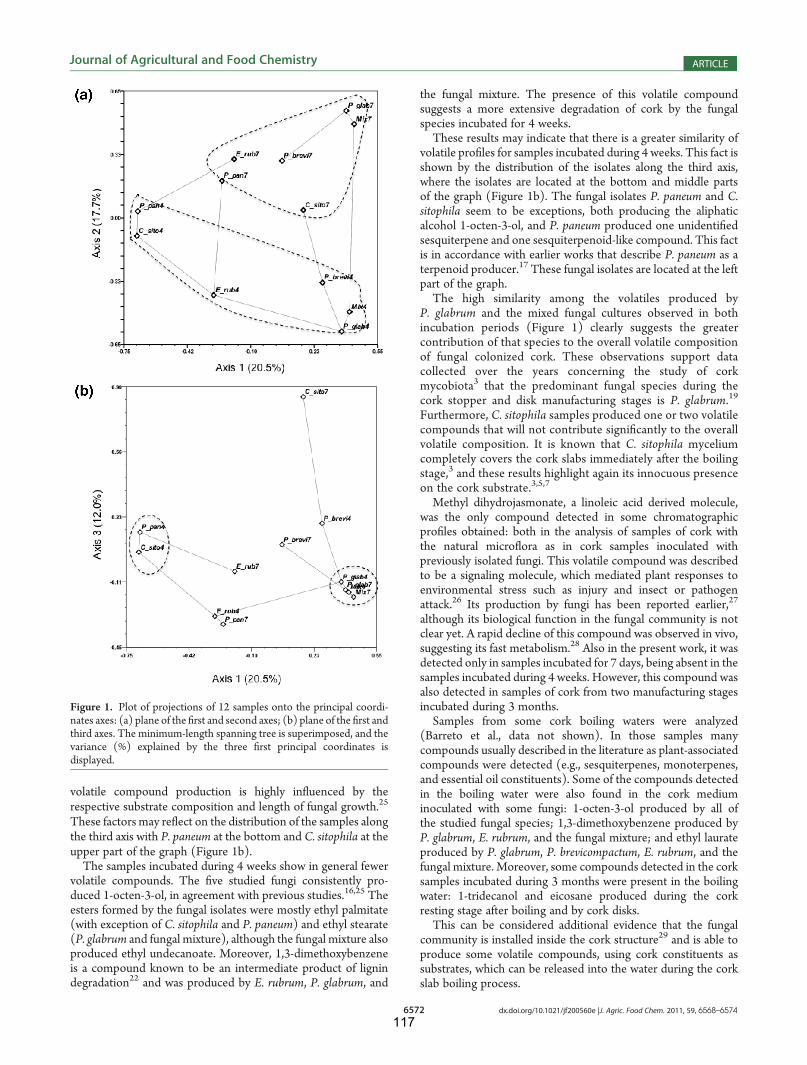
6572 dx.doi.org/10.1021/jf200560e |J. Agric. Food Chem. 2011, 59, 6568–6574
Journal of Agricultural and Food Chemistry ARTICLE
volatile compound production is highly in"uenced by therespective substrate composition and length of fungal growth.25
These factors may re"ect on the distribution of the samples alongthe third axis with P. paneum at the bottom and C. sitophila at theupper part of the graph (Figure 1b).The samples incubated during 4 weeks show in general fewer
volatile compounds. The !ve studied fungi consistently pro-duced 1-octen-3-ol, in agreement with previous studies.16,25 Theesters formed by the fungal isolates were mostly ethyl palmitate(with exception of C. sitophila and P. paneum) and ethyl stearate(P. glabrum and fungal mixture), although the fungal mixture alsoproduced ethyl undecanoate. Moreover, 1,3-dimethoxybenzeneis a compound known to be an intermediate product of lignindegradation22 and was produced by E. rubrum, P. glabrum, and
the fungal mixture. The presence of this volatile compoundsuggests a more extensive degradation of cork by the fungalspecies incubated for 4 weeks.These results may indicate that there is a greater similarity of
volatile pro!les for samples incubated during 4 weeks. This fact isshown by the distribution of the isolates along the third axis,where the isolates are located at the bottom and middle partsof the graph (Figure 1b). The fungal isolates P. paneum and C.sitophila seem to be exceptions, both producing the aliphaticalcohol 1-octen-3-ol, and P. paneum produced one unidenti!edsesquiterpene and one sesquiterpenoid-like compound. This factis in accordance with earlier works that describe P. paneum as aterpenoid producer.17 These fungal isolates are located at the leftpart of the graph.The high similarity among the volatiles produced by
P. glabrum and the mixed fungal cultures observed in bothincubation periods (Figure 1) clearly suggests the greatercontribution of that species to the overall volatile compositionof fungal colonized cork. These observations support datacollected over the years concerning the study of corkmycobiota3 that the predominant fungal species during thecork stopper and disk manufacturing stages is P. glabrum.19
Furthermore, C. sitophila samples produced one or two volatilecompounds that will not contribute signi!cantly to the overallvolatile composition. It is known that C. sitophila myceliumcompletely covers the cork slabs immediately after the boilingstage,3 and these results highlight again its innocuous presenceon the cork substrate.3,5,7
Methyl dihydrojasmonate, a linoleic acid derived molecule,was the only compound detected in some chromatographicpro!les obtained: both in the analysis of samples of cork withthe natural micro"ora as in cork samples inoculated withpreviously isolated fungi. This volatile compound was describedto be a signaling molecule, which mediated plant responses toenvironmental stress such as injury and insect or pathogenattack.26 Its production by fungi has been reported earlier,27
although its biological function in the fungal community is notclear yet. A rapid decline of this compound was observed in vivo,suggesting its fast metabolism.28 Also in the present work, it wasdetected only in samples incubated for 7 days, being absent in thesamples incubated during 4 weeks. However, this compound wasalso detected in samples of cork from two manufacturing stagesincubated during 3 months.Samples from some cork boiling waters were analyzed
(Barreto et al., data not shown). In those samples manycompounds usually described in the literature as plant-associatedcompounds were detected (e.g., sesquiterpenes, monoterpenes,and essential oil constituents). Some of the compounds detectedin the boiling water were also found in the cork mediuminoculated with some fungi: 1-octen-3-ol produced by all ofthe studied fungal species; 1,3-dimethoxybenzene produced byP. glabrum, E. rubrum, and the fungal mixture; and ethyl laurateproduced by P. glabrum, P. brevicompactum, E. rubrum, and thefungal mixture. Moreover, some compounds detected in the corksamples incubated during 3 months were present in the boilingwater: 1-tridecanol and eicosane produced during the corkresting stage after boiling and by cork disks.This can be considered additional evidence that the fungal
community is installed inside the cork structure29 and is able toproduce some volatile compounds, using cork constituents assubstrates, which can be released into the water during the corkslab boiling process.
Figure 1. Plot of projections of 12 samples onto the principal coordi-nates axes: (a) plane of the !rst and second axes; (b) plane of the !rst andthird axes. The minimum-length spanning tree is superimposed, and thevariance (%) explained by the three !rst principal coordinates isdisplayed.
117
6573 dx.doi.org/10.1021/jf200560e |J. Agric. Food Chem. 2011, 59, 6568–6574
Journal of Agricultural and Food Chemistry ARTICLE
Quantification of Releasable TCA Produced by FungalIsolates and a Fungal Mixture. The content of releasableTCA from samples inoculated with some fungal isolates in pureand mixed cultures was determined. The analysis conditions werevery similar to those used in the analysis of cork stoppers inindustrial quality control. The estimated detection limit (LD) andquantification limit (LQ) were, respectively, 1.6 and 5.4 ng L!1.The noninoculated cork media contained 1.90 and 1.78 ng L!1
of extractable TCA after 7 days and 4 weeks of incubation,respectively. TCA is formed by the O-methylation of the corre-sponding chlorophenol precursor.6 Previous studies showed thatat least some of the fungal species isolated from the cork possessthe S-adenosyl-L-methionine (SAM)-dependent chlorophenol-O-methyltransferase (CPOMT) enzyme, which in the presenceof TCP can metabolize TCA.30 To understand the origin oftheTCA values detected in our blank samples, the chlorophenolcontents of the cork were determined. The analysis showed thatcork contained an average of 2.7!3.3ng g!1 of TCA, 5.2!6.7 ng g!1
of 2,4,6-trichlorophenol (TCP), 0.6!1.3 ng g!1 of 2,3,4,6-tetrachlorophenol and 0.7!1.1 ng g!1 of 2,4,6-tribromophe-nol. These results show that TCA was detected in noninoculatedcork media and should have originated either from the corre-sponding trichlorophenol or from other chlorophenols alsopresent in the cork.Chlorophenols are common pollutants present in the envir-
onment due to earlier environmental contamination, and theirpresence was previously detected in cork.31
Under the conditions of our study, the TCA content deter-mined in the cork samples inoculated with the fungal isolates wassimilar to that of the noninoculated samples. Moreover, theextracts of cultures, both of 7 days and 4 weeks incubation time,showed similar values of releasable TCA. Applying the varianceanalysis with 5% signi!cance level (Supporting Information), inany case the TCA values obtained from the analysis of theinoculated samples could not be di#erentiated from thoseobtained from the noninoculated samples. It appears that underthe conditions of analysis, the releasable TCA on cork cannot beattributed to any of the assayed fungal isolates.To evaluate if cork dipped in the extracting medium could
retain part of the TCA eventually produced by fungi, a hydro-alcoholic solution (12% v/v) containing 800 ng L!1 of TCA andd5-TCA was placed in contact with cork granules at ca. 22 !C.The relative concentrations of TCA present in the solution afterdi#erent agitation times (5, 10, and 94 min) were evaluated bymeasurements of peak areas of TCA and d5-TCA in total ionchromatograms (TIC) obtained by GC-MS analysis. The resultsshowed that after 5 min of contact only !20% of the TCAcontent remained in the hydroalcoholic solution. After 94 min,only !11% of the initial TCA remained in the hydroalcoholicsolution. No exchange of TCA between the extracting solutionand the TCA originally present in cork was detected because nosigni!cant variation was observed in the ratio TCA/d5-TCA.This experiment con!rmed that cork had !xed most of the addedTCA, which is in agreement with other studies that have shownthat only ca. 3!5% of TCA contained in cork stoppers wasreleased to the wine.31,32 The amount of TCA adsorbed onto thecork granules depends on the cork surface, temperature, and timeof exposure. In this study cork granules were used, whichcorresponds to a higher contact area between solution and corkthan when entire cork stoppers are used in similar assays.Chlorophenol precursors present in the cork tree can be
converted into chloroanisoles by the existent colonizing fungal
species in a chemical reaction catalyzed by the SAM-dependentO-methyltransferase. However, the levels of chlorophenols in thecork forests are not very high, as seen by the cork analyses done inour study. Under these studied conditions it is improbable thatquantities of TCA produced by the fungal species present incork can be released into the wine to produce a signi!cantcontamination.To conclude, the levels of chlorophenols usually existing in
cork slabs in an industrial environment are not high enough toinduce biosynthesis of TCA by the existing fungi, even whengrown in more favorable conditions provided by laboratory tests.
’ASSOCIATED CONTENT
bS Supporting Information. Tables of data for the quanti-!cation of releasable TCA. This material is available free ofcharge via the Internet at http://pubs.acs.org.
’AUTHOR INFORMATION
Corresponding Author*Phone: #351 214469555. Fax: #351 214 421 161. E-mail:[email protected].
Funding SourcesM.C.B. thanks Fundac-~ao para a Ciencia e a Tecnologia for GrantBD/19264/2004.
’ACKNOWLEDGMENT
We thank Amorim & Irm~aos (Coruche, Portugal) for corksamples; also to Rob Samson and Jos Houbraken that help in thetaxonomic identi!cation of the fungal isolates used in this study.We gratefully acknowledge the cork irradiation done by theRadiation Technologies Unity (UTR) of Nuclear and Technolog-ical Institute (ITN, Portugal). CEVAQOE Laboratories, France,performed the cork chlorophenols analysis.
’REFERENCES(1) Pereira, H. Chemical composition and variability of cork from
Quercus suber L. Wood Sci. Technol. 1988, 22, 211–218.(2) Lacey, J. The air spora of a Portuguese cork factory. Ann. Occup.
Hyg. 1973, 16, 223–230.(3) Silva Pereira, C.; Pires, A.; Valle, M.; Vilas-Boas, L.; Figueiredo
Marques, J.; San Rom~ao, M. Role of Chrysonilia sitophila on the qualityfor cork stoppers for sealing wine bottle. J. Ind. Microbiol. Biotechnol.2000, 24, 256–261.
(4) Alvarez-Rodríguez, M. L.; L!opez-Oca~na, L.; L!opez-Coronado,J. M.; Rodríguez, E.; Martínez, M. J.; Larriba, G.; Coque, J.-J. R. Corktaint of wines: role of the !lamentous fungi isolated from cork in theformation of 2,4,6-trichloroanisole by O-methylation of 2,4,6-trichlor-ophenol. Appl. Environ. Microbiol. 2002, 68, 5860–5869.
(5) Prak, S.; Gunata, Z.; Guiraud, J. P.; Schorr-Galindo, S. Fungalstrains isolated from cork stoppers and the formation of 2,4,6-trichlor-oanisole involved in the cork taint of wine. Food Microbiol. 2007,24, 271–280.
(6) Thrane, U.; Andersen, B.; Frisvad, J. C.; Smedsgaard, J. The exo-metabolome in !lamentous fungi. Metabolomics. Topics in CurrentGenetics; Nielsen, J., Hewitt, J., Eds.; Springer: Berlin, Germany, 2007;Vol. 18, pp 235!252.
(7) Silva Pereira, C.; FigueiredoMarques, J.; San Rom~ao, M. V. Corktaint in wine: scienti!c knowledge and public perception: a criticalreview. Crit. Rev. Microbiol. 2000, 26, 147–162.
118

6574 dx.doi.org/10.1021/jf200560e |J. Agric. Food Chem. 2011, 59, 6568–6574
Journal of Agricultural and Food Chemistry ARTICLE
(8) Sefton, M. A.; Simpson, R. F. Compounds causing cork taint andthe factors a#ecting their transfer from natural cork closures to wine! areview. Aust. J. Grape Wine Res. 2005, 11, 226!240(9) Chatonnet, P.; Bonnet, S.; Boutou, S.; Labadie, M. D. Identi!ca-
tion and responsability of 2,4,6-tribromoanisole in musty, corked odorsin wine. J. Agric. Food Chem. 2004, 52, 1255–1262.
(10) Simpson, R. F.; Capone, D. L.; Sefton, M. A. Isolation andidenti!cation of 2-methoxy-3,5-dimethylpyrazine, a potent musty com-pound from wine corks. J. Agric. Food Chem. 2004, 52, 5425–5430.(11) Culler!e, L.; Cacho, J.; Ferreira, V. Comparative study of the
aromatic pro!le of di#erent kinds of wine cork stoppers. Food Chem.2009, 112, 381–387.(12) Pollnitz, A. P.; Pardon, K. H.; Liacoupos, D.; Skouroumounis,
G. K.; Sefton, M. A. The analysis of 2,4,6-trichloroanisole and otherchloroanisole in tainted wines. Aust. J. Grape Wine Res. 1996, 2,184–190.(13) Botelho, M. L.; Almeida, E.; Tenreiro, R.; Andrade, M. E.
Searching for a strategy to ! sterilize Portuguese cork stoppers !preliminary studies on bioburden radioresistance and sterility assurance.Int. J. Radiat. Appl. Instrum. C: Radiat. Phys. Chem. 1988, 31, 775–781.(14) Dias, J. A.An!alises Cromatogr!a!cas de Compostos Organoclorados
no Controlo e Garantia de Qualidade de Rolhas de Champanhe. M.Sc.Dissertation, Faculdade de Ciencias, Universidade de Lisboa, 2008.(15) Pherobase, available at http://www.pherobase.com (accessed
Jan 20, 2011).(16) Rocha, S.; Delgadillo, I.; Ferrer Correia, A. J. GC-MS of volatiles
of normal and microbiologically attacked cork from Quercus suber L.J. Agric. Food Chem. 1996, 44, 865–871.(17) Karlshof, K.; Larsen, T. O. Di#erentiation of species from the
Penicillium roqueforti group by volatile metabolite pro!ling. J. Agric. FoodChem. 2005, 53, 708–715.(18) Rohlf, J. F. NTSYS-pc ! Numerical Taxonomy and Multivariate
Analysis System, version 2.1; Exeter Software: Setauket, NY, 2000.(19) Soares, G. A. M.; Basílio, M. C.; Tenreiro, R.; San Rom~ao, M. V.
Diversity of Penicillium spp. colonizing cork slabs: a classical andmolecular approach. Biological Resource Centres and Use of Microbes !XXII ECO Meeting Proceedings Book; Lima, N., Smith, D., Eds.; MUM:Braga, Portugal, 2003; pp 161!170.(20) Peiris, D.; Dunn, W. B.; Brown, M.; Kell, D. B.; Roy, I.; Hedger,
J. N. Metabolite pro!les of interacting mycelial fronts di#er for pairingsof the wood decay basidiomycete fungus, Stereum hirsutum with itscompetitors Coprinus micaceus and Coprinus disseminatus. Metabolomics2008, 4, 52–62.(21) Bernards, M. A. Demystifying suberin. Can. J. Bot. 2002,
80, 227–240.(22) Javor, T.; Buchberger, W.; Tanzcos, I. Determination of low-
molecular-mass phenolics and non-phenolic lignin degradation com-pounds in wood digestion solutions by capillary electrophoresis. Micro-chim. Acta. 2000, 135, 45–53.(23) Tessman, D.; Dianese, J. C. Hentriacontrane: a leaf hydrocar-
bon from Syzygium jamboswith stimulatory e#ects on the germination ofurediniospores of Puccinia psidii. Fitopatol. Bras. 2002, 27, 538–542.(24) Tao, Y.; Li, H.; Wanga, H.; Zhang, L. Volatile compounds of
young Cabernet Sauvignon red wine from Changli County (China).J. Food Compos. Anal. 2008, 21, 689–694.
(25) Borjesson, T.; Stollman, U.; Schnurer, J. Volatile metabolitesand other indicators of Penicillium aurantiogriseum growth on di#erentsubstrates. Appl. Environ. Microbiol. 1990, 56, 3705–3710.
(26) Wasternack, C. Jasmonates: an update on biosynthesis. Signaltransduction and action in plant stress response, growth and develop-ment. Ann. Bot. 2007, 100, 1–17.(27) Miersch, O.; Porzel, A.;Wasternack, C.Microbial conversion of
jasmonates ! hydroxylations by Aspergillus niger. Phytochemistry 1999,50, 1147–1152.
(28) Mueller, M. J.; Brodschelm, W.; Spannagl, E.; Zenk, M. H.Signaling in the elicitation process is mediated through the octadecanoidpathway leading to jasmonic acid. Proc. Natl. Acad. Sci. U.S.A. 1993,90, 7490–7494.
(29) Silva Pereira, C.; Soares, G. A. M.; Oliveira, A. C.; Rosa, M. E.;Pereira, H.; Moreno, N.; San Rom~ao, M. V. E#ect of fungal colonizationon mechanical performance of cork. Int. Biodeterior. Biodegrad. 2006,57, 244–250.
(30) Coque, J.-J.; !Alvarez-Rodríguez, M. L.; Larriba, G. Character-ization of an inducible chlorophenolO-methyltransferase from Tricho-derma longibracheatum involved in the formation of chloroanisoles anddetermination of its role in cork taint of wines. Appl. Env. Microbiol.2003, 69, 5089–5095.
(31) Alvarez-Rodriguez, M. L.; Recio, E.; Coque, J. J. R. The analysisof natural cork in transversal sections as an e#ective tool to determinethe origin of the taint by 2,4,6-trichloroanisole. Eur. Food Res. Technol.2009, 230, 135–143.
(32) Juanola, R.; Subir!a, D.; Salvad!o, V.; Regueiro Garcia, J. A.;Antic!o, E. Migration of 2,4,6-trichloroanisole from cork stoppers towine. Eur. Food Res. Technol. 2005, 220, 347–352.
119

Supporting information

Table 17 days Mean Variance SquareBlank 0.7083 1.9137 2.7700 1.7973 1.0728 0.1780C. sitophila 3.9245 1.7651 2.0267 2.5721 1.3889 0.1245P. glabrum 0.6610 2.3867 0.6457 1.2311 1.0015 0.9764P. brevicompactum 1.6803 3.0726 0.8855 1.8795 1.2255 0.1155P. paneum 3.1725 4.6009 1.3306 3.0346 2.6880 0.6649E. rubrum 4.5808 0.9191 2.4334 2.6444 3.3854 0.1808Fungal mixture 1.1667 3.2347 2.7257 2.3757 1.1611 0.0245
2.2193 1.7033 2.2645average average sum
F=2.2645/1.7033= 1.329 < 2.848 (critical value of F for a one-‐tailed test) Probability (P=0.05) df(between samples)=6
df=degrees of freedom df(within samples)=14
Table 24 weeks Mean Variance SquaresBlank 3.4186 1.8893 0.0399 1.7826 2.8624 0.1858C. sitophila 3.5541 3.6682 1.7255 2.9826 1.1885 0.5913P. glabrum 1.2259 2.2230 3.6183 2.3557 1.4441 0.0202P. brevicompactum 2.5605 1.1630 0.9557 1.5597 0.7619 0.4277P. paneum 2.9761 0.5203 1.2211 1.5725 1.6004 0.4111E. rubrum 4.1644 0.3128 2.2386 7.4175 0.0006Fungal mixture 2.8960 0.4676 5.6731 3.0122 6.7844 0.6377
2.2137 2.2137 3.1513 2.2743average average sum
F=3.151/2.274= 1.386 < 2.915 (critical value of F for a one-‐tailed test) (P=0.05) df(between samples)=6
df(within samples)=13
triplicates
triplicates
Quantification of releasable TCA. Analysis of variance (ANOVA) Results (triplicates) of analysis of TCA for samples incubated during 7 days are presented in Table 1 and were treated by ANOVA to determine whether there were significant differences between blank and samples and also between samples. The results of calculations necessary for analysis of variance were included in table 1: mean and variance for each triplicate and square of diferences between the mean of each sample and the mean for all samples. The F-test was applied to values estimated for the variance between samples (2.2645) and variance within samples (1.7033) (ng/L).
The calculated value for F (1.329) is smaller than the critical value of F (2.848) (obtained from the tables of t-test): therefore no significant differences were observed in the results of analysis obtained for blank and for the various samples (ng/L). The same statistical treatment was applied to the samples incubated during 4 weeks.
The calculated value for F (1.386) is smaller than the critical value of F (2.915): therefore no significant differences were observed in the results of analysis obtained for blank and for the various samples (ng/L). The set of all samples (7 days and 4 weeks) was also considered and the same statistical treatment was applied.
121
KitaBarreto
Inserted Text
KitaBarreto
Typewritten Text

Table 37 days Means Variances SquaresBlank 0.7083 1.9137 2.7700 1.7973 1.0728 0.1098C. sitophila 3.9245 1.7651 2.0267 2.5721 1.3889 0.1966P. glabrum 0.6610 2.3867 0.6457 1.2311 1.0015 0.8057P. brevicompactum 1.6803 3.0726 0.8855 1.8795 1.2255 0.0621P. paneum 3.1725 4.6009 1.3306 3.0346 2.6880 0.8207E. rubrum 4.5808 0.9191 2.4334 2.6444 3.3854 0.2660Fungal mixture 1.1667 3.2347 2.7257 2.3757 1.1611 0.06104 weeksBlank 3.4186 1.8893 0.0399 1.7826 2.8624 0.1198C. sitophila 3.4186 1.8893 0.0399 1.7826 2.8624 0.1198P. glabrum 1.2259 2.2230 3.6183 2.3557 1.4441 0.0515P. brevicompactum 2.5605 1.1630 0.9557 1.5597 0.7619 0.3238P. paneum 2.9761 0.5203 1.2211 1.5725 1.6004 0.3094E. rubrum 4.1644 0.3128 2.2386 7.4175 0.0121Fungal mixture 2.8960 0.4676 5.6731 3.0122 6.7844 0.7805
2.1287 2.5469 4.0388average average sum
F=4.0387/2.5469 1.5858 < 2.1068 (critical value of F for a one-‐tailed test) (P=0.05) df(between samples)=13
df(within samples)=27
triplicates
Table 3 contains the complete collection of results obtained in the analysis of releasable TCA as well as the results of calculations necessary for analysis of variance .
The calculated value for F (1.5858) is smaller than the critical value of F (2.1068): therefore no significant differences were observed in the results of analysis obtained for blank and for the various samples
122

5
Discussion


D i s c u s s i o n
To address the study of the natural fungal community present in
the cork slabs one culture dependent and two culture independent
methods were employed. The isolation method provided indications
of the fungal quantity and diversity present in the samples taken in
some stages of the cork discs manufacturing. The methods
showed that the fungal concentrations present in each sample
have different quantities depending mainly on the stage of the cork
sample. Additionally, the mycobiota diversity is conditioned first by
the season on which the sample was made and at a later stage the
cork geographic origin. The diversity is higher in non-boiled cork.
Other works performed earlier to study the cork mycobiota isolated
in several stages of the cork manufacturing of stoppers gave
similar results although the fungal load present in those samples
were different of the ones considered in our work. Another
difference encountered is the fact that the fungal quantity present in
cork samples collected in the resting stage is higher than the
amount present in the raw cork samples (Alvarez-Rodriguez, 2002).
This is probably due to the fact that the period of time that the cork
slabs remained in the storeroom under conditions favoring the
fungal growth could be higher than the ones experienced in this
work (Alvarez-Rodriguez, 2002).
The culture-dependent technique combined the phenotypic and
molecular methods to identify the fungi isolated in this work. Most
of the fungal isolates belong to Penicillium and Aspergillus genera
and the most predominant species were P. glabrum and C.
sitophila. P. glabrum was isolated in most of the studied samples,
while C. sitophila appeared mostly in the resting stage, covering all
the cork slabs in that phase. In this work, this species was isolated
in one sample from the Spanish cork batch and in another from the
125

C h a p t e r 5
Portuguese cork batch; those samples were collected in the stages
before boiling and non-treated cork discs. These two fungal
species colonizing cork substrate were reported by other authors
(Davis, 1981; Daly, 1984; Lee, 1993; Danesh, 1997; Alvarez-
Rodriguez, 2002; Basílio, 2006).
The use of PCR specific primers, β-tubulin gene for all the
Penicillium and Aspergilus genera and calmodulin for the isolates
that belong to the Glabra series, allow the correct taxonomic
identification to species rank of most of the isolates. In addition a
more perceptive taxonomic study in the Glabra series was done
and the description of a new fungal species P.subericola was
obtained (Barreto, 2011b). Cork is an unique substrate that
possible allows new fungal species some of them already
described (Belloch, 2007).
The combination of two culture-independent methods to study the
fungal population resulted in a more precise knowledge of the
composition of this population. The fungal diversity that begins to
be known by the isolation technique was more unveiled using these
techniques. The cloning method revealed the existence of a variety
of fungi mostly in the raw cork composed mainly of uncultivable
fungi, Ascomycetes and endophytes. Most of them are present in
low relative frequencies. Cork mycobiota in samples taken
immediately after the first boiling was probably composed by
uncultivable species. Probably, the mycobiota present in these
stages are mainly composed by fungal species that colonised the
cork tree in the forest, as already being remarked in other studies
(Serra, 2008). However, at this stage one of the Portuguese
batches (38) was successful in isolating six different fungal species.
126

D i s c u s s i o n
Finally in at the remaining manufacturing stages the mycobiota
population present is similar to all of them. This population is
composed mainly by cultivable fungal species belonging to
Penicillium, Aspergillus and Neurospora species. Reinforcing the
fact that most of the fungal population present at these stages
originates from the environmental mycobiota present in the factory
and after the boiling have the necessary conditions (temperature
and humidity) to colonize the slabs and dominate the fungal
colonization. Although, the presence of at least one or two
phylotypes (Sporobolomyces sp. and Pezicula sp.) usually
associated to the plant material and that could be originated from
the cork tree and survived the whole manufacturing process was
detected.
The mycospores could be lodged inside the cork lenticels and due
to the cork’s impermeability to high temperatures and humidity
levels that occur during the boiling process does not reach all the
cork structure. Thus, a microhabitat is created inside the cork
structure and enables the survival of some microbiota that were
previously present.
The concern of cork quality was the starting point to study the exo-
metabolites and volatiles produced by some fungal species isolated
from the cork substrate. Some of these species are known to
produce exo-metabolites in semi-synthetic culture media; some of
them when growing in culture media are even mycotoxins
producers (e.g. P. citrinum, P. citreonigrum). The possible
production of exo-metabolites and volatiles by C. sitophila was
studied since this fungal species dominates the cork slabs mostly
during their resting stage.
127

C h a p t e r 5
The studied fungal isolates produced some exo-metabolites
specially when growing in the semi-synthetic media. When growing
on cork-based culture medium most of the isolates do not produce
any metabolite, while some (E. amstelodami, P. brevicompactum
and P. citrinum) produce few exo-metabolites. However, if C.
sitophila extracts are added to the cork-based culture medium only
two of the studied fungal isolates did not produce any metabolite in
this medium. This situation tries to mimic the cork at the resting
stage where the water activity (aw) decreases to values below 0.9.
Until this value C. sitophila mycelium completely covered the cork
slabs. Bellow 0.9 aw this fungus was replaced by the mycelium of
other fungal species (e.g. Penicillium, Aspergillus, Trichoderma).
This late mycobiota develops in the cork slabs that have the
remains of C. sitophila. The presence of this mycelium on the cork
slabs enhances the exo-metabolome of the late fungal colonizers
leading to a production of more metabolites (Barreto, 2011a). C.
sitophila, as expected did not produce any metabolite in any of the
used culture media, which is in accordance with previous studies
made on the safety of Neurospora crassa (Perkins, 2000).These
results suggested that the most predominant mycoflora present in
the resting stage of cork could be regarded as non-deleterious to
the cork discs (or cork stoppers) final product.
It appears that studies concerning the interactions that could exist
among cork colonizing fungi in such specific substrate as cork are
crucial. Also the different metabolic mechanisms used by those
fungal species to colonize/ survive in such environment should be a
subject of study.
The genome of some related species that colonize cork (e.g.
Neurospora crassa and Trichoderma viride) are fully sequenced.
128
D i s c u s s i o n
This fact could be the starting point to some further research of the
mechanisms of degradation of some main cork constituents by the
respective fungal species.
Furthermore, the production of volatile compounds was studied for
a) cork samples from one batch taken during several
manufacturing process and b) some fungal isolates growing in
cork-based culture medium, in both pure and mixed culture.
Most of the volatile compounds were produced by the mycobiota
that exists in two manufacturing stages: resting stage after the first
boiling and non-treated cork discs. Additionally, the volatile
compounds produced by the fungal isolates, either in pure or mixed
culture consisted mainly of 1-octen-3-ol and esters of fatty acids
(medium chain length C8 –C20). P. glabrum seems to be the main
contributor to the overall volatile composition observed in the mixed
culture. In general, no volatile compounds detected in this work will
influence negatively the cork’s final product (Barreto, 2011c).
The presence of chemical compounds that can produce
organoleptic defects on the wine is a major concern in the cork
Industry. These compounds can be present in very small quantities
(range of ng/L-1) and spoil the wine.
2,4,6–Trichloroanisole (TCA) was present in most of the cases of
“cork taint” in wines. TCA contaminates cork by many processes,
but usually results from the transformation of polychlorophenols,
used in some wood preservatives and in some pesticides, by the
action of fungal metabolism, e.g. Penicillium and Aspergillus.
Some fungal isolates, in pure and mixed culture, were screened to
investigate their capacity to produce TCA in cork-based medium.
To perform this experiment, Industrial conditions were considered
as well as the chlorophenols levels that usually exist in cork slabs.
129

C h a p t e r 5
The results show that the amount of TCA present in the non-
inoculated medium culture could not be distinguished from the
levels obtained in the studied samples, even if analysis of variance
with 5% significance level was applied (Supporting Information in
(Barreto, 2011c)).
Moreover, in the presence of a hydroalcoholic solution containing
known concentrations of TCA and 5d-TCA, the cork used in the
culture medium, retained most of the added chloroanisoles. After
the first 5 min of contact only ~20% of the added TCA remained in
the hydroalcoholic solution. No exchange of TCA between the
extracting solution and the TCA originally present in the cork was
observed (Barreto, 2011c).
These results can be the starting point to study the potential role of
cork granules in the treatment of polluted environments, namely
waters. Additionally, cork being an unique material with distinct
chemical constitution and exclusive physical properties can also
serve to investigate the growth and development of microbiota that
need to survive to extreme conditions to be used in bioremediation
situations. For instance, pentachlorophenol (PCP) is a chemical
compound widely used as herbicide, biocide and pesticide and thus
contaminating the terrestrial and aquatic environments. Recently,
few fungal species previously isolated from cork substrate showed
the capacity to fully degrade PCP (Carvalho, 2009). These results
reinforce the demand to study microbiota that colonizes recalcitrant
substrates and investigate their capacity to decontaminate polluted
environments.
The Industry needs to maintain the control of humidity of the cork
slabs (around 0.9 aw) during the slabs resting stage, to avoid the
germination of fungal species that can contribute to off-odors.
130
D i s c u s s i o n
However, the results obtained in this work point out that if the
humidity levels are controlled and the hygienic practices respected
(e.g. no contamination of the cork slabs from the environmental
and soil factory), no unpleasant sensory properties can be imputed
to the cork final product.
131


6
Bibliography


B i b l i o g r a p h y
AFGC (2007).Organohalogen taints in foods. In Supplement to
food Australia.
Alexopoulos, C. J., Mims, C. W., Blackwell, M. (1996).
Introductory Mycology, 4th edn. New York: Wiley.
Álvarez-Rodríguez, M. L., Belloch, C., Villa, M., Uruburu, F.,
Larriba, G., Coque, J-J R. (2003). Degradation of vanillic acid
and production of guaiacol by microorganisms isolated from cork
samples. FEMS Microbiology Letters 220, 49-55.
Alvarez-Rodriguez, M. L., Lopez-Ocana, L., Lopez-Coronado,
J. M., Rodriguez, E., Martinez, M. J., Larriba, G., Coque, J. J.
(2002). Cork taint of wines: role of the filamentous fungi isolated
from cork in the formation of 2,4,6-trichloroanisole by o
methylation of 2,4,6-trichlorophenol. Appl Environ Microbiol 68,
5860-5869.
Alvarez-Rodriguez, M. L., Recio, E., Coque, J. J. R. (2009).
The analysis of natural cork in transversal sections as an
effective tool to determine the origin of the taint by 2,4,6 –
trichloroanisole. EUR Food Res Technol 230, 135-143.
Amon, J. M., Vandepeer, J. M., Simpson, R. F. (1989).
Compounds responsable for the cork taint in wine. Wine Industry
Journal 2, 62-69.
Anderson, I. C., Campbell, C. D., Prosser, J. I. (2003a).
Potential bias of fungal 18S rDNA and internal transcribed spacer
135
C h a p t e r 6
polymerase chain reaction primers for estimating fungal
biodiversity in soil. Environ Microbiol 5, 36-47.
Anderson, I. C., Campbell, C. D., Prosser, J. I. (2003b).
Diversity of fungi in organic soils under a moorland- Scots pine
(Pinus sylvestris L.) gradient. Environ Microbiol 5, 1121-1132.
APCOR Other applications of cork. available at
http://www.cork.pt/other-applications-of-cork.html (acessed May
8, 2011)
Ávila, R., Lacey, J. (1974). The role of Penicillium frequentans in
suberosis (respiratory disease in workers in the cork industry).
Clinical Allergy 4, 109-117.
Barreto, M. C., Frisvad, J. C., Larsen, T. O., Mogensen, J.,
San-Romão, M. V. (2011a). Exo-metabolome of some fungal
isolates growing on cork-based medium. EUR Food Res Technol
232, 575-582.
Barreto, M. C., Houbraken, J., Samson, R., Frisvad, J. C.,
San-Romão, M. V. (2011b). Taxonomic studies of the Penicillium
glabrum complex and the description of a new species P.
subericola. Fungal Diversity.
Barreto, M. C., Vilas Boas, L., Carneiro, L. C., San Romão, M.
V. (2011c). Volatile compounds in samples of cork and also
produced by selected fungi. J Agric Food Chem 59, 6568-6574.
136

B i b l i o g r a p h y
Basílio, M. C. G., R.; Silva Pereira & San Romão, M. V. (2006).
Penicillium glabrum cork colonising isolates - preliminary analysis
of their genomic similarity. Rev Iberoam Micol 23, 151-154.
Belloch, C., Villa-Carvajal, M., Álvarez-Rodríguez, M. L.,
Coque, J. J. (2007). Rhodotorula subericola sp. nov., and
anamorphic basidiomycetous yeast speceis isolated from bark of
Quercus suber (cork oak). Int J Syst Evol Microbiol 57, 1668-
1671.
Bennet, J. W., Feibelman, T. (2001). Fungal bacteria
interacteractions. In The Mycota pp. 229 - 242. Edited by F. A.
Hock. Heidelberg, Berlin: Springer-Verlag.
Brown, S. P., Inglis, R. F., Taddei, F. (2009). Evolutionary
ecology of microbial wars: within-host competition and
(incidental) virulence. Evol Appl 2, 32-39.
Buser, H. R., Zanier, C., Tanner, H. (1982). Identification of
2,4,6-trichloroanisole as a potent component causing cork taint in
wine J Agric Food Chem 30, 359-362.
Caldentey, P., M.D. Fumi, Mazzoleni, V., Careri, M. (1998).
Volatile compounds produced by microorganisms isolated from
cork. Flavour Fragr 13, 185-188.
Calvo, A., Wilson, R. A., Bok, J. W., Keller, N. P. (2002).
Relationships between secondary metabolism and fungal
137
C h a p t e r 6
development. Microbiol Mol Biol Rev 66, 447-459.
Capone, D. L., Skouroumounis, G. K., Sefton, M. A. (2002).
Permeation of 2,4,6-trichloroanisole through cork closures in
wine bottles. Australian Journal of grape and wine research 8,
196-199.
Carlile, M. J., Watkinson, S. C., Gooday, G. W. (2001). Fungal
diversity, 2nd edition edn. London, UK: Academic Press.
Carvalho, M. B., Martins, I., Leitão, M. C., Garcia, H.,
Rodrigues, C., San-Romão, V., Hursthouse, A., Silva Pereira,
C. (2009). Screening pentachlorophenol degradation ability by
environmental fungal strains belonging to the phyla Ascomycota
and Zygomycota. J Ind Microbiol Biotechnol 36, 1249-1256.
Celiège (2006).International code of cork stopper manufacturing
practice.
Chatonnet, P., Bonnet, S., Bouton, S., Labadie, M-D. (2004).
Identification and responsibility of 2,4,6 - tribromoanisole in
musty, corked odors in wine. Journal of agricultural and food
chemistry 52, 1255-1262.
Chen, D. M., Cairney, J. W. G. (2002). Investigation of the
influence of prescribed burning on ITS profiles of ectomycorrhizal
and other soil fungi at three Australian sclerophyll forest sites.
Mycol Res 106, 532-540.
138

B i b l i o g r a p h y
Coque, J. J., Álvarez-Rodriguez, M. L., Larriba, G. (2003).
Characterization of an inducible chlorophenol O-
methyltransferase from Trichoderma longibracheatum involved in
the formation of chloroanisoles and determination of its role in
cork taint of wines. Appl Environ Microbiol 69, 5089-5095.
Coque, J. J., Rodríguez, M.L., Goswami, M., Martinez, R.F.
(2006). Causes and origins of wine contamination by
haloanisoles (chloroanisoles and bromoanisoles). León: Asecor -
Agrupación Sanvicenteña Empresarios del Corcho.
Coquet, C., Bauza, E., Oberto, G., Berghi, A., Farnet, A. M.,
Ferré, E., Peyronel, D., Dal Farra, C., Domloge, N. (2005).
Quercus suber cork extract displays a tensor and smoothing
effect on human skin: an in vivo study. Drugs Exptl Clin Res 31,
89-99.
Cork facts available at http://www.corkfacts.com/nchoice1.htm).
(acessed July 8, 2011)
Curtis, R. F., Dennis, C., Gee, J. M., Gee, M. G., Griffths, M.
N., Land, D. G., Peel, J. L., Robinson, D. (1974).
Chloroanisoles as a cause of musty taint in chickens and their
microbiological formation from chlorophenols in broiler house
liter. Journal Science Food Agriculture 25, 811-828.
Daly, N. M. L., T. H., Fleet, G. H. (1984). Growth of fungi on wine
corks and its contribution to cork taints in wine. Food Technology
139

C h a p t e r 6
in Australia 36, 22-24.
Danesh, P., Velez Caldas, F. M., Figueiredo Marques, J. J.,
San Romão, M. V. (1997). Mycobiota in portuguese "normal"
and "green" cork throughout te manufacturing process of
stoppers. J Appl Microbiol 82, 689-694.
Darriet, P., Pons, M., Lamy, S., Dubourdieu, D. (2000).
Identification and quantification of geosmin, an earthy odorant
contaminating wines. J Agric Food Chem 48, 4835-4838.
Davis, C. R., Fleet, G. H., Lee, T. H. (1981). The microflora of
wine corks. Aust Grapegrower winemaker 208, 42-44.
Dias, A. (2008). Análises cromatográficas de compostos
organoclorados no controlo e garantia de qualidade de rolhas de
champanhe. M.Sc. Dissertation, Faculdade de Ciências,
Universidade de Lisboa.
Dickie, I. A., Xu, B., Kolde, R. T. (2002). Vertical distribution of
ectomycorrhizal hyphae in soil as shown bt T-RFLP analysis.
New Phytol 156, 527-535.
Duncan, B. C., Gibson, R. L., Obradovic, D. (1997). 2,4,6-
trichloroanisole and cork production. Wine Industry Journal 12,
180-184.
Engel, G., Ernest von Milczewski, K., Prokopek, D., Teuber,
M. (1982). Strain-specific synthesis of mycophenolic acid by
140

B i b l i o g r a p h y
Penicillium roquefortii in blue-veined cheese. App Env Microbiol
43, 1034-1040.
Filtenborg, O., Frisvad, J. C., Thrane, U. (1990). The
significance of yeast extract composition on metabolite
production in Penicillium. In Modern concepts in Penicillium and
Aspergillus classification, pp. 433-440. Edited by R. Samson, Pitt,
J. New York: Plenum.
Flodin, C., Whitfield, F. B. (2000). Brominated anisoles and
cresols in the red alga Polysiphonia sphaerocarpa
Phytochemistry 53, 77-80.
Fortes, M. A., Rosa, M. E., Pereira, H. (2004). A cortiça. Lisboa:
IST Press.
Frisvad, J. C., Filtenborg, O., Thrane, U. (1989). Analysis and
screening for mycotoxins and other secondary metabolites in
fungal cultures by thin-layer chromatography and high-
performance liquid chromatography. Arch Environ Contam
Toxicol 18, 331-335.
Frisvad, J. C., Thrane, U., Filtenborg, O. (1998). Role and use
of secondary metabolites in fungal taxonomy. In Chemical fungal
taxonomy, pp. 289-319. Edited by J. C. Frisvad, Bridge, P. D.,
Arora, D. K. New York: Marcel Dekker.
Frisvad, J. C. S., R. A. (2004). Polyphasic taxonomy of
Penicillium subgenus Penicillium. A guide to identification of food
141
C h a p t e r 6
and air-borne terverticillate Penicilia and their mycotoxins. Stud
Mycol 49, 173.
Gil, A. M., Lopes, M. H., Neto, C. P., Callaghan, P. T. (2000).
An NMR microscopy study of water absorption in cork. J Mater
Sci 35, 1891-1900.
Gil , L. (1996). Densification of black agglomerate cork boards
and study of densified agglomerates. Wood Science and
Technology 30, 217-223.
Gil, L. (1998). A cortiça - Produção, Tecnologia e Aplicação.
Lisboa: INETI.
Gil, L. (2007). Cork composites: a review. Materials 2, 776-789.
Gourlay, I. D., Pereira, H. (1998). The effect of bark stripping on
wood production in cork oak (Quercus suber) and problems of
growth ring definition. In Cork oak and cork European
Conference on cork oak and cork 5 -7 May 1997, pp. 99-107.
Edited by H. Pereira. Lisboa: CEF - Centro de Estudos Florestais
- Instituto Superior de Agronomia.
Haas, H., Galler, H., Habib, J., Melkes, A., Schlacher, R.,
Buzina, W., Friedl, H., Marth, E., Reinthaler, F. F. (2010).
Concentrations of viable fungal spores and trichloroanisole in
wine cellars. Int J Food Microbiol 114, 126-132.
Hawksworth, D. L. (1991). The fungal dimension of biodiversity:
142
B i b l i o g r a p h y
magnitude, significance and conservation. Mycol Res 95, 641-
655.
Hawksworth, D. L., Kirk, P. M., Sutton, B. C., Pegler, D. L.
(1995). Ainsworth and Bisby's Dictionary of the fungi, 8th edn.
Wallingford: CAB International.
Hibbet, D. S. (1992). Ribosomal RNA and fungal systematics.
Trans Mycol Soc Japan 33, 533-556.
Hill, J. L., Hocking, A. D., Whitfield, F. B. (1995). The role of
fungi in the production of chloroanisoles in general purpoose
freight containers. Food Chemistry 54, 161-166.
Hitchcock, C. J., Chambers, S. M., Anderson, I. C., Cairney,
J. W. G. (2003). Development of markers for simple sequence
repeat-rich regions that discriminate between Pisolithus albus
and P. microcarpus. Mycol Res 107, 699-706.
Hoffman, A., Sponholz, W. R., David, F., Sandra, P. (2000).
Corkiness in wine-trace analysis of 2,4,6-trichloroanisole by stir
bar sorptive extraction (SBSE) and thermal desorption GC/MS.
AppNote 3, 1-5.
Houbraken, J. V., J.; Rico-Munoz, E.; Johnson, S. & Samson,
R. (2008). Sexual reproduction as the cause of heat resistance in
the food spoilage fungus Byssochlamys spectabilis (Anamorph
Paecilomyces variotii). Applied Environmental Microbiology 74, 7.
143
C h a p t e r 6
Howland, P. R., Pollnitz, A. P., Liacopoulos, D., Mclean, H. J.,
Sefton, M. A. (2008). The location of 2,4,6-trichloroanisole in a
batch of contaminated wine corks. Aust J Grape Wine Res 3, 1-5.
International, P. G. f. P. (2010). PAN International list of highly
hazardous pesticides (PAN list of HHP) available at www.pan-
germany.org/download/PAN_HHP-List_101216.pdf (accessed
July 8, 2010).
Juanola, R. S., D.; Salvado, V.; García Regueiro, J. A. &
Antico, E. (2005). Migration of 2,4,6-trichloroanisole from cork
stoppers to wine. EUR Food Res Technol 220, 347-352.
Karahadian, C., Josephson, D. B., Lindsay, R. C. (1985).
Volatile compounds from Penicillium sp. contributing musty-
earthy notes to brie and camembert cheese flavors. J Agric Food
Chem 33, 339-343.
Karbowiak, T., Gougeon, R. D., Alinc, J.-B., Brachais, L.,
Debeaufort, F., Voilley, A., Chassagne, D. (2010). Wine
oxidation and the role of cork. Crit Rev Food Sci Nutr 50, 20-52.
Karlovsky, P. (2008). Secondary metabolites in soil ecology. In
Soil Biology series, pp. 1-19. Edited by A. Varma: Springer.
Lacey, J. (1973). The air spora of a Portuguese cork factory. Ann
Occup Hyg 16, 223-230.
Landeweert, R., Leeflang, P., Smit, E., Kuyper, T. W. (2004).
144

B i b l i o g r a p h y
Diversity of an ectomycorrhizal fungal community studied by a
root tip and total soil DNA approach. Mycorrhiza 15, 1-6.
Larsen, T. O. (1998). Volatiles in fungal taxonomy. In Chemical
fungal taxonomy, pp. 263-288. Edited by J. C. Frisvad, Bridge, P.
D., Arora, D. K. New York: Marcel Dekker.
Larsen, T. O., Frisvad, J. C. (1995a). Characterization of volatile
metabolites from 47 Penicillium taxa. Mycol Res 99, 1153-1166.
Larsen, T. O., Frisvad, J. C. (1995b). Chemosystematics of
Penicillium based on profiles of volatile metabolites. Mycol Res
99, 1167-1174.
Larsen, T. O., Smedsgaard, J., Nielsen, K. F., Hansen, M. E.,
Frisvad, J. C. (2005). Phenotypic taxonomy and metabolite
profiling in microbial drug discovery. Nat Prod Rep 22, 672-695.
Lee, T. H., Simpson, R. F. (1993). Microbiology and chemistry of
cork taints in wine. In Wine, microbiology and Technology. Edited
by G. H. Fleet: Harwood Academic Publishers.
Lefebvre, A., Riboulet, J. -M., Boidron, J.-N. & Ribéreau-
Gayon, P. (1983). Incidence des micro-organismes du liège sur
les altérations olfactives du vin. Science des Aliments 3, 13.
Lopes, P., Saucier, C., Teissedre, P.-L, Glories, Y. (2007).
Main routes of oxygen ingress through different closures into
wine bottles. J Agric Food Chem 55, 5167-5170.
145

C h a p t e r 6
Macku, C., Gonzalez, L., Schleussner, C., Mesquita, A. C.,
Herwatt, J. W., Kirch, L. C., Schwartz, R. J. (2009). Sensory
screening for large-format natural corks by "dry soak" testing and
its correlation to headspace solid-phase microextraction (SPME)
gas chromatography/mass spectrometry (GC/MS) releasable
trichloroanisole (TCA) analysis. J Agric Food Chem 57, 7962-
7968.
Maga, J. A., Puech, J. L. (2005). Cork and alcoholic beverages.
Food Reviews International 21, 53-68.
Maggi, L., Mazzoleni, V., Fumi, M. D., Salinas, M. R. (2008).
Transformation ability of fungi isolated from cork and grape to
produce 2,4,6-trichloroanisole from 2,4,6-trichlorophenol. Food
Addit Contam Part A Chem Anal Control Expo Risk Assess 25,
265-269.
Mills, N., Lopes, P., Cabral, M. (2006). Oxygen brings new life
to closure debate. The Australian & New Zealand Grapegrower &
Winemaker, 112-114.
Muyzer, G. (1999). DGGE/TGGE a method for identifying genes
from natural ecosystems. Curr Opin Microbiol 2, 317-322.
Muyzer, G., Smalla, K. (1998). Application of denaturing
gradient gel electrophoresis (DGGE) and temperature gradient
gel electrophoresis (TGGE) in microbial ecology. Antonie Van
Leeuwenhoek 73, 127-141.
146

B i b l i o g r a p h y
Nannipieri, P., Ascher, J., Ceccherini, M. T., Landi, L.,
Pietramellara, G., Renella, G. (2003). Microbial diversity and
soil functions. Eur J Soil Science 54, 655-670.
Nielsen, D. S., Hønholt, S., Tano-Debrah, K., Jespersen, L.
(2005). Yeast populations associated with Ghanaian cocoa
fermentations analysed using denaturing gradient gel
electrophoresis (DGGE). Yeast 22, 271-284.
Oliveira, A. C., Peres, C. M., Correia Pires, J. M., Silva
Pereira, C., Vitorino, S., Figueiredo Marques, J. J., Crespo,
M. T. B., San Romão, M. V. (2003). Cork stoppers Industry:
defining appropriate mould colonization. Microb Res 158, 117-
124.
Oliveira, M. A., Oliveira, L. (2000). A cortiça. Casais de Mem
Martins, Rio de Mouro: Corticeira Amorim.
Pereira, C., Gil, L., Carriço, L. (2007a). Reduction of the 2,4,6-
trichloroanisole content in cork stoppers using gamma radiation.
Radiat Phys Chem 76, 729-732.
Pereira, H. (2007b). Cork: Biology, production and uses.
Amsterdam: Elsevier.
Pereira, M., Gil, L. (2006). O problema do odor a mofo nas
rolhas de cortiça e processos para a sua redução/eliminação.
Silva Lusitana 14, 101-111.
147

C h a p t e r 6
Perkins, D. D., Davis, R. H. (2000). Evidence for safety of
Neurospora speceis for academic and commercial uses. Appl
Environ Microbiol 66, 5107-5109.
Pires, J. M. C. P., H., San- Romão, M. V. (2007). Study of
humidity and water activity of cork slabs during cork stopper
manufacturing process - preliminary results. Ciência Téc Vitiv 22,
15-20.
Pitt, J. J., Hocking, A. D. (1997). Fungi and food spoilage.
London: Blackie Academic & Professional, Chapman & Hall.
Plutowska, B., Wardencki, W. (2008). Application of gas
chromatography-olfactometry (GC-O) in analysis and quality
assessment of alcoholic beverages - A review. Food Chemistry
107, 449-463.
Pollnitz, A. P. P., K. H.; Liacopoulos, D.; Skouroumounis, G.
K., Sefton, M. A. (1996). The analysis of 2,4,6-trichloroanisole
and other chloroanisoles in tainted wines and corks. Aust J
Grape Wine Res 2, 184-190.
Prak, S., Gunata, Z., Guiraud, J. -P, Schorr-Galindo, S. (2007).
Fungal strains isolated from cork stoppers and the formation of
2,4,6-trichloroanisole involved in the cork taint of wine. Food
Microbiol 24, 271-280.
Prat, C., Bañeras, L., Anticó, E. (2008). Screening of musty-
148

B i b l i o g r a p h y
earthy compounds from tainted cork using water-based soaks
followed by headspace solid -phase microextraction and gas
chromatography-mass spectrometry. EUR Food Res Technol
227, 1085-1090.
Prat., C., Trias, R., Culleré, L., Escudero, A., Anticó, E.,
Banñeras, L. (2009). Off-odor compounds produced in cork by
isolated bacteria and fungi: a gas chromatography-mass
spectrometry and gas chromatography-olfatometry study. J Agric
Food Chem 57, 7473-7479.
Ranjard, L., Poly, F., Nazaret, S. (2000). Monitoring complex
bacteria communities using culture-independent molecular
techniques: application to soil environment. Res Microbiol 151,
167-177.
Riboulet, J. M. (1982).Contribution a l'etude chimique et
microbiologique des "gouts de bouchon" dans le vins: Universite
de Bordeaux II.
Rigaud, J., Issanchou, S., Sarris, J., Langlois, D. (1984).
Incidence des composés volatils issus du liège sur le "goût de
bouchon" des vins. Science des Aliments 4, 81-93.
Sahgal, N., Monk, B., Wasil, M., Magan, N. (2006). Trycophyton
species: use of volatile fingerprints for rapid identification and
discrimination. British J Dermatol 155, 1209-1216.
Saxby, M. J. (1996). A survey of chemicals causing taints and
149

C h a p t e r 6
off-flavours in food. In Food and taints and off-flavors, pp. 326.
Edited by M. J. Saxby. London: Blackie Academic &
Professional.
Sefton, M., Simpson, R. (2005). Compounds causing cork taint
and factors affecting their transfer from natural cork closures to
wine - a review. Aust J Grape Wine Res 11, 226-240.
Serra, R., Peterson, S. W., Centro Tecnológico da Cortiça,
Venâncio, A. (2008). Multilocus sequence identification of
Penicillium species in cork bark during plank preparation for the
manufacture of stoppers. Res Microb 159, 178-186.
Silva Pereira, C., Figueiredo Marques, J. J., San Romão, M.
V. (2000a). Cork taint in wines: Scientific knowledge and public
perception - a critical review. Critical Rev Microbiol 26, 147-162.
Silva Pereira, C., Pires, A., Valle, M. J., Vilas Boas, L.,
Figueiredo Marques, J. J., San Romão, M. V. (2000b). Role of
Chrysonilia sitophila in the quality of cork stoppers for sealing
wine bottles. J Ind Microbiol Biotechnol 24, 256-261.
Silva, S. P., Sabino, M. A., Fernandes, E. M., Correlo, V. M.,
Boesel, L. F., Reis, R. L. (2005). Cork: properties, capabilities
and applications. International Material Revue 50, 345-365.
Simon, L., Levesque, R. C., Lalonde, M. (1993). Identification
of endomycorrhizal fungi colonising roots by fluorescent
single.strand conformation polymorphism-polymerase chain
150

B i b l i o g r a p h y
reaction. App Environ Microbiol 59, 4211-4215.
Simpson, R. F., Lee, T. H. (1990). The microbiology and taints
of cork and oak: E. and Figlestahler, E., Eds. International
Association for modern Winery Technology and Management.
Simpson, R. F., Sefton, M. A. (2007). Origin and fate of 2,4,6-
trichloroanisole in cork bark and wine corks. Aust J Grape Wine
Res 13, 106-116.
Skouroumounis, G. K., Kwiatkowski, M. J., Francis, I. L.,
Oakey, H., Capone, D. L., Duncan, B., Sefton, M. A., Waters,
E. J. (2005). The impact of closure type and storage conditions
on the composition, colour and flavour properties of a Riesling
and a wooded Chardonnay wine during five years' storage. Aust
J Grape Wine Res 11, 369-377.
Soleas, G. J., Yan, J., Seaver, T., Goldberg, D. M. (2002).
Method for the gas chromatographic assay with mass selective
detection of trichloro compounds in corks and wines applied to
elucidate the potential cause of cork taint. J Agri Food Chem 50,
1032-1039.
Soriano, J. M., Dragacci, S. (2004). Ocurrence of fumosins in
foods. Food Res Int 37, 985-1000.
Sousa, A. F., Pinto, P. C. R. O., Silvestre, A. J. D., Neto, C. P.
(2006). Triterpenic and other lipophilic components from
industrial ork byproducts. J Agric Food Chem 54, 6888-6893.
151
C h a p t e r 6
Spadone, J.-C., Takeoka, G., Liandron, R. (1990). Analytical
investigation of Rio off-flavour in green coffee. J Agr Food Chem
38, 226-233.
Sweeney, M. J., Dobson, A. D. (1998). Mycotoxin production by
Aspergillus, Fusarium and Penicillium species. Int J Food
Microbiol 43, 141-158.
Teixeira, M., San Romão, M. V., Bronze, M. R. , Vilas Boas, L.
(2006). 2,4,6-Trichloroanisole a consumer panel evaluation.
Ciência Téc Vitiv 21, 53-65.
Thrane, U., Andersen, B., Frisvad, J. C., Smedsgaard, J.
(2007). The exo-metabolome in filamentous fungi. In
Metabolomics: A powerful tool in systems biology, pp. 235-252.
Edited by J. Nielsen, Hewitt, J. Berlin: Springer.
Toit, W. J., Marais, J., Pretorius, I. S., Toit, M. (2006). Oxygen
in must and wine: a review. S Afr J Enol Vitic 27, 76-94.
Vainio, E. J., Hantula, J. (2000). Direct analysis of wood-
inhabiting fungi using denaturing gradient gel electrophoresis of
amplified DNA. Mycol Res 104, 927-936.
van Elsas, J. D., Duarte, G. F., Keijzer-Wolters, A., Smit, E.
(2000). Analysis of the dynamics of fungal communities in soil via
fungal-specific PCR of soil DNA followed by denaturing gradient
gel electrophoresis. J Microb Meth 43, 133-151.
152
B i b l i o g r a p h y
Vandenkoornhuyse, P., Baldauf, S. L., Leyval, C., Straczek,
J., Young, J. P. W. (2002). Extensive fungal diversity in plants.
Science 15, 2051.
Vandenkoornhuyse, P., Ridgway, K. P., Watson, I. J., Fitter,
A. H., Young, P. W. (2003). Co-existing grass species have
distictive arbuscular mycorrhizal communities. Mol Ecol 12, 3085-
3095.
Villa-Carvajal, M., Coque, J. J., Álvarez-Rodriguez, M. L.,
Uruburu, F., Belloch, M., (2004). Polyphasic identification of
yeasts isolated from bark of cork oak during the manufacturing
process of cork stoppers. FEMS Yeast Research 4, 745-750.
Watanabe, I., Kashimoto, T., Tatsukawa, R. (1983).
Polybrominated anisoles in marine fish, shellfish and sediments
in Japan. Arch Environ Contam Toxicol 12, 615-619.
Weindenboerner, M., Wieczorek, C., Appel, S., Kunz, B.
(2000). Whole wheat and white wheat flour- the mycobiota and
potencial mycotoxins. Food Mycotoxins 17, 103-107.
WWF (2006). Cork screwed? Environmental and economic
impacts of the cork stoppers market. In Report. Rome:
WWF/MEDPO
(http://archive.audubonmagazine.org/features0701/habitat.html
(accessed May 8, 2011).
153

C h a p t e r 6
“apoio financeiro da FCT e do FSE no âmbito do Quadro
Comunitário de Apoio, Grant No BD/19264/2004”
154