Duodeno-pancreatic and extrahepatic biliary tree trauma ...
23
REVIEW Open Access Duodeno-pancreatic and extrahepatic biliary tree trauma: WSES-AAST guidelines Federico Coccolini 1* , Leslie Kobayashi 2 , Yoram Kluger 3 , Ernest E. Moore 4 , Luca Ansaloni 5 , Walt Biffl 6 , Ari Leppaniemi 7 , Goran Augustin 8 , Viktor Reva 9 , Imitiaz Wani 10 , Andrew Kirkpatrick 11 , Fikri Abu-Zidan 12 , Enrico Cicuttin 5 , Gustavo Pereira Fraga 13 , Carlos Ordonez 14 , Emmanuil Pikoulis 15 , Maria Grazia Sibilla 5 , Ron Maier 16 , Yosuke Matsumura 17 , Peter T. Masiakos 18 , Vladimir Khokha 19 , Alain Chichom Mefire 20 , Rao Ivatury 21 , Francesco Favi 5 , Vassil Manchev 22 , Massimo Sartelli 23 , Fernando Machado 24 , Junichi Matsumoto 25 , Massimo Chiarugi 1 , Catherine Arvieux 26 , Fausto Catena 27 , Raul Coimbra 28 and WSES-AAST Expert Panel Abstract Duodeno-pancreatic and extrahepatic biliary tree injuries are rare in both adult and pediatric trauma patients, and due to their anatomical location, associated injuries are very common. Mortality is primarily related to associated injuries, but morbidity remains high even in isolated injuries. Optimal management of duodeno-bilio-pancreatic injuries is dictated primarily by hemodynamic stability, clinical presentation, and grade of injury. Endoscopic and percutaneous interventions have increased the ability to non-operatively manage these injuries. Late diagnosis and treatment are both associated to increased morbidity and mortality. Sequelae of late presentations of pancreatic injury and complications of severe pancreatic trauma are also increasingly addressed endoscopically and with interventional radiology procedures. However, for moderate and severe extrahepatic biliary and severe duodeno- pancreatic injuries, immediate operative intervention is preferred as associated injuries are frequent and commonly present with hemodynamic instability or peritonitis. The aim of this paper is to present the World Society of Emergency Surgery (WSES) and American Association for the Surgery of Trauma (AAST) duodenal, pancreatic, and extrahepatic biliary tree trauma management guidelines. Keywords: Pancreas, Bile duct, Biliary tree, Ampulla, Duodenum, Trauma, Adult, Pediatric, Classification, Guidelines, Injury, Surgery, Operative, Non-operative, Conservative, Endoscopic retrograde cholangiopancreatography (ERCP), Endoscopy Background Duodeno-pancreatic and extrahepatic biliary tree injuries are, by definition, transitional lesions that may involve one or more anatomical structures. Their management is multidisciplinary. The initial phase is best managed by trauma or emergency surgeons but the late reconstructive phase should involve hepatobiliary surgeons. Moreover, endoscopy, interventional radiology, and gastroenterology may be involved to improve success of non-operative management (NOM) and to manage early and late seque- lae of injury and complications. Transition of treatment strategies should occur as quickly and seamlessly as pos- sible as morbidity and mortality both increase with delays in treatment. Adult duodenal trauma has an incidence of 0.2–0.6% of all trauma patients and 1–4.7% of all cases of abdominal trauma [1–3]. Pediatric duodenal trauma is also rare, occurring in < 1% of all pediatric trauma and 2–10% of children with abdominal trauma [4–6]. Associated injuries are present in 68–86.5% of patients, with major vascular injury occurring in 23–40% of cases. Presence and type of associated injuries greatly impact the treatment of duo- denal trauma [1, 2, 7–12]. Penetrating trauma is the most common cause of duodenal injury (DI) in adult patients, accounting for 53.6–90% of cases [2, 8–10, 12, 13]. Pediatric DI is most frequently due to blunt trauma which © The Author(s). 2019 Open Access This article is distributed under the terms of the Creative Commons Attribution 4.0 International License (http://creativecommons.org/licenses/by/4.0/), which permits unrestricted use, distribution, and reproduction in any medium, provided you give appropriate credit to the original author(s) and the source, provide a link to the Creative Commons license, and indicate if changes were made. The Creative Commons Public Domain Dedication waiver (http://creativecommons.org/publicdomain/zero/1.0/) applies to the data made available in this article, unless otherwise stated. * Correspondence: [email protected] 1 General, Emergency and Trauma Surgery Department, Pisa University Hospital, Via Paradisa, 2, 56124 Pisa, Italy Full list of author information is available at the end of the article Coccolini et al. World Journal of Emergency Surgery (2019) 14:56 https://doi.org/10.1186/s13017-019-0278-6
Transcript of Duodeno-pancreatic and extrahepatic biliary tree trauma ...

REVIEW Open Access
Duodeno-pancreatic and extrahepaticbiliary tree trauma: WSES-AAST guidelinesFederico Coccolini1* , Leslie Kobayashi2, Yoram Kluger3, Ernest E. Moore4, Luca Ansaloni5, Walt Biffl6,Ari Leppaniemi7, Goran Augustin8, Viktor Reva9, Imitiaz Wani10, Andrew Kirkpatrick11, Fikri Abu-Zidan12,Enrico Cicuttin5, Gustavo Pereira Fraga13, Carlos Ordonez14, Emmanuil Pikoulis15, Maria Grazia Sibilla5, Ron Maier16,Yosuke Matsumura17, Peter T. Masiakos18, Vladimir Khokha19, Alain Chichom Mefire20, Rao Ivatury21,Francesco Favi5, Vassil Manchev22, Massimo Sartelli23, Fernando Machado24, Junichi Matsumoto25,Massimo Chiarugi1, Catherine Arvieux26, Fausto Catena27, Raul Coimbra28 and WSES-AAST Expert Panel
Abstract
Duodeno-pancreatic and extrahepatic biliary tree injuries are rare in both adult and pediatric trauma patients, anddue to their anatomical location, associated injuries are very common. Mortality is primarily related to associatedinjuries, but morbidity remains high even in isolated injuries. Optimal management of duodeno-bilio-pancreaticinjuries is dictated primarily by hemodynamic stability, clinical presentation, and grade of injury. Endoscopic andpercutaneous interventions have increased the ability to non-operatively manage these injuries. Late diagnosis andtreatment are both associated to increased morbidity and mortality. Sequelae of late presentations of pancreaticinjury and complications of severe pancreatic trauma are also increasingly addressed endoscopically and withinterventional radiology procedures. However, for moderate and severe extrahepatic biliary and severe duodeno-pancreatic injuries, immediate operative intervention is preferred as associated injuries are frequent and commonlypresent with hemodynamic instability or peritonitis. The aim of this paper is to present the World Society ofEmergency Surgery (WSES) and American Association for the Surgery of Trauma (AAST) duodenal, pancreatic, andextrahepatic biliary tree trauma management guidelines.
Keywords: Pancreas, Bile duct, Biliary tree, Ampulla, Duodenum, Trauma, Adult, Pediatric, Classification, Guidelines,Injury, Surgery, Operative, Non-operative, Conservative, Endoscopic retrograde cholangiopancreatography (ERCP),Endoscopy
BackgroundDuodeno-pancreatic and extrahepatic biliary tree injuriesare, by definition, transitional lesions that may involve oneor more anatomical structures. Their management ismultidisciplinary. The initial phase is best managed bytrauma or emergency surgeons but the late reconstructivephase should involve hepatobiliary surgeons. Moreover,endoscopy, interventional radiology, and gastroenterologymay be involved to improve success of non-operativemanagement (NOM) and to manage early and late seque-lae of injury and complications. Transition of treatment
strategies should occur as quickly and seamlessly as pos-sible as morbidity and mortality both increase with delaysin treatment.Adult duodenal trauma has an incidence of 0.2–0.6% of
all trauma patients and 1–4.7% of all cases of abdominaltrauma [1–3]. Pediatric duodenal trauma is also rare,occurring in < 1% of all pediatric trauma and 2–10% ofchildren with abdominal trauma [4–6]. Associated injuriesare present in 68–86.5% of patients, with major vascularinjury occurring in 23–40% of cases. Presence and type ofassociated injuries greatly impact the treatment of duo-denal trauma [1, 2, 7–12]. Penetrating trauma is the mostcommon cause of duodenal injury (DI) in adult patients,accounting for 53.6–90% of cases [2, 8–10, 12, 13].Pediatric DI is most frequently due to blunt trauma which
© The Author(s). 2019 Open Access This article is distributed under the terms of the Creative Commons Attribution 4.0International License (http://creativecommons.org/licenses/by/4.0/), which permits unrestricted use, distribution, andreproduction in any medium, provided you give appropriate credit to the original author(s) and the source, provide a link tothe Creative Commons license, and indicate if changes were made. The Creative Commons Public Domain Dedication waiver(http://creativecommons.org/publicdomain/zero/1.0/) applies to the data made available in this article, unless otherwise stated.
* Correspondence: [email protected], Emergency and Trauma Surgery Department, Pisa UniversityHospital, Via Paradisa, 2, 56124 Pisa, ItalyFull list of author information is available at the end of the article
Coccolini et al. World Journal of Emergency Surgery (2019) 14:56 https://doi.org/10.1186/s13017-019-0278-6

occurs in 70–78% of cases. Non-accidental trauma, motorvehicle crashes, and bicycle/handle bar injuries are themost common causes of pediatric DI [4–6]. Male genderis more commonly affected in both adult and pediatric DI.Adult pancreatic injury (PI) is rare, occurring in less than
1% of all traumas and 3.7–11% of abdominal trauma [1–7].Pediatric PI is also rare occurring in < 1% of children [8, 9].Blunt trauma is the most common cause among bothadults and children accounting for 61.1–89% of cases inmost series, with motor vehicle and bicycle crashes beingthe most frequent causes [5, 6, 10–16]. However, penetrat-ing mechanisms are much more common in studies fromSouth Africa, North America, and the military [2–4]. Asso-ciated injuries are frequent, occurring in 55–100% of cases,and are more common in patients requiring surgery andfollowing penetrating mechanisms of injury [1, 3, 6, 11, 12,14, 17]. Male gender is more commonly affected, account-ing for 63–79% of adults and 57–73% of pediatric PI [3, 5,6, 8, 10–12, 14–16].Extrahepatic biliary tree injury (EHBTI) is even rarer
than pancreatic injury. EHBTI occurs in 0.1% of adultand 0.009% of pediatric trauma. Isolated EHBTI is ex-tremely rare occurring in only 2–3% of cases [18–21].The most frequently associated injuries include the liver,pancreas, and duodenum. Blunt trauma is more com-mon than penetrating for all EHBTI except the gallblad-der, which is more frequently injured due to penetratingmechanisms [18, 21, 22]. Management of EHBTI in bothadults and children is primarily dictated by associatedinjuries and injury grade. The majority of EHBTI will re-quire surgical or endoscopic management.
Notes on the use of the guidelinesThe guidelines are evidence-based, with the grade of recom-mendation based on the evidence. The guidelines presentthe diagnostic and therapeutic methods for optimal manage-ment of duodenal-bilio-pancreatic trauma. The practiceguidelines promulgated in this work does not represent astandard of practice. They are suggested plans of care, basedon best available evidence and the consensus of experts, butthey do not exclude other approaches as being within thestandard of practice. For example, they should not be usedto compel adherence to a given method of medical manage-ment, which method should be finally determined aftertaking account of the conditions at the relevant medical in-stitution (staff levels, experience, equipment, etc.) and thecharacteristics of the individual patient. However, responsi-bility for the results of treatment rests with those who aredirectly engaged therein, and not with the consensus group.
MethodsA computerized search was done by the bibliographer indifferent databanks (MEDLINE, Scopus, EMBASE). Cita-tions were included for the period between January 1990
and March 2019 using the primary search strategy: duode-num, pancreas, bile duct, biliary tree, ampulla, trauma,adult, paediatric, classification, guidelines, injury, surgery,diagnosis, follow-up, operative, non-operative, conservative,endoscopic retrograde cholangiopancreatography (ERCP),endoscopic, management, combined with AND/OR. Nosearch restrictions were imposed. The dates were selectedto allow comprehensively published abstracts of clinical tri-als, consensus conference, comparative studies, congresses,guidelines, government publication, multicenter studies,systematic reviews, meta-analysis, large case series, originalarticles, and randomized controlled trials. Research detailsare summarized in Fig. 1. The level of evidence (LE) wasevaluated using the GRADE system (Table 1) [23]. A groupof experts in the field coordinated by a central coordinatorwas contacted to express their evidence-based opinion onseveral issues about the pediatric (< 16 years old) and adultduodeno-pancreatic and extrahepatic biliary tree trauma.Through the Delphi process, the different issues were dis-cussed in subsequent rounds. The central coordinator as-sembled the different answers derived from each round.Each version was then revised and improved. The definitiveversion was discussed during the World Society of Emer-gency Surgery (WSES) World Congress held in June 2019in Njimengen, The Netherlands, by a combined WSES-American Association for the Surgery for Trauma (AAST)expert group. The final version in which the agreementwas reached resulted in the present manuscript. Statementsare summarized in Table 2.
DefinitionsIn adults patients, hemodynamic instability is consideredthe condition in which admission systolic blood pressureis < 90 mmHg with evidence of skin vasoconstriction(cool, clammy, decreased capillary refill), altered level ofconsciousness and/or shortness of breath, or > 90 mmHgbut requiring bolus infusions/transfusions and/or vaso-pressor drugs and/or admission base excess (BE) > − 5mmol/L and/or shock index > 1 and/or transfusion re-quirement of at least 4–6 U of packed red blood cellswithin the first 24 h. Transient responder patients (adultand pediatric) are those showing an initial response to ad-equate fluid resuscitation, but then subsequent signs ofongoing blood loss and perfusion deficits. These patientshave an initial response to therapy but do not reach suffi-cient stabilization to undergo interventional radiology pro-cedures or NOM.In pediatric patients, hemodynamic stability is consid-
ered a systolic blood pressure of 90 mmHg plus twicethe child’s age in years (the lower limit is inferior to 70mmHg plus twice the child’s age in years, or inferior to50 mmHg in some studies). An acceptable hemodynamicstatus in children is considered a positive response tofluid resuscitation: 3 boluses of 20 mL/kg of crystalloid
Coccolini et al. World Journal of Emergency Surgery (2019) 14:56 Page 2 of 23

replacement should be administered before blood replace-ment leading to heart rate reduction, cleared sensorium,return of peripheral pulses, normal skin color, increase inblood pressure and urinary output, and an increase inwarmth of the skin in the extremities. Clinical judgment,however, is fundamental in evaluating children.
WSES classificationThe WSES classification divides duodenum, pancreas, andextrahepatic biliary tree injuries into four classes consider-ing the AAST-OIS classification (Tables 3, 4, and 5) andthe hemodynamic status (the final grade of the lesion de-pends on the higher grade lesion among the duodenal,pancreatic, and extrahepatic biliary tree) (Table 6):
– Minor (WSES class I)– Moderate (WSES class II)– Severe (WSES classes III and IV)
Minor duodeno-pancreatic and extrahepatic biliary treeinjuries:
– WSES class I includes:
◦ AAST-OIS grade I duodenal lesions◦ AAST-OIS grade I–II pancreatic lesions◦ AAST-OIS grade I–III extrahepatic biliarylesions
Moderate duodeno-pancreatic and extrahepatic biliarytree injuries:
– WSES class II includes:◦ AAST-OIS grade II duodenal lesions◦ AAST-OIS grade III pancreatic lesions◦ AAST-OIS grade IV extrahepatic biliary lesions
Severe duodeno-pancreatic and extrahepatic biliary treeinjuries:
– WSES class III includes:◦ AAST-OIS grade III–IV–V duodenal lesions◦ AAST-OIS grade IV–V pancreatic lesions◦ AAST-OIS grade V extrahepatic biliary tree lesions
– WSES class IV includes hemodynamicallyunstable AAST-OIS grade I–V duodeno-bilio-pancreatic lesions
Fig. 1 PRISMA flow chart
Coccolini et al. World Journal of Emergency Surgery (2019) 14:56 Page 3 of 23

Based on present classification, WSES and AAST sug-gest a diagnostic and management algorithm (Figs. 2and 3, respectively).
Diagnosis
– Management of pediatric patients with duodenal-pancreatic trauma requires specific skills; onlytrauma centers should take care of this cohort ofpatients. (GoR 1C)
– The choice of diagnostic technique at admissionmust be based on the hemodynamic status.(GoR 1A)
– Extended-Focused Assessment with Sonography forTrauma (E-FAST) is rapid, repeatable, and effectivefor detecting free fluid and solid organ injury.(GoR 1A)
– Ultrasonography is not recommended to routinelydiagnose duodeno-pancreatic trauma. Contrast-enhanced ultrasonography may have a diagnosticrole in stable trauma patients with suspectedpancreatic injury. (GoR 2B)
– Repeated and combined measurement of serumamylase and lipase levels, starting from 3 to 6 h afterthe initial injury, is a useful tool to support clinicalevaluation in suspicion of pancreatic injury. Elevatedand/or increasing levels of serum amylase and lipase,in the absence of definitive diagnosis, are indicationsfor more accurate investigation. (GoR 1B)
– Serial clinical examination is an important part offollow-up after biliary and pancreatic-duodenaltrauma. (GoR 2A)
– CT-scan with intravenous contrast is essential indiagnosing duodeno-pancreatic injuries inhemodynamically stable or stabilized traumapatients. (GoR 1A)
– Administration of oral contrast material does notimprove intravenous contrast-enhanced CT-scansensitivity in detecting duodeno-pancreatic injuries.(GoR 2A)
– A repeat CT-scan within 12–24 h from theinitial injury should be considered inhemodynamically stable patients with highclinical suspicion for duodeno-pancreatic injuryor pancreatic ductal injury with negative CT-
Table 1 GRADE system to evaluate the level of evidence and recommendation
Grade of recommendation Clarity of risk/benefit Quality of supporting evidence Implications
1A
Strong recommendation,high-quality evidence
Benefits clearly outweigh riskand burdens, or vice versa
RCTs without important limitationsor overwhelming evidence fromobservational studies
Strong recommendation, applies tomost patients in most circumstanceswithout reservation
1B
Strong recommendation,moderate-quality evidence
Benefits clearly outweigh riskand burdens, or vice versa
RCTs with important limitations(inconsistent results, methodologicalflaws, indirect analyses, or impreciseconclusions) or exceptionally strongevidence from observational studies
Strong recommendation, applies tomost patients in most circumstanceswithout reservation
1C
Strong recommendation,low-quality or very low-qualityevidence
Benefits clearly outweigh riskand burdens, or vice versa
Observational studies or case series Strong recommendation but subjectto change when higher qualityevidence becomes available
2A
Weak recommendation,high-quality evidence
Benefits closely balanced withrisks and burden
RCTs without important limitationsor overwhelming evidence fromobservational studies
Weak recommendation, best actionmay differ depending on thepatient, treatment circumstances, orsocial values
2B
Weak recommendation,moderate-quality evidence
Benefits closely balanced withrisks and burden
RCTs with important limitations(inconsistent results, methodologicalflaws, indirect, or imprecise) orexceptionally strong evidencefrom observational studies
Weak recommendation, best actionmay differ depending on thepatient, treatment circumstances, orsocial values
2C
Weak recommendation,low-quality or very low-qualityevidence
Uncertainty in the estimatesof benefits, risks, and burden;benefits, risk, and burden maybe closely balanced
Observational studies or case series Very weak recommendation;alternative treatments may beequally reasonable and meritconsideration
Coccolini et al. World Journal of Emergency Surgery (2019) 14:56 Page 4 of 23

Table 2 Statement summary
Statements
Diagnostic procedures - Management of pediatric patients with duodenal-pancreatic trauma requires specific skills;only trauma centers should take care of this cohort of patients. (GoR 1C)
- The choice of diagnostic technique at admission must be based on the hemodynamicstatus. (GoR 1A)
- E-FAST is rapid, repeatable, and effective for detecting free fluid and solid organ injury.(GoR 1A)
- Ultrasonography is not recommended to routinely diagnose duodeno-pancreatic trauma.Contrast-enhanced ultrasonography may have a diagnostic role in stable trauma patientswith suspected pancreatic injury. (GoR 2B)
- Repeated and combined measurement of serum amylase and lipase levels, starting from 3to 6 h after the initial injury, is a useful tool to support clinical evaluation in suspicion ofpancreatic injury. Elevated and/or increasing levels of serum amylase and lipase, in absenceof definitive diagnosis, are indications for more accurate investigation. (GoR 1B)
- Serial clinical examination is an important part of follow-up after biliary andpancreatic-duodenal trauma. (GoR 2A)
- CT-scan with intravenous contrast is essential in diagnosing duodeno-pancreatic injuries inhemodynamically stable or stabilized trauma patients. (GoR 1A)
- Administration of oral contrast material does not improve intravenous contrast-enhancedCT-scan sensitivity in detecting duodeno-pancreatic injuries. (GoR 2A)
- A repeat CT-scan within 12–24 h from the initial injury should be considered inhemodynamically stable patients with high clinical suspicion for duodeno-pancreatic injuryor pancreatic ductal injury with negative CT-scan or non-specific CT findings on admissionimaging, and/or elevated amylase and lipase, or persistent abdominal pain. (GoR 2A)
- Magnetic resonance cholangiopancreatography (MRCP) can be considered a second-linenon-invasive diagnostic modality to definitely rule out pancreatic parenchymal andpancreatic ductal injuries. It should be considered for the diagnosis of suspected biliaryinjuries when performed with hepatobiliary contrast. (GoR 1B)
- In pediatric patients and pregnant women, to detect pancreatic parenchymal or pancreaticduct lesions, MRI is preferred if available in the emergency setting. (GoR 2A)
- In adult and pediatric patients, the risks associated with the radiation burden of CT shouldbe balanced against the potential complications that may occur with a missed injury whenalternative diagnostic modalities for pancreaticoduodenal injury are not available. (GoR 1C)
- Abdominal plain films using water-soluble contrast in the early trauma scenario are notrecommended. (GoR 2A)
- Hepatobiliary scintigraphy is not recommended for detection of biliary leak in patients withsuspected gallbladder and biliary injuries in the trauma setting. (GoR 2B)
- Diagnostic peritoneal lavage does not improve the specificity of diagnosingduodeno-pancreatic injury. It is sensitive but not specific for biliary tract injury. (GoR 2B)
- Exploratory laparotomy is indicated in hemodynamically unstable (WSES class IV) patientswith a positive E-FAST. (GoR 1A)
- During surgical exploration of patients with abdominal trauma, the duodeno-pancreaticcomplex must be exposed and explored. (GoR 1A)
- During exploratory laparotomy, when biliary injury is suspected but not identified, anintraoperative cholangiogram is strongly recommended. (GoR 2A)
- In patients who are clinically suspected of having duodenal-pancreatic injuries, and aredeteriorating clinically, if the imaging is equivocal, a diagnostic laparotomy should beperformed. (GoR 2A)
- In suspected pancreatic duct and extrahepatic biliary tree injuries in hemodynamically stableor stabilized adults and pediatric patients, endoscopic retrograde cholangiopancreatography(ERCP) can be used for both diagnosis and treatment even in the early phase after trauma.(GoR 1B)
Non-operative management (NOM) - Hemodynamic stability is the key factor in determining management strategy. (GoR 1C)
Duodenum - Hemodynamically unstable (WSES class IV) patients should not be considered for NOM.(GoR 1C)
- NOM can be considered for hemodynamically stable or stabilized patients with duodenalwall hematomas (WSES class I–II, AAST-OIS grade I–II) in the absence of other abdominalorgan injuries requiring surgery. (GoR 2B)
- Patients with progressive symptoms or worsening findings on repeat imaging should beconsidered failures of NOM. (GoR 2C)
- Hematomas initially treated with NOM should be considered for operative management ifduodenal obstruction has not resolved within 14 days. (GoR 2C)
Pancreas, biliary tree - NOM should be the treatment of choice for all hemodynamically stable or stabilized minorPI WSES class I (AAST grade I and some grade II) and gallbladder hematomas withoutperforation WSES class I (AAST grade I) in the absence of other abdominal injuries requiringsurgery. (GoR 2C)
- Location of WSES class II (AAST grade III) PI is the primary determinant of treatment modalityin hemodynamically stable adult patients. (GoR 2C)
Coccolini et al. World Journal of Emergency Surgery (2019) 14:56 Page 5 of 23

scan or non-specific CT findings on admissionimaging, and/or elevated amylase and lipase, orpersistent abdominal pain. (GoR 2A)
– Magnetic resonance cholangiopancreatography(MRCP) can be considered a second-line non-invasive diagnostic modality to definitely rule out
Table 2 Statement summary (Continued)
Statements
- NOM may be considered only in selected hemodynamically stable or stabilized patientswith WSES class II (AAST grade III) very proximal pancreatic body injuries in the absence ofother abdominal injuries requiring surgery and only in higher level trauma centers; successof NOM may be increased with utilization of endoscopic and percutaneous interventions.(GoR 2C).
- Optimal management of hemodynamically stable or stabilized patients with WSES class III(AAST grade IV) PI is controversial. NOM management augmented by endoscopic orpercutaneous interventions may be used in selected patients. (GoR 2C)
- NOM of WSES class III (AAST grade IV) injuries should be considered only in an environmentthat provides around the clock capability for patient intensive monitoring, an immediatelyavailable endoscopy and interventional radiology suite, OR, and only in patients with stableor stabilized hemodynamic and absence of other abdominal injuries requiring surgery(GoR 2A).
- Sequelae of PI such as pancreatic fistulae and pseudocysts can frequently be addressed withimage-guided percutaneous drain placement, endoscopic stenting, internal drainage, andendoscopic cyst-gastrostomy or cyst-jejunostomy. (GoR 2C)
Operative management (OM) - Hemodynamically unstable (WSES class IV) patients and those with peritonitis or bowelevisceration or impalement should undergo immediate operative intervention. (GoR 1C)
Duodenum - Damage control techniques should be considered in hemodynamically unstable patientswith DI, particularly those with associated injuries and physiologic derangement. (GoR 2B)
- Primary repair of DI should be considered whenever technically possible regardless of gradeof injury. (GoR 2B)
- Ancillary procedures such as pyloric exclusion with and without gastrojejunostomy andbiliary diversion may be considered in WSES class III or higher DI (AAST grades III, IV, and V).(GoR 2C)
- Lesions requiring pancreaticoduodenectomy (Whipple procedure) are often accompaniedby severe associated injuries and shock. Damage control techniques and stagedreconstruction in subsequent phases performed by experienced surgeons should beconsidered. (GoR 2c)
Pancreas, biliary tree - In WSES class I (AAST grade I and some grade II) PI found during exploratory laparotomy,drainage may be considered (GoR 2B).
- Patients with distal WSES class II (AAST grade III) PI should undergo OM. (GoR 2C)- Distal pancreatectomy (with or without splenectomy) is the procedure of choice for distalWSES class II (AAST grade III) PI. (GoR 2C)
- Pancreatoduodenectomy may be needed in patients with destructive injuries of theduodenal-pancreatic complex. In such cases, the operation has better results whenperformed in a staged fashion. Pancreato-jejunostomy or pancreato-gastrostomyreconstructions are equally effective in selected cases performed by experienced surgeons.(GoR 2C)
- In extrahepatic biliary tree WSES class I injuries (AAST grade I, II, and III) with laceration,perforation, or avulsion of the gallbladder, cholecystectomy is the treatment of choice.(GoR 1C)
- EHBT injuries undergoing an initial damage control procedure may be drained with delayedreconstruction performed as a staged approach. (GoR 2B)
- EHBT WSES class II–III (AAST grades IV and V) injuries should undergo reconstruction withhepaticojejunostomy or choledochojejunostomy if there is no associated vascular injury.(GoR 2C)
- NOM failure of EHBT WSES class II–III (AAST grades IV and V) injuries, hepaticojejunostomyshould be considered during reconstruction. (GoR 2C)
Short- and long-term follow-up - After discharge, the necessity for follow-up imaging should be driven by clinical symptoms(i.e., onset of abdominal distention, tenderness, fever, vomiting, jaundice). (GoR 2B)
- In adults, CT-scan is usually the first-line follow-up imaging tool for new-onset signs andsymptoms. (GoR 2A)
- In pregnant females, the MRCP should be considered the diagnostic modality of choice fornew-onset signs and symptoms, wherever available. (GoR 2A)
- In pediatric patients, ultrasound or contrast-enhanced US should be the diagnostic modalityof choice for follow-up imaging. If cross-sectional imaging is required, MRI is preferred.(GoR 2A)
- Given the complexity and variability of traumatic injuries, the need for and choice offollow-up imaging should be made using a multidisciplinary approach. (GoR 2B)
Coccolini et al. World Journal of Emergency Surgery (2019) 14:56 Page 6 of 23
pancreatic parenchymal and pancreatic ductalinjuries. It should be considered for the diagnosis ofsuspected biliary injuries when performed withhepatobiliary contrast. (GoR 1B)
– In pediatric patients and pregnant women, to detectpancreatic parenchymal or pancreatic duct lesions,MRI is preferred if available in the emergencysetting. (GoR 2A)
– In adult and pediatric patients, the risks associatedwith the radiation burden of CT should be balancedagainst the potential complications that may occurwith a missed injury when alternative diagnosticmodalities for pancreatico-duodenal injury are notavailable. (GoR 1C)
– Abdominal plain films using water-soluble contrastin the early trauma scenario are not recommended.(GoR 2A)
– Hepatobiliary scintigraphy is not recommended fordetection of biliary leak in patients with suspectedgallbladder and biliary injuries in the trauma setting.(GoR 2B)
– Diagnostic peritoneal lavage does not improve thespecificity of diagnosing duodeno-pancreatic injury.It is sensitive but not specific for biliary tract injury.(GoR 2B)
– Exploratory laparotomy is indicated inhemodynamically unstable (WSES class IV) patientswith a positive E-FAST. (GoR 1A)
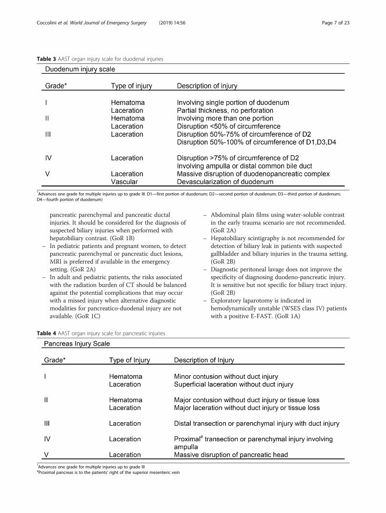
Table 4 AAST organ injury scale for pancreatic injuries
*Advances one grade for multiple injuries up to grade IIIaProximal pancreas is to the patients’ right of the superior mesenteric vein
Table 3 AAST organ injury scale for duodenal injuries
*Advances one grade for multiple injuries up to grade III. D1—first portion of duodenum; D2—second portion of duodenum; D3—third portion of duodenum;D4—fourth portion of duodenum)
Coccolini et al. World Journal of Emergency Surgery (2019) 14:56 Page 7 of 23

– During surgical exploration of patients withabdominal trauma, the duodeno-pancreatic complexmust be exposed and explored. (GoR 1A)
– During exploratory laparotomy, when biliary injuryis suspected but not identified, an intraoperativecholangiogram is strongly recommended. (GoR 2A)
– In patients who are clinically suspected of havingduodenal-pancreatic injuries, and are deterioratingclinically, if the imaging is equivocal, a diagnosticlaparotomy should be performed. (GoR 2A)
– In suspected pancreatic duct and extrahepatic biliarytree injuries in hemodynamically stable or stabilizedadults and pediatric patients, the endoscopicretrograde cholangiopancreatography (ERCP) can beused for both diagnosis and treatment even in theearly phase after trauma. (GoR 1B)
The diagnosis of duodeno-pancreatic injuries representsa challenge. In blunt trauma, evidence of direct impact onthe upper abdomen such as lower rib fractures, soft tissueecchymosis, supra-umbilical seat belt sign, and upper lum-bar spine fractures following a motor vehicle collisionshould suggest the involvement of the pancreas and duo-denum. Penetrating trauma of the front side or back sideof both the lower torso or upper abdomen should beconsidered highly suspicious for duodeno-pancreaticor extrahepatic biliary tree lesions if the diagnoseshave not been ruled out by other diagnostic means.
Clinical signs of traumatic DI are highly non-specific,especially in the early post-traumatic period. Patientsusually present with epigastric, right upper quadrant, orback pain 6–24 h after the injury, but the onset of painhas been reported as late as 5 days after injury [24, 25].The most common test is the analysis of serum amylaseand lipase [26]. However, in small-bowel injuries, initialamylase value does not differentiate between patientswith perforated and non-perforated DI [27]. A normalamylase level does not exclude DI [28].Persistently elevated or a rising level of amylase and
lipase may be of prognostic significance for both pancre-atic and duodenal injuries; therefore, measuring amylaseand lipase levels every 6 h is recommended [29, 30]. Ac-curacy may be improved if they are measured more than3 h after injury [31, 32].On E-FAST, the presence of free fluid in the absence
of solid organ injury may be a sign of hollow viscus in-jury; however, it has limited role in diagnosing acutepancreatic or duodenal injuries [28, 33, 34].Serum amylase levels are neither sensitive nor specific
for definitive screening or diagnosis of PI, particularlywithin 3–6 h after injury. Serum lipase is more specificthan amylase [35–37]; serum lipase may support tar-geted screening of patients with clinical suspicion of PI[10, 11, 16, 35, 37–71].Amylase is normal at admittance in up to 40% of pa-
tients with pancreatic trauma, and elevated levels are not
Table 5 AAST organ injury scale for extrahepatic biliary tree injuries
*Advances one grade for multiple injuries up to grade III
Coccolini et al. World Journal of Emergency Surgery (2019) 14:56 Page 8 of 23
specific for pancreatic trauma. Amylase can also be ele-vated in head, hepatic, and bowel injuries [61] and in alco-hol abuse and after hypo-perfusion of the pancreas [26].Lipase levels drawn on admission can be useful to excludepancreatic injury but not to guide further investigation:negative predictive value (NPV) of normal lipase is 99.8%,but with positive predictive value (PPV) of 3.3% [36].Amylase and lipase in association can reach sensitivity of85% and specificity of 100%, with PPV of 100% and NPVof 96% (after 6 h from injury) [26, 69, 72]. Decreasing en-zyme levels have been correlated with predicting successof NOM [16, 26, 35, 37, 40, 61, 70, 73]. Sensitivity of 88%and 96% NPV can be reached when amylase and lipaseare associated to ultrasonography (US) [26, 36]. In low-resource settings, amylase and lipase, in combination withUS, can be considered cost-effective methods to risk-stratify patients [26]. Persistently, elevated serum amylaseafter 10 days from the initial injury should be monitoredclosely given the increased risk of pseudocyst formation inboth adults and children [26, 40, 52, 63, 65, 70, 73–77].Contrast-enhanced CT-scan is the fastest and most com-
prehensive technique for evaluating duodeno-pancreatic in-juries [78–80]. In duodenal trauma, CT-scan has a sensitivityand specificity of 86% and 88%, respectively, in diagnosing
blunt hollow viscus injury [81–83]. However, missed bluntDI rates up to 27% have been described [84]. Of those withmissed DI, 83% had subtle CT findings on retrospective re-view [85]. Careful CT-scan interpretation with clinical correl-ation is mandatory to avoid delayed diagnosis and treatmentwith increased morbidity and mortality [28, 60, 61, 67, 79,80, 82, 86–90]. In fact, isolated periduodenal fluid orhematoma visualized on admission abdominal CT-scan doesnot necessitate immediate exploration [83, 91–94]. Intraperi-toneal or retroperitoneal extraluminal air is a relatively spe-cific sign of bowel perforation seen in 20–55% of patients;however, it may not be visible immediately after a traumaticperforation [95].In pancreatic trauma, contrast-enhanced CT-scan has
high specificity (90–95%) but low sensitivity (52–54%) forductal involvement. Up to 40% of PI can be missed ormisdiagnosed on abdominal CT-scan obtained within 12h of injury [96, 97]. PI becomes more evident 12–24 hafter trauma [41, 67, 98]. A repeat CT-scan with curvedmulti-planar reconstruction and specific pancreatic phase(35–40 s from iodine contrast injection) can help in diag-nosing pancreatic ductal (PD) injuries [61, 67, 82]. Aggres-sive resuscitation or prolonged hypovolemia can produceradiological changes in pancreatic imaging; fluid overload
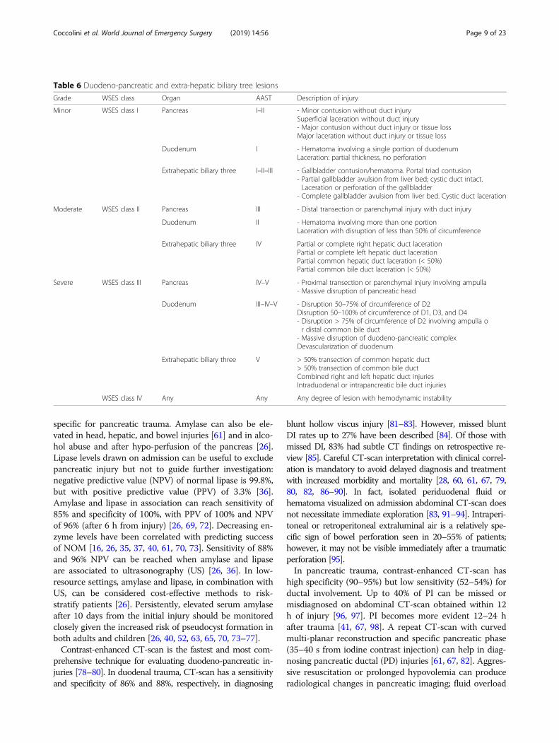
Table 6 Duodeno-pancreatic and extra-hepatic biliary tree lesions
Grade WSES class Organ AAST Description of injury
Minor WSES class I Pancreas I–II - Minor contusion without duct injurySuperficial laceration without duct injury- Major contusion without duct injury or tissue lossMajor laceration without duct injury or tissue loss
Duodenum I - Hematoma involving a single portion of duodenumLaceration: partial thickness, no perforation
Extrahepatic biliary three I–II–III - Gallbladder contusion/hematoma. Portal triad contusion- Partial gallbladder avulsion from liver bed; cystic duct intact.Laceration or perforation of the gallbladder
- Complete gallbladder avulsion from liver bed. Cystic duct laceration
Moderate WSES class II Pancreas III - Distal transection or parenchymal injury with duct injury
Duodenum II - Hematoma involving more than one portionLaceration with disruption of less than 50% of circumference
Extrahepatic biliary three IV Partial or complete right hepatic duct lacerationPartial or complete left hepatic duct lacerationPartial common hepatic duct laceration (< 50%)Partial common bile duct laceration (< 50%)
Severe WSES class III Pancreas IV–V - Proximal transection or parenchymal injury involving ampulla- Massive disruption of pancreatic head
Duodenum III–IV–V - Disruption 50–75% of circumference of D2Disruption 50–100% of circumference of D1, D3, and D4- Disruption > 75% of circumference of D2 involving ampulla or distal common bile duct
- Massive disruption of duodeno-pancreatic complexDevascularization of duodenum
Extrahepatic biliary three V > 50% transection of common hepatic duct> 50% transection of common bile ductCombined right and left hepatic duct injuriesIntraduodenal or intrapancreatic bile duct injuries
WSES class IV Any Any Any degree of lesion with hemodynamic instability
Coccolini et al. World Journal of Emergency Surgery (2019) 14:56 Page 9 of 23

can induce peripancreatic edema or collections. In pa-tients with severe shock both hypo- and hyper-perfusionof the gland have been described [99–101].A repeat CT-scan 12–48 h after admission in doubtful
cases of pancreatic-duodenal lesions should be considered[91, 102]. The follow-up scan sensitivity for bowel perfor-ation increases from 30 to 82% [103]. Moreover, the re-peat CT-scan sensitivity for identification of an operativeindication may increase up to 100% (67%). NPV for OMalso increases from 94 to 100% with no increase in mortal-ity or hospital length of stay [104, 105]. Complication rateis significantly higher only in those patients with delayedOM of more than 24 h [106].The MRCP may be used in pancreatic-duodenal trauma
to assess common bile duct/ampulla injury, and hepatobili-ary contrast agents can help in localizing associated bileleaks. Minor injuries may be more evident on MRI than onCT-scan [79]. In association with secretin-dynamic study,the MRCP may diagnose pancreatic leakage [107, 108] andgive additional information concerning parenchymaland proximal duct condition [71, 108, 109].
Oral contrast administration has not been shown tohave substantial benefits in depicting bowel injurieswhen compared with CT-scan alone at the initial evalu-ation and during follow-up (sensitivity 95%, specificity99.6%) [42, 102, 110–123].Radiation-related risks in children and young patients
must be considered. An increase in lifetime cancer-specific mortality of 801/4000 (20.00025%) to 800/4000(20%) after CT-scan has been reported for American chil-dren [124]. However, the consequences of missed injuryor delay in diagnosis on mortality and morbidity rates canbe grave particularly with duodeno-pancreatic injuries.Plain films of the abdomen are generally of little value in
diagnosing duodeno-pancreatic injuries [125]; the same istrue for upper gastrointestinal series using water-soluble con-trast. Duodenography (oral contrast–enhanced fluoroscopicevaluation) for blunt and penetrating duodenal trauma in pa-tients with equivocal CT-scan has an overall sensitivity of25% for blunt DI and 54% for those requiring repair [126].The ERCP may play a role in duodeno-pancreatic trauma
in order to avoid late-diagnosis and/or treatment both in
Fig. 2 Diagnostic algorithm for duodeno-pancreatic and extrahepatic biliary tree traumatic lesions
Coccolini et al. World Journal of Emergency Surgery (2019) 14:56 Page 10 of 23
adult and pediatric patients [10, 15, 41–43, 48–52, 58–60,62–64, 67, 68, 70, 76–78, 90, 97, 101, 127–149]. It is an in-vasive procedure with 3–14% risk of post-procedure pan-creatitis and 0.2–1% mortality rate [6, 10, 11, 40, 41, 45, 49,51–53, 58, 61–64, 67, 68, 70–72, 75, 77, 78, 97, 128, 130,133, 134, 137–140, 142, 144, 146, 148–157]. Moreover, insuspected duodenal perforations, the ERCP is not recom-mended. Failed cannulation of the papilla of Vater or inad-equate pancreatography can occur in up to 9–14% ofpatients [71, 137, 144, 152]. The small duct size in childrenis not an absolute contraindication for the ERCP in experthands as it is relatively safe and effective [16, 53, 63, 64, 70,76, 77, 134, 137, 139, 148, 152, 158]. Rates of PD cannula-tion may be influenced by duodenal mucosal edema and/orhematomas and anatomical changes [71]. Despite of theselimitations, the ERCP may have a role in decreasing timefrom definitive diagnosis of duct injury and first treatmentin selected cases [131, 159]. However, cross-sectional
imaging should be performed before proceeding with theERCP.Hepatobiliary scintigraphy (HIDA) is not frequently used
in the initial work-up of the acute trauma patient due tolong scan times and limited resource availability [128].Percutaneous transhepatic cholangiogram (PTC) could
be considered after non-feasible or unsuccessful ERCPfor diagnosis and treatment [21].Diagnostic peritoneal lavage (DPL) has sensitivity higher
than 99% for hemoperitoneum but it is neither specific norreliable for the assessment of retroperitoneal injuries, withundetected bowel perforation seen in up to 10% of cases[160–163]. DPL alone is associated with a high number ofunnecessary laparotomies [164], with consequent short-and long-term complications. Moreover, DPL is associatedwith a 0.8–2.3% risk of specific complications [165, 166].Diagnostic laparoscopy has both diagnostic and thera-
peutic potentials in a delayed setting. Whenever negative,
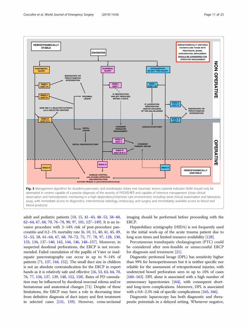
Fig. 3 Management algorithm for duodeno-pancreatic and extrahepatic biliary tree traumatic lesions (asterisk indicates NOM should only beattempted in centers capable of a precise diagnosis of the severity of PI/DI/EHBTI and capable of intensive management (close clinicalobservation and hemodynamic monitoring in a high dependency/intensive care environment, including serial clinical examination and laboratoryassay, with immediate access to diagnostics, interventional radiology, endoscopy, and surgery and immediately available access to blood andblood products)
Coccolini et al. World Journal of Emergency Surgery (2019) 14:56 Page 11 of 23

it may reduce the number of unnecessary laparotomies[167]. It has a growing role in the evaluation of penetrat-ing abdominal trauma but it has not been specificallystudied for the evaluation of pancreatic-duodenal injuries.The duodeno-pancreatic anatomy and the retroperitoneallocation increase the risk of missed injuries [168].Moreover, laparoscopy in trauma requires adequatetraining and experience as well as sufficient staffingand equipment [169, 170].Ultimately, in the patient with diagnostic uncertainty and
in the patient with persistent or worsening clinical signsand symptoms, radiologic and/or laboratory alterations dueto an intra-abdominal lesion, laparotomy should be stronglyconsidered [171]. For penetrating trauma, a thorough andmeticulous exploratory laparotomy with retroperitoneal ex-posure and assessment remains critical in detecting pancre-atic and duodenal injuries [172].If exploration is negative but there is still a strong suspi-
cion of DI, methylene blue administration through a naso-oro-gastric tube could be considered. During emergencylaparotomy, the use of intraoperative pancreatographydoes not add to the visual findings [145]. Intraoperativecholangiogram through the cystic duct may help in defin-ing EHBTI [87, 173]. Additional information can be pro-vided by the use of intraoperative US of the pancreas;however, the lack of strong evidence and the necessity oftrained surgeons make this technique not recommendedor routinely used in trauma [130].
TreatmentNon-operative management—duodenum
� Hemodynamic stability is the key factor indetermining management strategy. (GoR 1C)
� Hemodynamically unstable (WSES class IV) patientsshould not be considered for NOM. (GoR 1C)
� NOM can be considered for hemodynamically stableor stabilized patients with duodenal wall hematomas(WSES class I–II, AAST-OIS grade I–II) in absenceof other abdominal organ injuries requiring surgery.(GoR 2B)
� Patients with progressive symptoms or worseningfindings on repeat imaging should be consideredfailures of NOM. (GoR 2C)
� Hematomas initially treated with NOM shouldbe considered for operative management ifduodenal obstruction has not resolved within 14days. (GoR 2C)
Non-operative management—pancreatic and biliary tree
� NOM should be the treatment of choice for allhemodynamically stable or stabilized minor PIWSES class I (AAST grade I and some grade II) and
gallbladder hematomas without perforation WSESclass I (AAST grade I) in the absence of otherabdominal injuries requiring surgery. (GoR 2C)
� Location of WSES class II (AAST grade III) PI is theprimary determinant of treatment modality inhemodynamically stable adult patients. (GoR 2C)
� NOM may be considered only in selectedhemodynamically stable or stabilized patients withWSES class II (AAST grade III) very proximalpancreatic body injuries in the absence of otherabdominal injuries requiring surgery and only inhigher level trauma centers; success of NOM maybe increased with utilization of endoscopic andpercutaneous interventions. (GoR 2C)
� Optimal management of hemodynamically stable orstabilized patients with WSES class III (AAST gradeIV) PI is controversial. NOM managementaugmented by endoscopic or percutaneousinterventions may be used in selected patients.(GoR 2C)
� NOM of WSES class III (AAST grade IV) injuriesshould be considered only in an environment thatprovides around the clock capability for patientintensive monitoring, an immediately availableendoscopy and interventional radiology suite, OR,and only in patients with stable or stabilizedhemodynamic and absence of other abdominalinjuries requiring surgery. (GoR 2A)
� Sequelae of PI such as pancreatic fistulae andpseudocysts can frequently be addressed with image-guided percutaneous drain placement, endoscopicstenting, internal drainage, and endoscopiccyst-gastrostomy or cyst-jejunostomy. (GoR 2C)
NOM is similar between adult and pediatric patientsand is dependent on hemodynamic stability, clinicalpresentation, and associated injuries. Shock is generallydue to associated injuries, which are present in 55–100% of pancreatic-duodenal injuries, and are more fre-quent among patients with penetrating mechanism ofinjury [1, 3, 6, 7, 11, 12, 14, 17, 174–183].Physical exam findings associated with DI are non-
specific and may be more reliable in children. Serial ob-servations may increase the sensitivity of physical examfindings in diagnosing DI [57, 184]. CT-scan is generallythe standard of care in diagnosing DI. Patients with def-inite evidence of full thickness laceration such as ex-travasation of enteral contrast or free air should undergoimmediate operative intervention. These findings arerare, and in the vast majority of patients, findings areeither non-specific such as duodenal wall thickening,periduodenal edema, stranding, or free fluid, or they areentirely absent [62, 84, 91]. NOM should include serialabdominal exams, bowel rest, and nasogastric tube
Coccolini et al. World Journal of Emergency Surgery (2019) 14:56 Page 12 of 23

(NGT) decompression. Parenteral nutrition may be re-quired if obstruction persists beyond 7 days [185]. Obstruc-tion due to duodenal hematoma will generally resolvewithin 14 days; if not, operative decompression may be re-quired [185–188]. Operative evacuation can be done openor laparoscopically [188]. Percutaneous drainage of duo-denal hematomas is a viable alternative [185, 189–193].NOM of duodenal hematomas is generally successful
in both adults and children [62, 91, 105, 185, 194]. FailedNOM (fNOM) rates between 5 and 10.3% have been re-ported, with no differences in length of stay. In patientswith fNOM, a 0–3% complication rate and reduced mor-tality compared with the group undergoing immediateOM has been reported [91, 105].Minor PI is treated similarly in adults and children.
Hemodynamically stable patients without associated op-erative injury should undergo a trial of NOM. Total par-enteral nutrition (TPN) may be required in 62–73% ofpediatric and 22.6% of adults [8, 12, 15, 16]. NOM ofclass I injuries is successful in 96–100% of pediatric and80–92.2% of adults [6, 11, 15, 105, 195, 196] and is asso-ciated with reduced morbidity, mortality, and shorterlength of stay [3, 105].In WSES class II (AAST-OIS grade III) injuries in
hemodynamically stable or stabilized patients, locationof the injury largely determines optimal treatment.WSES class II injuries distal to the superior mesentericvein (AAST-OIS grade III) should be managed opera-tively by resection with or without splenectomy as OMis associated with improved recovery times, and reducedmorbidity in both adults and pediatrics [197–199]. Iso-lated proximal WSES class II and III injuries (AAST-OIS grade III and IV–V) may be considered for NOM.Although no randomized controlled trials exist, severallarge database studies and meta-analyses have demon-strated that NOM is pursued in 46% of pediatric and28–48.5% of adult patients [3, 6, 15].NOM of WSES moderate and severe PI (AAST-OIS
grade III and IV–V) has been reported more amongpediatric than adult patients with a success rate up to 89%[15]. NOM success rate in adults is about 30%. Pseudocystrate was higher among NOM patients and in 65–74% ofcases they were also managed non-operatively [15, 16].Length of stay was similar between NOM and OM [9, 200].Endoscopic and percutaneous interventions such as ERCP
with pancreatic stent and/or sphincterotomy or percutan-eous aspiration and drain placement for pancreatic duct in-jury have been reported in patients with class II and III PI(AAST-OIS grades III and IV–V) with success rates of 68–94% with or without the octreotide administration[15, 201–208]. However, some concerns exist regard-ing increased rates of pancreatic duct stricture [209].Many EHBTI will be diagnosed at the time of laparot-
omy. However, in patients undergoing NOM, concern
for EHBTI should prompt immediate investigation withMRCP or HIDA scan. Patients with gallbladder wallhematoma without perforation can be managed expect-antly [18]. NOM can be attempted in hemodynamicallystable patients with WSES grade II and III injuries(AAST-OIS grade IV–V) without definite indication forsurgical intervention. In these cases, fluid collectionsshould be drained percutaneously and the ERCP withstent placement should be attempted to address ductallacerations. Very little data exist about NOM of EHBTIbut a few small case series have demonstrated success inboth adult and pediatric patients [18, 19, 21].
Operative management—duodenum
� Hemodynamically unstable (WSES class IV) patientsand those with peritonitis or bowel evisceration orimpalement should undergo immediate operativeintervention. (GoR 1C)
� Damage control techniques should be considered inhemodynamically unstable patients with DI,particularly those with associated injuries andphysiologic derangement. (GoR 2B)
� Primary repair of DI should be considered whenevertechnically possible regardless of grade of injury.(GoR 2B)
� Ancillary procedures such as pyloric exclusion withand without gastrojejunostomy and biliary diversionmay be considered in WSES class III or higher DI(AAST grades III, IV, and V). (GoR 2C)
� Lesions requiring pancreaticoduodenectomy(Whipple procedure) are often accompanied bysevere associated injuries and shock. Damagecontrol techniques and staged reconstruction insubsequent phases performed by experiencedsurgeons should be considered. (GoR 2c)
Operative management—pancreas and biliary tree
� In WSES class I (AAST grade I and some grade II)PI found during exploratory laparotomy, drainagemay be considered. (GoR 2B)
� Patients with distal WSES class II (AAST grade III)PI should undergo OM. (GoR 2C)
� Distal pancreatectomy (with or withoutsplenectomy) is the procedure of choice for distalWSES class II (AAST grade III) PI. (GoR 2C)
� Pancreatoduodenectomy may be needed in patientswith destructive injuries of the duodenal-pancreaticcomplex. In such cases, the operation has betterresults when performed in a staged fashion.Pancreato-jejunostomy or pancreato-gastrostomyreconstructions are equally effective in selected casesperformed by experienced surgeons. (GoR 2C)
Coccolini et al. World Journal of Emergency Surgery (2019) 14:56 Page 13 of 23

� In extrahepatic biliary tree WSES class I injuries(AAST grades I, II, and III) with laceration,perforation, or avulsion of the gallbladder,cholecystectomy is the treatment of choice. (GoR 1C)
� EHBT injuries undergoing an initial damage controlprocedure may be drained with delayedreconstruction performed as a staged approach.(GoR 2B)
� EHBT WSES class II–III (AAST grades IV and V)injuries should undergo reconstruction withhepaticojejunostomy or choledochojejunostomy ifthere is no associated vascular injury. (GoR 2C)
� NOM failure of EHBT WSES class II–III (AASTgrades IV and V) injuries, hepaticojejunostomy shouldbe considered during reconstruction. (GoR 2C)
Due to the high percentage of associated injuries inpatients with duodeno-pancreatic and extrahepatic bil-iary three injuries, shock and peritonitis are common ator shortly after presentation. Hemodynamic instability ispresent in 10–44% of patients [210–215]. All patientswith hemodynamic instability or peritonitis shouldproceed immediately to OM. Hemodynamically stablepatients with CT findings of full thickness laceration, orclass III DI (AAST-OIS grade III–IV–V), such as free airor extravasation of enteral contrast from the duodenumor an associated operative injury should also undergoimmediate OM.Damage control surgery (DCS) is reported in 20–63%
of cases particularly in patients with associated vascularinjuries and/or higher grade duodeno-pancreatic lesions.DCS has been associated with improved survival andequivalent or improved complication rates [2, 211, 212,216, 217]. DCS is rarely needed for isolated DI, and theextent of the primary surgery will relate primarily to as-sociated vascular injuries. Once hemostasis has beenachieved, the DI can be addressed at the initial surgery ifthe patient’s physiology allows. The majority of DI foundat laparotomy are WSES class I–II lacerations (AAST-OIS grade I–II). They should be repaired primarily in atension-free transverse fashion after complete exposureand removal of all devitalized tissue. A nasogastric tube(NGT) should be placed to allow for proximal decom-pression. There is no evidence supporting routine peri-duodenal drain placement.Management of WSES class III lacerations (AAST-OIS
grade III–IV–V) not involving massive disruption of theduodeno-pancreatic complex is controversial. They are as-sociated with a high mortality and high duodenal-specificmorbidity (duodenal leak, fistula and anastomotic break-down) with consequent abdominal sepsis and poor out-comes [218, 219]. Duodenal diverticulization and tripletube decompression are no longer advocated for the treat-ment of DI [187, 218, 219]. Most modern studies advocate
primary repair, NGT decompression, and external drainplacement even with large, high-grade injuries. In caseswhere primary repair is not possible, segmental resectionand primary duodeno-duodenostomy could be performed.These more conservative techniques have demonstratedgood outcomes with similar or better mortality andduodenal-related morbidity compared with more complexdrainage and reconstructive procedures [57, 181, 194,211–213, 216, 217]. Pyloric exclusion (PE) is still utilizedalthough definite indications for it remains controversial[220]. Temporary PE has been described both with andwithout gastrojejunostomy. The pylorus can be stapledwithout transection or sutured internally with absorbablematerial so it will open spontaneously several weeks post-injury [221, 222], or sutures can be removed endoscopic-ally. Several studies reported no improvement in morbid-ity, mortality, and a prolonged length of stay with PEcompared with primary repair with NGT decompressionalone [212, 214, 215, 217, 223, 224]. Moreover, concernsexist regarding the possible PE increasing the length of theprocedure, complications, and risks of gastric suture lineand marginal ulcers [105, 222, 224–226].WSES class III injuries with massive disruption of the
duodeno-pancreatic complex (AAST-OIS grade III–IV–V for duodenum and AAST-OIS grade IV–V for pan-creas) are rare and require complex reconstruction. Inthe first or proximal second duodenal portion lesionswhere primary repair or resection and primary anasto-mosis are not possible, antrectomy and gastrojejunost-omy with closure of the duodenum is an option [186].In case of injuries located distal to the ampulla, a Roux-en-Y duodeno-jejunostomy can be performed [186, 187,212]. When the ampulla or distal common bile duct isinvolved, re-implantation into healthy adjacent duode-num or reconstruction with a Roux-en-Y jejunal limb isan option if the adjacent tissue loss and injury are min-imal [186]. When the duodenum and/or pancreatic headare severely devitalized or devascularized, pancreatico-duodenectomy (Whipple procedure) may be required.Associated injuries and severe physiologic derangementsare common with these injuries [227–230]. DCS is re-quired in 26–80% of cases and should be strongly con-sidered at the time of initial operation [227–230]. Itseems to improve survival and reduce complications intreating severe pancreatic-duodenal injuries requiringWhipple procedures [230]. Staged procedures have beensuggested to improve outcomes. The assistance of expe-rienced hepatobiliary surgeons should be defined on acase-by-case basis [187, 227–229]. Both classic Whippleprocedures and pylorus preserving reconstructions areoptions dependent on the location of the DI and associ-ated injuries [227, 231].Delayed bowel function and obstruction from duodenal
edema, hematoma, or stricture are common following DI
Coccolini et al. World Journal of Emergency Surgery (2019) 14:56 Page 14 of 23

[232]. To ensure adequate nutrition, a feeding jejunost-omy may be considered in patients with severe duodeno-pancreatic injuries requiring resection and reconstruction;however, jejunostomy-related complications can occur inup to 7% of patients and intolerance to enteral nutrition iscommon [211, 232]. Total parenteral nutrition (TPN) maybe required in 37–75% of patients [57, 185, 213].Patients with PI who are hemodynamically unstable
(33–50%) (WSES class IV) or have peritonitis shouldundergo immediate OM [1, 6, 14, 233]. Associated hol-low viscus injury or operative intra-abdominal injury willbe present in 24–82% of PI [4, 5, 11, 233]. DCS shouldbe considered in patients with shock and exsanguinatinghemorrhage. Surgical management of pancreatic injuryis dependent on grade, location, and extent of associatedinjuries. Intraoperatively diagnosed WSES class I PI DCS(AAST-OIS grade I–II) can be managed expectantly,and closed suction drain placement is recommended forlarger contusions and lacerations [234, 235]. Suture re-pair of lacerations should be avoided as it is associatedwith increased risk of pseudocyst formation [235]. WSESclass II PI injuries (AAST-OIS grade III) involving themain pancreatic duct distal to the superior mesentericvein (SMV) should be treated with distal pancreatectomywith or without splenectomy as OM is associated withimproved recovery times, and reduced morbidity in bothadult and pediatric PI [13, 197–199, 235]. Decreased inci-dence of pancreatic fistula when the pancreas was stapled ra-ther than sewn has been demonstrated; however, ductalligation made no difference [13]. Splenic preservation amongtrauma surgeons remains controversial. No significant in-crease in morbidity or mortality and a reduced length of stayassociated with spleen preservation has been demonstrated[236]. Spleen preservation is of great importance in pediatrictrauma patients; however, there is little data on splenic sal-vage in this cohort [237, 238]. Ultimately, the decision to pre-serve or remove the spleen will depend on the patient’sphysiology, associated splenic injury, and the surgeon’s levelof experience.Optimal management of WSES class III PI (AAST-
OIS grade IV–V) with transection of parenchyma/ductproximal to the SMV remains controversial. Subtotaland total pancreatectomy for proximal injuries may re-sult in endocrine and exocrine dysfunction. Because ofthis, initial management includes debridement, oversew-ing the proximal pancreatic stump, and distal drainagewith pancreaticojejunostomy (not well tolerated inphysiologically deranged patients). These procedures areassociated with high rates of pancreas-related (fistula)and overall complications. Modern studies predomin-antly utilize debridement and wide local drainage withgood success [2, 4, 14, 239]. Drainage alone for proximalPI has rates of pancreatic fistula of 12–13.8% [238, 240]which compares favorably with small series of more
complex reconstructions with pancreaticoenterostomy(11–20%) [241, 242].WSES class III PI (AAST-OIS grade IV–V) with
complete destruction or devascularization of the pancre-atic head and pancreatico-duodenal complex is a specificand rare circumstance. Most of these patients requirepancreaticoduodenectomy and present in shock andwith severe associated injuries and should be treatedwith DCS [243]. Mortality after trauma Whipple remainshigh varying from 12 to 33%, but it may be improvedwith DCS techniques and appropriate patient selection[231, 244, 245]. Mortality with more conservative surgi-cal treatments (duodenal reconstruction and drainage)appears to be similar, but complications, particularlypancreatic fistula, may be higher when compared withthe Whipple procedure [13, 246].Gallbladder WSES class I injuries (AAST-OIS grade I–
II–III) account for approximately 30–60% of EHBTI[18–20]. The majority of these injuries are noted at thetime of laparotomy. For all injuries except gallbladderwall hematomas, the treatment of choice is cholecystec-tomy [18, 19, 22]. Extrahepatic bile duct injuries oftenoccur in conjunction with severe liver, duodenal, andpancreatic injuries. In these instances, management isdictated as much by the severity of the associated injur-ies as by the grade of the bile duct injury itself. In mostcases, treatment of the injury with distal ligation and re-construction with a Roux-en-Y hepaticojejunostomy isrecommended [18, 19, 21]. Choledochojejunostomy maybe used for distal common bile duct injuries in the ab-sence of associated vascular injury that may compromisethe blood supply to the anastomosis. Primary repair ofWSES class II injuries (AAST-OIS grade IV) over a T-tube can be attempted but may result in strictures andneed for future reconstructive surgery [18]. OM withRoux-en-Y hepaticojejunostomy is also recommendedfor patients with WSES class II and III injuries (AAST-OIS grade IV–V) after fNOM [18, 21].
Follow-up
� After discharge, the necessity for follow-up imagingshould be driven by clinical symptoms (i.e., onset ofabdominal distention, tenderness, fever, vomiting,jaundice). (GoR 2B)
� In adults, CT-scan is usually the first-line follow-upimaging tool for new-onset signs and symptoms.(GoR 2A)
� In pregnant females, the MRCP should beconsidered the diagnostic modality of choice fornew-onset signs and symptoms, wherever available.(GoR 2A)
� In pediatric patients, ultrasound or contrast-enhanced US should be the diagnostic modality of
Coccolini et al. World Journal of Emergency Surgery (2019) 14:56 Page 15 of 23
choice for follow-up imaging. If cross-sectionalimaging is required, MRI is preferred. (GoR 2A)
� Given the complexity and variability of traumaticinjuries, the need for and choice of follow-upimaging should be made using a multidisciplinaryapproach. (GoR 2B)
CT-scan is usually the first-line imaging tool in the assess-ment of late complications of pancreatic trauma and veryuseful in driving management [39, 61, 71, 72, 76, 96, 135,145, 233, 247, 248]. MRI is a reliable alternative to CT-scanin children and pregnant women [40, 45, 52, 97, 249, 250].US or CEUS is used as an alternative to CT for follow-
up of fluid collections, pseudocysts, and pancreatic dis-ruptions after pancreatic trauma mainly in children or inlow-resource settings [16, 26, 40, 45, 49, 53, 55, 63, 71,75, 78, 133, 134, 138, 245, 247, 251–254]. CEUS may im-prove results of pancreatic imaging, being nearly as ac-curate as CT-scan and reducing radiation exposure inchildren [249, 255, 256].The ERCP is a useful tool in diagnosis, management,
and follow-up of late complications such as pseudocysts,pancreatic fistulas (i.e., trans-papillary stenting), or mainduct strictures secondary to injury or prolonged stenting(i.e., ERCP with pancreatic duct dilatation and stenting),even in pediatric patients [10, 39, 40, 45, 53, 67, 74, 137,138, 148, 152, 154, 247, 253].NOM of high-grade pancreatic lesions (WSES class III,
AAST-OIS grade IV–V) requires stringent follow-up forat least 6 months to detect early and late sequelae [45].
ComplicationsPseudocyst is the most frequent complication followingNOM [15, 52, 53, 64, 68, 69, 72, 154, 257, 258]. CT-scanis useful in evaluating pseudocysts and peripancreaticfluid collections following PI [96, 247, 259, 260] and inguiding percutaneous drainage [40]. US and endoscopicUS (EUS) can also be used for follow-up and to guidepercutaneous treatment of pseudocyst and abscess avoid-ing radiation exposure [45, 63, 70, 158, 247, 253]. Someauthors propose combined EUS-ERCP procedures even inchildren [152, 158, 247]. The use of EUS in the work-upof children with pancreatobiliary pathology may limit ex-posure to risk of adverse events from ERCP [152]. MRCPand ERCP may be used—the first to document the com-munication of the cyst with the main pancreatic duct [40,41, 45, 49, 68, 71, 97, 128, 138, 145, 154, 156, 247, 253,259, 261] and, the latter, for treating the disease.Abscess or intra-abdominal sepsis occurs in 7–25% of
patients with pancreatic injuries; CT-scan or MRI should beperformed for diagnosis and to guide treatment [40, 156].Pancreatic fistula occurs in 10–35% of major injuries
of the pancreas after operative drainage or resection. Acorrect diagnosis is very important in planning the
treatment. Preoperative cross-sectional imaging and pan-creatogram during ERCP are essential. The ERCP, whenfeasible, is the first step to treat persisting fistulas [11,40, 41, 48, 49, 61, 71, 156, 233, 262].The incidence of post-traumatic pancreatitis is 17%.
Patients with abdominal pain and hyperamylasemiashould undergo contrast-enhanced CT-scan for diagno-sis wherever possible [40, 156].
Post-traumatic exocrine or endocrine function Al-though transient post-operative glucose intolerance iscommon in all critically ill trauma patients, the inci-dence of persistent new-onset endocrine dysfunctionafter traumatic distal pancreatectomy is very low (< 4%)[263]; insulin requirement is more frequently associatedto proximal pancreatic resections [72, 263] or Whippleprocedure [264]. However, both exocrine and endocrineinsufficiencies are very rare [4, 10, 15, 16, 45, 52, 54, 58,69, 265] and no sufficient data exist to have definitiveanswers and indications [15, 68, 257]. Post-traumaticexocrine or endocrine function in the very long-termseems to be related to overall age and time from injuryrather than the surgical treatment [68, 69]. Long-termfollow-up is suggested for patients who underwent pan-creatic surgery for trauma due to the possibility that theonset of diabetes mellitus may be accelerated by pancre-atic resection [53, 264].
ConclusionsNon-operative management of bilio-duodeno-pancreaticinjuries without ductal involvement with or without endo-scopic adjuncts is recommended for hemodynamicallystable patients. EHBTI can be managed with cholecystec-tomy for minor injuries, although more severe injuriesrequire surgical reconstruction. Severe bilio-duodeno-pancreatic injuries are rare, often accompanied byhemodynamic instability and may benefit from DCStechniques. Many initial injuries as well as the seque-lae of injury may be addressed with percutaneous orendoscopic drainage, and endoscopic stenting. Despiteadvances in care, morbidity and mortality followingsevere bilio-duodeno-pancreatic trauma remain high.The management of duodenal, pancreatic, and extrahepaticbiliary tree injuries must be multidisciplinary. The manage-ment in the initial phase is best accomplished by thetrauma or emergency surgeon, and in the reconstructivephase, hepatobiliary surgeons may be helpful and should beconsulted.
AbbreviationsAAST: American Association for the Surgery for Trauma; BE: Base excess;CT: Computerized tomography; DCS: Damage control surgery; DI: Duodenalinjury; DPL: Diagnostic peritoneal lavage; E-FAST: Extended-FocusedAssessment with Sonography for Trauma; EHBTI: Extrahepatic biliary treeinjury; ERCP: Endoscopic retrograde cholangiopancreatography;EUS: Endoscopic US; fNOM: Failed non-operative management;
Coccolini et al. World Journal of Emergency Surgery (2019) 14:56 Page 16 of 23

HIDA: Hepatobiliary scintigraphy; LE: Level of evidence; MRCP: Magneticresonance cholangiopancreatography; MRI: Magnetic resonance imaging;NGT: Nasogastric tube; NOM: Non-operative management; NPV: Negativepredicting value; OIS: Organ injury scale; OM: Operative management;PD: Pancreatic duct; PE: Pyloric exclusion; PI: Pancreatic injury; PPV: Positivepredicting value; PTC: Percutaneous transhepatic cholangiogram;SMV: Superior mesenteric vein; TPN: Total parenteral nutrition;US: Ultrasound; WSES: World Society of Emergency Surgery
AcknowledgementsNoneContributors:WSES-AAST Expert PanelOffir Ben-Ishay (1), Matti Tolonen (2), Riccardo Bertelli (3), Tal Horer (4), PaulaFerrada (5), Isidoro Di Carlo (6), Bruno M Pereira (7), Dario Parini (8), GiuliaMontori (9), Belinda De Simone (10), Osvaldo Chiara (11), Andreas Hecker(12), Nicola DeAngelis (13), Carlos Augusto Gomes (14), Joseph Galante (15),Miklosh Bala (16), Konstantinos S Mylonas (17), Anastasia Pikoulis (18), PaolaPerfetti (19), Mircea Chirica (20), Joaquin Bado (21), Kenji Inaba (22), Neil Parry(23), Oreste Romeo (24), Martijn Stommel (25), Mohan Rajashekar (26),Edward Tan (27), Francesco Salvetti (28), Boris Sakakushev (29)(1) General and Trauma Surgery, Rambam Medical Centre, Tel Aviv, Israel(2) General Surgery Dept., Mehilati Hospital, Helsinki, Finland(3) General Emergency and Trauma Surgery, Bufalini Hospital, Cesena, Italy(4) Dept. of Cardiothoracic and Vascular Surgery & Dept. of Surgery ÖrebroUniversity Hospital and Örebro University, Sweden(5) General and Trauma Surgery, Virginia Commonwealth University,Richmond, Virginia, USA(6) Department of Surgical Sciences and Advanced Technologies “GFIngrassia”, University of Catania, Cannizzaro Hospital, Catania, Italy(7) Trauma/Acute Care Surgery & Surgical Critical Care, University ofCampinas, Campinas, Brazil(8) General Surgery Dept., Rovigo Hospital, Rovigo, Italy(9) General Surgery Dept., Aviano Hospital, Aviano, Italy(10) General Surgery Dept., Guastalla Hospital, Guastalla, Italy(11) Emergency and Trauma Surgery Dept., Niguarda Hospital, Milano, Italy(12) Department of General and Thoracic Surgery, University Hospital ofGiessen, Giessen, Germany.(13) Unit of Digestive Surgery, HPB Surgery and Liver Transplant, HenriMondor Hospital, Créteil, France(14) Hospital Universitário Terezinha De Jesus, Faculdade De CiênciasMédicas E Da Saúde De Juiz De Fora (Suprema) Brazil(15) Trauma and Acute Care Surgery and Surgical Critical Care Trauma,Department of Surgery University of California, Davis, USA(16) General Surgery Dept., Hadassah Medical Centre, Jerusalem, Israel(17) 3rd Department of Surgery, Attiko Hospital, National & KapodistrianUniversity of Athens, Greece(18) General Surgery, Medical School, NKUA, Athen, Greece(19) Emergency Medicine Dept., Verona Hospital, Verona, Italy(20) Chirurgie Digestive, CHUGA-CHU Grenoble Alpes, Grenoble, France(21) General and Emergency Surgery Dept., Montevideo Hospital,Montevideo, Paraguay(22) Division of Trauma & Critical Care, LAC+USC Medical Center, LosAngeles, CA, USA(23) General and Trauma Surgery Dept., London Health Sciences Centre,Victoria Hospital, London, ON, Canada(24) Trauma and Surgical Critical Care, University of Michigan Health System,East Medical Center Drive, Ann Arbor, MI, USA(25) Department of Surgery, Radboud University Medical Center, Nijmegen,The Netherlands.(26) General Surgery, Hegde Hospital, Mangaluru, Karnataka, India(27) Emergency Med. Dept., Radboud University Medical Center, Nijmegen,The Netherlands.(28) General Surgery, Pavia University Hospital, Pavia, Italy(29) General Surgery Department, Medical University, University Hospital StGeorge, Plovdiv, Bulgaria.
Authors’ contributionsFC, LK, YK, EEM, LA, WB, AL, GA, VR, IW, AK, FAZ, EC, GPF, CO, EP, MGS, RM,YM, PTM, VK, ACM, RI, FF, VM, MS, FM, JM, MC, CA, FC, and RC contributed inthe conception and drafting of the manuscript, critically revised the
manuscript, and contributed with important scientific knowledge giving thefinal approval.
FundingNone
Availability of data and materialsNot applicable
Ethics approval and consent to participateNot applicable
Consent for publicationNot applicable
Competing interestsThe authors declare that they have no competing interests.
Author details1General, Emergency and Trauma Surgery Department, Pisa UniversityHospital, Via Paradisa, 2, 56124 Pisa, Italy. 2Division of Trauma, Surgical CriticalCare, Burns and Acute Care Surgery, University of California San Diego, SanDiego, USA. 3Division of General Surgery, Rambam Health Care Campus,Haifa, Israel. 4Trauma Surgery, Denver Health, Denver, CO, USA. 5General,Emergency and Trauma Surgery Department, Bufalini Hospital, Cesena, Italy.6Trauma Surgery Department, Scripps Memorial Hospital, La Jolla, CA, USA.7General Surgery Department, Mehilati Hospital, Helsinki, Finland.8Department of Surgery, Zagreb University Hospital Centre and School ofMedicine, University of Zagreb, Zagreb, Croatia. 9General and EmergencySurgery, Sergei Kirov Military Academy, Saint Petersburg, Russia.10Department of Surgery, DHS Hospitals, Srinagar, Kashmir, India. 11General,Acute Care, Abdominal Wall Reconstruction, and Trauma Surgery, FoothillsMedical Centre, Calgary, Alberta, Canada. 12Department of Surgery, Collegeof Medicine and Health Sciences, UAE University, Al-Ain, United ArabEmirates. 13Trauma/Acute Care Surgery & Surgical Critical Care, University ofCampinas, Campinas, Brazil. 14Trauma and Acute Care Surgery, FundacionValle del Lili, Cali, Colombia. 153rd Department of Surgery, Attiko Hospital,National & Kapodistrian University of Athens, Athens, Greece. 16Departmentof Surgery, Harborview Medical Centre, Seattle, USA. 17Department ofEmergency and Critical Care Medicine, Chiba University Hospital, Chiba,Japan. 18Pediatric Trauma Service, Massachusetts General Hospital, Boston,MA, USA. 19General Surgery Department, Mozir City Hospital, Mazyr, Belarus.20Department of Surgery and Obstetrics and Gynecology, University of Buea,Buea, Cameroon. 21General and Trauma Surgery, Virginia CommonwealthUniversity, Richmond, VA, USA. 22General and Trauma Surgery Department,Pietermaritzburg Hospital, Pietermaritzburg, South Africa. 23General andEmergency Surgery, Macerata Hospital, Macerata, Italy. 24General andEmergency Surgery Department, Montevideo Hospital, Montevideo, Uruguay.25Department of Emergency and Critical Care Medicine, Saint-MariannaUniversity School of Medicine, Kawasaki, Japan. 26Clin. Univ. de ChirurgieDigestive et de l’Urgence, CHUGA-CHU Grenoble Alpes, UGA-UniversitéGrenoble Alpes, Grenoble, France. 27Emergency and Trauma Surgery,Maggiore Hospital, Parma, Italy. 28Department of General Surgery, RiversideUniversity Health System Medical Center, Moreno Valley, CA, USA.
Received: 7 November 2019 Accepted: 18 November 2019
References1. Vasquez JC, Coimbra R, Hoyt DB, Fortlage D. Management of penetrating
pancreatic trauma: an 11-year experience of a level-1 trauma center. Injury.2001;32:753–9.
2. Rickard MJFX, Brohi K, Bautz PC. Pancreatic and duodenal injuries: keep itsimple. ANZ J Surg. 2005;75:581–6.
3. Siboni S, Kwon E, Benjamin E, Inaba K, Demetriades D. Isolated bluntpancreatic trauma: a benign injury? J Trauma Acute Care Surg. 2016;81:855–9.
4. Vertrees A, Elster E, Jindal R, Ricordi C, Shriver C. Surgical management ofmodern combat-related pancreatic injuries: traditional management andunique strategies. Mil Med. 2014;179:315–9.
Coccolini et al. World Journal of Emergency Surgery (2019) 14:56 Page 17 of 23

5. Kantharia CV, Prabhu RY, Dalvi AN, Raut A, Bapat RD, Supe AN. Spectrumand outcome of pancreatic trauma. Trop Gastroenterol. 2007;28:105–8.
6. Menahem B, Lim C, Lahat E, Salloum C, Osseis M, Lacaze L, et al.Conservative and surgical management of pancreatic trauma in adultpatients. Hepatobiliary Surg Nutr. 2016;5:470–7.
7. Pata G, Casella C, Di Betta E, Grazioli L, Salerni B. Extension of nonoperativemanagement of blunt pancreatic trauma to include grade III injuries: asafety analysis. World J Surg. 2009;33:1611–7.
8. Houben CH, Ade-Ajayi N, Patel S, Kane P, Karani J, Devlin J, et al. Traumaticpancreatic duct injury in children: minimally invasive approach tomanagement. J Pediatr Surg. 2007;42:629–35.
9. Englum BR, Gulack BC, Rice HE, Scarborough JE, Adibe OO. Management ofblunt pancreatic trauma in children: review of the National Trauma DataBank. J Pediatr Surg. 2016;51:1526–31.
10. Kaman L, Iqbal J, Pall M, Bhukal I, Behera A, Singh G, et al. Currentmanagement of pancreatic trauma. Trop Gastroenterol. 2012;33:200–6.
11. Girard E, Abba J, Arvieux C, Trilling B, Sage PY, Mougin N, et al.Management of pancreatic trauma. J Visc Surg. 2016;153:259–68.
12. Addison P, Iurcotta T, Amodu LI, Crandall G, Akerman M, Galvin D, et al.Outcomes following operative vs. non-operative management of blunttraumatic pancreatic injuries: a retrospective multi-institutional study. BurnTrauma. 2016;4:39.
13. Byrge N, Heilburn M, Winkler N, Sommers D, Evans H, Cattin LM, et al. AnAAST-MITC analysis of pancreatic trauma: staple or sew? Resect or drain? JTrauma Acute Care Surg. 2018;85:1.
14. Antonacci N, Di Saverio S, Ciaroni V, Biscardi A, Giugni A, Cancellieri F, et al.Prognosis and treatment of pancreaticoduodenal traumatic injuries: whichfactors are predictors of outcome? J Hepatobiliary Pancreat Sci. 2011;18:195–201.
15. Koh EY, van Poll D, Goslings JC, Busch OR, Rauws EA, Oomen MW, et al.Operative versus nonoperative management of blunt pancreatic trauma inchildren. Pancreas. 2017;46:1091–7.
16. Naik-Mathuria BJ, Rosenfeld EH, Gosain A, Burd R, Falcone RA, Thakkar R,et al. Proposed clinical pathway for nonoperative management of high-grade pediatric pancreatic injuries based on a multicenter analysis: apediatric trauma society collaborative. J Trauma Acute Care Surg. 2017;83:589–96.
17. Young PR, Meredith JW, Baker CC, Thomason MH, Chang MC. Pancreaticinjuries resulting from penetrating trauma: a multi-institution review. AmSurg. 1998;64:838–43 discussion 843-4.
18. Thomson BNJ, Nardino B, Gumm K, Robertson AJ, Knowles BP, Collier NA,et al. Management of blunt and penetrating biliary tract trauma. J TraumaAcute Care Surg. 2012;72:1620–5.
19. Sawaya DE, Johnson LW, Sittig K, McDonald JC, Zibari GB. Iatrogenic andnoniatrogenic extrahepatic biliary tract injuries: a multi-institutional review.Am Surg. 2001;67:473–7.
20. Jurkovich GJ, Hoyt DB, Moore FA, Ney AL, Morris JA, Scalea TM, et al. Portaltriad injuries. J Trauma. 1995;39:426–34.
21. Soukup ES, Russell KW, Metzger R, Scaife ER, Barnhart DC, Rollins MD.Treatment and outcome of traumatic biliary injuries in children. J PediatrSurg. 2014;49:345–8.
22. Ball CG, Dixon E, Kirkpatrick AW, Sutherland FR, Laupland KB, Feliciano DV. Adecade of experience with injuries to the gallbladder. J Trauma ManagOutcomes. 2010;4:3.
23. Oxford Centre for Evidence-based Medicine - Levels of Evidence (March2009) - CEBM. Available from: http://www.cebm.net/oxford-centre-evidence-based-medicine-levels-evidence-march-2009/. Accessed June 2019.
24. Schneider R, Moebius C, Thelen A, Jonas S. Duodenal perforation afterabdominal trauma. Zentralbl Chir. 2009;134:567–9.
25. Jain S, Telang P, Joshi MA, Prabhakar S. Isolated pancreatic injury followingblunt abdominal trauma in a child. Indian J Crit Care Med. 2007;11:96–8.
26. Mahajan A, Kadavigere R, Sripathi S, Rodrigues GS, Rao VR, Koteshwar P. Utilityof serum pancreatic enzyme levels in diagnosing blunt trauma to thepancreas: a prospective study with systematic review. Injury. 2014;45:1384–93.
27. Fakhry SM, Watts DD, Luchette FA, EAST Multi-Institutional Hollow ViscusInjury Research Group. Current diagnostic approaches lack sensitivity in thediagnosis of perforated blunt small bowel injury: analysis from 275,557trauma admissions from the EAST Multi-Institutional HVI Trial. J Trauma InjInfect Crit Care. 2003;54:295–306.
28. Jurkovich G. The duodenum and pancreas. In: Mattox KL, Feliciano DV,Moore EE, editors. Trauma. 4th ed. New York: McGraw-Hill; 2000. p. 735–62.
29. Asensio JA, Forno W. Pancreatic injuries. In: Demetriades D, Asensio JA, editors.Trauma Handb. Georgetown: Landes Biosciences Co; 2000. p. 346–53.
30. Asensio JA, Forno W. Duodenal injuries. In: Demetriades D, Asensio JA, editors.Trauma Handb. Georgetown: Landes Biosciences Co; 2000. p. 354–60.
31. Takishima T, Sugimoto K, Hirata M, Asari Y, Ohwada T, Kakita A. Serumamylase level on admission in the diagnosis of blunt injury to the pancreas:its significance and limitations. Ann Surg. 1997;226:70–6.
32. Odom SR, Howell MD, Silva GS, Nielsen VM, Gupta A, Shapiro NI, et al.Lactate clearance as a predictor of mortality in trauma patients. J TraumaAcute Care Surg. 2013;74:999–1004.
33. Körner M, Krötz MM, Degenhart C, Pfeifer K-J, Reiser MF, Linsenmaier U.Current role of emergency us in patients with major trauma. Radiographics.2008;28:225–42.
34. Kunin JR, Korobkin M, Ellis JH, Francis IR, Kane NM, Siegel SE. Duodenalinjuries caused by blunt abdominal trauma: value of CT in differentiatingperforation from hematoma. AJR Am J Roentgenol. 1993;160:1221–3.
35. Rau C-S, Wu S-C, Chien P-C, Kuo P-J, Chen Y-C, Hsieh H-Y, et al.Identification of pancreatic injury in patients with elevated amylase or lipaselevel using a decision tree classifier: a cross-sectional retrospective analysisin a level I trauma center. Int J Environ Res Public Health. 2018;15:277.
36. Mitra B, Fitzgerald M, Raoofi M, Tan GA, Spencer JC, Atkin C. Serum lipasefor assessment of pancreatic trauma. Eur J Trauma Emerg Surg. 2014;40:309–13.
37. Yu DTY, Ngo TL, Goldstein M. Child abuse—a review of inflicted intraoral,esophageal, and abdominal visceral injuries. Clin Pediatr Emerg Med. 2016;17:284–95.
38. Gupta A, Kumar S, Yadav SK, Mishra B, Singhal M, Kumar A, et al. Magnitude,severity, and outcome of traumatic pancreatic injury at a level I traumacenter in India. Indian J Surg. 2017;79:515–20.
39. Girard E, Abba J, Cristiano N, Siebert M, Barbois S, Létoublon C, et al.Management of splenic and pancreatic trauma. J Visc Surg. 2016;153:45–60.
40. Kumar A, Panda A, Gamanagatti S. Blunt pancreatic trauma: a persistentdiagnostic conundrum? World J Radiol. 2016;8:159–73.
41. Potoka DA, Gaines BA, Leppäniemi A, Peitzman AB. Management of bluntpancreatic trauma: what’s new? Eur J Trauma Emerg Surg. 2015;41:239–50.
42. Daly KP, Ho CP, Persson DL, Gay SB. Traumatic retroperitoneal injuries:review of multidetector CT findings. Radiographics. 2008;28:1571–90.
43. Lynch T, Kilgar J, Al SA. Pediatric abdominal trauma. Curr Pediatr Rev. 2018;14:59–63.
44. Fisher RM. Paediatric trauma. Surgery. 2015;33:437–41.45. Hiremath B, Hegde N. Non-operative management of a grade IV pancreatic
injury. BMJ Case Rep. 2014;2014:bcr2014203805.46. Intravia JM, DeBerardino TM. Evaluation of blunt abdominal trauma. Clin
Sports Med. 2013;32:211–8.47. Memon AA, Zafar H, Raza R, Murtaza G. Traumatic pancreatic injury--an
elusive diagnosis: experience from a developing country urban traumareferral centre. J Pak Med Assoc. 2013;63:440–4.
48. Lahiri R, Bhattacharya S. Pancreatic trauma. Ann R Coll Surg Engl. 2013;95:241–5.
49. Debi U, Kaur R, Prasad KK, Sinha SK, Sinha A, Singh K. Pancreatic trauma: aconcise review. World J Gastroenterol. 2013;19:9003–11.
50. Rosenfeld EH, Vogel A, Russell RT, Maizlin I, Klinkner DB, Polites S, et al.Comparison of diagnostic imaging modalities for the evaluation ofpancreatic duct injury in children: a multi-institutional analysis from thePancreatic Trauma Study Group. Pediatr Surg Int. 2018;34:961–6.
51. Kohler JE, Chokshi NK. Management of abdominal solid organ injury afterblunt trauma. Pediatr Ann. 2016;45:e241–6.
52. Haugaard MV, Wettergren A, Hillingsø JG, Gluud C, Penninga L. Non-operative versus operative treatment for blunt pancreatic trauma inchildren. Cochrane Database Syst Rev. 2014;(2):CD009746.
53. Westgarth-Taylor C, Loveland J. Paediatric pancreatic trauma: a review ofthe literature and results of a multicentre survey on patient management. SAfr Med J. 2014;104:803–7.
54. Onder A, Kapan M, Tekbas G, Arikanoglu Z, Aliosmanoglu I, Taskesen F, et al.Blunt trauma - related isolated pancreatic injury. Bratisl Lek Listy. 2013;114:519–22.
55. Xie K-L, Liu J, Pan G, Hu W-M, Wan M-H, Tang W-F, et al. Pancreatic injuriesin earthquake victims: what have we learnt? Pancreatology. 2013;13:605–9.
56. Maguire SA, Upadhyaya M, Evans A, Mann MK, Haroon MM, Tempest V,et al. A systematic review of abusive visceral injuries in childhood--theirrange and recognition. Child Abuse Negl. 2013;37:430–45.
Coccolini et al. World Journal of Emergency Surgery (2019) 14:56 Page 18 of 23

57. Gutierrez IM, Mooney DP. Operative blunt duodenal injury in children: amulti-institutional review. J Pediatr Surg. 2012;47:1833–6.
58. Al-Ahmadi K, Ahmed N. Outcomes after pancreatic trauma: experience at asingle institution. Can J Surg. 2008;51:118–24.
59. Yang L, Zhang XM, Xu XX, Tang W, Xiao B, Zeng NL. MR imaging for bluntpancreatic injury. Eur J Radiol. 2010;75:e97–101.
60. Gordon RW, Anderson SW, Ozonoff A, Rekhi S, Soto JA. Blunt pancreatictrauma: evaluation with MDCT technology. Emerg Radiol. 2013;20:259–66.
61. Linsenmaier U, Wirth S, Reiser M, Körner M. Diagnosis and classification ofpancreatic and duodenal injuries in emergency radiology. Radiographics.2008;28:1591–602.
62. Spaniolas K, Velmahos GC. Nonoperative management of pancreato-duodenal injuries. Eur J Trauma Emerg Surg. 2007;33:221.
63. Bosboom D, Braam AWE, Blickman JG, Wijnen RMH. The role of imagingstudies in pancreatic injury due to blunt abdominal trauma in children. EurJ Radiol. 2006;59:3–7.
64. Cuenca AG, Islam S. Pediatric pancreatic trauma: trending towardnonoperative management? Am Surg. 2012;78:1204–10.
65. Herman R, Guire KE, Burd RS, Mooney DP, Ehlrich PF. Utility of amylase andlipase as predictors of grade of injury or outcomes in pediatric patients withpancreatic trauma. J Pediatr Surg. 2011;46:923–6.
66. Fenton SJ, Peterson DN, Connors RC, Hansen KW, Metzger RR, Scaife ER. AStandard pediatric trauma laboratory panel: a plea for a minimalistapproach. J Trauma Inj Infect Crit Care. 2009;66:703–6.
67. Stewart BT, Sandstrom CK, O’Keefe G, Balmadrid B, Johnson GE.Multidisciplinary diagnosis and management of pancreatic trauma. Dig DisInterv. 2018;02:179–92.
68. Søreide K, Weiser TG, Parks RW. Clinical update on management ofpancreatic trauma. HPB. 2018;20:1099–108.
69. Iurcotta T, Addison P, Amodu LI, Fatakhova K, Akerman M, Galvin D, et al.Patterns and outcomes of traumatic pancreatic injuries: a retrospectivereview from a large multi-institutional healthcare system. Trauma. 2018;20:11–29 SAGE PublicationsSage UK: London, England.
70. Kollár D, Molnár TF, Zsoldos P, Benedek-Tóth Z, Oláh A. Diagnosis andmanagement of blunt pancreatic trauma. Surg Gastroenterol Oncol.2018;23:23-25.
71. Krige J, Jonas E, Thomson S, Beningfield S. The management of pancreaticinjuries. Trauma. 2017;19:243–53 SAGE PublicationsSage UK: London, England.
72. Johnsen NV, Betzold RD, Guillamondegui OD, Dennis BM, Stassen NA,Bhullar I, et al. Surgical management of solid organ injuries. Surg Clin NorthAm. 2017;97:1077–105.
73. Aydogdu B, Arslan S, Zeytun H, Arslan MS, Basuguy E, İçer M, et al.Predicting pseudocyst formation following pancreatic trauma in pediatricpatients. Pediatr Surg Int. 2016;32:559–63.
74. Thomson DA, Krige JEJ, Thomson SR, Bornman PC. The role ofendoscopic retrograde pancreatography in pancreatic trauma: a criticalappraisal of 48 patients treated at a tertiary institution. J Trauma AcuteCare Surg. 2014;76:1362–6.
75. Iacono C, Zicari M, Conci S, Valdegamberi A, De Angelis M, Pedrazzani C,et al. Management of pancreatic trauma: a pancreatic surgeon’s point ofview. Pancreatology. 2016;16:302–8.
76. Dai L-N, Chen C-D, Lin X-K, Wang Y-B, Xia L-G, Liu P, et al. Abdominalinjuries involving bicycle handlebars in 219 children: results of 8-year follow-up. Eur J Trauma Emerg Surg. 2015;41:551–5 Springer Berlin Heidelberg.
77. Lesher A, Williams R. Pancreatic and duodenal trauma in children. J PediatrIntensive Care. 2015;4:21–6.
78. Melamud K, LeBedis CA, Soto JA. Imaging of pancreatic and duodenaltrauma. Radiol Clin North Am. 2015;53:757–71.
79. Iaselli F, Mazzei MA, Firetto C, D’Elia D, Squitieri NC, Biondetti PR, et al.Bowel and mesenteric injuries from blunt abdominal trauma: a review.Radiol Med. 2015;120:21–32.
80. Cinquantini F, Tugnoli G, Piccinini A, Coniglio C, Mannone S, Biscardi A,et al. Educational review of predictive value and findings of computedtomography scan in diagnosing bowel and mesenteric injuries after blunttrauma: correlation with trauma surgery findings in 163 patients. Can AssocRadiol J. 2017;68:276–85.
81. Joseph DK, Kunac A, Kinler RL, Staff I, Butler KL. Diagnosing blunt hollow viscusinjury: is computed tomography the answer? Am J Surg. 2013;205:414–8.
82. Choi AY, Bodanapally UK, Shapiro B, Patlas MN, Katz DS. Recentadvances in abdominal trauma computed tomography. SeminRoentgenol. 2018;53:178–86.
83. Rodriguez C, Barone JE, Wilbanks TO, Rha C-K, Miller K. Isolated free fluid oncomputed tomographic scan in blunt abdominal trauma: a systematicreview of incidence and management. J Trauma. 2002;53:79–85.
84. Ballard RB, Badellino MM, Eynon CA, Spott MA, Staz CF, Buckman RF. Bluntduodenal rupture: a 6-year statewide experience. J Trauma. 1997;43:229–32discussion 233.
85. Allen GS, Moore FA, Cox CS, Mehall JR, Duke JH. Delayed diagnosis of bluntduodenal injury: an avoidable complication. J Am Coll Surg. 1998;187:393–9.
86. Venkatesh SK, Wan JMC. CT of blunt pancreatic trauma: a pictorial essay. EurJ Radiol. 2008;67:311–20.
87. Gupta A, Stuhlfaut JW, Fleming KW, Lucey BC, Soto JA. Blunt trauma of thepancreas and biliary tract: a multimodality imaging approach to diagnosis.Radiographics. 2004;24:1381–95.
88. Brestas PS, Karakyklas D, Gardelis J, Tsouroulas M, Drossos C. Sequential CTevaluation of isolated non-penetrating pancreatic trauma. JOP. 2006;7:51–5.
89. Cirillo R, Koniaris LG. Detecting blunt pancreatic injuries. J Gastrointest Surg.2002;6:587–98 Springer-Verlag.
90. Moschetta M, Telegrafo M, Malagnino V, Mappa L, Ianora AAS, Dabbicco D,et al. Pancreatic trauma: the role of computed tomography for guidingtherapeutic approach. World J Radiol. 2015;7:415–20.
91. Bradley M, Bonds B, Dreizin D, Colton K, Shanmuganathan K, Scalea TM,et al. Indirect signs of blunt duodenal injury on computed tomography: Isnon-operative management safe? Injury. 2016;47:53–8.
92. Mahmood I, Tawfek Z, Abdelrahman Y, Siddiuqqi T, Abdelrahman H,El-Menyar A, et al. Significance of computed tomography finding ofintra-abdominal free fluid without solid organ injury after bluntabdominal trauma: time for laparotomy on demand. World J Surg.2014;38:1411–5.
93. Beierle EA, Chen MK, Whalen TV, Doolin EJ. Free fluid on abdominalcomputed tomography scan after blunt trauma does not mandateexploratory laparotomy in children. J Pediatr Surg. 2000;35:990–3.
94. Ng AKT, Simons RK, Torreggiani WC, Ho SGF, Kirkpatrick AW, Brown DRG.Intra-abdominal free fluid without solid organ injury in blunt abdominaltrauma: an indication for laparotomy. J Trauma. 2002;52:1134–40.
95. Brofman N, Atri M, Hanson JM, Grinblat L, Chughtai T, Brenneman F.Evaluation of bowel and mesenteric blunt trauma with multidetector CT.Radiographics. 2006;26:1119–31.
96. Elbanna KY, Mohammed MF, Huang S-C, Mak D, Dawe JP, Joos E, et al.Delayed manifestations of abdominal trauma: follow-up abdominopelvic CTin posttraumatic patients. Abdom Radiol. 2018;43:1642–55.
97. Rekhi S, Anderson SW, Rhea JT, Soto JA. Imaging of blunt pancreatictrauma. Emerg Radiol. 2010;17:13–9.
98. Leppäniemi AK, Haapiainen RK. Risk factors of delayed diagnosis ofpancreatic trauma. Eur J Surg. 1999;165:1134–7.
99. Higashi H, Tamada T, Kanki A, Yamamoto A, Ito K. Hypovolemic shockcomplex: does the pancreatic perfusion increase or decrease at contrast-enhanced dynamic CT? Clin Imaging. 2014;38:31–4.
100. Higashi H, Kanki A, Watanabe S, Yamamoto A, Noda Y, Yasokawa K, et al.Traumatic hypovolemic shock revisited: the spectrum of contrast-enhancedabdominal computed tomography findings and clinical implications for itsmanagement. Jpn J Radiol. 2014;32:579–84.
101. Stone TJ, Norbet C, Rhoades P, Bhalla S, Menias CO. Computed tomographyof adult blunt abdominal and pelvic trauma: implications for treatment andinterventions. Semin Roentgenol. 2014;49:186–201.
102. Stafford RE, McGonigal MD, Weigelt JA, Johnson TJ. Oral contrast solutionand computed tomography for blunt abdominal trauma: a randomizedstudy. Arch Surg. 1999;134:622–6 discussion 626-7.
103. Walker ML, Akpele I, Spence SD, Henderson V. The role of repeat computedtomography scan in the evaluation of blunt bowel injury. Am Surg. 2012;78:979–85.
104. Fakhry SM, Brownstein M, Watts DD, Baker CC, Oller D. Relatively shortdiagnostic delays (\8h) produce morbidity and mortality in blunt smallbowel injury: an analysis of time to operative interventions in 198patients from a multicenter experience. J Trauma. 2000;48:408–14discussion 414-5.
105. Velmahos GC, Tabbara M, Gross R, Willette P, Hirsch E, Burke P, et al. Bluntpancreatoduodenal injury: a multicenter study of the Research Consortiumof New England Centers for Trauma (ReCONECT). Arch Surg. 2009;144:413–9discussion 419-20.
106. Fang JF, Chen RJ, Lin BC, Hsu YB, Kao JL, Kao YC, et al. Small bowelperforation: is urgent surgery necessary? J Trauma. 1999;47:515–20.
Coccolini et al. World Journal of Emergency Surgery (2019) 14:56 Page 19 of 23

107. Gillams AR, Kurzawinski T, Lees WR. Diagnosis of duct disruption andassessment of pancreatic leak with dynamic secretin-stimulated MRcholangiopancreatography. AJR Am J Roentgenol. 2006;186:499–506.
108. Sahni VA, Mortelé KJ. The bloody pancreas: MDCT and MRI features ofhypervascular and hemorrhagic pancreatic conditions. AJR Am JRoentgenol. 2009;192:923–35.
109. Ragozzino A, Manfredi R, Scaglione M, De Ritis R, Romano S, Rotondo A.The use of MRCP in the detection of pancreatic injuries after blunt trauma.Emerg Radiol. 2003;10:14–8.
110. Megibow AJ, Babb JS, Hecht EM, Cho JJ, Houston C, Boruch MM, et al.Evaluation of bowel distention and bowel wall appearance by using neutraloral contrast agent for multi-detector row CT. Radiology. 2006;238:87–95.
111. Ramsay DW, Markham DH, Morgan B, Rodgers PM, Liddicoat AJ. The use ofdilute Calogen as a fat density oral contrast medium in upper abdominalcomputed tomography, compared with the use of water and positive oralcontrast media. Clin Radiol. 2001;56:670–3.
112. Thompson SE, Raptopoulos V, Sheiman RL, McNicholas MM, Prassopoulos P.Abdominal helical CT: milk as a low-attenuation oral contrast agent.Radiology. 1999;211:870–5.
113. Zwaan M, Gmelin E, Borgis KJ, Rinast E. Non-absorbable fat-dense oral contrastagent for abdominal computed tomography. Eur J Radiol. 1992;14(3):189-91.
114. Malik N, Khandelwal N, Garg K, Suri S. Computed tomography of theabdomen with fat density oral contrast medium. Australas Radiol.1992;36:31–3.
115. Raptopoulos V, Davis MA, Davidoff A, Karellas A, Hays D, D’Orsi CJ, et al. Fat-density oral contrast agent for abdominal CT. Radiology. 1987;164:653–6.
116. Allen TL, Mueller MT, Bonk RT, Harker CP, Duffy OH, Stevens MH. Computedtomographic scanning without oral contrast solution for blunt bowel andmesenteric injuries in abdominal trauma. J Trauma. 2004;56:314–22.
117. Stuhlfaut JW, Soto JA, Lucey BC, Ulrich A, Rathlev NK, Burke PA, et al. Bluntabdominal trauma: performance of CT without oral contrast material.Radiology. 2004;233:689–94.
118. Lee CH, Haaland B, Earnest A, Tan CH. Use of positive oral contrast agentsin abdominopelvic computed tomography for blunt abdominal injury:meta-analysis and systematic review. Eur Radiol. 2013;23:2513–21.
119. Victoria T, Mahboubi S. Normal appendiceal diameter in children: doeschoice of CT oral contrast (VoLumen versus Gastrografin) make a difference?Emerg Radiol. 2010;17:397–401.
120. Berther R, Patak MA, Eckhardt B, Erturk SM, Zollikofer CL. Comparison ofneutral oral contrast versus positive oral contrast medium in abdominalmultidetector CT. Eur Radiol. 2008;18:1902–9.
121. Erturk SM, Mortelé KJ, Oliva M-R, Ichikawa T, Silverman SG, Cantisani V, et al.Depiction of normal gastrointestinal anatomy with MDCT: comparison oflow- and high-attenuation oral contrast media. Eur J Radiol. 2008;66:84–7.
122. Hebert JJ, Taylor AJ, Winter TC, Reichelderfer M, Weichert JP. Low-attenuation oral GI contrast agents in abdominal-pelvic computedtomography. Abdom Imaging. 2006;31:48–53.
123. Halsted MJ, Racadio JM, Emery KH, Kreymerman P, Poe SA, Bean JA, et al.Oral contrast agents for CT of abdominal trauma in pediatric patients: acomparison of dilute hypaque and water. AJR Am J Roentgenol. 2004;182:1555–9.
124. Brody AS, Guillerman RP. Don’t let radiation scare trump patient care: 10ways you can harm your patients by fear of radiation-induced cancer fromdiagnostic imaging. Thorax. 2014;69:782–4.
125. Asensio JA, Petrone P, Roldán G, Pak-art R, Salim A. Pancreatic andduodenal injuries. complex and lethal. Scand J Surg. 2002;91:81–6.
126. Timaran CH, Daley BJ, Enderson BL. Role of duodenography in the diagnosisof blunt duodenal injuries. J Trauma. 2001;51:648–51.
127. Keil R, Drabek J, Lochmannova J, Stovicek J, Rygl M, Snajdauf J, et al. What isthe role of endoscopic retrograde cholangiopancreatography in assessingtraumatic rupture of the pancreatic in children? Scand J Gastroenterol. 2016;51:218–24.
128. Gagne S, O Sullivan-Murphy B, Lo HS, McIntosh LJ. Pancreaticobiliarytrauma: a multimodality imaging update. Semin Ultrasound CT MR. 2018;39:355–62.
129. Shah S, Khosa F, Rai S, McLaughlin P, Louis L, Nicolaou S. Imaging bluntpancreatic and duodenal trauma. Appl Radiol. 2016;45(11):22-28.
130. Hofmann LJ, Learn PA, Cannon JW. Intraoperative ultrasound to assess forpancreatic duct injuries. J Trauma Acute Care Surg. 2015;78:888–91.
131. Jeroukhimov I, Zoarets I, Wiser I, Shapira Z, Abramovich D, Nesterenko V,et al. Diagnostic use of endoscopic retrograde
cholangiopancreatectography for pancreatic duct injury in trauma patients.Isr Med Assoc J. 2015;17:401–4.
132. Panda A, Kumar A, Gamanagatti S, Bhalla AS, Sharma R, Kumar S, et al.Evaluation of diagnostic utility of multidetector computed tomography andmagnetic resonance imaging in blunt pancreatic trauma: a prospectivestudy. Acta Radiol. 2015;56:387–96.
133. Lv F, Tang J, Luo Y, Nie Y, Liang T, Jiao Z, et al. Emergency contrast-enhanced ultrasonography for pancreatic injuries in blunt abdominaltrauma. Radiol Med. 2014;119:920–7.
134. Maeda K, Ono S, Baba K, Kawahara I. Management of blunt pancreatictrauma in children. Pediatr Surg Int. 2013;29:1019–22.
135. Hamidian Jahromi A, D’Agostino HR, Zibari GB, Chu QD, Clark C, Shokouh-Amiri H. Surgical versus nonsurgical management of traumatic majorpancreatic duct transection: institutional experience and review of theliterature. Pancreas. 2013;42:76–87.
136. Kokabi N, Shuaib W, Xing M, Harmouche E, Wilson K, Johnson J-O, et al.Intra-abdominal solid organ injuries: an enhanced management algorithm.Can Assoc Radiol J. 2014;65:301–9.
137. Rosenfeld EH, Vogel AM, Klinkner DB, Escobar M, Gaines B, Russell R,et al. The utility of ERCP in pediatric pancreatic trauma. J Pediatr Surg.2017;53:146–51.
138. Lin TK, Troendle DM, Wallihan DB, Barth B, Fox VL, Fishman DS, et al.Specialized imaging and procedures in pediatric pancreatology: a NorthAmerican Society for Pediatric Gastroenterology, Hepatology, and NutritionClinical Report. J Pediatr Gastroenterol Nutr. 2017;64:472–84.
139. Garvey EM, Haakinson DJ, McOmber M, Notrica DM. Role of ERCP inpediatric blunt abdominal trauma: a case series at a level one pediatrictrauma center. J Pediatr Surg. 2015;50:335–8.
140. Lee PH, Lee SK, Kim GU, Hong S-K, Kim J-H, Hyun Y-S, et al. Outcomes ofhemodynamically stable patients with pancreatic injury after bluntabdominal trauma. Pancreatology. 2012;12:487–92.
141. Sharma AK. Management of pancreaticoduodenal injuries. Indian J Surg.2012;74:35–9.
142. Soto JA, Anderson SW. Multidetector CT of blunt abdominal trauma.Radiology. 2012;265:678–93.
143. Scaglione M, Romano L, Bocchini G, Sica G, Guida F, Pinto A, et al.Multidetector computed tomography of pancreatic, small bowel, andmesenteric traumas. Semin Roentgenol. 2012;47:362–70.
144. Halefoglu A-M. Magnetic resonance cholangiopancreatography: a usefultool in the evaluation of pancreatic and biliary disorders. World JGastroenterol. 2007;13:2529–34.
145. Schellenberg M, Inaba K, Bardes JM, Cheng V, Matsushima K, Lam L, et al.Detection of traumatic pancreatic duct disruption in the modern era. Am JSurg. 2018;216:299–303 Elsevier.
146. Bates DDB, LeBedis CA, Soto JA, Gupta A. Use of magnetic resonancein pancreaticobiliary emergencies. Magn Reson Imaging Clin N Am.2016;24:433–48.
147. Biffl WL, Moore EE, Croce M, Davis JW, Coimbra R, Karmy-Jones R, et al.Western Trauma Association critical decisions in trauma: management ofpancreatic injuries. J Trauma Acute Care Surg. 2013;75:941–6.
148. Lin TK, Barth BA. Endoscopic retrograde cholangiopancreatography inpediatrics. Tech Gastrointest Endosc. 2013;15:41–6.
149. Holalkere N-S, Soto J. Imaging of miscellaneous pancreatic pathology(trauma, transplant, infections, and deposition). Radiol Clin North Am. 2012;50:515–28.
150. Cazauran J-B, Muller A, Hengy B, Valette P-J, Gruner L, Monneuse O.Preliminary report of percutaneous cholecystostomy as diagnosis andtreatment of biliary tract trauma. World J Surg. 2018;42:3705–14.
151. Melamud K, LeBedis CA, Anderson SW, Soto JA. Biliary imaging:multimodality approach to imaging of biliary injuries and theircomplications. Radiographics. 2014;34:613–23.
152. Enestvedt BK, Tofani C, Lee DY, Abraham M, Shah P, ChandrasekharaV, et al. Endoscopic retrograde cholangiopancreatography in thepediatric population is safe and efficacious. J Pediatr GastroenterolNutr. 2013;57:649–54.
153. Choron RL, Efron DT. Isolated and combined duodenal and pancreaticinjuries: a review and update. Curr Surg Reports. 2018;6:65-68.
154. Lin B-C, Wong Y-C, Chen R-J, Liu N-J, Wu C-H, Hwang T-L, et al. Majorpancreatic duct continuity is the crucial determinant in the management ofblunt pancreatic injury: a pancreatographic classification. Surg Endosc. 2017;31:4201–10.
Coccolini et al. World Journal of Emergency Surgery (2019) 14:56 Page 20 of 23

155. Ho VP, Patel NJ, Bokhari F, Madbak FG, Hambley JE, Yon JR, et al.Management of adult pancreatic injuries: a practice management guidelinefrom the Eastern Association for the Surgery of Trauma. J Trauma AcuteCare Surg. 2017;82:185–99.
156. DiGiacomo L, Schreiber MA. Penetrating pancreatic injury. Curr Trauma Rep.2015;1:85–91.
157. Notrica DM. Pediatric blunt abdominal trauma: current management. CurrOpin Crit Care. 2015;21:531–7.
158. Scheers I, Ergun M, Aouattah T, Piessevaux H, Borbath I, Stephenne X, et al.Diagnostic and therapeutic roles of endoscopic ultrasound in pediatricpancreaticobiliary disorders. J Pediatr Gastroenterol Nutr. 2015;61:238–47.
159. Hayward SR, Lucas CE, Sugawa C, Ledgerwood AM. Emergent endoscopicretrograde cholangiopancreatography: a highly specific test for acutepancreatic trauma. Arch Surg. 1989;124:745–6.
160. Wisner DH, Chun Y, Blaisdell FW. Blunt intestinal injury. Keys to diagnosisand management. Arch Surg. 1990;125:1319–22 discussion 1322-3.
161. Munns J, Richardson M, Hewett P. A review of intestinal injury from bluntabdominal trauma. Aust N Z J Surg. 1995;65:857–60.
162. Fang JF, Chen RJ, Lin BC. Cell count ratio: new criterion of diagnosticperitoneal lavage for detection of hollow organ perforation. J Trauma. 1998;45:540–4.
163. Catre MG. Diagnostic peritoneal lavage versus abdominal computedtomography in blunt abdominal trauma: a review of prospective studies.Can J Surg. 1995;38:117–22.
164. Bain IM, Kirby RM. 10 year experience of splenic injury: an increasing placefor conservative management after blunt trauma. Injury. 1998;29:177–82.
165. Falcone RE, Thomas B, Hrutkay L. Safety and efficacy of diagnosticperitoneal lavage performed by supervised surgical and emergencymedicine residents. Eur J Emerg Med. 1997;4:150–5.
166. Chereau N, Wagner M, Tresallet C, Lucidarme O, Raux M, Menegaux F. CTscan and diagnostic peritoneal lavage: towards a better diagnosis in thearea of nonoperative management of blunt abdominal trauma. Injury. 2016;47:2006–11.
167. Zantut LF, Ivatury RR, Smith RS, Kawahara NT, Porter JM, Fry WR, et al.Diagnostic and therapeutic laparoscopy for penetrating abdominal trauma:a multicenter experience. J Trauma. 1997;42:825–9 discussion 829-31.
168. Johnson JJ, Garwe T, Raines AR, Thurman JB, Carter S, Bender JS, et al. Theuse of laparoscopy in the diagnosis and treatment of blunt and penetratingabdominal injuries: 10-year experience at a level 1 trauma center. Am JSurg. 2013;205:317–20 discussion 321.
169. Zafar SN, Onwugbufor MT, Hughes K, Greene WR, Cornwell EE, Fullum TM,et al. Laparoscopic surgery for trauma: the realm of therapeuticmanagement. Am J Surg. 2015;209:627–32.
170. Justin V, Fingerhut A, Uranues S. Laparoscopy in blunt abdominal trauma:for whom? when? and why? Curr Trauma Rep. 2017;3:43–50.
171. Chen G, Yang H. Management of duodenal trauma. Chinese J Traumatol.2011;14:61–4.
172. Brotman S, Cisternino S, Myers RA, Cowley RA. A test to help diagnosis ofrupture in the injured duodenum. Injury. 1981;12:464–5.
173. Parks RW, Diamond T. Non-surgical trauma to the extrahepatic biliary tract.Br J Surg. 1995;82:1303–10.
174. Ivatury RR, Nallathambi M, Gaudino J, Rohman M, Stahl WM. Penetratingduodenal injuries. Ann Surg. 1985;202:153–8.
175. Ivatury RR, Nallathambi M, Rao P, Stahl WM. Penetrating pancreatic injuries.Analysis of 103 consecutive cases. Am Surg. 1990;56:90–5.
176. Ivatury RR, Simon RJ, Guignard J, Kazigo J, Gunduz Y, Stahl WM. The spleen atrisk after penetrating trauma. J Trauma Inj Infect Crit Care. 1993;35:409–14.
177. Nassoura ZE, Ivatury RR, Simon RJ, Kihtir T, Stahl WM. A prospectivereappraisal of primary repair of penetrating duodenal injuries. Am Surg.1994;60:35–9.
178. Ivatury RR, Nassoura ZE, Simon RJ, Rodriguez A. Complex duodenal injuries.Surg Clin North Am. 1996;76:797–812.
179. Fulcher AS, Turner MA, Yelon JA, McClain LC, Broderick T, Ivatury RR, et al.Magnetic resonance cholangiopancreatography (MRCP) in the assessmentof pancreatic duct trauma and its sequelae: preliminary findings. J Trauma.2000;48:1001–7.
180. Ivatury RR, Malhotra AK, Aboutanos MB, Duane TM. Duodenal injuries: areview. Eur J Trauma Emerg Surg. 2007;33:231–7.
181. Ferrada P, Wolfe L, Duchesne J, Fraga GP, Benjamin E, Alvarez A, et al.Management of duodenal trauma: a retrospective review from thePanamerican Trauma Society. J Trauma Acute Care Surg. 2019;86:392–6.
182. Ivatury RR, Gaudino J, Ascer E, Nallathambi M, Ramirez-Schon G, Stahl WM.Treatment of penetrating duodenal injuries: primary repair vs. repair withdecompressive enterostomy/serosal patch. J Trauma. 1985;25:337–41.
183. Ivatury RR, Rohman M, Nallathambi M, Rao PM, Gunduz Y, Stahl WM. Themorbidity of injuries of the extra-hepatic biliary system. J Trauma Inj InfectCrit Care. 1985;25:967–73.
184. O’Reilly DA, Bouamra O, Kausar A, Malde DJ, Dickson EJ, Lecky F. Theepidemiology of and outcome from pancreatoduodenal trauma in the UK,1989-2013. Ann R Coll Surg Engl. 2015;97:125–30.
185. Peterson ML, Abbas PI, Fallon SC, Naik-Mathuria BJ, Rodriguez JR.Management of traumatic duodenal hematomas in children. J Surg Res.2015;199:126–9.
186. Carrillo EH, Richardson JD, Miller FB. Evolution in the management ofduodenal injuries. J Trauma. 1996;40:1037–45 discussion 1045-6.
187. Malhotra A, Biffl WL, Moore EE, Schreiber M, Albrecht RA, Cohen M,et al. Western Trauma Association Critical Decisions in Trauma:diagnosis and management of duodenal injuries. J Trauma Acute CareSurg. 2015;79:1096–101.
188. Nolan GJ, Bendinelli C, Gani J. Laparoscopic drainage of an intramuralduodenal haematoma: a novel technique and review of the literature.World J Emerg Surg. 2011;6:42.
189. Patterson VM, Goettler CE, Thomas CC. Traumatic duodenal hematoma in apediatric patient treated by percutaneous drainage. Am Surg. 2011;77:E220–1.
190. Moon S-B, Jung S-E, Cheon J-E, Kim W-S, Park K-W. Very large traumaticduodenal hematoma: successful percutaneous drainage in a child. Eur JPediatr Surg. 2009;19:412–3.
191. Yang JC, Rivard DC, Morello FP, Ostlie DJ. Successful percutaneous drainageof duodenal hematoma after blunt trauma. J Pediatr Surg. 2008;43:e13–5.
192. Gullotto C, Paulson EK. CT-guided percutaneous drainage of a duodenalhematoma. AJR Am J Roentgenol. 2005;184:231–3.
193. Hanish SI, Pappas TN. CT guided drainage of a duodenal hematoma aftertrauma. J Trauma. 2007;63:E10–2.
194. Clendenon JN, Meyers RL, Nance ML, Scaife ER. Management of duodenalinjuries in children. J Pediatr Surg. 2004;39:964–8.
195. Jurić I, Pogorelić Z, Biocić M, Todorić D, Furlan D, Susnjar T. Management ofblunt pancreatic trauma in children. Surg Today. 2009;39:115–9.
196. Duchesne JC, Schmieg R, Islam S, Olivier J, McSwain N. Selectivenonoperative management of low-grade blunt pancreatic injury: are wethere yet? J Trauma. 2008;65:49–53.
197. Mattix KD, Tataria M, Holmes J, Kristoffersen K, Brown R, Groner J, et al.Pediatric pancreatic trauma: predictors of nonoperative management failureand associated outcomes. J Pediatr Surg. 2007;42:340–4.
198. Iqbal CW, St Peter SD, Tsao K, Cullinane DC, Gourlay DM, Ponsky TA, et al.Operative vs nonoperative management for blunt pancreatic transection inchildren: multi-institutional outcomes. J Am Coll Surg. 2014;218:157–62.
199. Lin B-C, Chen R-J, Fang J-F, Hsu Y-P, Kao Y-C, Kao J-L. Management of bluntmajor pancreatic injury. J Trauma. 2004;56:774–8.
200. Wood JH, Partrick DA, Bruny JL, Sauaia A, Moulton SL. Operative vsnonoperative management of blunt pancreatic trauma in children. J PediatrSurg. 2010;45:401–6.
201. Björnsson B, Kullman E, Gasslander T, Sandström P. Early endoscopictreatment of blunt traumatic pancreatic injury. Scand J Gastroenterol. 2015;50:1435–43.
202. Telford JJ, Farrell JJ, Saltzman JR, Shields SJ, Banks PA, Lichtenstein DR, et al.Pancreatic stent placement for duct disruption. Gastrointest Endosc. 2002;56:18–24.
203. Kong Y, Zhang H, He X, Liu C, Piao L, Zhao G, et al. Endoscopicmanagement for pancreatic injuries due to blunt abdominal traumadecreases failure of nonoperative management and incidence ofpancreatic-related complications. Injury. 2014;45:134–40.
204. Rogers SJ, Cello JP, Schecter WP. Endoscopic retrogradecholangiopancreatography in patients with pancreatic trauma. J Trauma InjInfect Crit Care. 2010;68:538–44.
205. Coelho DE, Ardengh JC, Carbalo MT, de Lima-Filho ER, Baron TH, Coelho JF.Clinicopathologic characteristics and endoscopic treatment of post-traumatic pancreatic pseudocysts. Pancreas. 2011;40:469–73.
206. Malick KJ. Endoscopic management of pancreatic pseudocysts.Gastroenterol Nurs. 2005;28:298–303 quiz 304–5.
207. Lin B-C, Fang J-F, Wong Y-C, Liu N-J. Blunt pancreatic trauma andpseudocyst: management of major pancreatic duct injury. Injury. 2007;38:588–93.
Coccolini et al. World Journal of Emergency Surgery (2019) 14:56 Page 21 of 23

208. Koganti SB, Kongara R, Boddepalli S, Mohammad NS, Thumma V, Nagari B,et al. Predictors of successful non-operative management of grade III &IV blunt pancreatic trauma. Ann Med Surg. 2016;10:103–9.
209. Lin B-C, Liu N-J, Fang J-F, Kao Y-C. Long-term results of endoscopic stent inthe management of blunt major pancreatic duct injury. Surg Endosc. 2006;20:1551–5.
210. Bozkurt B, Ozdemir BA, Kocer B, Unal B, Dolapci M, Cengiz O.Operative approach in traumatic injuries of the duodenum. Acta ChirBelg. 2006;106(4):405-8.
211. Talving P, Nicol AJ, Navsaria PH. Civilian duodenal gunshot wounds: surgicalmanagement made simpler. World J Surg. 2006;30:488–94.
212. Ordoñez C, García A, Parra MW, Scavo D, Pino LF, Millán M, et al. Complexpenetrating duodenal injuries: less is better. J Trauma Acute Care Surg.2014;76:1177–83.
213. Cogbill TH, Moore EE, Feliciano DV, Hoyt DB, Jurkovich GJ, Morris JA, et al.Conservative management of duodenal trauma: a multicenter perspective. JTrauma. 1990;30:1469–75.
214. Velmahos GC, Constantinou C, Kasotakis G. Safety of repair for severeduodenal injuries. World J Surg. 2008;32:7–12.
215. DuBose JJ, Inaba K, Teixeira PGR, Shiflett A, Putty B, Green DJ, et al. Pyloricexclusion in the treatment of severe duodenal injuries: results from theNational Trauma Data Bank. Am Surg. 2008;74:925–9.
216. Lopez PP, Benjamin R, Cockburn M, Amortegui JD, Schulman CI, Soffer D,et al. Recent trends in the management of combined pancreatoduodenalinjuries. Am Surg. 2005;71:847–52.
217. Mayberry J, Fabricant L, Anton A, Ham B, Schreiber M, Mullins R.Management of full-thickness duodenal laceration in the damage controlera: evolution to primary repair without diversion or decompression. AmSurg. 2011;77:681–5.
218. Stone HH, Fabian TC. Management of duodenal wounds. J Trauma.1979;19:334–9.
219. Berne CJ, Donovan AJ, White EJ, Yellin AE. Duodenal “diverticulization” forduodenal and pancreatic injury. Am J Surg. 1974;127:503–7.
220. Vaughan GD, Frazier OH, Graham DY, Mattox KL, Petmecky FF, Jordan GL.The use of pyloric exclusion in the management of severe duodenalinjuries. Am J Surg. 1977;134:785–90.
221. Ginzburg E, Carrillo EH, Sosa JL, Hertz J, Nir I, Martin LC. Pyloric exclusion inthe management of duodenal trauma: is concomitant gastrojejunostomynecessary? Am Surg. 1997;63:964–6.
222. Martin TD, Feliciano DV, Mattox KL, Jordan GL. Severe duodenal injuries:treatment with pyloric exclusion and gastrojejunostomy. Arch Surg. 1983;118:631–5.
223. Cruvinel Neto J, Pereira BMT, Ribeiro MAF, Rizoli S, Fraga GP, Rezende-NetoJB. Is there a role for pyloric exclusion after severe duodenal trauma? RevCol Bras Cir. 2014;41(3):228-31.
224. Seamon MJ, Pieri PG, Fisher CA, Gaughan J, Santora TA, Pathak AS, et al. Aten-year retrospective review: does pyloric exclusion improve clinicaloutcome after penetrating duodenal and combined pancreaticoduodenalinjuries? J Trauma. 2007;62:829–33.
225. Girgin S, Gedik E, Yağmur Y, Uysal E, Baç B. Management of duodenal injury:our experience and the value of tube duodenostomy. Ulus Travma AcilCerrahi Derg. 2009;15:467–72.
226. Degiannis E, Boffard K. Duodenal injuries. Br J Surg. 2000;87:1473–9.227. Krige JE, Nicol AJ, Navsaria PH. Emergency pancreatoduodenectomy for
complex injuries of the pancreas and duodenum. HPB (Oxford). 2014;16:1043–9.
228. Krige JE, Navsaria PH, Nicol AJ. Damage control laparotomy and delayedpancreatoduodenectomy for complex combined pancreatoduodenal andvenous injuries. Eur J Trauma Emerg Surg. 2016;42:225–30.
229. Krige JE, Kotze UK, Setshedi M, Nicol AJ, Navsaria PH. Surgical managementand outcomes of combined pancreaticoduodenal injuries: analysis of 75consecutive cases. J Am Coll Surg. 2016;222:737–49.
230. Thompson CM, Shalhub S, DeBoard ZM, Maier RV. Revisiting thepancreaticoduodenectomy for trauma: a single institution’s experience. JTrauma Acute Care Surg. 2013;75:225–8.
231. Gulla A, Tan WP, Pucci MJ, Dambrauskas Z, Rosato EL, Kaulback KR, et al.Emergent pancreaticoduodenectomy: a dual institution experience andreview of the literature. J Surg Res. 2014;186:1–6.
232. Dickerson RN, Voss JR, Schroeppel TJ, Maish GO, Magnotti LJ, Minard G,et al. Feasibility of jejunal enteral nutrition for patients with severe duodenalinjuries. Nutrition. 2016;32:309–14.
233. Krige JEJ, Kotze UK, Nicol AJ, Navsaria PH. Morbidity and mortality afterdistal pancreatectomy for trauma: a critical appraisal of 107 consecutivepatients undergoing resection at a Level 1 Trauma Centre. Injury. 2014;45:1401–8.
234. Fabian TC, Kudsk KA, Croce MA, Payne LW, Mangiante EC, Voeller GR, et al.Superiority of closed suction drainage for pancreatic trauma. A randomized,prospective study. Ann Surg. 1990;211:724–8 discussion 728-30.
235. Malgras B, Douard R, Siauve N, Wind P. Management of left pancreatictrauma. Am Surg. 2011;77:1–9.
236. Schellenberg M, Inaba K, Cheng V, Bardes JM, Lam L, Benjamin E, et al.Spleen-preserving distal pancreatectomy in trauma. J Trauma Acute CareSurg. 2018;84:118–22.
237. Nikfarjam M, Rosen M, Ponsky T. Early management of traumatic pancreatictransection by spleen-preserving laparoscopic distal pancreatectomy. JPediatr Surg. 2009;44:455–8.
238. Leva E, Huscher C, Rode H, Fava G, Napolitano M, Maestri L, et al.Management of traumatic complete pancreatic fracture in a child: casereport and review of literature. J Laparoendosc Adv Surg Tech A. 2008;18:321–3.
239. Sharpe JP, Magnotti LJ, Weinberg JA, Zarzaur BL, Stickley SM, Scott SE, et al.Impact of a defined management algorithm on outcome after traumaticpancreatic injury. J Trauma Acute Care Surg. 2012;72:100–5.
240. Krige JEJ, Kotze UK, Hameed M, Nicol AJ, Navsaria PH. Pancreatic injuriesafter blunt abdominal trauma: an analysis of 110 patients treated at a level1 trauma centre. S Afr J Surg. 2011;49:58–60 62-4 passim.
241. Buccimazza I, Thomson SR, Anderson F, Naidoo NM, Clarke DL. Isolatedmain pancreatic duct injuries spectrum and management. Am J Surg. 2006;191:448–52.
242. Chinnery GE, Thomson SR, Ghimenton F, Anderson F. Pancreatico-enterostomy for isolated main pancreatic duct disruption. Injury. 2008;39:50–6.
243. Seamon MJ, Kim PK, Stawicki SP, Dabrowski GP, Goldberg AJ, Reilly PM,et al. Pancreatic injury in damage control laparotomies: Is pancreaticresection safe during the initial laparotomy? Injury. 2009;40:61–5.
244. Asensio JA, Petrone P, Roldán G, Kuncir E, Demetriades D.Pancreaticoduodenectomy: a rare procedure for the management ofcomplex pancreaticoduodenal injuries. J Am Coll Surg. 2003;197:937–42.
245. Bao W-Y, She G, Duan Y-F, Liu S-Y, Sun D-L, Yang Y, et al. Diagnosis andmanagement of high-grade pancreatic trauma: report of 14 cases. Indian JSurg. 2015;77:1222–6.
246. van der Wilden GM, Yeh DD, Hwabejire JO, Klein EN, Fagenholz PJ, King DR,et al. Trauma Whipple: do or don’t after severe pancreaticoduodenalinjuries? An analysis of the National Trauma Data Bank (NTDB). World J Surg.2014;38:335–40.
247. Nabi Z, Lakhtakia S, Basha J, Chavan R, Gupta R, Ramchandani M, et al.Endoscopic drainage of pancreatic fluid collections: long-term outcomes inchildren. Dig Endosc. 2017;29:790–7.
248. Almeida RR, Lo GC, Patino M, Bizzo B, Canellas R, Sahani DV. Advances inpancreatic CT imaging. AJR Am J Roentgenol. 2018;211:52–66.
249. Miele V, Piccolo CL, Galluzzo M, Ianniello S, Sessa B, Trinci M. Contrast-enhanced ultrasound (CEUS) in blunt abdominal trauma. Br J Radiol. 2016;89:20150823.
250. Tkacz JN, Anderson SA, Soto J. MR imaging in gastrointestinal emergencies.Radiographics. 2009;29:1767–80.
251. Nicolau C, Ripollés T. Contrast-enhanced ultrasound in abdominal imaging.Abdom Imaging. 2012;37:1–19.
252. Valentino M, De Luca C, Galloni SS, Branchini M, Modolon C, Pavlica P, et al.Contrast-enhanced US evaluation in patients with blunt abdominal trauma.J Ultrasound. 2010;13:22–7.
253. Makin E, Harrison PM, Patel S, Davenport M. Pancreatic pseudocysts inchildren: treatment by endoscopic cyst gastrostomy. J Pediatr GastroenterolNutr. 2012;55:556–8.
254. Zhou J, Huang J, Wu H, Jiang H, Zhang H, Prasoon P, et al. Screeningultrasonography of 2,204 patients with blunt abdominal trauma in theWenchuan earthquake. J Trauma Acute Care Surg. 2012;73:890–4.
255. Valentino M, Ansaloni L, Catena F, Pavlica P, Pinna AD, Barozzi L. Contrast-enhanced ultrasonography in blunt abdominal trauma: considerations after5 years of experience. Radiol Med. 2009;114:1080–93.
256. Weiser R, Pencovich N, Mlynarsky L, Berliner-Senderey A, Lahat G, Santo E,et al. Management of endoscopic retrograde cholangiopancreatography-related perforations: experience of a tertiary center. Surgery. 2017;161:920–9.
Coccolini et al. World Journal of Emergency Surgery (2019) 14:56 Page 22 of 23

257. Abbo O, Lemandat A, Reina N, Bouali O, Ballouhey Q, Carfagna L, et al.Conservative management of blunt pancreatic trauma in children: a singlecenter experience. Eur J Pediatr Surg. 2013;23:470–3.
258. Beres AL, Wales PW, Christison-Lagay ER, McClure ME, Fallat ME, Brindle ME.Non-operative management of high-grade pancreatic trauma: is it worththe wait? J Pediatr Surg. 2013;48:1060–4.
259. Krige JEJ, Kotze UK, Nicol AJ, Navsaria PH. Isolated pancreatic injuries: ananalysis of 49 consecutive patients treated at a Level 1 Trauma Centre. JVisc Surg. 2015;152:349–55.
260. Krige JEJ, Kotze UK, Setshedi M, Nicol AJ, Navsaria PH. Prognosticfactors, morbidity and mortality in pancreatic trauma: a critical appraisalof 432 consecutive patients treated at a Level 1 Trauma Centre. Injury.2015;46:830–6.
261. Braha J, Tenner S. Fluid collections and pseudocysts as a complication ofacute pancreatitis. Gastrointest Endosc Clin N Am. 2018;28:123–30.
262. Mutignani M, Dokas S, Tringali A, Forti E, Pugliese F, Cintolo M, et al.Pancreatic leaks and fistulae: an endoscopy-oriented classification. Dig DisSci. 2017;62:2648–57.
263. Mansfield N, Inaba K, Berg R, Beale E, Benjamin E, Lam L, et al. Earlypancreatic dysfunction after resection in trauma: an 18-year report from aLevel I trauma center. J Trauma Acute Care Surg. 2017;82:528–33.
264. Morita T, Takasu O, Sakamoto T, Mori S, Nakamura A, Nabeta M, et al. Long-term outcomes of pancreatic function following pancreatic trauma. KurumeMed J. 2017;63:53–60.
265. Katz MG, Fenton SJ, Russell KW, Scaife ER, Short SS. Surgical outcomes ofpancreaticoduodenal injuries in children. Pediatr Surg Int. 2018;34:641–5.
Publisher’s NoteSpringer Nature remains neutral with regard to jurisdictional claims inpublished maps and institutional affiliations.
Coccolini et al. World Journal of Emergency Surgery (2019) 14:56 Page 23 of 23