Dual-color fluorescence imaging of tumor-host …metamouse.com/Oncobrite 69.Yang-2004.SPIE.pdf ·...
7
Dual-color fluorescence imaging of tumor-host interaction with green and red fluorescent proteins Meng Yang 1 , Yasuyuki Amoh 1,2 , Lingna Li 1 , Eugene Baranov 1 , Jinwei Wang 1 , Ping Jiang 1 , A. R. Moossa 2 , Robert M. Hoffman 1,2 1. AntiCancer, Inc., 7917 Ostrow St., San Diego, CA 92111, USA 2. Department of Surgery, University of California, 200 W. Arbor Dr., San Diego, CA 92103-8220, USA ABSTRACT Dual-color fluorescence imaging using red fluorescent protein (RFP)-expressing tumors transplanted in green fluorescent protein (GFP) expressing transgenic mice has been shown to be a powerful technology to study tumor-host interaction. Host animals include mice which express the GFP transgene in essentially all cells as well as animals in which the regulatory elements of the stem cell marker nestin drive GFP. The general GFP-transgenic mouse is available in both the normal and athymic nude (nu/nu) background. These models show with great clarity the details of the tumor-stroma interaction especially tumor induced angiogenesis, tumor-infiltrating lymphocytes, stromal fibroblasts and macrophages. GFP- expressing tumor vasculature could be visualized interacting with the RFP-expressing tumor cells transplanted to the nestin- driven GFP transgenic mice which expressed nestin-GFP in nascent blood vessels was shown as a marker of nascent tumor angiogenesis. Dual-color fluorescence imaging, which visualizes the tumor-host interaction by whole-body imaging and at the cellular level in fresh tissues, dramatically expanding previous studies in fixed and stained preparations (1). Keywords: GFP, RFP, metastasis, orthotopic, transgenic, tumor-stroma 1.0 INTRODUCTION One of the earliest indications of the importance of host tissue to tumor growth was the selectivity of metastatic seeding. Target tissues most often were characteristic of the originating tumor. Such metastasis was described in the “seed and soil” hypothesis by Paget (2) more than 100 years ago. Paget (2) proposed that tumor cells, or “seeds,” were randomly disseminated by vascular routes but metastatic deposits grew only on permissive organs, i.e., the “soil.” Paget hypothesized that tumors act together with the distant organ to effect tumor metastases. Fidler (3-6) developed the concept of the tumor microenvironment in host tissues necessary for growth promotion. The metastatic host microenvironment consists of critical host endothelial cells that form new blood vessels, epithelial cells, lymphocytes, platelets, macrophages, fibroblasts, and other cell types interacting with tumor cells that enable a metastasis to grow. Fidler (3-6) noted that the micro-environments of different organs are biologically unique and that growth of potentially metastatic cells depends on interaction of these cells with specific host cells. The host may also resist tumor growth by both immune and other mechanisms as well (7). Thus, solid tumors proliferate in a complex association with the stromal tissue, which provides the vascular supply to the tumor as the result of angiogenesis. Unfortunately, the factors regulating stromal element induction, as well as the influences these elements have on tumor growth, are poorly understood. The paucity of information about the interaction between tumor and host is due largely to the absence of suitable models that allow visualization and precise study of the tumor-host interaction in the living state. A number of attempts have been made to visualize tumor-host interaction. To study tumor angiogenesis, Fukumura et al. (8) and Brown et al. (9) have used transgenic mice that express the green fluorescent protein (GFP) under the control of the human vascular endothelial cell growth factor (VEGF) promoter. Following implantation of solid tumors, highly fluorescent fibroblasts were observed surrounding and infiltrating the tumor mass. When spontaneous mammary tumors developed in these mice, GFP was visualized in fibroblasts surrounding the neoplastic nodules but not in the tumor cells themselves. Thus, the VEGF promoter of nontransformed cells is strongly activated by the tumor microenvironment (8, 9). Okabe et al. (10) produced transgenic mice with GFP under the control of a chicken beta-actin promoter and cytomegalovirus enhancer. With the GFP host mouse, it became possible to visualize all the host cells that can interact with the tumor. All of 54 Genetically Engineered and Optical Probes for Biomedical Applications II, edited by Alexander P. Savitsky, et al., Proc. of SPIE Vol. 5329 (SPIE, Bellingham, WA, 2004) ⋅ 1605-7422/04/$15 ⋅ doi: 10.1117/12.528023
Transcript of Dual-color fluorescence imaging of tumor-host …metamouse.com/Oncobrite 69.Yang-2004.SPIE.pdf ·...

Dual-color fluorescence imaging of tumor-host interaction with green and red fluorescent proteins
Meng Yang1, Yasuyuki Amoh1,2, Lingna Li1, Eugene Baranov1,
Jinwei Wang1, Ping Jiang1, A. R. Moossa2, Robert M. Hoffman1,2 1. AntiCancer, Inc., 7917 Ostrow St., San Diego, CA 92111, USA 2. Department of Surgery, University of California, 200 W. Arbor Dr., San Diego, CA 92103-8220, USA
ABSTRACT
Dual-color fluorescence imaging using red fluorescent protein (RFP)-expressing tumors transplanted in green fluorescent protein (GFP) expressing transgenic mice has been shown to be a powerful technology to study tumor-host interaction. Host animals include mice which express the GFP transgene in essentially all cells as well as animals in which the regulatory elements of the stem cell marker nestin drive GFP. The general GFP-transgenic mouse is available in both the normal and athymic nude (nu/nu) background. These models show with great clarity the details of the tumor-stroma interaction especially tumor induced angiogenesis, tumor-infiltrating lymphocytes, stromal fibroblasts and macrophages. GFP-expressing tumor vasculature could be visualized interacting with the RFP-expressing tumor cells transplanted to the nestin-driven GFP transgenic mice which expressed nestin-GFP in nascent blood vessels was shown as a marker of nascent tumor angiogenesis. Dual-color fluorescence imaging, which visualizes the tumor-host interaction by whole-body imaging and at the cellular level in fresh tissues, dramatically expanding previous studies in fixed and stained preparations (1). Keywords: GFP, RFP, metastasis, orthotopic, transgenic, tumor-stroma
1.0 INTRODUCTION One of the earliest indications of the importance of host tissue to tumor growth was the selectivity of metastatic seeding. Target tissues most often were characteristic of the originating tumor. Such metastasis was described in the “seed and soil” hypothesis by Paget (2) more than 100 years ago. Paget (2) proposed that tumor cells, or “seeds,” were randomly disseminated by vascular routes but metastatic deposits grew only on permissive organs, i.e., the “soil.” Paget hypothesized that tumors act together with the distant organ to effect tumor metastases. Fidler (3-6) developed the concept of the tumor microenvironment in host tissues necessary for growth promotion. The metastatic host microenvironment consists of critical host endothelial cells that form new blood vessels, epithelial cells, lymphocytes, platelets, macrophages, fibroblasts, and other cell types interacting with tumor cells that enable a metastasis to grow. Fidler (3-6) noted that the micro-environments of different organs are biologically unique and that growth of potentially metastatic cells depends on interaction of these cells with specific host cells. The host may also resist tumor growth by both immune and other mechanisms as well (7). Thus, solid tumors proliferate in a complex association with the stromal tissue, which provides the vascular supply to the tumor as the result of angiogenesis. Unfortunately, the factors regulating stromal element induction, as well as the influences these elements have on tumor growth, are poorly understood. The paucity of information about the interaction between tumor and host is due largely to the absence of suitable models that allow visualization and precise study of the tumor-host interaction in the living state. A number of attempts have been made to visualize tumor-host interaction. To study tumor angiogenesis, Fukumura et al. (8) and Brown et al. (9) have used transgenic mice that express the green fluorescent protein (GFP) under the control of the human vascular endothelial cell growth factor (VEGF) promoter. Following implantation of solid tumors, highly fluorescent fibroblasts were observed surrounding and infiltrating the tumor mass. When spontaneous mammary tumors developed in these mice, GFP was visualized in fibroblasts surrounding the neoplastic nodules but not in the tumor cells themselves. Thus, the VEGF promoter of nontransformed cells is strongly activated by the tumor microenvironment (8, 9). Okabe et al. (10) produced transgenic mice with GFP under the control of a chicken beta-actin promoter and cytomegalovirus enhancer. With the GFP host mouse, it became possible to visualize all the host cells that can interact with the tumor. All of
54 Genetically Engineered and Optical Probes for Biomedical Applications II, edited by Alexander P. Savitsky, et al., Proc. of SPIE Vol. 5329 (SPIE, Bellingham, WA, 2004) ⋅ 1605-7422/04/$15 ⋅ doi: 10.1117/12.528023

the tissues from these transgenic mice, with the exception of erythrocytes and hair, fluoresce green. Transgenic mice with the regulatory elements of the stem-cell marker nestin-driving GFP (11) have also been shown to be an important host to study early angiogenesis since nascent blood vessels express nestin (12). These models have been shown to enable general tumor-host interaction to be visualized by dual-color imaging upon transplantation of red fluorescent protein (RFP)-expressing tumors (12, 13). Dual-color imaging makes it possible to visualize the tumor growth in the host by whole-body imaging as well as to visibly distinguish interacting tumor and host cells in fresh tissue. The results reported here afford a powerful means of both visualizing and distinguishing the components of the host-tumor interaction.
2.0 MATERIALS AND METHODS
2.1 Transgenic animals Transgenic C57/B6-GFP mice (10) were obtained from Research Institute for Microbial Diseases, Osaka University, Osaka, Japan. The C57/B6-GFP mice express GFP under the control of the chicken beta-actin promoter and cytomegalovirus enhancer. All of the tissues from this transgenic line, with the exception of erythrocytes and hair, fluoresce green under excitation light. The GFP gene, regulated as described above, was crossed in to nude mice on the C57/B6 background. Both immunocompetent and nude GFP transgenic mice were used in the present study. 2.2 Nestin-GFP transgenic mice. Nestin is an intermediate filament (IF) gene that is a marker for central nervous system (CNS) progenitor cells and neuroepithelial stem cells, hair follicle stem cells (11) as well as blood vessels (12). Transgenic mice carrying enhanced GFP under the control of the nestin second-intron enhancer were also used as hosts to visualize nascent angiogenesis in transplanted RFP-expressing tumors. 2.3 Expression Vectors The pLNCX2 vectors were purchased from Clontech Laboratories, Inc. (Palo Alto, CA). The pLNCX2 vector contains the neomycin resistance gene for antibiotic selection in eukaryotic cells. The red fluorescent protein (RFP), (DsRed2, Clontech Laboratories, Inc., Palo Alto, CA), was inserted in the pLNCX2 vector at the Egl II and Not I sites. 2.4 RFP vector production For retroviral transduction, PT67, an NIH3T3-derived packaging cell line, expressing the 10 Al viral envelope, was purchased from Clontech Laboratories, Inc. PT67 cells were cultured in DME (Irvine Scientific, Santa Ana, CA) supplemented with 10% heat-inactivated fetal bovine serum (FBS) (Gemini Bio-products, Calabasas, CA). For vector production, packaging cells (PT67), at 70% confluence, were incubated with a precipitated mixture of DOTAPTM reagent (Boehringer Mannheim), and saturating amounts of pLNCX2-DsRed2 plasmid for 18 hours. Fresh medium was replenished at this time. The cells were examined by fluorescence microscopy 48 hours post-transfection. For selection of brightly fluorescing cells producing high titer retroviral supernatants, the RFP-expressing packaging cells were cultured in the presence of 500 µg/ml- 2000 µg/ml of G418 increased in a step-wise manner (Life Technologies, Grand Island, NY) for seven days. 2.5 RFP gene transduction of tumor cell lines For RFP gene transduction, 20% confluent rodent B16F0 melanoma cells, Dunning prostrate carcinoma cells, as well as human PC-3 prostate carcinoma cells and HCT-116 colon cancer cells were incubated with a 1:1 precipitated mixture of retroviral supernatants of PT67 cells and RPMI 1640 or other culture media (GIBCO) containing 10% fetal bovine serum (FBS) (Gemini Bio-products, Calabasas, CA) for 72 hours. Fresh medium was replenished at this time. Tumor cells were harvested with trypsin/EDTA and subcultured at a ratio of 1:15 into selective medium, which contained 50 µg/ml of G418. To select brightly fluorescent cells, the level of G418 was increased to 800 µg/ml in a step-wise manner. Clones expressing RFP were isolated with cloning cylinders (Bel-Art Products, Pequannock, NJ) by trypsin/EDTA and were amplified and transferred by conventional culture methods in the absence of selective agent. 2.6 RFP-expressing cutaneous melanoma model Six-week-old male C57/B6-GFP or nestin-GFP mice were injected subcutaneously with 106 RFP-expressing mouse B16F0 melanoma cells. Cells were first harvested by trypsinization and washed 3 times with cold serum-containing medium, then kept on ice. Cells were inoculated by intradermal injection of the dorsal skin of the animal in a total volume of 50 µl within 40 minutes of harvesting.
Proc. of SPIE Vol. 5329 55

2.7 RFP-expressing orthotopic prostate cancer models Six-week-old male C57/B6-GFP immunocompetent mice were injected orthotopically with a single dose of 5x10⁵ RFP-expressing rat Dunning prostate cancer cells. Similarly, C57/B6 GFP nude mice were injected with 106 PC-3-RFP human prostate cancer cells. Cells were first harvested by trypsinization and washed 3 times with cold serum-containing medium, then kept on ice. The cells were injected in a total volume of 30 µm within 40 minutes of harvesting. The bladder and prostate were exposed after a lower midline abdominal incision. The incision in the abdominal wall was closed with a 6–0 surgical suture in one layer. The animals were kept under isoflurane anesthesia during surgery. All procedures of the operation described above were performed with a 7X magnification stereomicroscope. 2.8 RFP-expressing orthotopic colon cancer model Six-week-old transgenic female GFP nude mice were used as the host for HCT-116-RFP human colon cancer cells. HCT-116-RFP cells were first harvested by trypsinization and washed three times with cold serum-free medium and then re-suspended with serum-free 1640 RBMI. The cells were injected within 40 minutes of harvesting. After proper exposure of the colon through a lower left abdominal incision, 106 HCT-116-RFP cells in 50 µl were injected under the serosa of the descending colon using a 25 microl syringe (Hamilton, Nevada). The incision in the abdominal wall was closed with a 6–0 surgical suture in one layer. The animals were kept under ketamine anesthesia during surgery. 2.9 Tumor tissue sampling Tumor tissue biopsies were processed from three days to four weeks after inoculation of tumor cells. Fresh tissue were cut into ~1mm3 pieces and pressed on slides for fluorescence microscopy. For analyzing tumor angiogenesis, the tissues were digested with trypsin/EDTA at 37°C for 5 minutes before examination. After trypsinization, tissues were put on pre-cleaned microscope slides (Fisher Scientific, Pittsburgh, PA 15219) and covered with another microscope slide. 2.10 Whole-body imaging. Whole-body imaging (1, 13) was performed in a fluorescent light box illuminated by friberoptic lighting at 470 nm (Lightools Research, Encinitas, CA). Emitted fluorescence was collected through a long-pass GG475 filter (Chroma Technology, Battleboro, VT) on a Hamamatsu C5810 3-chip cooled color CCD camera (Hamamatsu Photonics Systems, Bridgewater, NJ). High resolution images of 1024/724 pixels were captured directly on an IBM PC or continuously through video output on a high resolution Sony VCR model SLV-R1000 (Sony Corp., Tokyo, Japan). Images were processed for contrast and brightness and analyzed with the use of Image Pro Plus 4.0 software (Media Cybernetics, Silver Springs, MD). 2.11 Fluorescence imaging Microscopy. An Olympus BH 2-RFCA fluorescence microscope equipped with a mercury 100W lamp power supply was used (13). To visualize both GFP and RFP fluorescence at the same time, excitation light was produced through a D425/60 band pass filter with a 470 DCXR dichroic mirror. Emitted fluorescence light was collected through the long pass GG475 filter. High-resolution images of 1024/724 pixels were captured by the Hamamatsu C5810 3-chip cool color CCR camera and directly stored on an IBM PC. Images were processed for contrast and brightness and analyzed with the use of Image Pro Plus 4.0 software. All animal studies were conducted in accordance with the principles and procedures outlined in the National Institute of Health Guide for the Care and Use of Animals under assurance number A3873-1. Animals were kept in a barrier facility under HEPA filtration. Mice were fed with autoclaved laboratory rodent diet (Tecklad LM-485, Western Research Products, Orange, CA).
3.0 RESULTS
3.1 Whole-body imaging of RFP-expressing colon tumor in GFP mouse Whole-body imaging in a fluorescence lightbox visualized the RFP-expressing HCT-116 human colon cancer with contrast to the GFP-expressing nude mouse host (Figure 1). The dual-color whole-body imaging system readily distinguishes the tumor from the host suggesting that dual-color fluorescence microscopy of fresh tissue would differentiate tumor and host at the cellular level.
56 Proc. of SPIE Vol. 5329

Figure 1. Dual-color whole-body image of orthotopically-growing HT 29-RFP human colon cancer in GFP nude mouse. Whole-body image of HT-29-RFP human colon cancer orthotopically transplanted in GFP-nude mice. Image was acquired 10 weeks after orthotopic injection of 5 X 10⁶ HT 29-RFP cells (1).
3.2 Visualization of interaction of host dendritic cells and tumor cells in live tumor tissue Figure 2 shows many host-derived GFP-expressing dendritic cells (DCs) intimately contacting B16F10-RFP melanoma cells with their dendrites.
50µm
Figure 2. Visualization of the interaction of host dendritic cells and tumor cells in fresh tumor tissue. Many host-derived GFP-expressing dendritic cells intimately contacting B16F10-RFP melanoma cells with their dendrites (arrows) are visualized under dual-color fluorescence microscopy. Dendritic cell–lymphocyte clusters can be seen in fresh tissue (arrowheads) obtained 3 weeks after tumor implantation. (Scale bar, 50 mm) (1).
3.3 Host lymphocyte infiltration visualized in live tumor tissue Numerous host-derived GFP-expressing lymphocytes are seen infiltrating the RFP-expressing Dunning prostate tumor mass (Figure 3). This apparent immune response is consistent with this tumor’s eventual regression.
Proc. of SPIE Vol. 5329 57

Figure 3. Tumor-infiltrating lymphocytes in live tumor tissue. Numerous GFP host immune cells infiltrating into the orthotopically-implanted Dunning-RFP rodent prostate tumor visualized under dual-color fluorescence microscopy. Image of fresh tissue was acquired on day 7 after tumor implantation. (Scale bar, 50 mm) (1).
3.4 Visualization of host macrophage – tumor cell interaction in live tumor tissue Figure 4 shows a host macrophage expressing GFP 35 days after orthotopic implantation of human PC-3 prostate carcinoma cells in the transgenic GFP nude mouse. The host macrophages, identified by morphology and expressing GFP are observed under dual-color fluorescence microscopy after engulfing human PC-3 prostate cancer cells expressing RFP.
Figure 4. Macrophage-tumor cell interation in live tumor tissue. Image of fresh tissue shows host macrophage expressing GFP 35 days after orthotopic implantation of human PC-3 prostate carcinoma cells in the transgenic GFP nude mouse. Dual-color fluorescence microscopy shows that the GFP host macrophage has engulfed the RFP PC-3 human prostate cancer cells. (Scale bar, 20 mm) (1).
58 Proc. of SPIE Vol. 5329
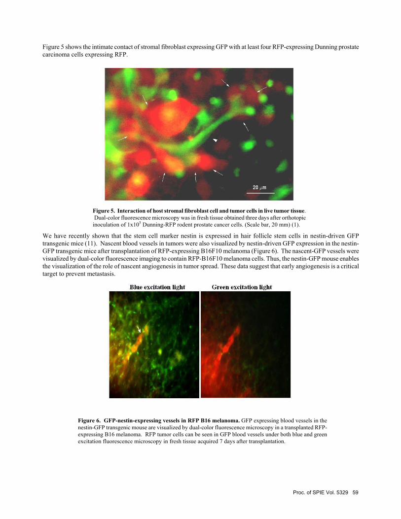
Figure 5 shows the intimate contact of stromal fibroblast expressing GFP with at least four RFP-expressing Dunning prostate carcinoma cells expressing RFP.
Figure 5. Interaction of host stromal fibroblast cell and tumor cells in live tumor tissue. Dual-color fluorescence microscopy was in fresh tissue obtained three days after orthotopic inoculation of 1x10⁵ Dunning-RFP rodent prostate cancer cells. (Scale bar, 20 mm) (1).
We have recently shown that the stem cell marker nestin is expressed in hair follicle stem cells in nestin-driven GFP transgenic mice (11). Nascent blood vessels in tumors were also visualized by nestin-driven GFP expression in the nestin-GFP transgenic mice after transplantation of RFP-expressing B16F10 melanoma (Figure 6). The nascent-GFP vessels were visualized by dual-color fluorescence imaging to contain RFP-B16F10 melanoma cells. Thus, the nestin-GFP mouse enables the visualization of the role of nascent angiogenesis in tumor spread. These data suggest that early angiogenesis is a critical target to prevent metastasis.
Figure 6. GFP-nestin-expressing vessels in RFP B16 melanoma. GFP expressing blood vessels in the nestin-GFP transgenic mouse are visualized by dual-color fluorescence microscopy in a transplanted RFP-expressing B16 melanoma. RFP tumor cells can be seen in GFP blood vessels under both blue and green excitation fluorescence microscopy in fresh tissue acquired 7 days after transplantation.
Proc. of SPIE Vol. 5329 59

4.0 DISCUSSION
This dual-color tumor-host interaction model system allows visualization of tumor host interaction by whole-body imaging as well as in fresh tissue. Both the tumor and the host cells are uniquely identified by their fluorescence color—RFP for the tumor, GFP for the host. The model has shown the specificity of various types of host cells for the tumor. For example, we have visualized tumor cells being contacted by host dendritic cells and stromal fibroblasts, macrophages engulfing tumor cells, and lymphocytes attacking the tumor which eventually regressed. The dual-color, tumor-host interaction model system allows observation of tumor-host interaction at the single-cell level in fresh tissue affording further insights of the role of host cells in tumor growth and progression. This may be particularly important for understanding the angiogenic process. To specifically visualize angiogenesis, the nestin-GFP transgenic mouse was used since nascent blood vessels express nestin-GFP. A remarkable image was acquired in fresh tissue of an RFP-expressing melanoma with tumor cells filling a GFP-expressing tumor blood vessel (Figure 6). These models can also be used to develop specific therapeutics that attack or support host cells that affect tumor growth and progression.
5.0 ACKNOWLEDGEMENT
This study was funded in part by National Cancer Institute grant number 1 R43 CA101600-01 and 1 R43 CA099258-01.
REFERENCES
1. Yang, M., Li, L., Jiang, P., Moossa, A.R., Penman, S., and Hoffman, R.M. Dual-color fluorescence imaging
distinguishes tumor cells from induced host angiogenic vessels and stromal cells. Proc. Natl. Acad. Sci. USA 100, 14259-14262, 2003.
2. Paget, S. The distribution of secondary growths in caner of the breast. Lancet 1, 571-573, 1889. 3. Fidler, I.J. The pathogenesis of cancer metastasis: the 'seed and soil' hypothesis revisited. Nature Reviews
Cancer 3, 453-458, 2003. 4. Fidler, I.J. The organ microenvironment and cancer metastasis. Differentiation 70, 498-505, 2002. 5. Fidler, I.J. Angiogenic heterogeneity: regulation of neoplastic angiogenesis by the organ microenvironment. J.
Natl. Cancer Inst. 93, 1040-1041, 2001. 6. Fidler, I.J. Seed and soil revisited: contribution of the organ microenvironment to cancer metastasis. Surg. Oncol.
Clin. N. Am. 10, 257-269, 2001. 7. Krutovskikh, V. Implication of direct host-tumor intercellular interactions in non-immune host resistance to
neoplastic growth. Semin. Cancer Biol. 12, 267-276, 2002. 8. Fukumura, D., Xavier, R., Sugiura, T., Chen, Y., Park, E.C., Lu, N., Selig, M., Nielsen, G., Taksir, T., Jain, R.K.,
Seed, B. Tumor induction of VEGF promoter activity in stromal cells. Cell 94, 715-725, 1998. 9. Brown EB, Campbell RB, Tsuzuki Y, Xu L, Carmeliet P, Fukumura D, Jain RK. In vivo measurement of gene
expression, angiogenesis and physiological function in tumors using multiphoton laser scanning microscopy. Nature Med. 7, 864-868, 2001.
10. Okabe M, Ikawa M, Kominami K, Nakanishi T, Nishimune T. 'Green mice' as a source of ubiquitous green cells. FEBS Letters 407, 313-319, 1997.
11. Li, L., Mignone, J., Yang, M., Matic, M., Penman, S., Enikolopov, G., Hoffman, R.M. Nestin expression in hair follicle sheath progenitor cells. Proc. Natl. Acad. Sci. USA 100, 9658-9662, 2003.
12. Sugawara, K., Kurihara, H., Negishi, M., Saito, N., Nakazato, Y., Sasaki, T., and Takeuchi, T. Nestin as a marker for proliferative endotheliums in gliomas. Laboratory Investigation 82, 345-351, 2002.
13. Hoffman, R.M. Green fluorescent protein imaging of tumour growth, metastasis, and angiogenesis in mouse models. Lancet Oncology 3, 546-556, 2002.
60 Proc. of SPIE Vol. 5329


![Cancer Research - Containment of Tumor …Thus, bacteria-mediated tumor therapy can be amplified by depletion of host neutrophils. [Cancer Res 2008;68(8):2952–60] Introduction Since](https://static.fdocuments.net/doc/165x107/5f51e8a666b2ad36e87145e9/cancer-research-containment-of-tumor-thus-bacteria-mediated-tumor-therapy-can.jpg)


