Dry Heat as a Decontamination Method for N95 Respirator Reuse
39
doi.org/10.26434/chemrxiv.12290621.v2 Dry Heat as a Decontamination Method for N95 Respirator Reuse Chamteut Oh, Elbashir Araud, Joseph V. Puthussery, Hezi Bai, Gemma G. Clark, Vishal Verma, Thanh H. Nguyen Submitted date: 12/06/2020 • Posted date: 18/06/2020 Licence: CC BY-NC 4.0 Citation information: Oh, Chamteut; Araud, Elbashir; Puthussery, Joseph V.; Bai, Hezi; Clark, Gemma G.; Verma, Vishal; et al. (2020): Dry Heat as a Decontamination Method for N95 Respirator Reuse. ChemRxiv. Preprint. https://doi.org/10.26434/chemrxiv.12290621.v2 A pandemic such as COVID-19 can cause a sudden depletion in the worldwide supply of respirators, forcing healthcare providers to reuse them. In this study, we systematically evaluated dry heat treatment as a viable option for the safe decontamination of N95 respirators (1860, 3M) before its reuse. We found that the dry heat generated by an electric cooker (100°C, 5% relative humidity, 50 min) effectively inactivated Tulane virus (>5.2-log 10 reduction), rotavirus (>6.6-log 10 reduction), adenovirus (>4.0-log 10 reduction), and transmissible gastroenteritis virus (>4.7-log 10 reduction). The respirator integrity (determined based on the particle filtration efficiency and quantitative fit testing) was not compromised after 20 cycles of 50-min dry heat treatment. Based on these results, we propose dry heat decontamination generated by an electric cooker (e.g., rice cookers, instant pots, ovens) to be an effective and accessible decontamination method for the safe reuse of N95 respirators. File list (2) download file view on ChemRxiv Manuscript.pdf (651.56 KiB) download file view on ChemRxiv Supporting information.pdf (736.71 KiB)
Transcript of Dry Heat as a Decontamination Method for N95 Respirator Reuse
doi.org/10.26434/chemrxiv.12290621.v2
Dry Heat as a Decontamination Method for N95 Respirator ReuseChamteut Oh, Elbashir Araud, Joseph V. Puthussery, Hezi Bai, Gemma G. Clark, Vishal Verma, Thanh H.Nguyen
Submitted date: 12/06/2020 • Posted date: 18/06/2020Licence: CC BY-NC 4.0Citation information: Oh, Chamteut; Araud, Elbashir; Puthussery, Joseph V.; Bai, Hezi; Clark, Gemma G.;Verma, Vishal; et al. (2020): Dry Heat as a Decontamination Method for N95 Respirator Reuse. ChemRxiv.Preprint. https://doi.org/10.26434/chemrxiv.12290621.v2
A pandemic such as COVID-19 can cause a sudden depletion in the worldwide supply of respirators, forcinghealthcare providers to reuse them. In this study, we systematically evaluated dry heat treatment as a viableoption for the safe decontamination of N95 respirators (1860, 3M) before its reuse. We found that the dry heatgenerated by an electric cooker (100°C, 5% relative humidity, 50 min) effectively inactivated Tulane virus(>5.2-log10 reduction), rotavirus (>6.6-log10 reduction), adenovirus (>4.0-log10 reduction), and transmissiblegastroenteritis virus (>4.7-log10 reduction). The respirator integrity (determined based on the particle filtrationefficiency and quantitative fit testing) was not compromised after 20 cycles of 50-min dry heat treatment.Based on these results, we propose dry heat decontamination generated by an electric cooker (e.g., ricecookers, instant pots, ovens) to be an effective and accessible decontamination method for the safe reuse ofN95 respirators.
File list (2)
download fileview on ChemRxivManuscript.pdf (651.56 KiB)
download fileview on ChemRxivSupporting information.pdf (736.71 KiB)
1
Dry heat as a decontamination method for N95 respirator reuse 1
2
Chamteut Oh1, Elbashir Araud2, Joseph V. Puthussery1, Hezi Bai1, Gemma G. Clark1, Vishal 3
Verma1, Thanh H. Nguyen1,3 4
1: Department of Civil and Environmental Engineering, University of Illinois at Urbana-5
Champaign 6
2: Holonyak Micro & Nanotechnology Lab, University of Illinois at Urbana-Champaign 7
3: Institute of Genomic Biology, University of Illinois at Urbana-Champaign 8
9
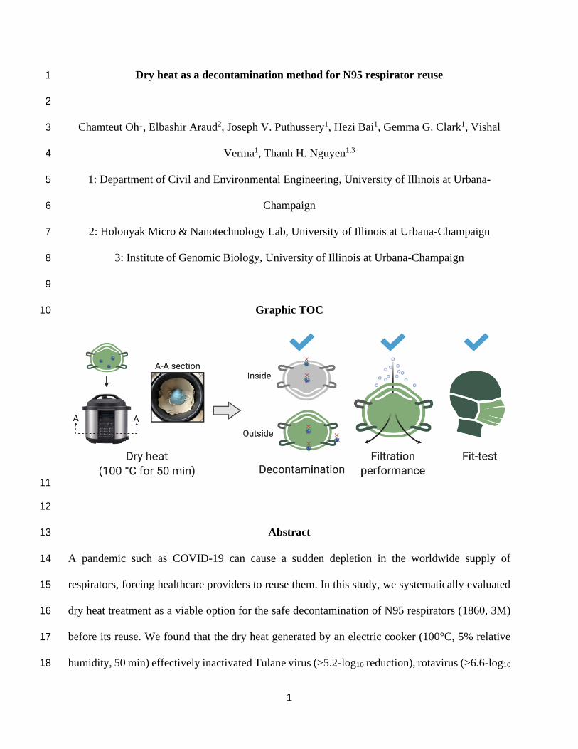
Graphic TOC 10
11
12
Abstract 13
A pandemic such as COVID-19 can cause a sudden depletion in the worldwide supply of 14
respirators, forcing healthcare providers to reuse them. In this study, we systematically evaluated 15
dry heat treatment as a viable option for the safe decontamination of N95 respirators (1860, 3M) 16
before its reuse. We found that the dry heat generated by an electric cooker (100°C, 5% relative 17
humidity, 50 min) effectively inactivated Tulane virus (>5.2-log10 reduction), rotavirus (>6.6-log10 18
2
reduction), adenovirus (>4.0-log10 reduction), and transmissible gastroenteritis virus (>4.7-log10 19
reduction). The respirator integrity (determined based on the particle filtration efficiency and 20
quantitative fit testing) was not compromised after 20 cycles of 50-min dry heat treatment. Based 21
on these results, we propose dry heat decontamination generated by an electric cooker (e.g., rice 22
cookers, instant pots, ovens) to be an effective and accessible decontamination method for the safe 23
reuse of N95 respirators. 24
25
Introduction 26
An N95 respirator is an essential piece of personal protection equipment (PPE) during an outbreak 27
of infectious disease. Although the respirator is designed to be disposable, the high demand during 28
a pandemic such as COVID-19 can force healthcare providers to reuse respirators. The primary 29
problem with respirator reuse is that once a respirator is contaminated, it can act as a potential 30
transmission route of pathogens to both patients and healthcare providers. The safety of healthcare 31
providers depends on respirators being effectively decontaminated prior to reuse.1 3M, the main 32
respirator manufacturer, has issued four recommendations for reuse.2 First, the decontamination 33
should be virucidal under relevant conditions. For example, the Food and Drug Administration 34
(FDA) requires at least 3-log10 virucidal efficacy for multiple viruses, including coronaviruses in 35
soiling agents (e.g., blood, mucus, or sebum).3 Second, the filtration performance (filtration 36
efficiency and breathability) should be maintained after the decontamination process. Third, the 37
treated respirator must be leak-tight, fitting closely against the user’s face such that there are no 38
obvious gaps that permit air to enter between the respirator and the user’s face. Fourth, the 39
decontamination method must not leave residual harmful chemicals that affect the user’s safety. 40
3
We recommend an additional requirement that the decontamination technology should be easily 41
accessible. 42
Dry heat has the potential to satisfy the five requirements mentioned above. Heat is one of 43
the most conventional disinfection technologies, so the thermal inactivation efficacies for various 44
pathogens are known.4 Dry heat is the least likely to reduce the filtration efficiency when compared 45
with other available decontamination methods (moist heat, ethanol, isopropanol solution, bleach, 46
and UV).5,6 In addition, dry heat can be generated by electric heating appliances (e.g., rice cookers, 47
instant pots, and ovens) without using toxic materials. However, no experimental conclusions 48
about dry heat have been made for N95 respirator reuse in terms of these five requirements. In this 49
research, we conducted experiments for viral decontamination, filtration performance, and 50
quantitative fit testing. Based on the results, we determined that dry heat is an appropriate 51
decontamination technology for N95 respirator reuse. 52
53
Materials and method 54
Respirator and cooker 55
We used N95 respirators (1860, 3M) and an electric cooker (WM-CS60004W, Farberware), which 56
is an inexpensive and commonly available kitchen appliance. The pot was 22 cm in diameter, 15 57
cm in height, and 5.7 L in volume. The pot could fit a stack of about 5 respirators while maintaining 58
at least 3 cm between the respirators and the interior sides and lid of the pot. The surface 59
temperatures of the pot and the respirator were monitored every 5 to 13 min during the dry heat 60
treatment using an infrared thermometer (IRT205, General Tools). The temperature and relative 61
humidity of the air inside the pot were measured using a thermo-hygrometer (A600FC, General 62
Tools). 63
4
64
Testing viruses 65
To fulfill the FDA requirements for viral inactivation, we used four different viruses with different 66
virus genomes and capsid structures that included: dsDNA virus (respiratory human adenovirus 67
type 2, single-layered non-enveloped virion),7 dsRNA virus (rotavirus OSU, triple-layered non-68
enveloped virion),8 7 kb ssRNA virus (Tulane virus, surrogate for human norovirus, single-layered 69
non-enveloped virion),9 and 28.5 kb ssRNA virus (porcine transmissible gastroenteritis virus 70
(TGEV),10 single-layered enveloped virion). TGEV is categorized into Coronaviridae, the same 71
family as SARS-CoV-2. It has the same viral structure and genome as SARS-CoV-2 (enveloped 72
and (+)ssRNA virus), but it primarily infects pigs.11 Tulane virus, rotavirus (OSU strain), and 73
human adenovirus type 2 belong to Caliciviridae, Reoviridae, and Adenoviridae, respectively. 74
Details of the virus preparation methods are described in Text S1 and Table S1. 75
The virus suspension was mixed with artificial saliva at a 1:1 ratio before use. The artificial 76
saliva was used as a soiling agent and prepared following ASTM E2720-16 with a slight 77
modification (Table S2).12 All of the experiments were replicated three times. 78
79
Decontamination test 80
We performed three separate procedures to test inactivation efficacy. First, we inoculated Tulane 81
virus in five different locations (the inside edge, the inside center, the outside edge, the outside 82
center, and the strap) on one whole respirator to see the effect of the inoculation site on Tulane 83
virus inactivation efficacy. We applied dry heat and then cut the respirators into pieces at the 84
inoculation sites (Figure S1). Second, we cut a clean respirator into 5 mm diameter pieces, 85
inoculated them with Tulane virus, and surrounded them with polycotton lab coat (Fisher 86
5
Scientific, USA) in the pot to simulate a case where the dominant heat transfer method is 87
convective heat instead of radiation heat from the interior walls of the pots (Figure S2). Third, we 88
inoculated 5 mm diameter clean pieces with each of the four viruses (Tulane virus, rotavirus, 89
adenovirus, and TGEV) and used dry heat for various time spans. Details are described in Text S2. 90
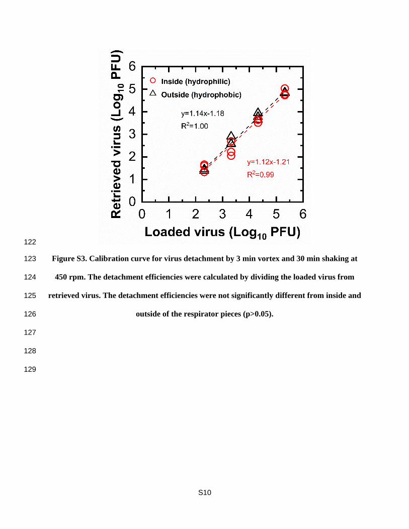
We submerged respirator pieces in 1 mL of fresh culture media and detached the viruses 91
from the respirator fragments by vortexing them for 3 min and shaking them for 30 min at 450 92
rpm (Figure S3). The supernatant was used for the plaque assay and the molecular assays to 93
determine the inactivation efficacy and mechanisms, respectively. We calculated the reduction in 94
virus infectivity by dividing the infectivity of the negative control by that of the treated sample 95
(i.e., log10 (N0/N)). We used the previously established molecular assays with a slight modification 96
to analyze the primary structural target of Tulane virus by the dry heat treatment.13,14 An RNase 97
assay, a binding assay using magnetic beads coated with the host cell receptors, and a two-step 98
RT-qPCR assay were applied to examine the integrity of capsid proteins, binding proteins, and 99
viral genomes, respectively. Details of the molecular assays are explained in Text S3. 100
101
Filtration performance test 102
The particle filtration efficiency test of the filters was performed using a slightly modified version 103
of the NIOSH 42 CFR 84 regulations.15 The detailed experimental setup and procedure are 104
provided in the SI (Text S4 and Figure S4). Briefly, a small portion (47 mm diameter) of the N95 105
mask fabric was cut and loaded onto a 47 mm filter holder (URG, Carrboro, NC, USA). A solution 106
of 2% NaCl was aerosolized using a constant output atomizer (TSI Model 3076, MN, USA).15 The 107
polydisperse NaCl aerosols generated from the atomizer were first dried and charge neutralized; 108
after which they were passed into a polypropylene chamber, which houses the filter holder. We 109
6
used a condensation particle counter (CPC, TSI Model 3022A; flow rate = 1.5 lpm) to measure the 110
particle concentration before and after loading the test filter (i.e., a section of the mask) in the filter 111
holder. We tested the filters for a face velocity of 9.4 cm/s (equivalent to NIOSH recommended 112
test flow rate of 85 lpm). A pressure gauge (Magnehelic 1-10 inches of water) was also connected 113
in parallel and downstream of the filter-holder using a T-connector to measure the pressure drop 114
across the mask. The particle number concentration was measured before and after connecting the 115
filter holder, and particle removal efficiency of the mask was measured by the following equation: 116
117
𝑃𝑎𝑟𝑡𝑖𝑐𝑙𝑒 𝑟𝑒𝑚𝑜𝑣𝑎𝑙 𝐸𝑓𝑓𝑖𝑐𝑖𝑒𝑛𝑐𝑦 (%)118
= (1 −𝑝𝑎𝑟𝑡𝑖𝑐𝑙𝑒 𝑛𝑢𝑚𝑏𝑒𝑟 𝑐𝑜𝑛𝑐𝑒𝑛𝑡𝑟𝑎𝑡𝑖𝑜𝑛 𝑎𝑓𝑡𝑒𝑟 𝑝𝑙𝑎𝑐𝑖𝑛𝑔 𝑡ℎ𝑒 𝑚𝑎𝑠𝑘 (
#𝑐𝑚3)
𝑝𝑎𝑟𝑡𝑖𝑐𝑙𝑒 𝑛𝑢𝑚𝑏𝑒𝑟 𝑐𝑜𝑛𝑐𝑒𝑛𝑡𝑟𝑎𝑡𝑖𝑜𝑛 𝑏𝑒𝑓𝑜𝑟𝑒 𝑝𝑙𝑎𝑐𝑖𝑛𝑔 𝑡ℎ𝑒 𝑚𝑎𝑠𝑘 (#
𝑐𝑚3)) × 100 119
120
The filtration performance test was performed on each respirator after 1, 2, 3, 5, 10, and 20 cycles 121
of dry heat decontamination. All the experiments were replicated three times. 122
123
Quantitative Fit testing 124
Quantitative fit testing was performed by the Office of Occupational Safety and Health at 125
University of Illinois at Urbana-Champaign following the modified ambient aerosol condensation 126
nuclei counter quantitative fit testing protocol (1910.134 App A, OSHA). The purpose was to 127
check the overall integrity of the respirators. Three respirators treated by 20 cycles of 50 min of 128
dry heat were prepared. The testing room was filled with a NaCl aerosol, which was produced by 129
a particle generator (8026, TSI, USA). A test taker donned each respirator connected to a respirator 130
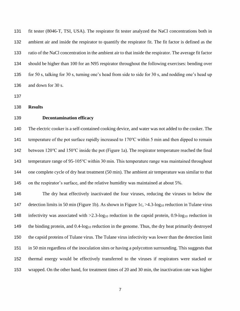
7
fit tester (8046-T, TSI, USA). The respirator fit tester analyzed the NaCl concentrations both in 131
ambient air and inside the respirator to quantify the respirator fit. The fit factor is defined as the 132
ratio of the NaCl concentration in the ambient air to that inside the respirator. The average fit factor 133
should be higher than 100 for an N95 respirator throughout the following exercises: bending over 134
for 50 s, talking for 30 s, turning one’s head from side to side for 30 s, and nodding one’s head up 135
and down for 30 s. 136
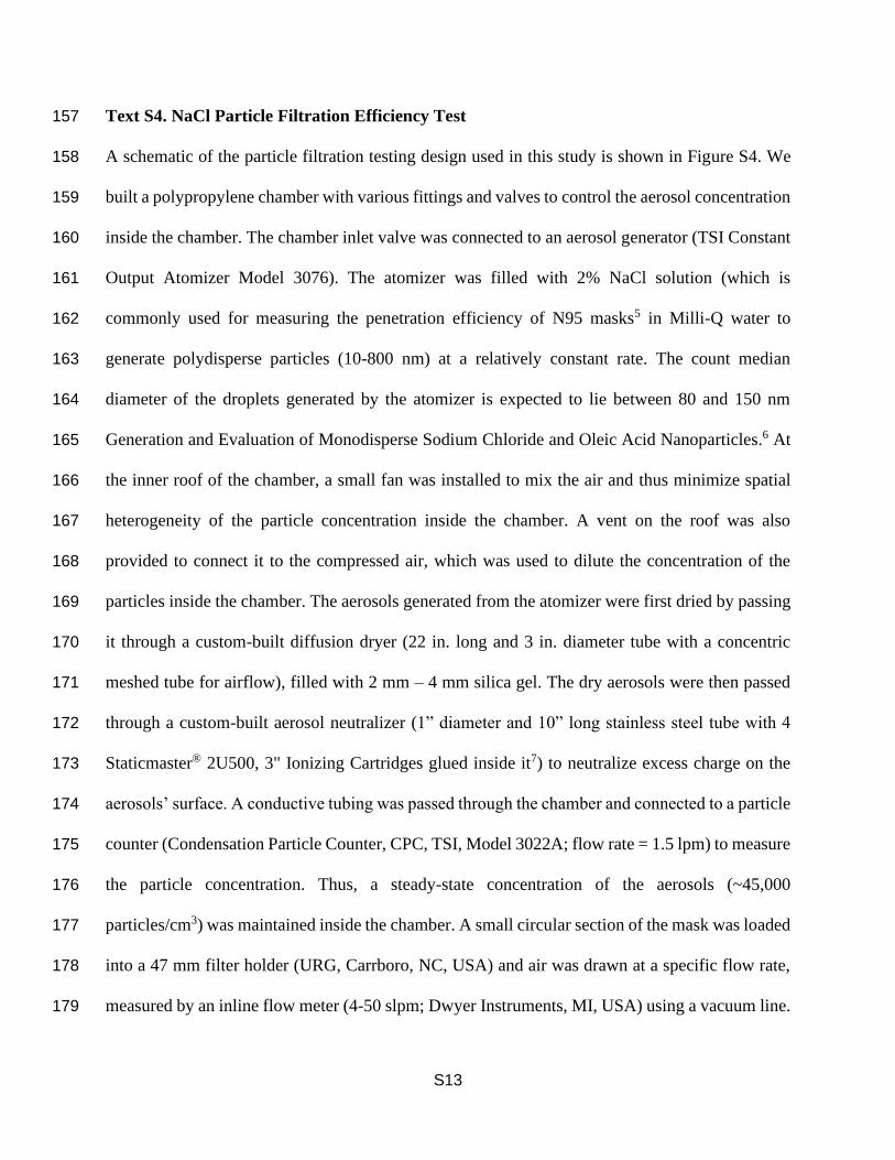
137
Results 138
Decontamination efficacy 139
The electric cooker is a self-contained cooking device, and water was not added to the cooker. The 140
temperature of the pot surface rapidly increased to 170℃ within 5 min and then dipped to remain 141
between 120℃ and 150℃ inside the pot (Figure 1a). The respirator temperature reached the final 142
temperature range of 95-105℃ within 30 min. This temperature range was maintained throughout 143
one complete cycle of dry heat treatment (50 min). The ambient air temperature was similar to that 144
on the respirator’s surface, and the relative humidity was maintained at about 5%. 145
The dry heat effectively inactivated the four viruses, reducing the viruses to below the 146
detection limits in 50 min (Figure 1b). As shown in Figure 1c, >4.3-log10 reduction in Tulane virus 147
infectivity was associated with >2.3-log10 reduction in the capsid protein, 0.9-log10 reduction in 148
the binding protein, and 0.4-log10 reduction in the genome. Thus, the dry heat primarily destroyed 149
the capsid proteins of Tulane virus. The Tulane virus infectivity was lower than the detection limit 150
in 50 min regardless of the inoculation sites or having a polycotton surrounding. This suggests that 151
thermal energy would be effectively transferred to the viruses if respirators were stacked or 152
wrapped. On the other hand, for treatment times of 20 and 30 min, the inactivation rate was higher 153
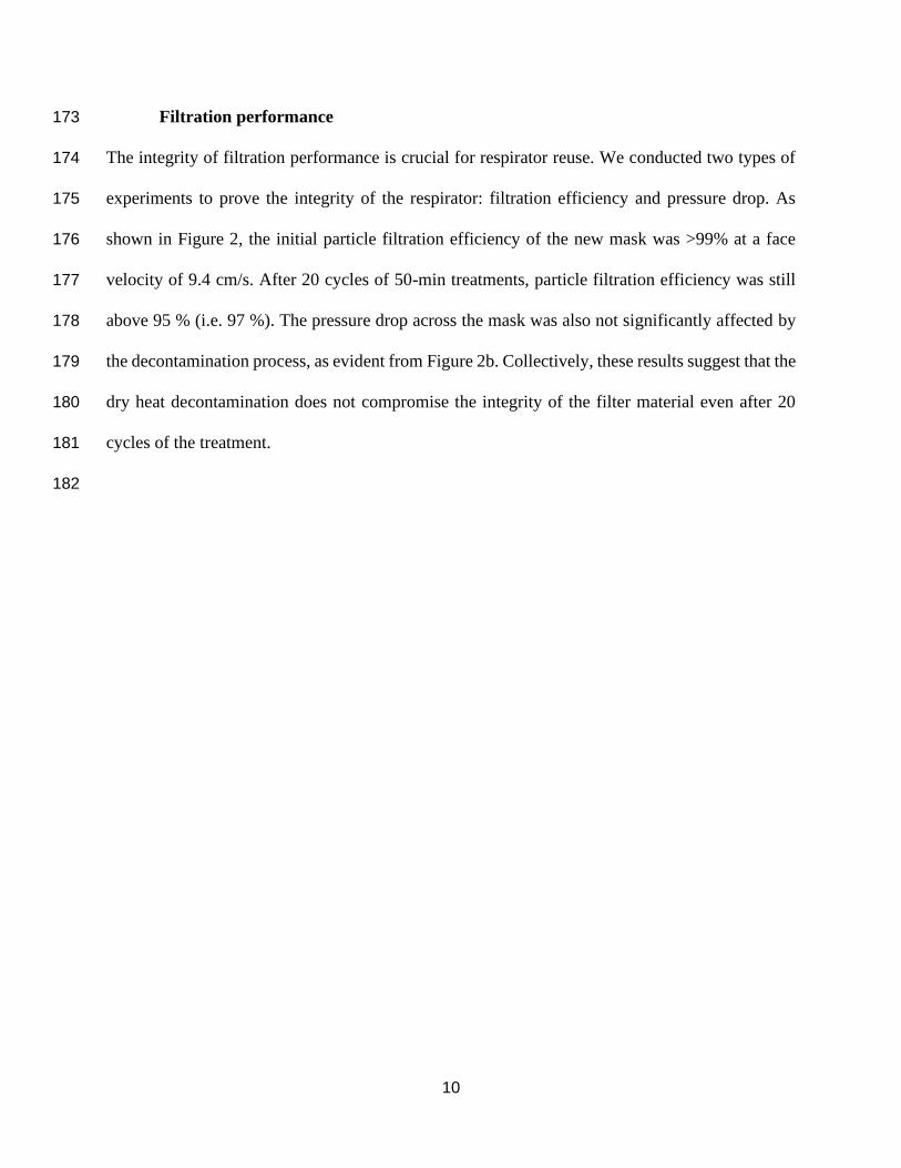
8
when the viruses were inoculated on the hydrophilic surface (p<0.05). This can be explained by 154
how different respirator materials held the virus solution. When the viruses were inoculated, the 155
testing solution was absorbed by the inside of the respirator that faces the user (the hydrophilic 156
surface). At the same time, a droplet was formed on the outside of the respirator that faces the 157
user’s surroundings (the hydrophobic surface). After the droplet evaporated, the virus and saliva 158
were evenly distributed inside of the respirator. In contrast, there was a high concentration of saliva 159
remaining on the outside of the respirator, which could shield the viruses from the dry heat.16 160
161
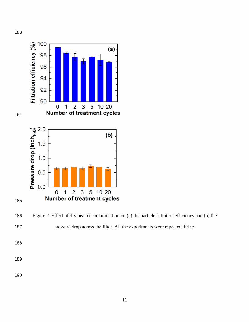
9
162
163
164
165
Figure 1. Effect of dry heat treatment on (a) temperature profiles for the surfaces of the pot and 166
the respirator and (b) virus inactivation rates. Tulane viruses were inoculated on the hydrophobic 167
(outside) and hydrophilic (inside) surfaces, while the other viruses were inoculated only on the 168
hydrophilic surfaces. (c) Molecular assay results from Tulane virus samples treated by the dry 169
heat for 30 min. Arrows indicate the detection limit. The detection limit varied depending on the 170
initial infectivity of the virus solution (Log10 N0). 171
172
10
Filtration performance 173
The integrity of filtration performance is crucial for respirator reuse. We conducted two types of 174
experiments to prove the integrity of the respirator: filtration efficiency and pressure drop. As 175
shown in Figure 2, the initial particle filtration efficiency of the new mask was >99% at a face 176
velocity of 9.4 cm/s. After 20 cycles of 50-min treatments, particle filtration efficiency was still 177
above 95 % (i.e. 97 %). The pressure drop across the mask was also not significantly affected by 178
the decontamination process, as evident from Figure 2b. Collectively, these results suggest that the 179
dry heat decontamination does not compromise the integrity of the filter material even after 20 180
cycles of the treatment. 181
182
11
183
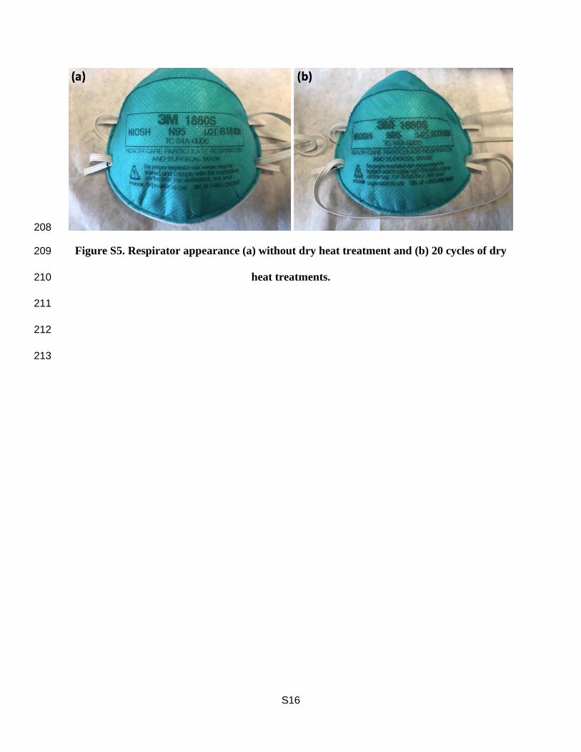
184
185
Figure 2. Effect of dry heat decontamination on (a) the particle filtration efficiency and (b) the 186
pressure drop across the filter. All the experiments were repeated thrice. 187
188
189
190
12
Quantitative fit testing 191
The respirator treated with 20 cycles of the dry heat had an average fit factor of 139±18, which is 192
higher than the fit factor required of the N95 respirator. In addition, no visible deformations (such 193
as burning signs, nose form detachment, loss of elasticity in the band, or deformation of the entire 194
shape) except for ink spread (Figure S5) were noticed on the respirator after 20 cycles of dry heat. 195
196
Discussion 197
Our experiments simulated droplets or aerosols of an infected patient’s saliva depositing on a 198
healthcare provider’s respirator and then evaporating. The inside of the respirator could also be 199
contaminated by the viruses while handling the respirator with contaminated hands. The dry heat 200
generated by the cooker was confirmed to satisfy the five requirements for respirator reuse 201
(decontamination efficacy, filtration performance, fit testing, no toxic residual chemicals, and 202
accessibility). 203
The dry heat (100°C for 50 min) successfully conveyed the thermal energy to the viruses 204
and denatured capsid proteins, resulting in >5.2-log10 reduction for Tulane virus, >6.6-log10 205
reduction for rotavirus, >4.0-log10 reduction for adenovirus, >4.7-log10 reduction for TGEV. Since 206
the protein denaturation follows first-order reaction and Arrhenius equation, the virus inactivation 207
will be significantly affected by treatment temperature and time.4 A recent study showed that dry 208
heat (82°C, 30 min) using a lab oven was not enough to achieve 3-log10 reduction of MS2, Phi6, 209
and murine hepatitis virus.17 Also, the inactivation efficacy of dry heat (100°C, 15 min) for MS2 210
was no greater than 1-log10 reduction.18 This result aligned with our findings that the dry heat 211
(100°C) for 10 min inactivated Tulane virus by a factor of less than 1-log. However, the virus 212
infectivity reduced rapidly by a factor of greater than 3-log10 after 30 min. Those results 213
13
collectively can be explained by the fact that the optimum temperature and time should be provided 214
for the proper decontamination. The dry heat generated by the cooker (100°C) for 50 min was the 215
optimal condition for the inactivation of tested viruses. Because about 4-log10 reduction of SARS-216
CoV-2 on the respirator’s surface was achieved by applying dry heat (70°C for 60 min),19 the dry 217
heat used in this study (100°C for 50 min) should be adequate to inactivate SARS-CoV-2. 218
The respirator integrity (filtration performance and fit testing) was not degraded by 20-219
cycles of the dry heat treatment.6 Although the temperature of the respirator’s surface was higher 220
than the maximum operating temperature (50°C) that is provided by the manufacturer,20 the 221
primary materials for the respirator (polyester, polypropylene, polyurethane, polyisoprene) can 222
withstand a temperature as high as 150°C.21,22 Note that the temperature of the pot surface is higher 223
than the allowable temperature for the outside surface of the respirator (polypropylene), so direct 224
contact between the respirator and the pot surface must be avoided using a towel or some other 225
item to create a barrier and insulate the respirators. It was reported that N95 respirators partially 226
melt when subjected to dry heat generated by a lab oven at 100-120°C (Isotemp 500 Series, Fisher 227
Scientific).23,24 In these studies, however, the respirators were placed directly on the metal pan. 228
We confirmed that the respirator filtration efficiency (98.5±0.1%) and the pressure drop (0.7±0.0 229
inchH2O) were still acceptable for the N95 respirator after the dry heat generated by the lab oven 230
(Isotemp 650G, Fisher Scientific) set at 120°C (the temperature of the respirator’s surface was 231
110°C) for 24 hours. 232
The dry heat can be produced using readily available heating appliances. The respirator 233
can also be reused after dry heat treatment without further treatment because no toxic chemicals 234
were involved. Given the filtration efficiency of the respirators after being treated by the dry heat 235
generated by the cooker (20 cycles) and the lab oven (24 hours), we believe that any device 236
14
providing dry heat and holding the respirator temperature at 100℃ for 50 min would work for 237
respirator reuse. Note that at temperatures higher than 100℃, the dry heat could reduce the 238
respirator integrity while temperatures lower than 100℃ may require a longer treatment time to 239
inactivate the viruses. 240
In conclusion, dry heat treatment of 100℃ for 50 min is an appropriate method for 241
preparing N95 respirators (1860, 3M) for reuse. Further studies for other types of respirator reuse 242
are needed because different materials may require different temperatures and treatment times to 243
produce the same treatment result. 244
Acknowledgement 245
This research was supported jointly by the EPA/NIFA grant on water reuse #2017-39591-246
27313. Its contents are solely the responsibility of the grantee and do not necessarily represent the 247
official views of the EPA. Further, the EPA does not endorse the purchase of any commercial 248
products or services mentioned in the publication. We acknowledge Dr Leyi Wang (Veterinary 249
Diagnostic Lab), Dr Carleigh Hebbard, Dr Lyndon Goodly, and Mr Jeremy Neighbors 250
(Occupational Safety and Health) for their support and feedback on this project. 251
252
15
References 253
(1) Farsi, D.; Mofidi, M.; Mahshidfar, B.; Hafezimoghadam, P. Consider the Options ; Can 254
Decontamination and Reuse Be the Answer to N95 Respirator Shortage in COVID-19 255
Pandemic ? 2020, No. 10, 1–2. https://doi.org/10.22114/ajem.v0i0.378. 256
(2) 3M. Decontamination Methods for 3M Filtering Facepiece Respirators Such as N95 257
Respirators; 2020. 258
(3) U.S. Food and Drug Administration. Enforcement Policy for Face Masks and Respirators 259
During the Coronavirus Disease ( COVID-19 ) Public Health Emergency ( Revised ) 260
Guidance for Industry And. 2020, No. April. 261
(4) Yap, T. F.; Liu, Z.; Shveda, R. A.; Preston, D. J. A Predictive Model of the Temperature-262
Dependent Inactivation of Coronaviruses. ChemRxiv 2020, 1–40. 263
(5) Lin, T. H.; Chen, C. C.; Huang, S. H.; Kuo, C. W.; Lai, C. Y.; Lin, W. Y. Filter Quality of 264
Electret Masks in Filtering 14.6–594 Nm Aerosol Particles: Effects of Five 265
Decontamination Methods. PLoS One 2017, 12 (10), 1–15. 266
https://doi.org/10.1371/journal.pone.0186217. 267
(6) Liao, L.; Xiao, W.; Zhao, M.; Yu, X.; Wang, H.; Wang, Q.; Chu, S.; Cui, Y. Can N95 268
Respirators Be Reused after Disinfection? And for How Many Times? medRxiv 2020, 2, 269
2020.04.01.20050443. https://doi.org/10.1101/2020.04.01.20050443. 270
(7) NCBI. Human Adenovirus C, Complete Genome (NC_001405.1); 2018. 271
(8) Taube, S.; Jiang, M.; Wobus, C. E. Glycosphingolipids as Receptors for Non-Enveloped 272
Viruses. Viruses. 2010. https://doi.org/10.3390/v2041011. 273
(9) NCBI. Tulane Virus Nonstructural Polyprotein, Capsid Protein, and Minor Structural 274
Protein VP2 Genes, Complete Cds (NC_043512.1); 2019. 275
16
(10) NCBI. Transmissible Gastroenteritis Virus Complete Genome, Genomic RNA 276
(NC_038861.1); 2018. 277
(11) Ashour, H. M.; Elkhatib, W. F.; Rahman, M. M.; Elshabrawy, H. A. Insights into the 278
Recent 2019 Novel Coronavirus (Sars-CoV-2) in Light of Past Human Coronavirus 279
Outbreaks. Pathogens. 2020. https://doi.org/10.3390/pathogens9030186. 280
(12) E2720-16, A. Standard Practice for Evaluation of Effectiveness of Decontamination 281
Procedures for Air-Permeable Materials When Challenged with Biological Aerosols 282
Containing Human Pathogenic Viruses; West Conshohocken, PA, 2016. 283
https://doi.org/10.1520/E2720-16. 284
(13) Fuzawa, M.; Araud, E.; Li, J.; Shisler, J. L.; Nguyen, T. H. Free Chlorine Disinfection 285
Mechanisms of Rotaviruses and Human Norovirus Surrogate Tulane Virus Attached to 286
Fresh Produce Surfaces. Environ. Sci. Technol. 2019, 53 (20), 11999–12006. 287
https://doi.org/10.1021/acs.est.9b03461. 288
(14) Araud, E.; Fuzawa, M.; Shisler, J. L.; Li, J.; Nguyen, T. H. UV Inactivation of Rotavirus 289
and Tulane Virus Targets Different Components of the Virions. Appl. Environ. Microbiol. 290
2020. https://doi.org/10.1128/AEM.02436-19. 291
(15) NIOSH. Determination of Particulate Filter Efficiency Level for N95 Series Filters 292
Against Solid Particulates for Non-Powered, Air-Purifying Respirators; 2019. 293
https://doi.org/TEB-APR-STP-0059. 294
(16) Waldman, P.; Meseguer, A.; Lucas, F.; Moulin, L.; Wurtzer, S. Interaction of Human 295
Enteric Viruses with Microbial Compounds: Implication for Virus Persistence and 296
Disinfection Treatments. Environ. Sci. Technol. 2017. 297
https://doi.org/10.1021/acs.est.7b03875. 298
17
(17) Wigginton, K. R.; Arts, P. J.; Clack, H.; Fitzsimmons, W. J.; Gamba, M.; Katherine, R.; 299
Lebar, W.; Lauring, A. S.; Li, L.; Roberts, W. W.; Rockey, N.; Young, C.; Anderegg, L. 300
G.; Cohn, A. M.; Doyle, J. M.; Cole, M.; Kaye, K.; Love, N. G. Validation of N95 301
Filtering Facepiece Respirator Decontamination Methods Available at a Large University 302
Hospital. 2020. 303
(18) Li, D. F.; Cadnum, J. L.; Redmond, S. N.; Jones, L. D.; Donskey, C. J. It’s Not the Heat, 304
It’s the Humidity: Effectiveness of a Rice Cooker-Steamer for Decontamination of Cloth 305
and Surgical Face Masks and N95 Respirators. Am. J. Infect. Control 2020. 306
https://doi.org/10.1016/j.ajic.2020.04.012. 307
(19) Fischer, R.; Morris, D. H.; Doremalen, N. van; Sarchette, S.; Matson, J.; Bushmaker, T.; 308
Yinda, C. K.; Seifert, S.; Gamble, A.; Williamson, B.; Judson, S.; Wit, E. de; Lloyd-309
Smith, J.; Munster, V. Assessment of N95 Respirator Decontamination and Re-Use for 310
SARS-CoV-2. medRxiv 2020, 2020.04.11.20062018. 311
https://doi.org/10.1101/2020.04.11.20062018. 312
(20) 3M. 3MTM Disposable Respirator 1860, 1860S, N95; 2018. https://doi.org/AV011466081. 313
(21) Lokensgard, E. Industrial Plastics: Theory and Applications; Cengage Learning, 2016. 314
(22) Hutten, I. M.; Wadsworth, L. Handbook of Nonwoven Filter Media; 2007. 315
https://doi.org/10.1016/B978-1-85617-441-1.X5015-X. 316
(23) Viscusi, D. J.; King, W. P.; Shaffer, R. E. Effect of Decontamination on the Filtration 317
Efficiency of Two Filtering Facepiece Respirator Models. Int. Soc. Respir. Prot. 2007, 24 318
(3/4), 93. 319
(24) Viscusi, D. J.; Bergman, M. S.; Eimer, B. C.; Shaffer, R. E. Evaluation of Five 320
Decontamination Methods for Filtering Facepiece Respirators. Ann. Occup. Hyg. 2009, 53 321
18
(8), 815–827. https://doi.org/10.1093/annhyg/mep070. 322
323

download fileview on ChemRxivManuscript.pdf (651.56 KiB)
S1
Supporting information for 1
2
Dry heat as a decontamination method for N95 respirator reuse 3
4
Chamteut Oh1, Elbashir Araud2, Joseph V. Puthussery1, Hezi Bai1, Gemma G. Clark1, Vishal 5
Verma1, Thanh H. Nguyen1,3 6
7
1: Department of Civil and Environmental Engineering, University of Illinois at Urbana-8
Champaign 9
2: Holonyak Micro & Nanotechnology Lab, University of Illinois at Urbana-Champaign 10
3: Institute of Genomic Biology, University of Illinois at Urbana-Champaign 11
12
Corresponding author: Thanh H. Nguyen. [email protected] 13
14
2 Tables 15
5 Figures 16
4 Texts 17
18 pages 18
19
S2
Text S1. Testing virus preparation 20
Tulane virus was received from Cincinnati Children’s Hospital Medical Center1 and rotavirus OSU 21
strain was obtained from ATCC (VR-892). The MA104 cell line was used to propagate Tulane 22
virus and rotavirus. The culture medium for the MA104 cells was prepared by mixing 1X minimum 23
essential medium (MEM; Thermo Fisher Scientific, MA, USA) with 10% fetal bovine serum 24
(FBS; Thermo Fisher Scientific, MA, USA), 1X antibiotic-antimycotic (Thermo Fisher Scientific, 25
MA, USA), 17 mM of NaHCO3, 10 mM of HEPES, and 1 mM of sodium pyruvate. MA 104 cells 26
with 80-90% confluency were washed with PBS and inoculated with Tulane virus or rotavirus 27
(OSU strain) in 175 cm2 flasks at a multiplicity of infection (MOI) of 0.1. The inoculated cells 28
were incubated at 37°C in a 5% CO2 environment for an hour with gentle shaking every 10 to 15 29
min. Then, 20 mL of the culture medium were added to each flask. For Tulane virus, FBS was 30
added to the culture medium at a final concentration of 2%. For rotavirus (OSU strain), trypsin 31
was added to the culture medium at a final concentration of 10 µg/mL while FBS was not added. 32
The infected flasks were incubated until an 80% cytopathic effect (CPE) was reached. The viruses 33
were harvested after three freeze-thaw cycles. Both viruses were purified in 1 mM NaCl and 0.1 34
mM CaCl2 solution using an ultracentrifuge (Optima XPN-90 Ultracentrifuge, Beckman Coulter, 35
CA, USA). The ultracentrifuge was run at 1000 rpm (116 g) at 4°C for 5 min followed by 36000 36
rpm (150700 g) at 4°C for 3 hours. The final concentrations of Tulane virus and rotavirus were 37
about 107 and 108 PFU/mL, respectively. The decontamination efficacy of both viruses was 38
determined by plaque assay using the MA104 cell line. The incubation time for the plaque assay 39
was 2 and 3 days for Tulane virus and rotavirus, respectively. Detailed information is described in 40
our previous work.2,3 41
S3
Adenovirus was obtained from ATCC (VR-846). They were propagated in A549 cells 42
using Ham F-12 media with 10% FBS (Thermo Fisher Scientific, MA, USA) and 1X antibiotic-43
antimycotic (Thermo Fisher Scientific, MA, USA). The adenovirus was purified in 1X PBS 44
(Thermo Fisher Scientific, MA, USA) using the ultracentrifuge and had a final infectivity of about 45
106 PFU/mL. A volume of 2 mL of overlay solution for the plaque assay was prepared by mixing 46
1.31 mL of 2X MEM, 0.5 mL of 1% agarose solution, 0.1 mL of FBS, 0.05 mL of 15 mM HEPES, 47
0.03 mL of 7.5% sodium bicarbonate, and 0.01 mL of 100X antibiotic-antimycotic. The incubation 48
time for the plaque assay was 5 days. Detailed information is described in our previous work.4 49
Transmissible Gastroenteritis Virus (TGEV) was obtained from the Veterinary Diagnostic 50
Laboratory at the University of Illinois at Urbana-Champaign. Swine testis (ST) cells were used 51
as a host cell for the virus to grow in and for the plaque assay. The same culture medium described 52
for Tulane virus was also used for the ST cells. TGEV was harvested in the culture medium by 53
centrifugation at 2000 rpm (556 g) for 10 min (Sorvall Legend RT Plus, Thermo Fisher Scientific, 54
MA, USA), followed by filtration through a 0.45 μm filter (Millipore Sigma, MA, USA). The 55
infectivity of TGEV was determined by the plaque assay; ST cell monolayers were prepared in 6-56
well plates (USA Scientific, FL, USA). The 750 μL of virus solution was inoculated to the cells 57
followed by incubation at 37°C with 5% CO2 for 60 min. The virus solution was replaced with 2 58
mL of the MEM containing 1% agarose, 7.5% sodium bicarbonate, 15 mM HEPES, and 1X 59
antibiotic-antimycotic. The overlay was solidified at 4°C for 20 min followed by the incubation at 60
37°C with 5% CO2 for 4 days. The cellular monolayers were fixed with 10% formaldehyde for 1 61
hour. The plaques were visualized after the fixed cells were dyed with 0.05% crystal violet in 10% 62
ethanol for 20 min. The initial infectivity of TGEV solution was about 106 PFU/mL. 63
64
S4
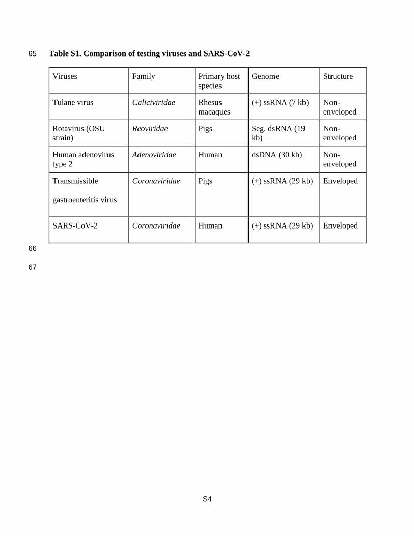
Table S1. Comparison of testing viruses and SARS-CoV-2 65
Viruses Family Primary host
species
Genome Structure
Tulane virus Caliciviridae Rhesus
macaques
(+) ssRNA (7 kb) Non-
enveloped
Rotavirus (OSU
strain)
Reoviridae Pigs Seg. dsRNA (19
kb)
Non-
enveloped
Human adenovirus
type 2
Adenoviridae Human dsDNA (30 kb) Non-
enveloped
Transmissible
gastroenteritis virus
Coronaviridae Pigs (+) ssRNA (29 kb) Enveloped
SARS-CoV-2 Coronaviridae Human (+) ssRNA (29 kb) Enveloped
66
67
S5
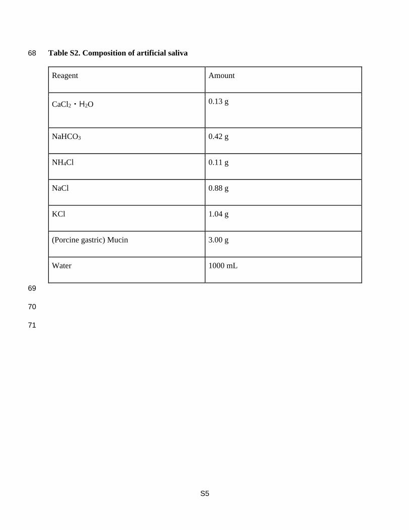
Table S2. Composition of artificial saliva 68
Reagent Amount
CaCl2・H2O 0.13 g
NaHCO3 0.42 g
NH4Cl 0.11 g
NaCl 0.88 g
KCl 1.04 g
(Porcine gastric) Mucin 3.00 g
Water 1000 mL
69
70
71
S6
72
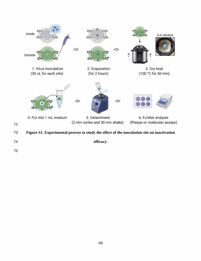
Figure S1. Experimental process to study the effect of the inoculation site on inactivation 73
efficacy. 74
75
S7
76
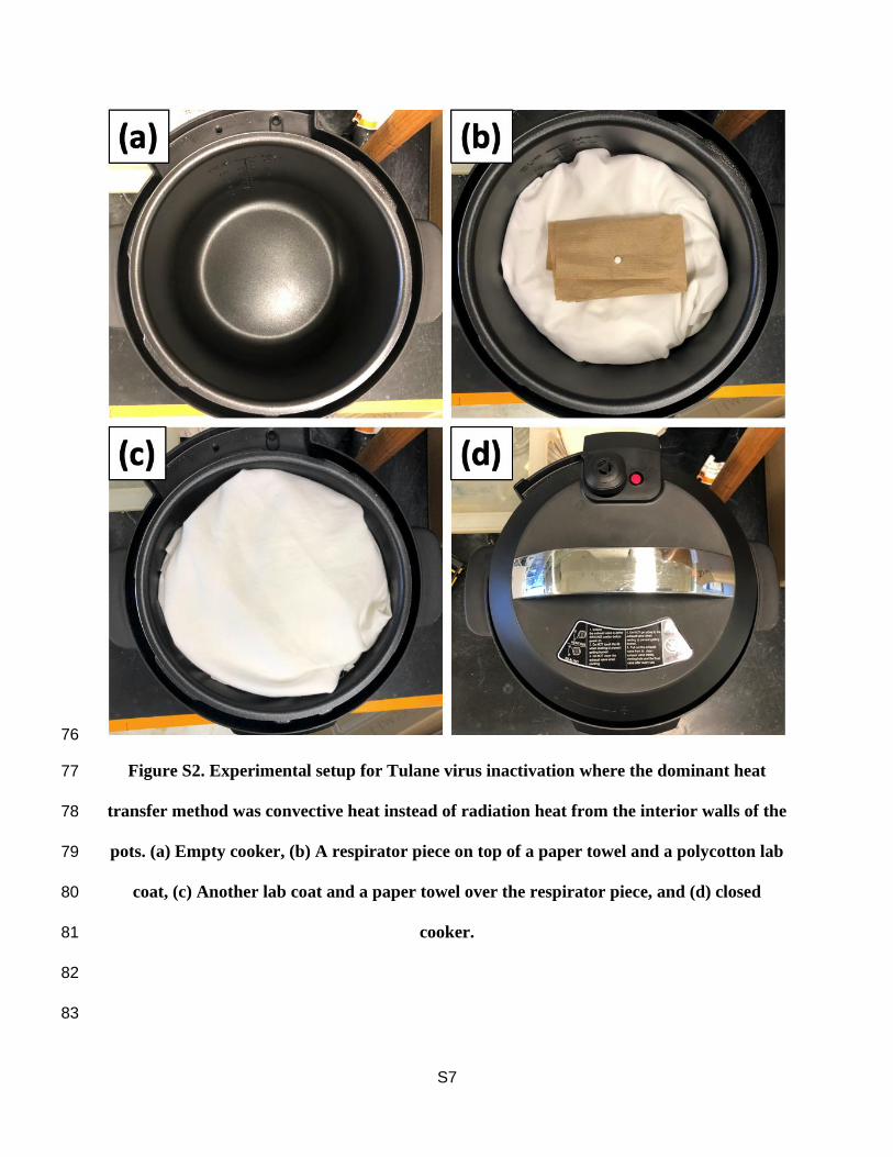
Figure S2. Experimental setup for Tulane virus inactivation where the dominant heat 77
transfer method was convective heat instead of radiation heat from the interior walls of the 78
pots. (a) Empty cooker, (b) A respirator piece on top of a paper towel and a polycotton lab 79
coat, (c) Another lab coat and a paper towel over the respirator piece, and (d) closed 80
cooker. 81
82
83
S8
Text S2. Experimental procedures for decontamination test 84
We followed three different procedures to test (1) the effect of the inoculation site on inactivation 85
efficacy using Tulane virus, (2) the effect of heat transfer method on inactivation efficacy using 86
Tulane virus, and (3) the inactivation efficacy of dry heat over treatment time for each surrogate 87
virus. (1) We inoculated each respirator with five separate 30 µL droplets of the Tulane virus and 88
saliva mixture in five different locations: the inside edge, inside center, the outside edge, outside 89
center, and the strap. The respirator was left in a biosafety cabinet until the testing solution had 90
thoroughly evaporated (about 2 hours). We placed the contaminated respirator in the center of the 91
electric cooker on top of paper towels so that the respirator was 3 cm above the bottom surface of 92
the pot. These paper towels prevented direct contact between the respirator and the pot’s hot 93
surface. The respirator was subject to one 50-min cycle of 100°C dry heat. We then cut the treated 94
respirator into 5 mm diameter pieces and submerged each in 1 mL of fresh culture medium. (2) 95
We cut a clean respirator into 5 mm diameter pieces. We inoculated these pieces with 30 µL 96
droplets of Tulane virus and saliva mixture, left the droplets to evaporate, and then wrapped the 97
inoculated respirator pieces in a paper towel. We lined the interior of the pot with layers of 98
polycotton fabric, placed the paper-towel-wrapped inoculated pieces in the center of the pot, and 99
then covered the pieces with another layer of polycotton. The polycotton lining simulates 100
respirators being stacked or enclosed in a bag so the dominant heat transfer method is convective 101
heat instead of radiation heat from the interior walls of the pots. After the dry heat application, we 102
added each piece to 1 mL of fresh culture medium. (3) For each of the four viruses (Tulane virus, 103
rotavirus, adenovirus, and TGEV), we inoculated 5 mm diameter pieces of a clean respirator with 104
30 µL droplets of the virus and saliva mixture. After being left to evaporate in the biosafety cabinet, 105
the inoculated respirator pieces were placed on paper towels in the electric cooker and subjected 106
S9
to one 50-min cycle of 100°C dry heat. We then submerged each piece in 1 mL of fresh culture 107
medium. 108
We detached the viruses from the respirator fragments by vortexing them in the culture 109
medium for 3 min and shaking them for 30 min at 450 rpm (Figure S3). We followed the same 110
procedure for the negative controls except that they were left in the biosafety cabinet instead of 111
the electric cooker for the same amount of time as the dry heat treatment. The supernatant was 112
used for the plaque assay and the molecular assays to determine the inactivation efficacy and 113
mechanisms, respectively. We calculated the reduction in virus infectivity by dividing the 114
infectivity of the negative control by that of the treated sample (i.e., log10 (N0/N)). We used the 115
three molecular assays with a slight modification to analyze the primary structural target of Tulane 116
virus by the dry heat treatment.2,3 An RNase assay, a binding assay, and a two-step RT-qPCR assay 117
were developed to examine the integrity of capsid proteins, binding proteins, and viral genomes, 118
respectively (Text S3). 119
120
121
S10
122
Figure S3. Calibration curve for virus detachment by 3 min vortex and 30 min shaking at 123
450 rpm. The detachment efficiencies were calculated by dividing the loaded virus from 124
retrieved virus. The detachment efficiencies were not significantly different from inside and 125
outside of the respirator pieces (p>0.05). 126
127
128
129
S11
Text S3. Molecular assays to determine the primary damage of Tulane virus 130
The two-step RT-qPCR assay was designed to quantify intact genomic RNA of Tulane virus. This 131
assay consisted of RT-PCR which synthesized cDNA covering 80% of the genomic RNA and 132
qPCR which quantified the cDNA. We hypothesize that the viruses that had intact genomes in the 133
range of the template for the cDNA will be quantified by this assay. The RNA was extracted from 134
the viruses using QIAmp Viral RNA Mini Kit (Qiagen, Germany) following the manufacturer’s 135
protocol. The cDNA was synthesized using (ProtoScript First Strand cDNA Synthesis Kit, New 136
England BioLabs, USA) by the reverse primer which was designed to cover 5534 bp of the 137
genomic RNA. Finally, the cDNA was quantified by qPCR (PowerUp SYBR™ Green Master 138
Mix, Applied Biosystems, USA). 139
The RNase assay was developed to examine the integrity of capsid proteins. The RNase 140
(A/T1 mix, Thermo Fisher Scientific, USA) was incubated with the viruses at 37°C for 30 min. 141
We assumed that the RNase would be able to penetrate the damaged-capsid and degrade the RNA 142
if the capsid proteins were damaged. RNase inhibitor (SupeRNase inhibitor, Sigma Aldrich, USA) 143
reacted with the RNase treated solution at room temperature for 30 min to inhibit the RNase 144
activity. The remaining intact RNA was quantified by RT-qPCR, which represented the integrity 145
of capsid proteins. 146
The binding assay measures the integrity of binding proteins. Magnetic beads (MagnaBind 147
carboxyl-derivatized beads, Thermo Fisher Scientific, USA) loaded by porcine gastric mucin 148
(Sigma Aldrich, USA) were mixed with the virus solution. The viruses with intact binding proteins 149
were bound to the magnetic beads while the viruses that lost binding ability were washed out. The 150
viruses bound to the magnetic beads were quantified by one-step RT-qPCR (iTaq universal SYBR 151
S12
green reaction mix, Bio-Rad Laboratories, USA) following the manufacturer’s protocol. Detailed 152
information for the two-step RT-qPCR, RNase, binding assay including reagent amount, reaction 153
time, PCR cycles, and primers are described in our previous work.2,3 154
155
156
S13
Text S4. NaCl Particle Filtration Efficiency Test 157
A schematic of the particle filtration testing design used in this study is shown in Figure S4. We 158
built a polypropylene chamber with various fittings and valves to control the aerosol concentration 159
inside the chamber. The chamber inlet valve was connected to an aerosol generator (TSI Constant 160
Output Atomizer Model 3076). The atomizer was filled with 2% NaCl solution (which is 161
commonly used for measuring the penetration efficiency of N95 masks5 in Milli-Q water to 162
generate polydisperse particles (10-800 nm) at a relatively constant rate. The count median 163
diameter of the droplets generated by the atomizer is expected to lie between 80 and 150 nm 164
Generation and Evaluation of Monodisperse Sodium Chloride and Oleic Acid Nanoparticles.6 At 165
the inner roof of the chamber, a small fan was installed to mix the air and thus minimize spatial 166
heterogeneity of the particle concentration inside the chamber. A vent on the roof was also 167
provided to connect it to the compressed air, which was used to dilute the concentration of the 168
particles inside the chamber. The aerosols generated from the atomizer were first dried by passing 169
it through a custom-built diffusion dryer (22 in. long and 3 in. diameter tube with a concentric 170
meshed tube for airflow), filled with 2 mm – 4 mm silica gel. The dry aerosols were then passed 171
through a custom-built aerosol neutralizer (1” diameter and 10” long stainless steel tube with 4 172
Staticmaster® 2U500, 3" Ionizing Cartridges glued inside it7) to neutralize excess charge on the 173
aerosols’ surface. A conductive tubing was passed through the chamber and connected to a particle 174
counter (Condensation Particle Counter, CPC, TSI, Model 3022A; flow rate = 1.5 lpm) to measure 175
the particle concentration. Thus, a steady-state concentration of the aerosols (~45,000 176
particles/cm3) was maintained inside the chamber. A small circular section of the mask was loaded 177
into a 47 mm filter holder (URG, Carrboro, NC, USA) and air was drawn at a specific flow rate, 178
measured by an inline flow meter (4-50 slpm; Dwyer Instruments, MI, USA) using a vacuum line. 179
S14
The surface area of the N95 mask was measured manually (~150 cm2) to calculate face velocity 180
for the NIOSH recommended flow rate (i.e. 85 lpm). The face velocity for this recommended flow 181
rate is 9.4 cm/s. Since, we used only a small section (47 mm diameter) of this mask, we drew only 182
10 lpm through the filter holder, which yielded an equivalent face velocity of 9.4 cm/s. Out of the 183
total flow through the filter, CPC used 1.5 lpm, while the rest was by-passed through a T-184
connector. A pressure gauge (Magnehelic 1-10 inches of water) was also connected in parallel, 185
right downstream of the filter holder using a T-connector to measure the pressure drop. The particle 186
number concentration was measured before and after connecting the filter holder, and particle 187
removal efficiency of the mask was measured by the following equation: 188
189
𝑃𝑎𝑟𝑡𝑖𝑐𝑙𝑒 𝑟𝑒𝑚𝑜𝑣𝑎𝑙 𝐸𝑓𝑓𝑖𝑐𝑖𝑒𝑛𝑐𝑦 (%)190
= (1 −𝑝𝑎𝑟𝑡𝑖𝑐𝑙𝑒 𝑛𝑢𝑚𝑏𝑒𝑟 𝑐𝑜𝑛𝑐𝑒𝑛𝑡𝑟𝑎𝑡𝑖𝑜𝑛 𝑎𝑓𝑡𝑒𝑟 𝑝𝑙𝑎𝑐𝑖𝑛𝑔 𝑡ℎ𝑒 𝑚𝑎𝑠𝑘 (
#𝑐𝑚3)
𝑝𝑎𝑟𝑡𝑖𝑐𝑙𝑒 𝑛𝑢𝑚𝑏𝑒𝑟 𝑐𝑜𝑛𝑐𝑒𝑛𝑡𝑟𝑎𝑡𝑖𝑜𝑛 𝑏𝑒𝑓𝑜𝑟𝑒 𝑝𝑙𝑎𝑐𝑖𝑛𝑔 𝑡ℎ𝑒 𝑚𝑎𝑠𝑘 (#
𝑐𝑚3)) × 100 191
192
Note, the NIOSH testing protocol recommends performing the filtration tests until the respirator 193
reaches a loading of 200 mg NaCl (this takes around 90-100 min);8 however, in our current study 194
we stopped the testing once a constant particle filtration value was obtained (10 - 15 min of total 195
sampling time). We assume this reduced sampling time would not significantly influence our 196
results based on several past studies showing that the filtration efficiencies obtained by measuring 197
initial penetration (average of the first min) of N95 masks were similar to the penetration levels 198
obtained at full loading conditions (i.e. 200 mg).8,9 NISOH recommends N95 masks should not 199
exceed peak air flow resistance of 35 mm (1.37 inches of water). Here, in addition to the particle 200
S15
filtration efficiency we also report the pressure drop across the filter after every cycle of rice cooker 201
decontamination to observe any effect on the inhalation resistance. 202
203
Figure S4. Experimental setup for testing the NaCl particle filtration efficiency of the 204
respirator. 205
206
207
S16
208
Figure S5. Respirator appearance (a) without dry heat treatment and (b) 20 cycles of dry 209
heat treatments. 210
211
212
213
S17
References 214
(1) Tan, M.; Wei, C.; Huang, P.; Fan, Q.; Quigley, C.; Xia, M.; Fang, H.; Zhang, X.; Zhong, 215
W.; Klassen, J. S.; Jiang, X. Tulane Virus Recognizes Sialic Acids as Cellular Receptors. 216
Sci. Rep. 2015, 5, 1–14. https://doi.org/10.1038/srep11784. 217
(2) Fuzawa, M.; Araud, E.; Li, J.; Shisler, J. L.; Nguyen, T. H. Free Chlorine Disinfection 218
Mechanisms of Rotaviruses and Human Norovirus Surrogate Tulane Virus Attached to 219
Fresh Produce Surfaces. Environ. Sci. Technol. 2019, 53 (20), 11999–12006. 220
https://doi.org/10.1021/acs.est.9b03461. 221
(3) Araud, E.; Fuzawa, M.; Shisler, J. L.; Li, J.; Nguyen, T. H. UV Inactivation of Rotavirus 222
and Tulane Virus Targets Different Components of the Virions. Appl. Environ. Microbiol. 223
2020. https://doi.org/10.1128/AEM.02436-19. 224
(4) Oh, C.; Sun, P. P.; Araud, E.; Nguyen, T. H. Mechanism and Efficacy of Viruses 225
Inactivation by a Microplasma UV Lamp Generating Monochromatic UV Irradiation at 226
222 Nm. Under Rev. 2020. 227
(5) NIOSH. Determination of Particulate Filter Efficiency Level for N95 Series Filters 228
Against Solid Particulates for Non-Powered, Air-Purifying Respirators; 2019. 229
https://doi.org/TEB-APR-STP-0059. 230
(6) Chen, T. M.; Chein, H. M. Generation and Evaluation of Monodisperse Sodium Chloride 231
and Oleic Acid Nanoparticles. Aerosol Air Qual. Res. 2006. 232
https://doi.org/10.4209/aaqr.2006.09.0007. 233
S18
(7) Covert, D.; Wiedensohler, A.; Russell, L. Particle Charging and Transmission Efficiencies 234
of Aerosol Charge Neutralizes. Aerosol Sci. Technol. 1997. 235
https://doi.org/10.1080/02786829708965467. 236
(8) Rengasamy, S.; Shaffer, R.; Williams, B.; Smit, S. A Comparison of Facemask and 237
Respirator Filtration Test Methods. J. Occup. Environ. Hyg. 2017. 238
https://doi.org/10.1080/15459624.2016.1225157. 239
(9) Viscusi, D. J.; Bergman, M. S.; Eimer, B. C.; Shaffer, R. E. Evaluation of Five 240
Decontamination Methods for Filtering Facepiece Respirators. Ann. Occup. Hyg. 2009, 53 241
(8), 815–827. https://doi.org/10.1093/annhyg/mep070. 242
243

download fileview on ChemRxivSupporting information.pdf (736.71 KiB)