Drug-loaded electrospun nanofibrous sheets as barriers ... · PDF fileNanomed Res J...
9
Nanomed Res J 2(1):64-72, Winter 2017 RESEARCH ARTICLE Drug-loaded electrospun nanofibrous sheets as barriers against postsurgical adhesions in mice model Safieh Boroumand 1 ; Sara Hosseini 2 ; Mohammad Salehi 2 **; Reza Faridi-Majidi 1 * 1 Department of Medical Nanotechnology, School of Advanced Technology in Medicine, Tehran University of Medical science, Tehran, Iran 2 Department of Biotechnology, School of Advanced Technologies in Medicine, Shahid Behesh University of Medical Sciences, Tehran, Iran Corresponding Author Email: * [email protected] ** [email protected] Objecve(s): Postsurgical adhesion is one of the common complicaons aſter surgery. Some an-adhesion barriers are commercially available which are not customarily used by physicians as much as expected because of ineffecveness. Recently, nanofibers have been introduced as an-adhesion barriers with the potenal of drug delivery. In the light of role of inflammaon and oxidave stress in adhesion formaon, it is supposed that curcumin as an an-oxidant and an- inflammatory agent is able to prevent postsurgical adhesions. Methods: In the present study, curcumin-loaded nanofibrous sheets were prepared using electrospinning and evaluated for its an-adhesion profile in a mice model. Scanning Electron Microscopy (SEM), Aenuated Total Reflectance Fourier Transformed Infrared Spectroscopy (ATR-FTIR), drug release and degradaon invesgaons and also, in vivo studies were performed. Results: curcumin-loaded nanofibers were successfully prepared and shown significant prevenon of postsurgical adhesion formaon in mice model. Release study indicated that aſter 30 days, about 30% of the drug is released from the electrospun nanofibers. In vivo experiments showed that postsurgical adhesion has been reduced about 50% compared to the control group. Conclusions: Curcumin-loaded nanofibers have the potenal to decrease postsurgical adhesion formaon as a barrier. This system supports the sustained release of curcumin from the nanofibers. ARTICLE INFO Arcle History: Received 04 December 2016 Accepted 12 January 2017 Published 20 January 2017 Keywords: Postsurgical adhesion Electrospinning Curcumin ABSTRACT How to cite this article Boroumand S, Hosseini S, Salehi M, Faridi-Majidi R, Drug-loaded electrospun nanofibrous sheets as barriers against postsurgical adhesions in mice model. Nanomed Res J, 2017; 2(1):64-72. DOI: 10.22034/nmrj.2017.23780 INTRODUCTION Postsurgical adhesion is an unavoidable complication that can cause morbidity and mortality to patients aſter medical surgeries. Formation of adhesions leads to various clinical disorders including small bowel obstruction, reoperations and its difficulties, infertility in females and also, chronic pain [1, 2]. In a mechanistic view, an imbalance in fibrin band formation and fibrinolysis processes results in the persistence of the fibrinous mass at the site of surgery. Consequently, fibroblasts foray to that area and secrete extracellular matrix materials such as collagen that results in the formation of adhesions [3, 4]. Surgeons try to decrease adhesion formation with less invasive surgical techniques like Laparoscopy instead of Laparotomy to reduce injury to the parietal peritoneum and serosal surfaces [1]. It was estimated by Ouaïssi et al. that peritoneal adhesion occurs aſter 93-100% of upper abdominal
Transcript of Drug-loaded electrospun nanofibrous sheets as barriers ... · PDF fileNanomed Res J...
Nanomed Res J 2(1):64-72, Winter 2017
RESEARCH ARTICLE
Drug-loaded electrospun nanofibrous sheets as barriers against postsurgical adhesions in mice modelSafieh Boroumand1; Sara Hosseini2; Mohammad Salehi2**; Reza Faridi-Majidi1*
1Department of Medical Nanotechnology, School of Advanced Technology in Medicine, Tehran University of Medical science, Tehran, Iran2Department of Biotechnology, School of Advanced Technologies in Medicine, Shahid Beheshti University of Medical Sciences, Tehran, Iran
Corresponding Author Email: *[email protected] **[email protected]
Objective(s): Postsurgical adhesion is one of the common complications after surgery. Some anti-adhesion barriers are commercially available which are not customarily used by physicians as much as expected because of ineffectiveness. Recently, nanofibers have been introduced as anti-adhesion barriers with the potential of drug delivery. In the light of role of inflammation and oxidative stress in adhesion formation, it is supposed that curcumin as an anti-oxidant and anti-inflammatory agent is able to prevent postsurgical adhesions.
Methods: In the present study, curcumin-loaded nanofibrous sheets were prepared using electrospinning and evaluated for its anti-adhesion profile in a mice model. Scanning Electron Microscopy (SEM), Attenuated Total Reflectance Fourier Transformed Infrared Spectroscopy (ATR-FTIR), drug release and degradation investigations and also, in vivo studies were performed.
Results: curcumin-loaded nanofibers were successfully prepared and shown significant prevention of postsurgical adhesion formation in mice model. Release study indicated that after 30 days, about 30% of the drug is released from the electrospun nanofibers. In vivo experiments showed that postsurgical adhesion has been reduced about 50% compared to the control group.
Conclusions: Curcumin-loaded nanofibers have the potential to decrease postsurgical adhesion formation as a barrier. This system supports the sustained release of curcumin from the nanofibers.
ARTICLE INFO
Article History:Received 04 December 2016 Accepted 12 January 2017Published 20 January 2017
Keywords:Postsurgical adhesionElectrospinning Curcumin
ABSTRAC T
How to cite this articleBoroumand S, Hosseini S, Salehi M, Faridi-Majidi R, Drug-loaded electrospun nanofibrous sheets as barriers against postsurgical adhesions in mice model. Nanomed Res J, 2017; 2(1):64-72. DOI: 10.22034/nmrj.2017.23780
INTRODUCTIONPostsurgical adhesion is an unavoidable
complication that can cause morbidity and mortality to patients after medical surgeries. Formation of adhesions leads to various clinical disorders including small bowel obstruction, reoperations and its difficulties, infertility in females and also, chronic pain [1, 2]. In a mechanistic view, an imbalance in fibrin band formation and fibrinolysis processes results in the persistence of the fibrinous
mass at the site of surgery. Consequently, fibroblasts foray to that area and secrete extracellular matrix materials such as collagen that results in the formation of adhesions [3, 4].Surgeons try to decrease adhesion formation with less invasive surgical techniques like Laparoscopy instead of Laparotomy to reduce injury to the parietal peritoneum and serosal surfaces [1]. It was estimated by Ouaïssi et al. that peritoneal adhesion occurs after 93-100% of upper abdominal

65
S. Boroumand et al./ Drug-loaded electrospun nanofibrous sheets
Nanomed Res J 2(1): 64-72, Winter 2017
laparotomies and after 63-93% of lower abdominal laparotomies. In laparoscopy, the incidence of peritoneal adhesion decreases to 45% [5].Some commercial barriers for prevention of adhesion have been introduced. However, clinical utilization of these products have not been extensive [6]. Some of the barriers used after abdominal surgery include oxidized regenerated cellulose [7], hyaluronate carboxymethylcellulose [8, 9], Icodextrin [10], and polyethylene glycol [11]. Broek et al. have recently reviewed the benefits and harms of adhesion barriers [6]. Lack of awareness and also, underestimating the burden of morbidities of postsurgical adhesions have resulted in the neglect of applying the barriers for adhesion prevention. In spite of severe complications of postsurgical adhesions, there is no suitable medical strategy to prevent adhesion formation [12].In the light of recent advances in nanomedicine, new strategies for managing complicated medical problems like postsurgical adhesion have been developed. Over-ally, anti-adhesion agents proposed for postsurgical adhesion are hydrogels [13-16], gels [2, 17], nanofibers [18-20], nanosheets [21]. Nanofibers can be fabricated by self-assembly, phase separation and electrospinning and each one has its own advantages and disadvantages [22-24]. Among different preparation techniques, electrospinning is vastly considered for fabrication of nanofibers for tissue engineering [9, 25, 26], antibacterial membranes [27] and drug delivery [28] applications. Different kinds of polymers have been used to prepare electrospun nanofibrous sheets for anti-adhesion barriers [3, 18-20, 29-31]. It is expressed in many research that drug-loaded nanofibers are more admissible for an anti-adhesion barrier. For decades, scientists are interested in curcumin (Cur) as an antioxidant, anti-inflammatory and anticancer agent [32-34]. In this regard, Cur has been previously loaded into different kinds of nanofibers, such as Polycaprolactone (PCL), PCL/PEG (Polyethylene glycol), PVA (polyvinyl alcohol), PLA (polylactic acid), various medical applications [35-39]. In the present study, electrospun Cur-loaded PCL (Cur-PCL) sheets were successfully prepared to be utilized as an adhesion barrier in mice model. The obtained electrospun sheets were characterized using SEM and ART-FTIR. Also in vitro
degradation and drug release were investigated. Finally, the resultant fibrous mats were evaluated in mice model.
MATERIALS AND METHODSMaterilas
Cur (purity 99%) was purchased from sigma-aldrich, Germany. PCL (Mw 80,000) was purchased Hangzhou Ruijiang Chemical CO., Ltd, China. Chloroform and methanol were obtained from Merck Co., Germany. All of the materials were used without further purification. Electrospinning process was performed by a machine provided by Fanavaran Nano-Meghyas Co. Ltd, Tehran, Iran.
Electrospining processPCL solutions with the 6.0, 7.0, 8.0 and 10%
(wt.) concentrations were prepared by dissolving the polymer in a mixture of chloroform/methanol (4/1 v/v) as a solvent system and stirring for 2 hours at room temperature. After complete dissolution of the polymers, Cur (6.0 wt% of total polymer weight) was added to the stirring solutions after 2 h. The stirring process continued for 1h. To electrospun the prepared solutions, a 5ml syringe with a blunt-end 18-gauge needle was used. A rotating drum (300 rpm) covered with an aluminum foil was used as a collector. Applied voltage was 22 kV, the distance between the collector and injecting syringe pump was adjusted to 10 cm and pumping rate was set at 0.7 ml/h. All of the processes were performed at about 30 °C.
CharacterizationFor morphological studies, scanning electron
microscopy (SEM, XL 30, Philips, USA) was utilized. The samples were observed after gold sputter-coating. Then, the average diameter of about 50 nanofibers in each image was calculated using ImageJ software.ATR-FTIR spectra were conducted to study the structure of Cur-PCL nanofiber sheets. The scanning range was 600 to 4000 cm -1 and resolution was 2 cm-1 (ATR-FTIR-Tensor 27, Bruker Co, and Germany).
Degradation studyDegradation profile of Cur-PCL nanofibers was
done in PBS solution (pH: 7.4). For this purpose, pieces of Cur-PCL nanofibers were immersed in
66Nanomed Res J 2(1): 64-72, Winter 2017
S. Boroumand et al./ Drug-loaded electrospun nanofibrous sheets
PBS and SEM images were conducted weekly up to 4 weeks.
In vitro Release studyA calibration curve of standard concentrations of
Cur was utilized to calculate the amount of released drug in obtained serial samples. For evaluation of the release profile of fabricated nanofibers, the sheets (45 mg) were immersed in 5 ml of PBS/DMSO mixture (9/1 and pH 7.4) and kept in thermostatic shaking incubator in 37 C at 100 rpm. Sampling was done at intended times for 30 days and 1 ml of release buffer was taken and replaced again with the same fresh solution in order to keep the volume of releasing medium constant. A UV spectrophotometer (Bio Aquarius, Cecil, US) was used to analyze the absorbance of obtained samples at 450 nm.
In vivo studyTwenty female Balb/c mice at the age of 8 –10
weeks and 24-28 g were utilized to evaluate anti-adhesion properties of Cur-PCL nanofibers. All of the mice were kept in a controlled environment for humidity and temperature in the animal lab at Stem Cell Technology Research Center. The animal ethical issue was considered in order to avoid pain and unsuitable conditions. Induction of adhesion formation was conducted with simulating a sterile abdominal surgery process in two groups, treatment and control. To stimulate an adhesion animal model, a vertical incision created in the middle of the abdomen of anesthetized mice (100 mg/kg Ketamine, 10 mg/kg Xylazine, intra peritoneal) and abdominal walls were reflected. Then, peritoneum was injured roughly by a blade in vertical and horizontal lines. The outer surface of internal organs was abraded by rubbing with gauze. Cur-PCL nanofibers were cut into 1.5 ×1.5 cm2 pieces with the 30 µm thickness and were exposed to UV radiation overnight. Thereafter, the samples were fixed with four sutures (at four corners) to the peritoneum of treatment group, and the control groups were left without any treatment. Reoperation was done after 28 days to evaluate and score the probable adhesions in both groups.
Adhesion scoring systemDifferent scoring systems have been previously
used to evaluate various anti-adhesion agents in
studies [31, 40, 41]. In the present study, the scoring system which is introduced by Haney et al. [41] was utilized. Table 1 represents the scoring data. The total score of both groups was calculated and compared.
RESULTS AND DISCUSSION Morphology
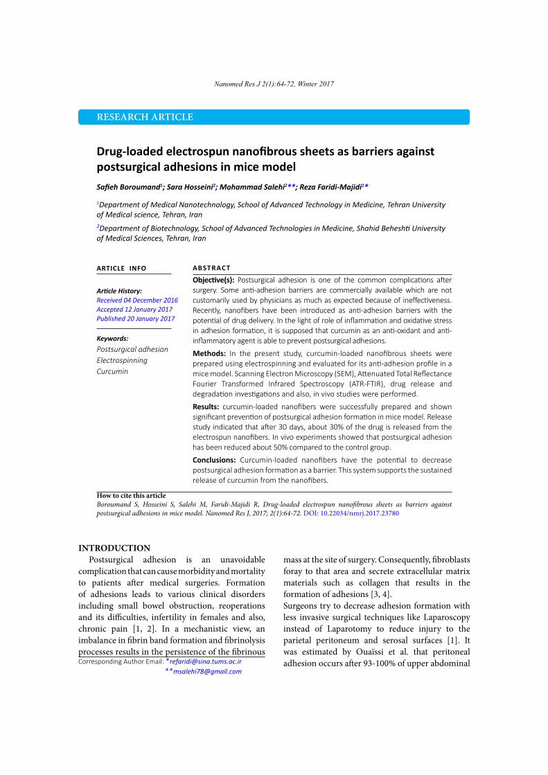
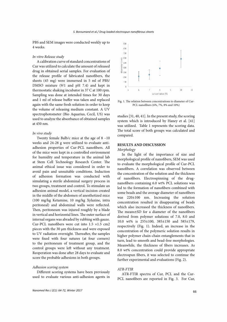
In the light of the importance of size and morphological profile of nanofibers, SEM was used to evaluate the morphological profile of Cur-PCL nanofibers. A correlation was observed between the concentration of the solution and the thickness of nanofibers. Electrospinning of the drug-nanofibers containing 6.0 wt% PCL solutions was led to the formation of nanofibers combined with some beads and the average diameter of nanofibers was 220±100 nm. Increasing the solution concentration resulted in disappearing of beads which also increased the thickness of nanofibers. The means±SD for a diameter of the nanofibers derived from polymer solutions of 7.0, 8.0 and 10.0 wt% is 255±100, 385±150 and 585±179, respectively (Fig. 1). Indeed, an increase in the concentration of the polymeric solution results in higher polymer chain-chain entanglements that in turn, lead to smooth and bead-free morphologies. Meanwhile, the thickness of fibers increases. As 8.0 wt% concentration could provide appropriate electrospun fibers, it was selected to continue the further experimental and evaluations (Fig. 2).
ATR-FTIRATR-FTIR spectra of Cur, PCL and the Cur-
PCL nanofibers are reported in Fig. 3. For Cur,
Fig. 1. The relation between concentrations to diameter of Cur-PCL nanofibers (6%, 7%, 8% and 10%)
67
S. Boroumand et al./ Drug-loaded electrospun nanofibrous sheets
Nanomed Res J 2(1): 64-72, Winter 2017
stretching of the phenolic O-H, stretching vibration of benzene ring, C=C vibration and C-O-C stretching, have bands at 3085–3552 cm−1, 1588 cm−1, 1512 cm−1, and 1143 cm−1. Spectral bands of PCL include asymmetric CH2 stretching which appears in 2938 cm-1, symmetric CH2 stretching at 2865 cm-1 and C=O at 1724 cm-1 [38, 42].In ATR-FTIR for Cur-PCL nanofibers there are little changes in 3085-3552 cm−1 which is related to phenolic O-H stretching, also there are changes about 2865 cm-1 which is for symmetric CH2 stretching. As observed in ATR-FTIR spectra of Cur-PCL nanofibers, there are bands which are citing to the PCL and Cur.
In vitro degradation As increasing attention to nanofibers for medical
purposes, the degradation rate of nanofibers in the body is an important issue. Studies on the rate of degradation have been done by weighing or imaging for defined pieces of nanofibers. In this study, we used SEM for evaluating degradation rate,
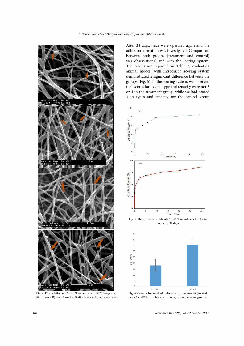
weekly for 4 weeks. According to the SEM images (Fig. 4), little alterations in the morphology and structure of the nanofibers after immersion in PBS (pH 7.4) were observed. The rate of degradation was slow as expected for PCL polymer. However, some disruptions in the integrity of nanofibers are shown by arrows in Fig. 4.
In vitro release studyAs well as an increasing usage of nanofibers as
a drug delivery system in the body, investigating release profile of drug-loaded nanofibers is important. One of the purposes for using nanofibers as drug delivery system in the body is having the drug for an extended time with a sustained release profile, without several prescribed doses of drug. Cur has been considered by researchers for different applications in medicine such as wound healing and anti-inflammatory, in this regard, Cur loaded nanofibers has been designed with different kinds of polymers and resulted in different release profiles.
Fig. 2. Morphology of Cur-PCL nanofibers in deferent concentrations of solution A) 6% B) 7% C) 8 % and D) 10%
68Nanomed Res J 2(1): 64-72, Winter 2017
S. Boroumand et al./ Drug-loaded electrospun nanofibrous sheets
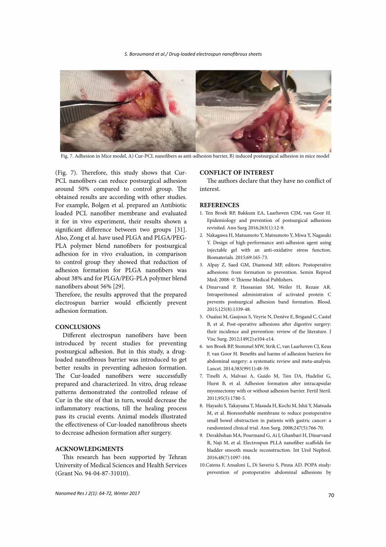
In the study of Sun et al., polyvinyl alcohol (PVA) was used for Cur loading and approximately, 100% of the Cur in Cur-PVA nanofibers was released until 160 min [38]. Thangaraju et al. have used Poly L-Lactide (PLLA) to load Cur, in their study about 81%-86% of Cur was released until 8 days [39]. In another report, Merrell et al. have utilized PCL to deliver Cur for diabetic wound healing and they studied release profile of their Cur-PCL nanofibers for 72 hours (3% and 17% amount of Cur in Cur-PCL nanofibers). Their results demonstrated that about 35% of Cur release occurred until 72 h [36]. Briefly, based on these studies we can conclude that the release profile of Cur from different kinds of polymeric nanofibers depends on the kind of polymers and molecular weight of polymers. Release profile of Cur-PCL nanofibers, during 30 days is reported in fig. 5. A burst release of about 12% of the loaded drug was observed in the first 30 min of the study which was not unexpected. Then, the study was followed by 25% release of Cur at 2 days. The continuous investigation till 30 days after immersion of fibers in PBS demonstrated that about 35% of the loaded drug was released. A burst release of Cur in the site of surgery is an advantage for this drug delivery system as it would be able to control the inflammatory responses at the beginning of the process and cause a decrease in adhesion formation.In this respect, the barrier system that is designed in this system supports sustained release of Cur that is more beneficial to suppress postsurgical adhesions.
In vivo studyPostsurgical adhesion was successfully
simulated in 20 female Balb/c, 8-10 weeks old.
Table 2. Total score of applied scoring system to control and treatment groups
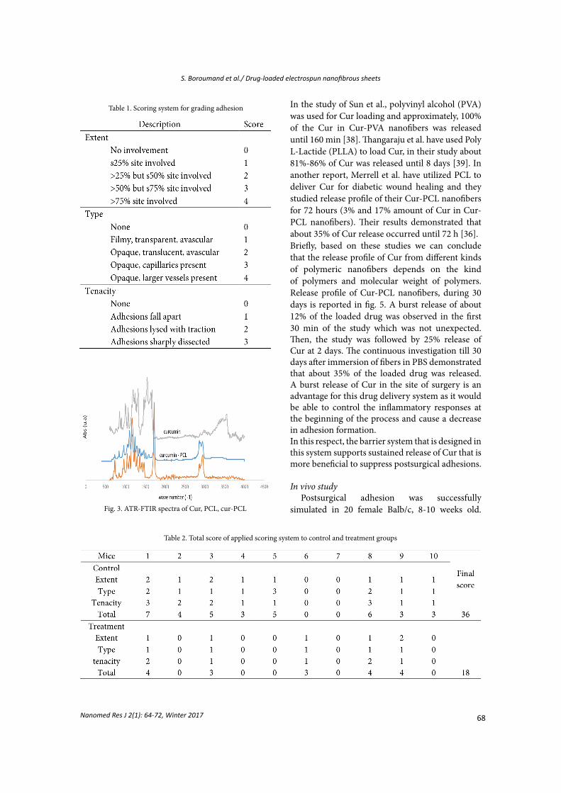
Table 1. Scoring system for grading adhesion
Fig. 3. ATR-FTIR spectra of Cur, PCL, cur-PCL
69
S. Boroumand et al./ Drug-loaded electrospun nanofibrous sheets
Nanomed Res J 2(1): 64-72, Winter 2017
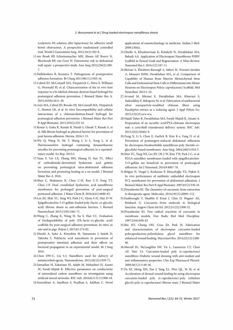
After 28 days, mice were operated again and the adhesion formation was investigated. Comparison between both groups (treatment and control) was observational and with the scoring system. The results are reported in Table 2, evaluating animal models with introduced scoring system demonstrated a significant difference between the groups (Fig. 6). In the scoring system, we observed that scores for extent, type and tenacity were not 3 or 4 in the treatment group, while we had scored 3 in types and tenacity for the control group
Fig. 4. Degradation of Cur-PCL nanofibers in SEM images A) after 1 week B) after 2 weeks C) after 3 weeks D) after 4 weeks.
Fig. 5. Drug release profile of Cur-PCL nanofibers for A) 24 hours, B) 30 days
Fig. 6. Comparing total adhesion score of treatment (treated with Cur-PCL nanofibers after surgery) and control groups
70Nanomed Res J 2(1): 64-72, Winter 2017
S. Boroumand et al./ Drug-loaded electrospun nanofibrous sheets
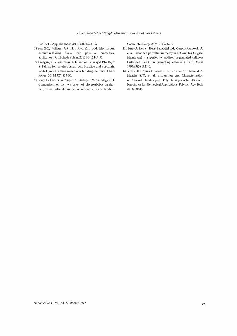
(Fig. 7). Therefore, this study shows that Cur-PCL nanofibers can reduce postsurgical adhesion around 50% compared to control group. The obtained results are according with other studies. For example, Bolgen et al. prepared an Antibiotic loaded PCL nanofiber membrane and evaluated it for in vivo experiment, their results shown a significant difference between two groups [31]. Also, Zong et al. have used PLGA and PLGA/PEG-PLA polymer blend nanofibers for postsurgical adhesion for in vivo evaluation, in comparison to control group they showed that reduction of adhesion formation for PLGA nanofibers was about 38% and for PLGA/PEG-PLA polymer blend nanofibers about 56% [29]. Therefore, the results approved that the prepared electrospun barrier would efficiently prevent adhesion formation.
CONCLUSIONSDifferent electrospun nanofibers have been
introduced by recent studies for preventing postsurgical adhesion. But in this study, a drug-loaded nanofibrous barrier was introduced to get better results in preventing adhesion formation. The Cur-loaded nanofibers were successfully prepared and characterized. In vitro, drug release patterns demonstrated the controlled release of Cur in the site of that in turn, would decrease the inflammatory reactions, till the healing process pass its crucial events. Animal models illustrated the effectiveness of Cur-loaded nanofibrous sheets to decrease adhesion formation after surgery.
ACKNOWLEDGMENTS
This research has been supported by Tehran University of Medical Sciences and Health Services (Grant No. 94-04-87-31010).
Fig. 7. Adhesion in Mice model, A) Cur-PCL nanofibers as anti-adhesion barrier, B) induced postsurgical adhesion in mice model
CONFLICT OF INTERESTThe authors declare that they have no conflict of
interest.
REFERENCES1. Ten Broek RP, Bakkum EA, Laarhoven CJM, van Goor H.
Epidemiology and prevention of postsurgical adhesions revisited. Ann Surg 2016;263(1):12-9.
2. Nakagawa H, Matsumoto Y, Matsumoto Y, Miwa Y, Nagasaki Y. Design of high-performance anti-adhesion agent using injectable gel with an anti-oxidative stress function. Biomaterials. 2015;69:165-73.
3. Alpay Z, Saed GM, Diamond MP, editors. Postoperative adhesions: from formation to prevention. Semin Reprod Med; 2008: © Thieme Medical Publishers.
4. Dinarvand P, Hassanian SM, Weiler H, Rezaie AR. Intraperitoneal administration of activated protein C prevents postsurgical adhesion band formation. Blood. 2015;125(8):1339-48.
5. Ouaïssi M, Gaujoux S, Veyrie N, Denève E, Brigand C, Castel B, et al. Post-operative adhesions after digestive surgery: their incidence and prevention: review of the literature. J Visc Surg. 2012;149(2):e104-e14.
6. ten Broek RP, Stommel MW, Strik C, van Laarhoven CJ, Keus F, van Goor H. Benefits and harms of adhesion barriers for abdominal surgery: a systematic review and meta-analysis. Lancet. 2014;383(9911):48-59.
7. Tinelli A, Malvasi A, Guido M, Tsin DA, Hudelist G, Hurst B, et al. Adhesion formation after intracapsular myomectomy with or without adhesion barrier. Fertil Steril. 2011;95(5):1780-5.
8. Hayashi S, Takayama T, Masuda H, Kochi M, Ishii Y, Matsuda M, et al. Bioresorbable membrane to reduce postoperative small bowel obstruction in patients with gastric cancer: a randomized clinical trial. Ann Surg. 2008;247(5):766-70.
9. Derakhshan MA, Pourmand G, Ai J, Ghanbari H, Dinarvand R, Naji M, et al. Electrospun PLLA nanofiber scaffolds for bladder smooth muscle reconstruction. Int Urol Nephrol. 2016;48(7):1097-104.
10.Catena F, Ansaloni L, Di Saverio S, Pinna AD. POPA study: prevention of postoperative abdominal adhesions by
71
S. Boroumand et al./ Drug-loaded electrospun nanofibrous sheets
Nanomed Res J 2(1): 64-72, Winter 2017
icodextrin 4% solution after laparotomy for adhesive small bowel obstruction. A prospective randomized controlled trial. World J Gastrointest Surg. 2012;16(2):382-8.
11.ten Broek RP, Schreinemacher MH, Jilesen AP, Bouvy N, Bleichrodt RP, van Goor H. Enterotomy risk in abdominal wall repair: a prospective study. Ann Surg 2012;256(2):280-7.
12.Hellebrekers B, Kooistra T. Pathogenesis of postoperative adhesion formation. Br J Surg 2011;98(11):1503-16.
13.Cabral JD, McConnell MA, Fitzpatrick C, Mros S, Williams G, Wormald PJ, et al. Characterization of the in vivo host response to a bi‐labeled chitosan‐dextran based hydrogel for postsurgical adhesion prevention. J Biomed Mater Res A. 2015;103(8):2611-20.
14.Aziz MA, Cabral JD, Brooks HJ, McConnell MA, Fitzpatrick C, Hanton LR, et al. In vitro biocompatibility and cellular interactions of a chitosan/dextran‐based hydrogel for postsurgical adhesion prevention. J Biomed Mater Res Part B Appl Biomater. 2015;103(2):332-41.
15.Konar S, Guha R, Kundu B, Nandi S, Ghosh T, Kundu S, et al. Silk fibroin hydrogel as physical barrier for prevention of post hernia adhesion. Hernia. 2016:1-13.
16.Wu Q, Wang N, He T, Shang J, Li L, Song L, et al. Thermosensitive hydrogel containing dexamethasone micelles for preventing postsurgical adhesion in a repeated-injury model. Sci Rep. 2015;5.
17.Yuan F, Lin LX, Zhang HH, Huang D, Sun YL. Effect of carbodiimide‐derivatized hyaluronic acid gelatin on preventing postsurgical intra‐abdominal adhesion formation and promoting healing in a rat model. J Biomed Mater Res A. 2016.
18.Sheu C, Shalumon K, Chen C-H, Kuo C-Y, Fong YT, Chen J-P. Dual crosslinked hyaluronic acid nanofibrous membranes for prolonged prevention of post-surgical peritoneal adhesion. J Mater Chem B. 2016;4(41):6680-93.
19.Lee JH, Shin YC, Yang WJ, Park J-C, Hyon S-H, Han D-W. Epigallocatechin-3-O-gallate-loaded poly (lactic-co-glycolic acid) fibrous sheets as anti-adhesion barriers. J Biomed Nanotechnol. 2015;11(8):1461-71.
20.Wang C, Zhang K, Wang H, Xu S, Han CC. Evaluation of biodegradability of poly (DL-lactic-co-glycolic acid) scaffolds for post-surgical adhesion prevention: In vitro, in rats and in pigs. Polym J. 2015;61:174-82.
21.Hinoki A, Saito A, Kinoshita M, Yamamoto J, Saitoh D, Takeoka S. Polylactic acid nanosheets in prevention of postoperative intestinal adhesion and their effects on bacterial propagation in an experimental model. Br J Surg 2016.
22.Chen DW-C, Liu S-J. Nanofibers used for delivery of antimicrobial agents. Nanomedicine. 2015;10(12):1959-71.
23.Samadian H, Zakariaee SS, Adabi M, Mobasheri H, Azami M, Faridi-Majidi R. Effective parameters on conductivity of mineralized carbon nanofibers: an investigation using artificial neural networks. RSC Adv. 2016;6(113):111908-18.
24.Surendiran A, Sandhiya S, Pradhan S, Adithan C. Novel
applications of nanotechnology in medicine. Indian J Med. 2009;130(6).
25.Gheibi A, Khoshnevisan K, Ketabchi N, Derakhshan MA, Babadi AA. Application of Electrospun Nanofibrous PHBV Scaffold in Neural Graft and Regeneration: A Mini-Review. Nanomed Res J. 2016;1(2):107-11.
26.Shirian S, Ebrahimi-Barough S, Saberi H, Norouzi-Javidan A, Mousavi SMM, Derakhshan MA, et al. Comparison of Capability of Human Bone Marrow Mesenchymal Stem Cells and Endometrial Stem Cells to Differentiate into Motor Neurons on Electrospun Poly(ε-caprolactone) Scaffold. Mol Neurobiol. 2015:1-10.
27.Arvand M, Mirzaei E, Derakhshan MA, Kharrazi S, Sadroddiny E, Babapour M, et al. Fabrication of antibacterial silver nanoparticle-modified chitosan fibers using Eucalyptus extract as a reducing agent. J Appl Polym Sci. 2015;132(25):n/a-n/a.
28.Najafi-Taher R, Derakhshan MA, Faridi-Majidi R, Amani A. Preparation of an ascorbic acid/PVA-chitosan electrospun mat: a core/shell transdermal delivery system. RSC Adv. 2015;5(62):50462-9.
29.Zong X, Li S, Chen E, Garlick B, Kim K-s, Fang D, et al. Prevention of postsurgery-induced abdominal adhesions by electrospun bioabsorbable nanofibrous poly (lactide-co-glycolide)-based membranes. Ann Surg. 2004;240(5):910-5.
30.Shin YC, Yang WJ, Lee JH, Oh J-W, Kim TW, Park J-C, et al. PLGA nanofiber membranes loaded with epigallocatechin-3-O-gallate are beneficial to prevention of postsurgical adhesions. Int J Nanomed. 2014;9:4067-78.
31.Bölgen N, Vargel I, Korkusuz P, Menceloğlu YZ, Pişkin E. In vivo performance of antibiotic embedded electrospun PCL membranes for prevention of abdominal adhesions. J Biomed Mater Res Part B Appl Biomater. 2007;81(2):530-43.
32.Priyadarsini KI. The chemistry of curcumin: from extraction to therapeutic agent. Molecules. 2014;19(12):20091-112.
33.Esatbeyoglu T, Huebbe P, Ernst I, Chin D, Wagner AE, Rimbach G. Curcumin—from molecule to biological function. Angew Chem Int Ed. 2012;51(22):5308-32.
34.Priyadarsini KI. Free radical reactions of curcumin in membrane models. Free Radic Biol Med Discipline. 1997;23(6):838-43.
35.Bui HT, Chung OH, Cruz JD, Park JS. Fabrication and characterization of electrospun curcumin-loaded polycaprolactone-polyethylene glycol nanofibers for enhanced wound healing. Macromol Res. 2014;22(12):1288-96.
36.Merrell JG, McLaughlin SW, Tie L, Laurencin CT, Chen AF, Nair LS. Curcumin‐loaded poly (ε‐caprolactone) nanofibres: Diabetic wound dressing with anti‐oxidant and anti‐inflammatory properties. Clin Exp Pharmacol Physiol. 2009;36(12):1149-56.
37.Fu SZ, Meng XH, Fan J, Yang LL, Wen QL, Ye SJ, et al. Acceleration of dermal wound healing by using electrospun curcumin‐loaded poly (ε‐caprolactone)‐poly (ethylene glycol)‐poly (ε‐caprolactone) fibrous mats. J Biomed Mater
72Nanomed Res J 2(1): 64-72, Winter 2017
S. Boroumand et al./ Drug-loaded electrospun nanofibrous sheets
Res Part B Appl Biomater 2014;102(3):533-42.38.Sun X-Z, Williams GR, Hou X-X, Zhu L-M. Electrospun
curcumin-loaded fibers with potential biomedical applications. Carbohydr Polym. 2013;94(1):147-53.
39.Thangaraju E, Srinivasan NT, Kumar R, Sehgal PK, Rajiv S. Fabrication of electrospun poly l-lactide and curcumin loaded poly l-lactide nanofibers for drug delivery. Fibers Polym. 2012;13(7):823-30.
40.Ersoy E, Ozturk V, Yazgan A, Ozdogan M, Gundogdu H. Comparison of the two types of bioresorbable barriers to prevent intra-abdominal adhesions in rats. World J
Gastrointest Surg. 2009;13(2):282-6.41.Haney A, Hesla J, Hurst BS, Kettel LM, Murphy AA, Rock JA,
et al. Expanded polytetrafluoroethylene (Gore-Tex Surgical Membrane) is superior to oxidized regenerated cellulose (Interceed TC7+) in preventing adhesions. Fertil Steril. 1995;63(5):1021-6.
42.Pereira IH, Ayres E, Averous L, Schlatter G, Hebraud A, Mendes STO, et al. Elaboration and Characterization of Coaxial Electrospun Poly (ε‐Caprolactone)/Gelatin Nanofibers for Biomedical Applications. Polymer Adv Tech. 2014;33(S1).