
Draft protocol to guide the assessment of [name of …File/1098.1-FINAL-DAP.docx · Web viewd for...
43
1098.1: Final Decision Analytic Protocol (DAP) to guide the assessment of: (a) the review of interim funded items 63464, 63467 - Breast Magnetic Resolution Imaging (MRI) for screening of high risk women (b) inclusion of additional high risk patient populations in MBS item 63464 May 2012
Transcript of Draft protocol to guide the assessment of [name of …File/1098.1-FINAL-DAP.docx · Web viewd for...
1098.1:
Final DecisionAnalytic Protocol (DAP) to guide the assessment of:
(a) the review ofinterim funded items63464, 63467 - Breast MagneticResolution Imaging(MRI) for screening of high risk women(b) inclusion of additional high risk patient populations in MBS item 63464
May 2012
Table of Contents1098.1:............................................................................................................................................ 1
Final Decision.................................................................................................................................. 1
Analytic Protocol (DAP)................................................................................................................... 1
to guide the assessment of:............................................................................................................1
MSAC and PASC...............................................................................................................................3
Purpose of this document............................................................................................................3
Purpose of application.....................................................................................................................4
Intervention.....................................................................................................................................4
Description...................................................................................................................................4
Administration, dose, frequency of administration, duration of treatment................................5
Co-administered interventions....................................................................................................5
Background..................................................................................................................................... 6
Current arrangements for public reimbursement.......................................................................6
Regulatory status.........................................................................................................................8
Patient population...........................................................................................................................8
Proposed MBS listing...................................................................................................................8
Clinical place for proposed intervention....................................................................................10
Comparator................................................................................................................................... 12
Mammography outside the BreastScreen Australia program....................................................12
Ultrasound................................................................................................................................. 12
Clinical claim..................................................................................................................................13
Outcomes and health care resources affected by introduction of proposed intervention.........16
Outcomes...................................................................................................................................... 16
Health care resources....................................................................................................................17
Proposed structure of economic evaluation (decision-analytic)...................................................19
Reference List................................................................................................................................25
Page 2 of 39
MSAC and PASC
The Medical Services Advisory Committee (MSAC) is an independent expert committee appointed by the Minister for Health and Ageing (the Minister) to strengthen the role of evidence in health financing decisions in Australia. MSAC advises the Minister on the evidence relating to the safety, effectiveness, and cost-effectiveness of new and existing medical technologies and procedures and under what circumstances public funding should be supported.
The Protocol Advisory Sub-Committee (PASC) is a standing sub-committee of MSAC. Its primary objective is the determination of protocols to guide clinical and economic assessments of medical interventions proposed for public funding.
Purpose of this document
This document is intended to provide a draft decision analytic protocol that will be used to guide the assessment of an intervention for a particular population of patients. The draft protocol that will be finalised after inviting relevant stakeholders to provide input to the protocol. The final protocol will provide the basis for the assessment of the intervention.
The protocol guiding the assessment of the health intervention has been developed using the widely accepted “PICO” approach. The PICO approach involves a clear articulation of the following aspects of the research question that the assessment is intended to answer:
Patients – specification of the characteristics of the patients in whom the intervention is to be considered for use;Intervention – specification of the proposed interventionComparator – specification of the therapy most likely to be replaced by the proposed interventionOutcomes – specification of the health outcomes and the healthcare resources likely to beaffected by the introduction of the proposed intervention
Page 3 of 39
Purpose of application
An application requesting the review of the Medicare Benefits Schedule (MBS) listing of breast magnetic resonance imaging (MRI) for screening of young women at high risk of breast cancer was received from The Royal Australian and New Zealand College of Radiologists by the Department of Health and Ageing in May 2011. The application is requesting a review of:
• interim funded items 63464 and 63467 - breast magnetic resolution imaging (MRI) for screening of high risk women in terms of effectiveness and cost-effectiveness; and
• the inclusion of additional new high risk patient populations in MBS item 63464.
Intervention
Description
Magnetic resonance imaging (MRI) uses a strong external magnetic field to produce images of biological tissues. This magnetic field acts on hydrogen protons (elementary particles) in body tissues and a radiofrequency pulse to produce signals that vary according to their local chemical, structural and magnetic environment. MRI is particularly well suited to distinguishing between blood vessels, other fluid filled structures and surrounding soft tissues, and as such is especially useful in imaging the brain, muscles and the heart as well as detecting abnormal tissues such as tumours.
Breast MRI is performed in a dedicated MRI room using an MRI machine with a minimum magnet strength of 1.5 Teslar. A dedicated breast coil, compromising of 7 or more channels is also required and intravenous contrast is administered by powered or electronic injector. As breast tissue generally has similar signal intensity to tumour tissue on routine MRI, the intravenous administration of a contrast agent containing gadolinium chelate is used to enhance breast lesions.
During the examination the patient lies prone on the MRI table with the breast dependant in the dedicated breast coil. A number of imaging sequences are obtained, prior to the administration of the contrast agent gadolinium. Following contrast injection further, sequences are obtained including evaluation of the uptake and washout of contrast by breast tissue and any focal lesion over several minutes.
Page 4 of 39
The MRI sequences obtained are interpreted by a radiologist to analyse the findings on the various sequences, including enhancement patterns. The aim is to distinguish between normal, benign and malignant findings. Malignant lesions usually display an enhancement pattern with rapid uptake and
Page 5 of 39

washout of contrast. In benign masses the contrast uptake is usually slower and more prolonged. Some lesions have atypical or indeterminate findings.
MRI can be used in both screening and diagnosis of breast cancer. This includes the identification of breast cancer in women with a high risk of breast cancer due to family history or genetic predisposition. Breast MRI is also used in preoperative staging, evaluating response to treatment, screening of women with breast augmentation or reconstruction and identification of occult breast cancer in women with metastatic disease. This assessment is concerned with the first group, the screening of breast cancer in asymptomatic women at high risk of breast cancer.
Administration, dose, frequency of administration, duration of treatment
BreastScreen Australia is the national population-based screening program which is targeted to asymptomatic women at average risk of breast cancer. It provides free screening mammograms at two-yearly intervals for women aged 50-59, although women aged 40-49 and 70 years and older are also eligible for screening.
Women at high risk of breast cancer due to family history of genetic predisposition make up 5–10% of breast cancers (National Breast Ovarian Cancer Centre now Cancer Australia [NBOCC 2009]). These women often develop breast cancer early in life, need more frequent screening and earlier commencement of screening than asymptomatic women at average risk of breast cancer. This occurs outside of the BreastScreen Australia program and can include MRI and/or mammography, with or without the use of ultrasound. Currently women less than 50 years of age and assessed at high risk are offered annual screening with most commencing no earlier than 25 years of age. However this can vary dependent on the age of cancers in the family and potential nature of gene mutation.
To perform breast MRI, a radiographer is required with specialised training for setup and scanning. The supervising radiologist should have expertise in breast imaging and MRI interpretation. In addition, for an MRI scan to attract a Medicare rebate, the patient must be 50 years of age or less and fulfil the specified risk criteria (see Table 2). The scan must be requested by a specialist or consultant physician (not a GP) and be performed on a Medicare-eligible MRI unit by a Medicare- eligible provider, and be an MRI service listed in the MBS.
Co-administered interventions
Women would first have a medical consultation including a clinical breast examination (CBE) (MBS items 3, 23, 36 and 44) and then be referred for a mammogram or a specialist appointment. Breast MRI is currently used in addition to mammography with or
Page 6 of 39
without the use of ultrasound (MBS items59300-59318), and as such, MRI and mammography may be given on the same day or within a week
Page 7 of 39
or so of one another. Factors such as menstrual cycles, availability of staff and equipment may have an impact on the length of time between tests but should not result in only one test being given to eligible women as this may lead to women having a biopsy with less information than would have been supplied by having both tests.
To attract a rebate for a breast MRI, women will need to have a referral from a specialist medical practitioner or consultant physician (MBS items 104 and 110). Appendix 1 outlines the MBS items associated with breast MRI.
Background
Current arrangements for public reimbursement
In 2007, MSAC recommended interim public funding for breast MRI in the diagnosis of breast cancer in asymptomatic women with a high risk of developing breast cancer when used as part of an organised surveillance program. In February 2009, the Government acted on MSAC advice and listed breast MRI on the MBS – item numbers 63464 and 63467 (see Table 1).
To be eligible for the rebate, the patient must be a woman who is less than 50 years of age, with no current signs or symptoms of breast cancer and who has been identified as at high risk of breastcancer due to one of the following:
• three or more first or second degree relatives on the same side of the family diagnosed with breast or ovarian cancer;
• two or more first or second degree relatives on the same side of the family diagnosed with breast or ovarian cancer, if any of the following applies to at least one of the relatives
- has been diagnosed with bilateral breast cancer;- had onset of breast cancer before the age of 40 years;- had onset of ovarian cancer before the age of 50 years;- has been diagnosed with breast and ovarian cancer, at the same time or
at different times;- has Ashkenazi Jewish ancestry;- is a male relative who has been diagnosed with breast cancer;
• one first or second degree relative diagnosed with breast cancer at age 45 years or younger, plus another first or second degree relative on the same side of the family with bone or soft tissue sarcoma at age 45 years or younger; or
• that genetic testing has identified the presence of a high risk breast cancer gene mutation.
In addition, for an MRI scan to attract a Medicare rebate, the scan must be requested by a specialist or consultant physician and be performed on a Medicare-eligible MRI unit by a Medicare-eligible provider, and be an MRI service listed in the MBS. Unlicensed sites that are ineligible for MBS funding may also provide breast MRI however the patient will
Page 8 of 39
need to pay for the scan themselves (around$700).
Page 9 of 39
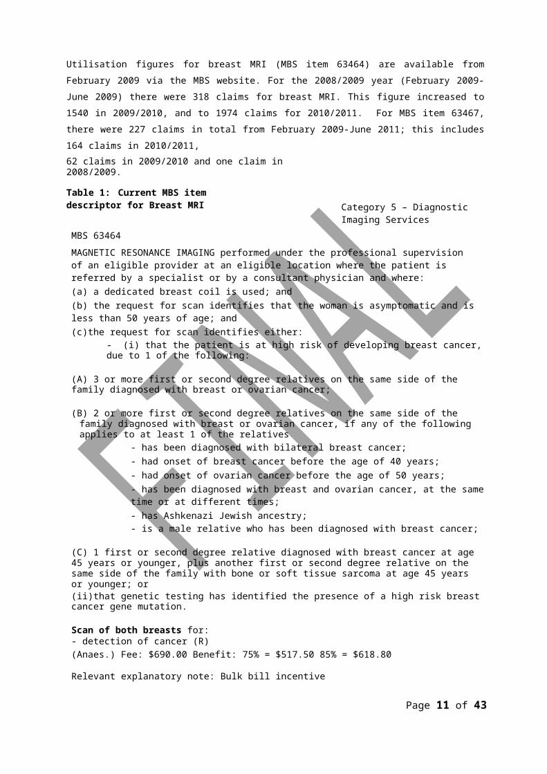
Utilisation figures for breast MRI (MBS item 63464) are available from February 2009 via the MBS website. For the 2008/2009 year (February 2009-June 2009) there were 318 claims for breast MRI. This figure increased to 1540 in 2009/2010, and to 1974 claims for 2010/2011. For MBS item 63467, there were 227 claims in total from February 2009-June 2011; this includes 164 claims in 2010/2011,62 claims in 2009/2010 and one claim in 2008/2009.Table 1: Current MBS item descriptor for Breast MRI
MBS 63464
Category 5 – Diagnostic Imaging Services
MAGNETIC RESONANCE IMAGING performed under the professional supervision of an eligible provider at an eligible location where the patient is referred by a specialist or by a consultant physician and where:(a) a dedicated breast coil is used; and(b) the request for scan identifies that the woman is asymptomatic and is less than 50 years of age; and(c)the request for scan identifies either:
- (i) that the patient is at high risk of developing breast cancer, due to 1 of the following:
(A) 3 or more first or second degree relatives on the same side of the family diagnosed with breast or ovarian cancer;
(B) 2 or more first or second degree relatives on the same side of the family diagnosed with breast or ovarian cancer, if any of the following applies to at least 1 of the relatives
- has been diagnosed with bilateral breast cancer;- had onset of breast cancer before the age of 40 years;- had onset of ovarian cancer before the age of 50 years;- has been diagnosed with breast and ovarian cancer, at the same time or at different times;- has Ashkenazi Jewish ancestry;- is a male relative who has been diagnosed with breast cancer;
(C) 1 first or second degree relative diagnosed with breast cancer at age 45 years or younger, plus another first or second degree relative on the same side of the family with bone or soft tissue sarcoma at age 45 years or younger; or(ii)that genetic testing has identified the presence of a high risk breast cancer gene mutation.
Scan of both breasts for:- detection of cancer (R)(Anaes.) Fee: $690.00 Benefit: 75% = $517.50 85% = $618.80
Relevant explanatory note: Bulk bill incentive
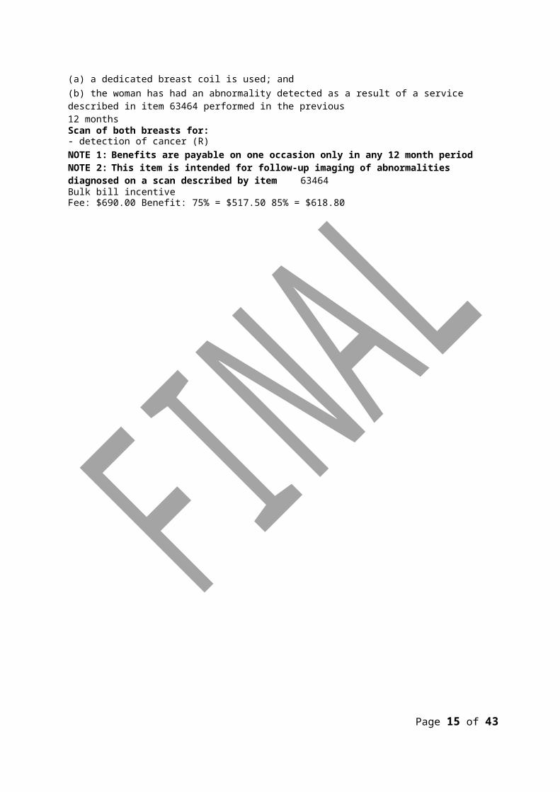
MBS 63467MAGNETIC RESONANCE IMAGING performed under the professional supervision of an eligible provider at an eligible location where the patient is referred by a specialist or by a consultant physician and where:(a) a dedicated breast coil is used; and(b) the woman has had an abnormality detected as a result of a service described in item 63464 performed in the previous12 months
Scan of both breasts for:- detection of cancer (R)NOTE 1: Benefits are payable on one occasion only in any 12 month periodNOTE 2: This item is intended for follow-up imaging of abnormalities diagnosed on a scan described by item 63464Bulk bill incentive(Anaes.)Fee: $690.00 Benefit: 75% = $517.50 85% = $618.80
Page 10 of 39
Regulatory status
MRI is currently available in public and private facilities in major centres in each state and territory. One hundred and thirty sites have been licensed by the Department of Health and Ageing to provide services that are eligible for funding under the MBS.
Breast MRI requires both a breast coil and the use of a gadolinium-containing contrast agent. The Australian Register of Therapeutic Goods lists several coils and gadolinium-containing contrast agents that have been approved by the Therapeutic Goods Administration for use in diagnostic imaging procedures.
Patient population
Proposed MBS listing
As previously mentioned, breast MRI for screening of high risk women is already listed on the MBS. This assessment addresses the following issues:
• a review of interim funded items 63464 and 63467 - breast MRI for screening of high risk women in terms of effectiveness and cost effectiveness; and
• the inclusion of new high risk patient population(s) in MBS item 63464 namely women:
- with a prior history of invasive breast cancer;- with a prior history of treatment for lobular carcinoma in situ (LCIS)
or ductal carcinoma in situ (DCIS); and- who have a history of radiotherapy to the chest area undertaken between
the ages of 10-35 years.
Page 11 of 39
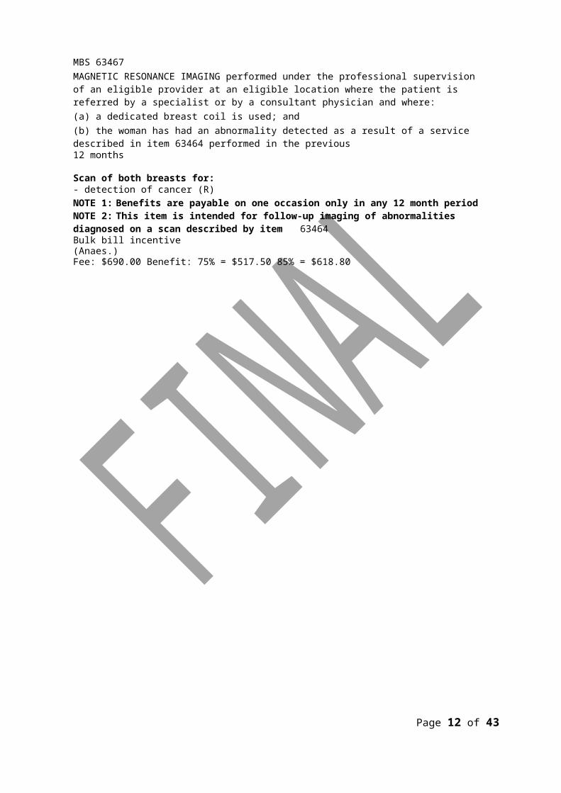
Table 2: Proposed MBS item descriptor for Breast Magnetic resonance ImagingCategory 5 – Diagnostic Imaging Services
MBS : 63464MAGNETIC RESONANCE IMAGING performed under the professional supervision of an eligible provider at an eligible location where the patient is referred by a specialist or by a consultant physician and where:(a) a dedicated breast coil is used; and(b) the request for scan identifies that the woman is asymptomatic and is less than 50 years of age; and(c)the request for scan identifies either:(i) that the patient is at high risk of developing breast cancer, due to 1 of the following:
(A) 3 or more first or second degree relatives on the same side of the family diagnosed with breast or ovarian cancer;
(B) 2 or more first or second degree relatives on the same side of the family diagnosed with breast or ovarian cancer, if any of the following applies to at least 1 of the relatives
- has been diagnosed with bilateral breast cancer;- had onset of breast cancer before the age of 40 years;- had onset of ovarian cancer before the age of 50 years;- has been diagnosed with breast and ovarian cancer, at the same time or at different times;- has Ashkenazi Jewish ancestry;- is a male relative who has been diagnosed with breast cancer;
(C) 1 first or second degree relative diagnosed with breast cancer at age 45 years or younger, plus another first or second degree relative on the same side of the family with bone or soft tissue sarcoma at age 45 years or younger; or(ii)that genetic testing has identified the presence of a high risk breast cancer gene mutation.
(D) prior history of treatment for invasive breast cancer
(E) prior history of treatment for Ductal Carcinoma In Situ (DCIS) or Lobular Carcinoma In Situ (LCIS)
(F) with a history of radiotherapy to the chest area undertaken between the ages of 10-35 years
Scan of both breasts for:- detection of cancer (R)Fee: $690.00 Benefit: 75% = $517.50 85% = $618.80Relevant explanatory note: Bulk bill incentive
MBS 63467MAGNETIC RESONANCE IMAGING performed under the professional supervision of an eligible provider at an eligible location where the patient is referred by a specialist or by a consultant physician and where:(a) a dedicated breast coil is used; and(b) the woman has had an abnormality detected as a result of a service described in item 63464 performed in the previous12 monthsScan of both breasts for:- detection of cancer (R)NOTE 1: Benefits are payable on one occasion only in any 12 month periodNOTE 2: This item is intended for follow-up imaging of abnormalities diagnosed on a scan described by item 63464Bulk bill incentiveFee: $690.00 Benefit: 75% = $517.50 85% = $618.80
Page 12 of 39

Clinical place for proposed intervention
Following a 2006 MSAC assessment, breast MRI was conditionally recommended for use as an additional test in the diagnosis of breast cancer in asymptomatic women with a high risk of developing breast cancer when used as part of organised surveillance. Since February 2009, breast MRI has been available on the MBS as an interim funded item.
The 2006 assessment also assessed breast MRI as a replacement to CBE, mammography ± ultrasound, however this was not recommended by MSAC at the time and is not being considered here.
What is being assessed is:
• use of breast MRI for the screening of high risk women in addition to an organised surveillance program, as recommended by MSAC in 2007 (MSAC 2006). As such no change is being proposed to the use of breast MRI in the clinical pathway from what was recommended in this review;and
• the inclusion of additional high risk patient populations in MBS item 63464. The change in the proposed patient population will see additional patients screened who would not previously have had access to this item.
This inclusion of the additional high risk patient populations are consistent with those risk factors that have been identified by the NBOCC 2009 associated with a strong Relative Risk (RR >4) increased risk of breast cancer.
Page 13 of 39
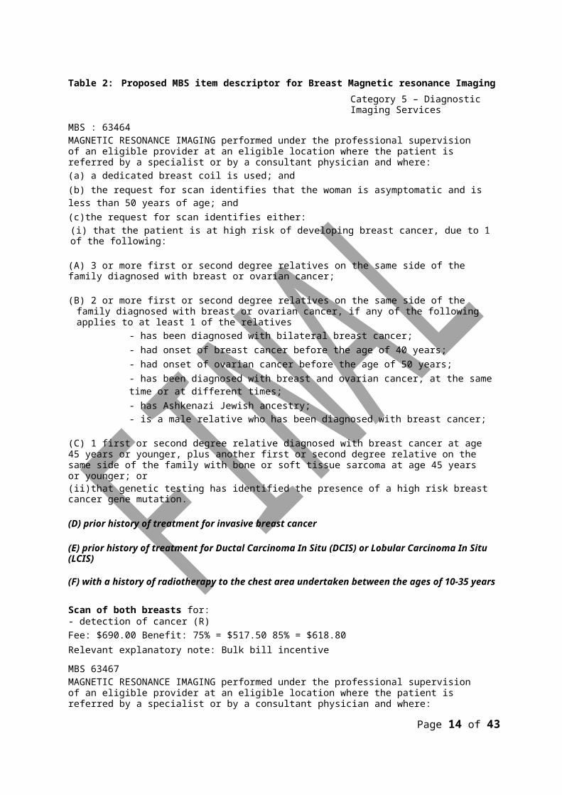
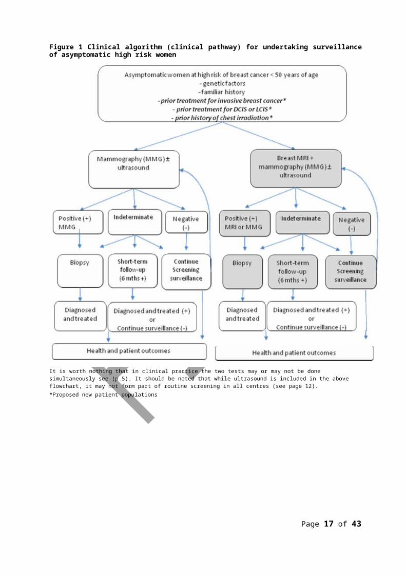
Figure 1 Clinical algorithm (clinical pathway) for undertaking surveillance of asymptomatic high risk women
It is worth nothing that in clinical practice the two tests may or may not be done simultaneously see (p.5). It should be noted that while ultrasound is included in the above flowchart, it may not form part of routine screening in all centres (see page 12).*Proposed new patient populations
Page 14 of 39
Comparator
Mammography is the most common form of breast imaging for asymptomatic and symptomatic women and may be used for screening or diagnosis. BreastScreen Australia is the national population- based screening program which is targeted to asymptomatic women at average risk of breast cancer. It provides free screening mammograms at two-yearly intervals for women aged 50-59, although women aged 40-49 and 70 years and older are also eligible for screening. A screening mammogram consists of two sets of low dose x-rays which gives a view from the slide (medio-lateral oblique) and the top (cranio-caudal).
Mammography outside the BreastScreen Australia program
Diagnostic mammograms are recommended for women who have symptoms which may be due to breast cancer, a previous history of breast cancer or who are at familial risk of developing breast cancer. The MBS provides a rebate for diagnostic mammography where there is a reason to suspect the presence of a malignancy, for example in women with breast symptoms and women with a personal or family history of breast cancer (MBS items 59300 and 59301).
The MBS specifically excludes rebates for mammography for screening purposes except for personal or family history. However, it is apparent that some mammography services accessed through the MBS are for non-diagnostic purposes (IMS Health 2009b).
Ultrasound
Breast ultrasound may be used to complement mammography (MBS items 55070, 55073 and 55076). However, the role of breast ultrasound in screening young women at high risk of breast cancer has not been established (NBCC 2002) and its use varies by centre in Australia. Some clinicians use it routinely to screen all young high risk women; others use it selectively, for example in young women with increased mammographic density (Advisory Panel, March 2006). A systematic review of the accuracy of screening tests for breast cancer has identified evidence that ultrasound increases the sensitivity of mammography in detecting cancers for women with mammographically dense breasts and those assessed as at high risk of breast cancer, but also results in an increase in the rate of false positive findings (Irwig et al 2004).
Page 15 of 39
Clinical claim
Breast MRI
The 2006 MSAC report (Medical Services Advisory Committee 2006) made the following conclusions in respect to the safety and effectiveness of breast MRI.
Safety
Breast MRI is a safe procedure in patients without contraindications to exposure to magnetic fields.
Effectiveness
No randomised controlled trials have assessed MRI in breast screening for evidence about its impact on patient outcomes. Accuracy studies have provided strong evidence that MRI is a more sensitive and less specific test than mammography for detecting breast cancer. There was consistent evidence that adding MRI to mammography provides a 2.6-fold increase in test sensitivity (MRI+mammography sensitivity 94% [95% CI 86-98%]; mammography sensitivity 36% [95% CI 25-48%; incremental sensitivity of MRI 58% [95% CI 46-70%]). Estimates of test specificity using MRIvaried, but one study showed a 3-fold increase in the rate of investigations for false positive findings. Existing evidence that mammography has a higher sensitivity in older women suggests the incremental accuracy of MRI is likely to be lower in this age group. There was a lack of clinical evidence to determine the health benefits gained by earlier detection of breast cancer in women at high risk.
Since the 2006 report, a number of health technology assessments (HTAs) and systematic reviews have been published assessing the role of breast MRI in women at high risk of breast cancer (Bermejo-Perez et al 2008) (Davidson & Hancock 2007; Dunfield & Severn 2007; Gilbert et al 2009; Warner et al 2008; Washington State Care Authority 10 A.D.).
These reports all had similar conclusions to the 2006 MSAC report, for example MRI in addition to mammography in women at high risk of breast cancer appeared to increase the number of tumours detected, but also leads to an increase in false positive outcomes. It was also noted that trial evidence is lacking evaluating long term outcomes (mortality) in women at high risk of breast cancer who have undergone surveillance with mammography +/- MRI.
Recently however, it has been reported that there is some evidence that MRI is associated with a reduction in the incidence of advanced-staged breast cancer (Warner et al 2011)
Clinical claims for inclusion of additional patient populations:
Page 16 of 39
Breast MRI in women who have prior personal history of breast cancerWomen with a prior history of breast cancer are also considered a high risk group for developing subsequent cancer (NBOCC 2009) with a recent study estimating the risk of a second breast cancer to be 5.4 to 6.6 per 1000 woman-years (Buist et al 2010). Despite this, there seems to be little consensus whether screening (or surveillance) with MRI is of benefit in this population perhaps
Page 17 of 39
because much of the research has focused on women at high risk due to genetic mutations or family history (Houssami et al 2011) . The 2007 American Cancer Society Guidelines (Saslow, 2007) advised that there was insufficient evidence to recommend for or against MRI imaging in patients with a personal history of breast cancer. However, recommendations from the EUSOMA working group (Sardanelli et al 2010) recommended that women who have already been diagnosed and treated for breast cancer should be included in screening programmes including MRI. This is in agreement with a recent Australian study (Price & Chen 2009) which suggested that MRI should be available to a wider group of women considered at high risk.
Breast MRI in women who have been diagnosed with ductal carcninoma In Situ (DCIS)or Lobular Carcinoma In Situ (LCIS)In a report published by the NBOCC in 2009, breast conditions such as lobular carcinoma in situ and ductal carcinoma in situ were identified as risk factors associated with a moderately to strongly increased risk of breast cancer. DCIS is generally thought to be a precursor lesion of invasive breast cancer, and compared with women in the general population, women diagnosed with DCIS have a2.0 fold to 8.6 fold higher risk of developing invasive breast cancer (Australian Institute of Health andWelfare and National Breast Ovarian Cancer Centre 2010) (Li et al 2006). A diagnosis of LCIS is also associated with an increased risk of breast cancer, resulting in a 7 to 12-fold increased relative risk (Sung et al 2011b). While the 2007 American Cancer Society Guidelines (Saslow, 2007) advised that there was insufficient evidence to recommend for or against MRI imaging in patients with LCIS and DCIS, the 2009 National Comprehensive Cancer Network Guidelines (Bevers et al 2011) recommended an annual MRI in women with a diagnosis of LCIS. This is supported by recent papers (Fridlander et al 2011) (Sung et al 2011b) that report MRI in addition to mammography detected additional cancers over mammography alone.Breast MRI in women who have received therapeutic radiation to the chest area undertaken between ages 10 and 35.
Both the US and European peak bodies recommend screening MRI for women who were treated with radiotherapy for mediastinal Hodgkin’s lymphoma as young adults or children (Howell et al 2009; Saslow et al 2007). The recommendations however appear not to be based on the demonstration of the effectiveness of MRI in this population but rather evidence that this subgroup of women have a higher risk of breast cancer (Henderson et al 2010) and thus should be included when defining women at high risk of developing breast cancer. The relative risk is greater in women treated with radiation in adolescence and young adulthood (Howell et al 2009) which explains why 35 years is the upper age limit for radiation exposure (and does not refer to when MRI should be undertaken).
For women treated with radiotherapy for mediastinal Hodgkin’s lymphoma, breast cancer is the most common secondary malignancy (Taylor et al 2007). When diagnosed with
Page 18 of 39

breast cancer, women with prior Hodgkin’s Lymphoma are more likely to be younger than the average breast cancer patient, and to have bilateral disease. In a recent report from the NBOCC (NBOCC 2009) it was noted that for women diagnosed with Hodgkin’s Lymphoma at age 60 or more, breast cancer risk is up to 2-fold that of the general population, and increases to more than 4-fold for women diagnosed with Hodgkin’s lymphoma at age less than 30. However, it is stated that the excess breast cancer risk
Page 19 of 39

associated with Hodgkin’s Lymphoma appears to be lower for patients receiving new treatment regimens (Faculty of Radiation Oncology 2010).
Recently papers have been published (Sung et al 2011a); (Howell et al 2009) looking at the role of MRI in women with a history of chest irradiation. In the more recent study (Sung et al 2011a) which was specifically designed to assess the utility of screening MRI in this subpopulation, it was found that MRI resulted in an increased cancer detection rate, however there was a increase in additional biopsies due to false positive results.
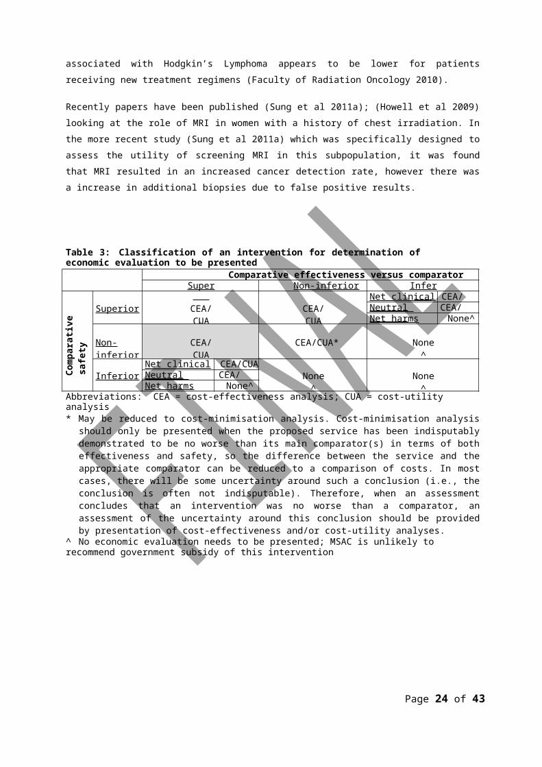
Table 3: Classification of an intervention for determination of economic evaluation to be presentedComparative effectiveness versus comparator
Superior Non-inferior Inferior
Com
para
tive
safe
ty
vers
us c
ompa
rato
r Superior CEA/CUA CEA/CUANet clinic a l ben e fit CEA/CUANeutral benefit CEA/CUA*Net harms None^
Non-inferior CEA/CUA CEA/CUA* None^
InferiorNet clinic a l ben e fit CEA/CUA
None^ None^Neutral benefit CEA/CUA*Net harms None^
Abbreviations: CEA = cost-effectiveness analysis; CUA = cost-utility analysis* May be reduced to cost-minimisation analysis. Cost-minimisation analysis should only be presented when the proposed
service has been indisputably demonstrated to be no worse than its main comparator(s) in terms of both effectiveness and safety, so the difference between the service and the appropriate comparator can be reduced to a comparison of costs. In most cases, there will be some uncertainty around such a conclusion (i.e., the conclusion is often not indisputable). Therefore, when an assessment concludes that an intervention was no worse than a comparator, an assessment of the uncertainty around this conclusion should be provided by presentation of cost-effectiveness and/or cost-utility analyses.
^ No economic evaluation needs to be presented; MSAC is unlikely to recommend government subsidy of this intervention
Page 20 of 39
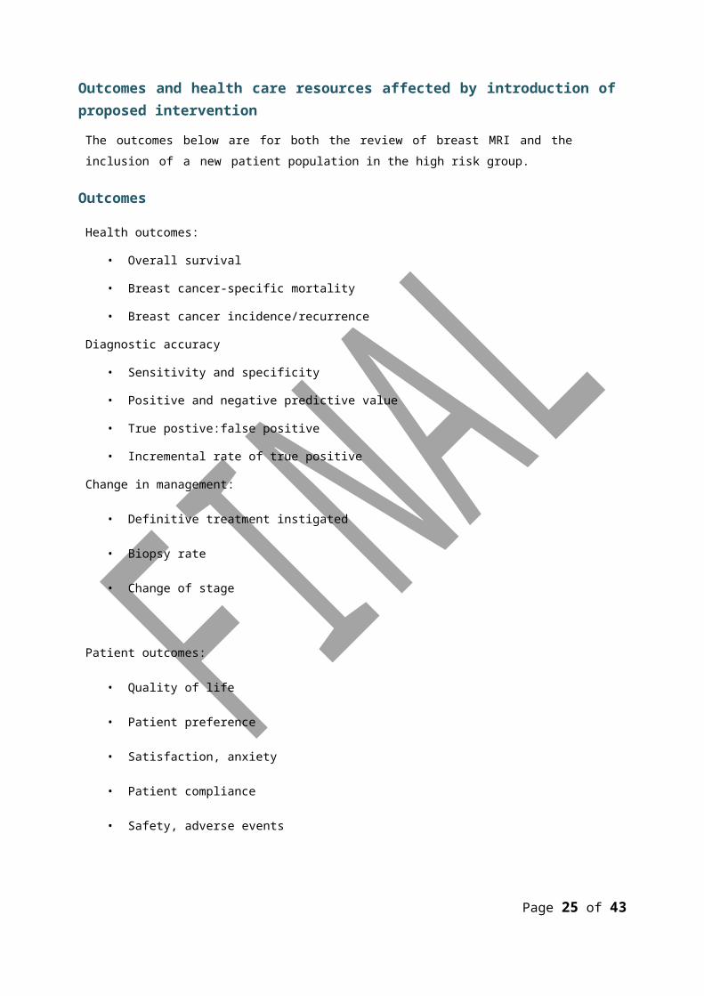
Outcomes and health care resources affected by introduction of proposed interventionThe outcomes below are for both the review of breast MRI and the inclusion of a new patient population in the high risk group.
Outcomes
Health outcomes:
• Overall survival
• Breast cancer-specific mortality
• Breast cancer incidence/recurrence
Diagnostic accuracy
• Sensitivity and specificity
• Positive and negative predictive value
• True postive:false positive
• Incremental rate of true positive
Change in management:
• Definitive treatment instigated
• Biopsy rate
• Change of stage
Patient outcomes:
• Quality of life
• Patient preference
• Satisfaction, anxiety
• Patient compliance
• Safety, adverse events
Page 21 of 39

Health care resources
Breast MRI for the screening of high risk women is funded in Australia under the MBS as an interim item(s). The 2006 MSAC review estimated that implementing MRI as an additional test for screening young high risk women, (defined as women who have a genetic predisposition and 20% of women25-50 years who are eligible for screening) would cost around $3.2 million more than mammographyalone. This not only refers to the cost of MRI, but the additional general practitioner and specialist appointments, and procedures associated with referral, screening and diagnosis.
The above only relates to public screening of MRI and it would seem likely that around the same number of women are accessing MRI through private means which would need to be taken into account. The model will need to consider that there may be a public to private shift in the provision of existing tests.
As mentioned above, MRI appears to be more sensitive (detects more true cancers) and as such will result in a corresponding change in the utilisation of the health care resources used to treat or manage these cancers. Breast MRI also results in lower test specificity, leading to an increase in follow-up investigations (biopsies and additional screening tests) to assess these false positive results. Ongoing funding of breast MRI will result in a continuation of these costs which are likely to increase with further uptake of the procedure as shown by the utilisation figures and with any change in the eligible patient population.
Implications of inclusion of a new patient population(s)
The broadening of the patient group will also lead to greater health care utilisation particularly if the descriptor would include those women with a personal history of invasive breast cancer, LCIS or DCIS.
In 2007 there were 3060 women (25-49 years) diagnosed with invasive breast cancer (AIHW, 2010), the largest proportion being those 40-49 years. This of course is an underestimate of the number of women who would potentially access MRI in a given year as it does not take into account the past cases of breast cancer among women, i.e. prevalence of invasive breast cancer in women aged 25-49 years. Part of the recent BreastScreen Australia Evaluation was to estimate the amount of non- diagnostic mammography funded through the MBS (IMS Health 2009b). It was estimated that in2006, 33,920 women between 40-49 years of age who were considered at high risk due to personal history of breast cancer underwent a mammography. Should all of these women go on to have MRI this would be a substantial increase in utilisation.
Women with DCIS and LCIS are a much smaller group. In a recent report (Australian Institue ofHealth and Welfare and National Breast Ovarian Cancer Centre 2010) there were 326
Page 22 of 39
women under49 years of age diagnosed with DCIS in 2005. Figures for LCIS are more difficult to determine as LCIS is rarely diagnosed as a sole pathology, rather, it is more commonly diagnosed at biopsy for other pathology. However it is unlikely that prevalence would be greater than those with DCIS.
Page 23 of 39
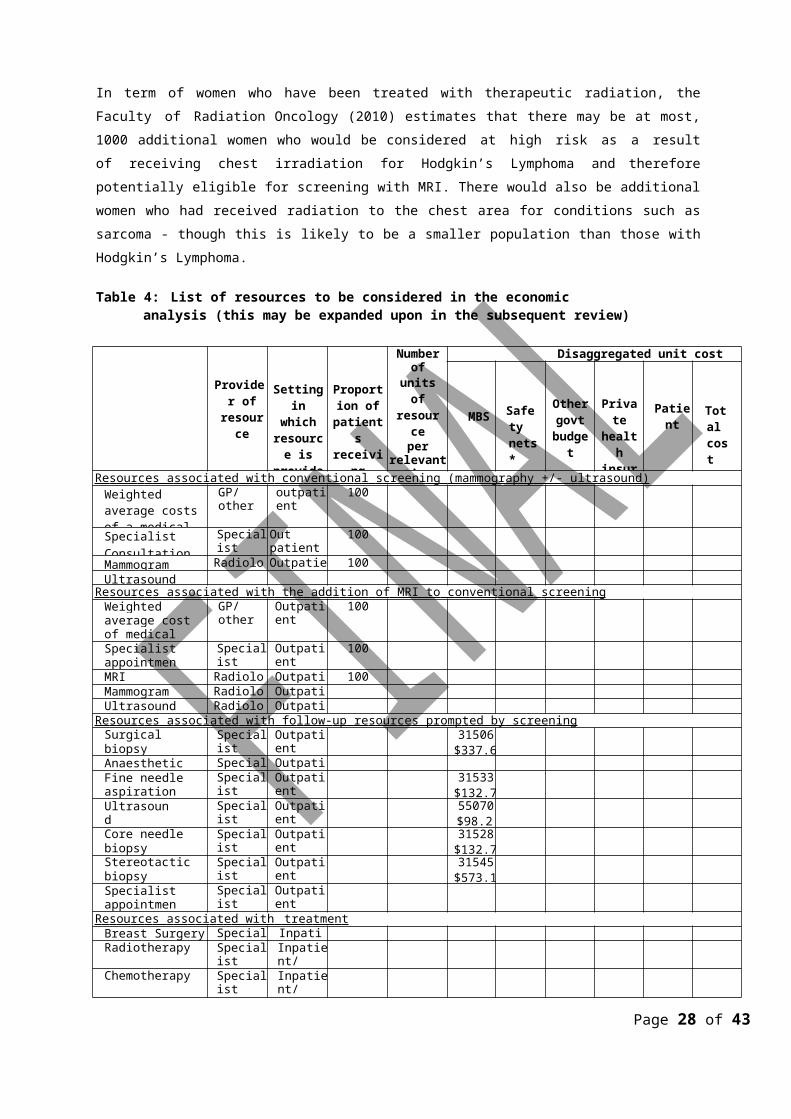
In term of women who have been treated with therapeutic radiation, the Faculty of Radiation Oncology (2010) estimates that there may be at most, 1000 additional women who would be considered at high risk as a result of receiving chest irradiation for Hodgkin’s Lymphoma and therefore potentially eligible for screening with MRI. There would also be additional women who had received radiation to the chest area for conditions such as sarcoma - though this is likely to be a smaller population than those with Hodgkin’s Lymphoma.
Table 4: List of resources to be considered in the economic analysis (this may be expanded upon in the subsequent review)
Provider of resource
Setting in which
resource is provided
Proportion of patients receiving resource
Number ofunits of
resourceper relevant
timehorizon per
patient receivingresource
Disaggregated unit cost
MBS Safety nets*
Other govt
budget
Private health insurer
Patient Total cost
Re s our c e s a ss o c iated with c on v en ti on a l sc reen i ng (m am m ogr a phy +/- u ltra s o u nd) Weighted average costs of a medicalconsultation
GP/other outpatient 100
SpecialistConsultation
Specialist Out patient 100
Mammogram Radiologist Outpatient 100Ultrasound
Re s our c e s a ss o c iated with t h e a ddi t ion of MRI to c on v ent i o n a l sc ree n ing Weighted average cost of medicalconsultation
GP/other Outpatient 100
Specialistappointment
Specialist Outpatient 100
MRI Radiologist Outpatient 100Mammogram Radiologist OutpatientUltrasound Radiologist Outpatient
Re s our c e s a ss o c iated with fo l low- u p re s our c e s pr o m p t ed by sc ree n ing Surgical biopsy Specialist Outpatient 31506
$337.60Anaesthetic costs Specialist OutpatientFine needle aspiration biopsy
Specialist Outpatient 31533$132.70
Ultrasound guidance
Specialist Outpatient 55070$98.25
Core needle biopsy Specialist Outpatient 31528$132.70
Stereotactic biopsy Specialist Outpatient 31545$573.10
Specialist appointment
Specialist Outpatient
R e s ou rc e s a ss o ci a t e d with tre a tme n t Breast Surgery Specialist InpatientRadiotherapy Specialist Inpatient/
OutpatientChemotherapy Specialist Inpatient/
OutpatientHormone therapy Specialist Outpatient
‐ MRI compatible needles and disposables may also need to be considered
Page 24 of 39
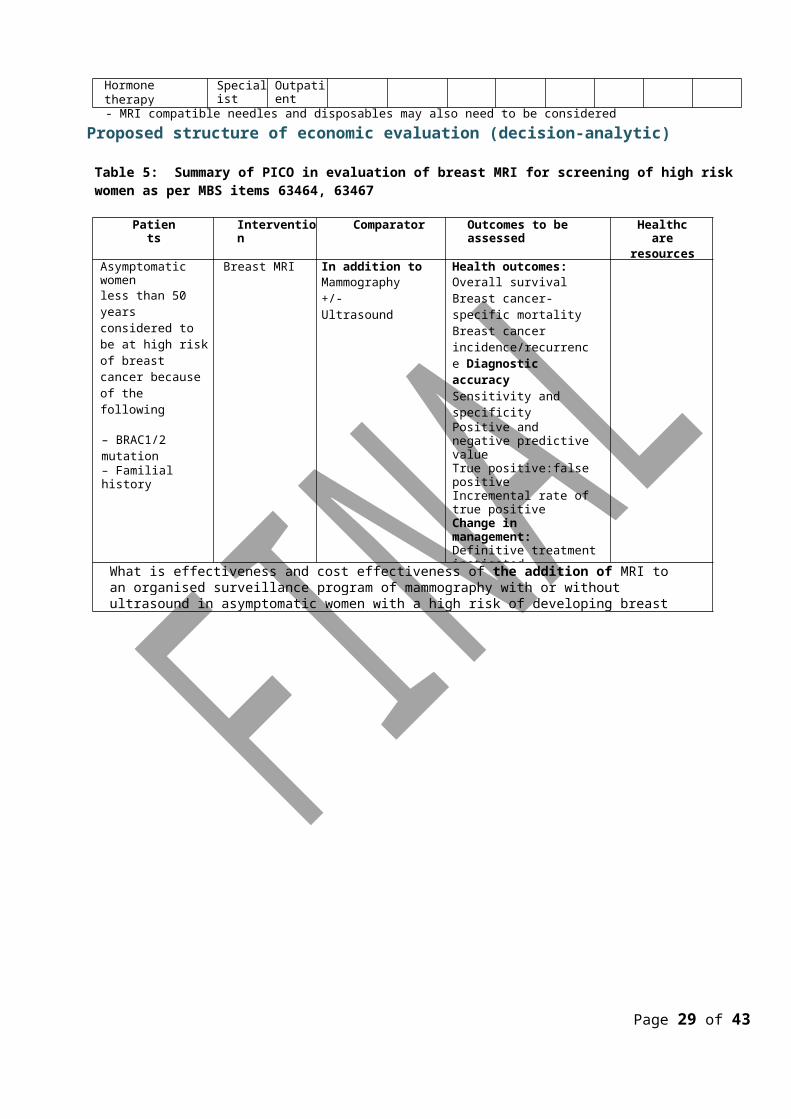
Proposed structure of economic evaluation (decision-analytic)
Table 5: Summary of PICO in evaluation of breast MRI for screening of high risk women as per MBS items 63464, 63467
Patients Intervention Comparator Outcomes to be assessed Healthcareresources to be
consideredAsymptomatic womenless than 50 years considered to be at high risk of breast cancer because of the following
– BRAC1/2 mutation– Familial history
Breast MRI In addition toMammography +/- Ultrasound
Health outcomes:Overall survivalBreast cancer-specific mortality Breast cancer incidence/recurrence Diagnostic accuracy Sensitivity and specificityPositive and negative predictive valueTrue positive:false positive Incremental rate of true positive Change in management:Definitive treatment instigatedBiopsy rateChange of stage Patient outcomes: Quality of life Patient preferenceSatisfaction, anxiety Patient compliance Safety, adverse events
What is effectiveness and cost effectiveness of the addition of MRI to an organised surveillance program of mammography with or without ultrasound in asymptomatic women with a high risk of developing breast cancer due?
Page 25 of 39
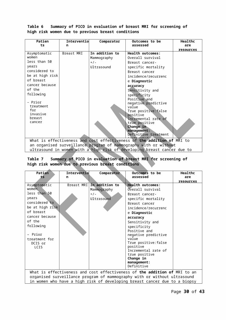
Table 6 Summary of PICO in evaluation of breast MRI for screening of high risk women due to previous breast conditions
Patients Intervention Comparator Outcomes to be assessed Healthcareresources to be
consideredAsymptomatic womenless than 50 years considered to be at high risk of breast cancer because of the following
– Prior treatment for invasive breast cancer
Breast MRI In addition toMammography +/- Ultrasound
Health outcomes:Overall survivalBreast cancer-specific mortality Breast cancer incidence/recurrence Diagnostic accuracy Sensitivity and specificityPositive and negative predictive valueTrue positive:false positive Incremental rate of true positive Change in management:Definitive treatment instigatedBiopsy rate Change of stage Patient outcomes: Quality of life Patient preferenceSatisfaction, anxiety Patient compliance Safety, adverse events
What is effectiveness and cost effectiveness of the addition of MRI to an organised surveillance program of mammography with or without ultrasound in women with a high risk of developing breast cancer due to a history of invasive breast cancer?
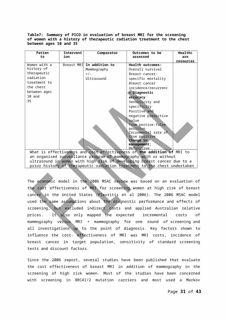
Table 7 Summary of PICO in evaluation of breast MRI for screening of high risk women due to previous breast conditions
Patients Intervention Comparator Outcomes to be assessed Healthcareresources to be
consideredAsymptomatic womenless than 50 years considered to be at high risk of breast cancer because of the following
– Prior treatment forDCIS or LCIS
Breast MRI In addition toMammography +/- Ultrasound
Health outcomes:Overall survivalBreast cancer-specific mortality Breast cancer incidence/recurrence Diagnostic accuracy Sensitivity and specificityPositive and negative predictive valueTrue positive:false positive Incremental rate of true positive Change in management: Definitive treatment instigated Biopsy rateChange of stage Patient outcomes: Quality of life Patient preferenceSatisfaction, anxiety Patient compliance Safety, adverse events
What is effectiveness and cost effectiveness of the addition of MRI to an organised surveillance program of mammography with or without ultrasound in women who have a high risk of developing breast cancer due to a biopsy proven diagnosis of DCIS or
Page 26 of 39
Table7: Summary of PICO in evaluation of breast MRI for the screening of women with a history of therapeutic radiation treatment to the chest between ages 10 and 35
Patients Intervention Comparator Outcomes to be assessed Healthcareresources to
be consideredWomen with a history oftherapeutic radiation treatment to the chest between ages 10 and35
Breast MRI In addition toMammography +/- Ultrasound
Health outcomes:Overall survivalBreast cancer-specific mortality Breast cancer incidence/recurrence Diagnostic accuracy Sensitivity and specificityPositive and negative predictive valueTrue postive:false positive Incremental rate of true positive Change in management: Definitive treatment instigated Biopsy rateChange of stage Patient outcomes: Quality of lifePatient preference Satisfaction, anxiety Patient compliance Safety, adverse events
What is effectiveness and cost effectiveness of the addition of MRI to an organised surveillance program of mammography with or without ultrasound in women with high risk of developing breast cancer due to a prior history of therapeutic radiation treatment to the chest undertaken between the ages 10 and 35?
The economic model in the 2006 MSAC review was based on an evaluation of the cost effectiveness of MRI for screening women at high risk of breast cancer in the United States (Plevritis et al 2006). The 2006 MSAC model used the same assumptions about the diagnostic performance and effects of screening, but excluded indirect costs and applied Australian relative prices. It also only mapped the expected incremental costs of mammography versus MRI + mammography for one round of screening and all investigations up to the point of diagnosis. Key factors shown to influence the cost- effectiveness of MRI was MRI costs, incidence of breast cancer in target population, sensitivity of standard screening tests and discount factors.
Since the 2006 report, several studies have been published that evaluate the cost effectiveness of breast MRI in addition of mammography in the screening of high risk women. Most of the studies have been concerned with screening in BRCA1/2 mutation carriers and most used a Markov modelling approach
In these cost effectiveness studies of women with BRCA1/2 and MRI, the following points were made:
• annual screening with combined mammography and MRI provides BRCA1 mutation carriers with the greatest life expectancy gain and breast cancer
Page 27 of 39
mortality reduction (Norman et al2007);
Page 28 of 39
• however, this needs to be balanced against a strategy with an increased rate of false-positive screening results and biopsies performed for benign disease (Grann et al 2011) (Lee et al2010);
• breast MRI screening is more cost-effective for BRACA1 than BRAC2; and
• results were dependent on key parameters such as the cost of MRI, and age at first screen.
These parameters varied by study setting and, therefore results also varied significantly among studies.
Two additional papers, one that was included in an addendum to the 2006 report (Griebsch et al2006) and one published in 2009 (Taneja et al 2009) look at a broader group of women at high risk of breast cancer. Both these papers concluded that the addition of MRI to the screening regime was cost-effective for women with BRCA1/2 mutations but for other groups may depend on the expected prevalence of undiagnosed breast cancer.
Modelling using Monte Carlo simulation is considered to be the most appropriate modelling approach for the current question. Markov modelling using Monte Carlo simulations enables simulations to be made of hypothetical cohorts of individuals with particular characteristics under specific scenarios (such as age groups and screening intervals). Hypothetical individuals progress from one health state to the next based on pre-determined transition probabilities over a series of discrete time periods (cycles). Stage-specific costs and health outcomes are accumulated dependent on the time spent in that state. This enables overall calculation to be made of costs and outcomes over the specified model time span (IMS Health 2009a).
One such model was developed by Lee et al (Lee et al 2010) in their study to evaluate the cost effectiveness of screening of MRI in women with BRCA1 mutation. The model consists of three linked modules: (a) breast cancer development and detection, (b) treatment and follow-up, and (c) screening. Individual women entered the breast cancer development and detection module at the beginning of the simulation.
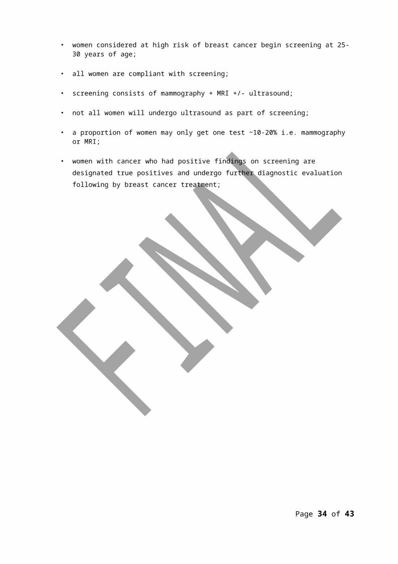
For any such model in the Australian context the following parameters may need to be considered:
• women considered at high risk of breast cancer begin screening at 25-30 years of age;
• all women are compliant with screening;
• screening consists of mammography + MRI +/- ultrasound;
• not all women will undergo ultrasound as part of screening;
• a proportion of women may only get one test ~10-20% i.e. mammography or MRI;
Page 29 of 39
• women with cancer who had positive findings on screening are designated true positives and undergo further diagnostic evaluation following by breast cancer treatment;
Page 30 of 39
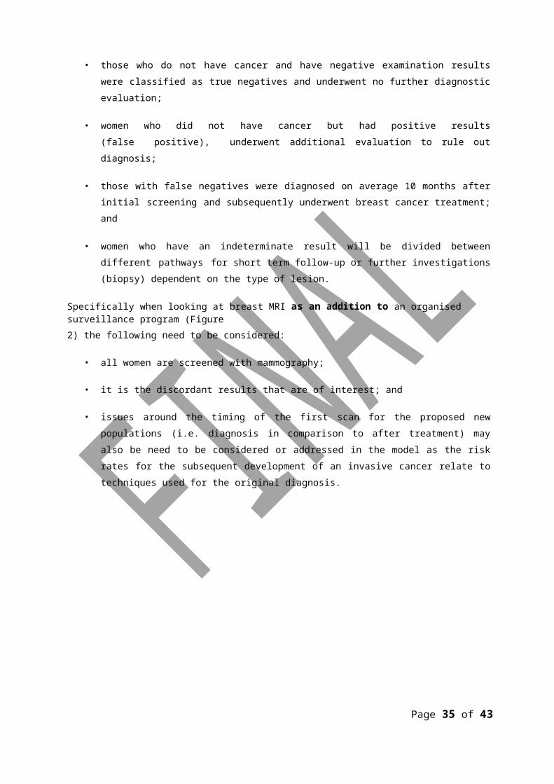
• those who do not have cancer and have negative examination results were classified as true negatives and underwent no further diagnostic evaluation;
• women who did not have cancer but had positive results (false positive), underwent additional evaluation to rule out diagnosis;
• those with false negatives were diagnosed on average 10 months after initial screening and subsequently underwent breast cancer treatment; and
• women who have an indeterminate result will be divided between different pathways for short term follow-up or further investigations (biopsy) dependent on the type of lesion.
Specifically when looking at breast MRI as an addition to an organised surveillance program (Figure2) the following need to be considered:
• all women are screened with mammography;
• it is the discordant results that are of interest; and
• issues around the timing of the first scan for the proposed new populations (i.e. diagnosis in comparison to after treatment) may also be need to be considered or addressed in the model as the risk rates for the subsequent development of an invasive cancer relate to techniques used for the original diagnosis.
Page 31 of 39
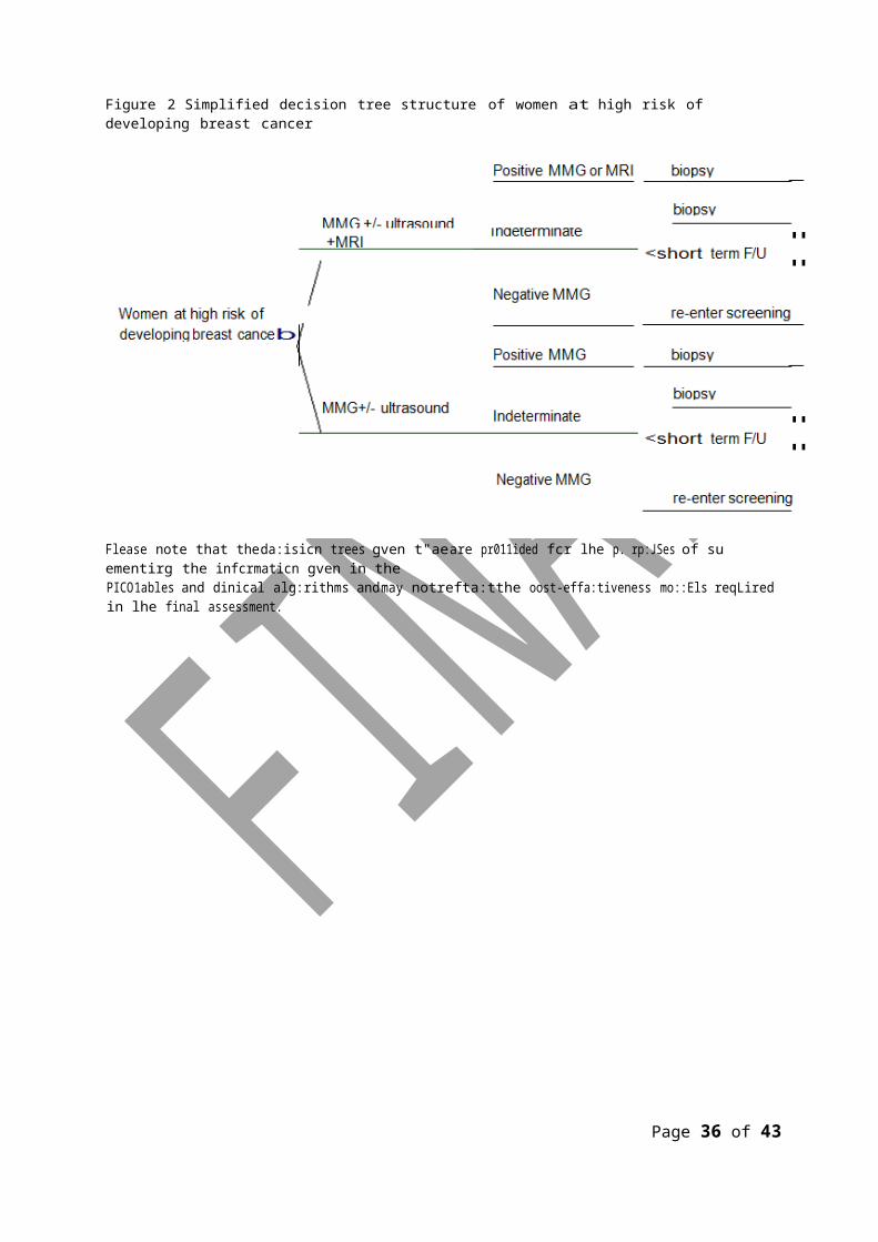
Figure 2 Simplified decision tree structure of women at high risk of developing breast cancer
Flease note that theda:isicn trees gven t"aeare pr011ided fcr lhe p. rp:JSes of su ementirg the infcrmaticn gven in thePICO1ables and dinical alg:rithms andmay notrefta:tthe oost-effa:tiveness mo::Els reqLired in lhe final assessment.
Page 32 of 39
Reference List
1. Australian Institue of Health and Welfare and National Breast Ovarian Cancer Centre 2010, Risk of invasive breat cancer in women diagnosed with ductal carcinoma in situ in Australia between1995 and 2005. 51.
2. BERMEJO-PEREZ, MJ, MARQUEZ-CALDERON, S et al 1-3-2008. Cancer surveillance based on imaging techniques in carriers of BRCA1/2 gene mutations: a systematic review, The British Journal of Radiology, 81 (963), 172-179.
3. Bevers, TB, Anderson, BOBE et al 2011. NCCN clinical practice guidelines in oncology: breast cancer screening and diagnosis, Journal of the National Comprehensive Cancer Network, 2009 (7), 1060-1096.
4. Buist, D, Abraham, L et al 1-12-2010. Diagnosis of second breast cancer events after initial diagnosis of early stage breast cancer, Breast Cancer Research and Treatment, 124 (3), 863-873.
5. Davidson, E and Hancock, S 2007, Surveillance of women at high risk of breast cancer. 6.
Christchurch: NZHTA.
6. Dunfield, L and Severn, M 2007, Effectiveness of magnetic resonance imaging (MRI) screening for women at high risk of breast cancer. Ottawa: Canadian Agency for Drugs and Technologies in Health (CADTH).
7. Faculty of Radiation Oncology 2010, Position Paper: breast cancer and late effects following radiotherapy and chemotherapy for Hodgkin's Lymphoma. Sydney: The Royal Australian and New Zealand College of Radiologists.
8. Fridlander, L, Orel Roth, S et al 2011. Results of MR Imaging Screening for Breast Cancer in
High-Risk Patiens with Lobular Carcinoma in Situ, Radiology, 261 (2), 421-427.
9. Gilbert, FJ, Warren, RML et al 2009. Cancers in BRCA1 and BRCA2 carriers and in women at high risk for breast cancer: MR imaging and mammographic features, Radiology, 252 (2), 358-368.
10. Grann, VR, Patel, PR et al 2011. Comparative effectiveness of screening and prevention strategies among BRCA1/2-affected mutation carriers, Breast Cancer Research & Treatment,125 (3), 837-847.
11. Griebsch, I, Brown, J et al 9-10-2006. Cost-effectiveness of screening with contrast enhanced magnetic resonance imaging vs X-ray mammography of women at a high familial risk of breast cancer, British Journal of Cancer, 95 (7), 801-810.
12. Henderson, TO, Amsterdam, A et al 2010. Systematic review: surveillance for breast cancer in women treated with chest radiation for childhood, adolescent, or young adult cancer. [Review] [82 refs], Annals of Internal Medicine, 152 (7), 444-455.
Page 33 of 39
13. Houssami, N, Abraham, LA et al 23-2-2011. Accuracy and outcomes of screening mammography in women with a personal history of early-stage breast cancer, JAMA, 305 (8),790-799.
Page 34 of 39
14. Howell, SJ, Searle, C et al 18-8-2009. The UK national breast cancer screening programme for survivors of Hodgkin lymphoma detects breast cancer at an early stage, British Journal of Cancer, 101 (4), 582-588.
15. IMS Health 2009a, Breastscreen Australia Evaluation: economic evaluation and modellling study. 9/2009. ACT: Department of Health and Ageing.
16. IMS Health 2009b, Breastscreen Australia: MBS Mammography Analysis Project. 11/2009. ACT: Department of Health and Ageing.
17. Irwig, L, Houssami, N et al 1-6-2004. New technologies in screening for breast cancer: a systematic review of their accuracy. [Review] [23 refs], British Journal of Cancer.90(11):2118-22,
18. Lee, JM, McMahon, PM et al 2010. Cost-effectiveness of breast MR imaging and screen-film mammography for screening BRCA1 gene mutation carriers, Radiology, 254 (3), 793-800.
19. Li, C, Malone, K et al 2006. Risk of invasive breast carninoma among women diagnosed with
Ductal Carcinoma in Situ and Lobular Carcinoma in Situ, 1988-2001, Cancer, 106 (10), 2104-2112.
20. Medical Services Advisory Committee 2006, Breast Magnetic Resonance Imaging. Canberra: Department of Health and Ageing.
21. NBOCC 2009, Breast cancer risk factors: a review of the evidence. Surry Hills: National Breast and Ovarian Cancer Centre.
22. Norman, RPA, Evans, DG et al 1-6-2007. The cost-utility of magnetic resonance imaging for breast cancer in BRCA1 mutation carriers aged 30ΓÇô49, The European Journal of Health Economics, 8 (2), 137-144.
23. Plevritis, SK, Kurian, AW et al 2006. Cost-effectivness of Screening BRCA1/2 Mutation Carries with Breast Magnetic Resonance Imaging, Journal of American Medical Association, 295 (20),2374-2384.
24. Price, J and Chen, SW 2009. Screening for breast cancer with MRI: recent experience from the
Australian Capital Territory, Journal of Medical Imaging & Radiation Oncology, 53 (1), 69-80.
25. Sardanelli, F, Boetes, C et al 2010. Magnetic resonance imaging of the breast:recommendations from the EUSOMA working group. [223 refs], European Journal of Cancer, 46 (8), 1296-1316.
26. Saslow, D, Boetes, C et al 2007. American Cancer Society Guidelines for Breast Screening with
MRI as an Adjunct to Mammography, CA: A Cancer Journal for Clinicians, 57 (2), 75-89.
27. Sung, JS, Lee, CH et al 2011a. Screening breast MR imaging in women with a history of chest irradiation, Radiology, 259 (1), 65-71.
Page 35 of 39
28. Sung, JS, Malak, S et al 2011b. Screening Breast MR Imaging in Women with History of Lobular
Carcinoma in Situ, Radiology, 261 (2), 414-420.
29. Taneja, C, Edelsberg, J et al 2009. Cost Effectiveness of Breast Cancer Screening With Contrast-Enhanced MRI in High-Risk Women, JACR Journal of the American College of Radiology, 6 (3), 171-179.
Page 36 of 39
30. Taylor, AJ, Winter, DL et al 15-1-2007. Risk of breast cancer in female survivors of childhood
Hodgkin's disease in Britain: a population-based study, International Journal of Cancer, 120 (2),384-391.
31. Warner, E, Hill, KA et al 2011. Prospective study of breast cancer incidence in women with a BRCA1 or BRCA2 Mutation under surveillance with and without magnetic resonance imaging, Journal of Clinical Oncology, 29 (13), 1664-1669.
32. Warner, E, Messersmith, H et al 6-5-2008. Systematic review: using magnetic resonance imaging to screen women at high risk for breast cancer. [Review] [35 refs], Annals of Internal Medicine, 148 (9), 671-679.
33. Washington State Care Authority 23-7-0010, HTA Report: Breast MRI in diagnosis adn treatment of cancer in women at high risk. Olympia: Washington State Health Care Authority.
Page 37 of 39
Appendix 1
Table 8 MBS items associated with Breast MRIMammography Category 5 – Diagnostic Imaging Services
MBS 59300
MAMMOGRAPHY OF BOTH BREASTS, if there is a reason to suspect the presence of malignancy because of:
(i) the past occurrence of breast malignancy in the patient or members of the patient's family; or(ii) symptoms or indications of malignancy found on an examination of the patient by a medical practitioner.
Unless otherwise indicated, mammography includes both breasts (R)Bulk bill incentiveFee: $89.50 Benefit: 75% = $67.15 85% = $76.10
MBS 59301*
MAMMOGRAPHY OF BOTH BREASTS, if there is a reason to suspect the presence of malignancy because of:
(i) the past occurrence of breast malignancy in the patient or members of the patient's family; or(ii) symptoms or indications of malignancy found on an examination of the patient by a medical practitioner.
Unless otherwise indicated, mammography includes both breasts (R) (NK)Bulk bill incentiveFee: $44.75 Benefit: 75% = $33.60 85% = $38.05
MBS 59303
MAMMOGRAPHY OF ONE BREAST, if:(a) the patient is referred with a specific request for a unilateral mammogram; and(b) there is reason to suspect the presence of malignancy because of:
(i) the past occurrence of breast malignancy in the patient or members of the patient's family; or(ii) symptoms or indications of malignancy found on an examination of the patient by a medical practitioner (R)
Bulk bill incentiveFee: $53.95 Benefit: 75% = $40.50 85% = $45.90
MBS 59304*
MAMMOGRAPHY OF ONE BREAST, if:(a) the patient is referred with a specific request for a unilateral mammogram; and(b) there is reason to suspect the presence of malignancy because of:
(i) the past occurrence of breast malignancy in the patient or members of the patient's family; or(ii) symptoms or indications of malignancy found on an examination of the patient by a medical practitioner (R)
(NK)Bulk bill incentiveFee: $27.00 Benefit: 75% = $20.25 85% = $22.95
* From 1 July 2011 all services listed in the Diagnostic Imaging Services Table of the Medicare Benefits Schedule (MBS), excluding Positron Emission Tomography (PET) services, preparation items 60918 and 60927 and MRI modifier items in subgroup 22, will have a mirror NK item (50% of the Schedule Fee) for diagnostic imaging services provided on aged equipment. This rule, known as ‘capital sensitivity’, is currently in place for computed tomography (CT) and angiography and will be extended to improve the quality of diagnostic imaging services by encouraging providers to upgrade and replace aged equipment as appropriate.
Page 38 of 39
Ultrasound Category 5 – Diagnostic Imaging Services
MBS 55070BREAST, one, ultrasound scan of, where:(a) the patient is referred by a medical practitioner; and(b) the service is not associated with a service to which an item in Subgroup 2 or 3 of this group applies; and(c) the referring medical practitioner is not a member of a group of practitioners of which the providing practitioner is a
member (R)Bulk bill incentiveFee: $98.25 Benefit: 75% = $73.70 85% = $83.55
MBS 55073BREAST, one, ultrasound scan of, where:(a) the patient is not referred by a medical practitioner; and(b) the service is not associated with a service to which an item in Subgroup 2 or 3 of this group applies (NR)Bulk bill incentiveFee: $34.05 Benefit: 75% = $25.55 85% = $28.95
MBS 55076
BREASTS, both, ultrasound scan of, where:(a) the patient is referred by a medical practitioner; and(b) the service is not associated with a service to which an item in Subgroup 2 or 3 of this group applies; and(c) the referring medical practitioner is not a member of a group of practitioners of which the providing practitioner is a
member (R)Bulk bill incentiveFee: $109.10 Benefit: 75% = $81.85 85% = $92.75
MBS 55079BREASTS, both, ultrasound scan of, where:(a) the patient is not referred by a medical practitioner; and(b) the service is not associated with a service to which an item in Subgroup 2 or 3 of this group applies (NR)
Bulk bill incentiveFee: $37.85 Benefit: 75% = $28.40 85% = $32.20
Specialist Consultation Category 1 - PROFESSIONAL ATTENDANCES
MBS 104SPECIALIST, REFERRED CONSULTATION - SURGERY OR HOSPITAL(Professional attendance at consulting rooms or hospital by a specialist in the practice of his or her specialty where the patient is referred to him or her)- INITIAL attendance in a single course of treatment, not being a service to which ophthalmology items 106, 109 or obstetric item 16401 apply.Fee: $82.30 Benefit: 75% = $61.75 85% = $70.0
CONSULTANT PHYSICIAN (OTHER THAN IN PSYCHIATRY), REFERRED CONSULTATION - SURGERY OR HOSPITAL(Professional attendance at consulting rooms or hospital by a consultant physician in the practice of his or her specialty(other than in psychiatry) where the patient is referred to him or her by a medical practitioner)- INITIAL attendance in a single course of treatmentFee: $145.20 Benefit: 75% = $108.90 85% = $123.45
Page 39 of 39


