Draft Benefit Definition: ST-Elevation Myocardial … Definition Project...Draft Benefit Definition:...
23
Draft Benefit Definition: ST-Elevation Myocardial Infarct 01 September 2014 907E Acute and sub-acute ischaemic heart disease including myocardial infarction and unstable angina. Treatment: Medical management; surgery; percutaneous procedures
Transcript of Draft Benefit Definition: ST-Elevation Myocardial … Definition Project...Draft Benefit Definition:...

Draft Benefit Definition: ST-Elevation Myocardial Infarct
01 September 2014
907E Acute and sub-acute ischaemic heart disease including myocardial infarction and
unstable angina.
Treatment: Medical management; surgery; percutaneous procedures

2
Contents
1 Introduction..................................................................................................................................................... 4
2 Scope ............................................................................................................................................................. 4
3 Burden of Disease .......................................................................................................................................... 4
4 Emergency Diagnosis and Care for ST-Segment Elevation Myocardial Infarct ............................................. 5
5 Logistical considerations for Management of Patients with STEMI .............................................................. 10
6 Reperfusion Strategies ................................................................................................................................. 11
6.1 Percutaneous procedures ................................................................................................................... 11
6.2 Pharmacological reperfusion ............................................................................................................... 17
6.3 Acute Phase coronary by-pass graft .................................................................................................... 18
7 Care post emergency reperfusion ................................................................................................................ 18
8 Post discharge follow-up .............................................................................................................................. 19
9 Secondary prevention for STEMI Patients ................................................................................................... 22
10 Bibliography ............................................................................................................................................. 23
3
Abbreviations
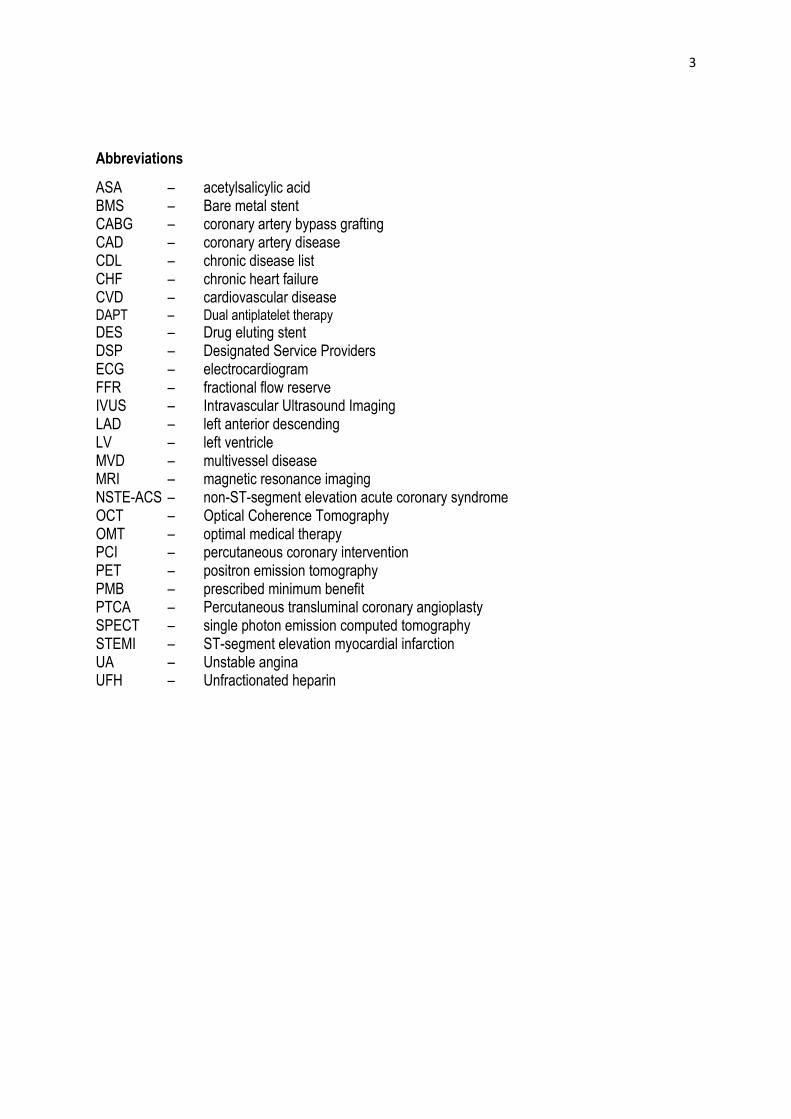
ASA – acetylsalicylic acid BMS – Bare metal stent CABG – coronary artery bypass grafting CAD – coronary artery disease CDL – chronic disease list CHF – chronic heart failure CVD – cardiovascular disease DAPT – Dual antiplatelet therapy DES – Drug eluting stent DSP – Designated Service Providers ECG – electrocardiogram FFR – fractional flow reserve IVUS – Intravascular Ultrasound Imaging LAD – left anterior descending LV – left ventricle MVD – multivessel disease MRI – magnetic resonance imaging NSTE-ACS – non-ST-segment elevation acute coronary syndrome OCT – Optical Coherence Tomography OMT – optimal medical therapy PCI – percutaneous coronary intervention PET – positron emission tomography PMB – prescribed minimum benefit PTCA – Percutaneous transluminal coronary angioplasty SPECT – single photon emission computed tomography STEMI – ST-segment elevation myocardial infarction UA – Unstable angina UFH – Unfractionated heparin

4
1 Introduction
This benefit definition does not explicitly endorse one medicine/medical device within a particular
therapeutic class over another. However due to the emergency nature of STEMI and to avoid delays
associated with pre-authorisation and consultation with scheme formularies, this Benefit definition is
highly specific on which treatment and classes may be used during ST elevation myocardial infarct
(STEMI). This is to safe guard members against any possible co-payments that may arise from failure
to use formularies and to protect the schemes of unplanned expenditure that may arise in a setting
where it is impossible to obtain scheme authorization.
Provision must be made for appropriate exceptions where this benefit definition has been ineffective, or
causes, or would cause harm to a beneficiary, without penalty to that beneficiary. Health care providers
must provide written documentation for exceptions.
All patients who are treated successfully in an emergency setting must register with their scheme for
chronic management of ischaemic heart disease. Scheme protocols and formularies should be
developed and applied while taking into consideration evidence-based medicine, cost-effectiveness and
affordability.
It should be noted that benefit definitions are a minimum set of benefits and schemes may enrich the
benefits but not offer benefits less than those stated here.
It should also be noted that management of Ischaemic heart disease takes into consideration many
clinical aspects of the patient. This benefit definition does not address specific circumstances of high
risk and complicated patients who may need more care than specified here.
Alternatives must be made for patients in whom treatment stated here or in the scheme formulary may
cause harm.
Due to high variability of clinical presentation and possible outcomes in patients with Ischaemic heart
disease it was difficult to quantify frequency of tests and interventions in an acute setting.
Procedure codes serve as a guideline for billing and may not include all relevant procedure
codes.
2 Scope
These benefit definitions include the management of ST-Elevation Myocardial Infarct (STEMI). The
benefit definition covers out of hospital emergency care, in-hospital care and long term follow-up
including secondary prevention. Coronary artery bypass graft is not included.
3 Burden of Disease
According to results of the INTERHEART study, the five most important risk factors for myocardial
infarction operate similarly in different ethnic groups and geographical locations worldwide. These risk
factors are smoking history, diabetes history, hypertension, abdominal obesity and the ratio of 74
apolipoprotein B to apolipoprotein A-1 (1). The emergence of risk factors for atherosclerotic vascular

5
disease in South Africa has been noted for several decades (2). Population based surveys in the early
1990s showed that 13-31% of the population have at least one risk factor for atherosclerotic disease.
Later in the 2000s, surveys confirmed high population prevalence of hypertension, diabetes, smoking
as well as a high prevalence of obesity affecting about 50% of the female population in Limpopo and
Mpumalanga provinces (2). Heart disease, diabetes and stroke together constitute the second most
important cause of death in the adult population in South Africa (3). Cardiovascular disease is
increasing amongst all age groups in South Africa and is predicted to become the prime contributor to
overall morbidity and mortality in the over 50-year age group (4).
4 Emergency Diagnosis and Care for ST-Segment Elevation Myocardial Infarct
Patients may present with a history of chest pain for more than 20 minutes. The pain may radiate to the
left arm, lower jaw and neck. Sometimes, patients may present with atypical symptoms such as fatigue,
nausea and vomiting, palpitations or syncope. This atypical presentation is common in the elderly,
women and diabetic patients.
The key to successful management is Timely diagnosis of STEMI. ECG monitoring should be initiated
as soon as possible in all patients with suspected STEMI to detect life-threatening arrhythmias and
allow prompt defibrillation if indicated. A 12-lead ECG should be obtained and interpreted as soon as
possible.
Management of STEMI; including diagnosis and treatment, start at the point of first medical contact.
Point of first medical contact in South Africa includes general practice, emergency rooms, paramedics
and other specialists other than physicians and cardiologists
STEMI is typically diagnosed when there is ST-segment elevation in two consecutive leads on the
ECG.
The highest priority in STEMI is to restore coronary blood flow as soon as possible. Due to successful
outcomes associated with early intervention (5); pre-authorisation should not be a pre-requisite
for initiating care.
The aim of emergency medical care is
i. To establish diagnosis using ECG and blood sampling for cardiac enzymes
ii. Initiate management depending on the logistical arrangements (ability to refer to a specialist
centre without delay, scope of practice of the first contact health provider, availability of
resources etc).
iii. Reduce pain
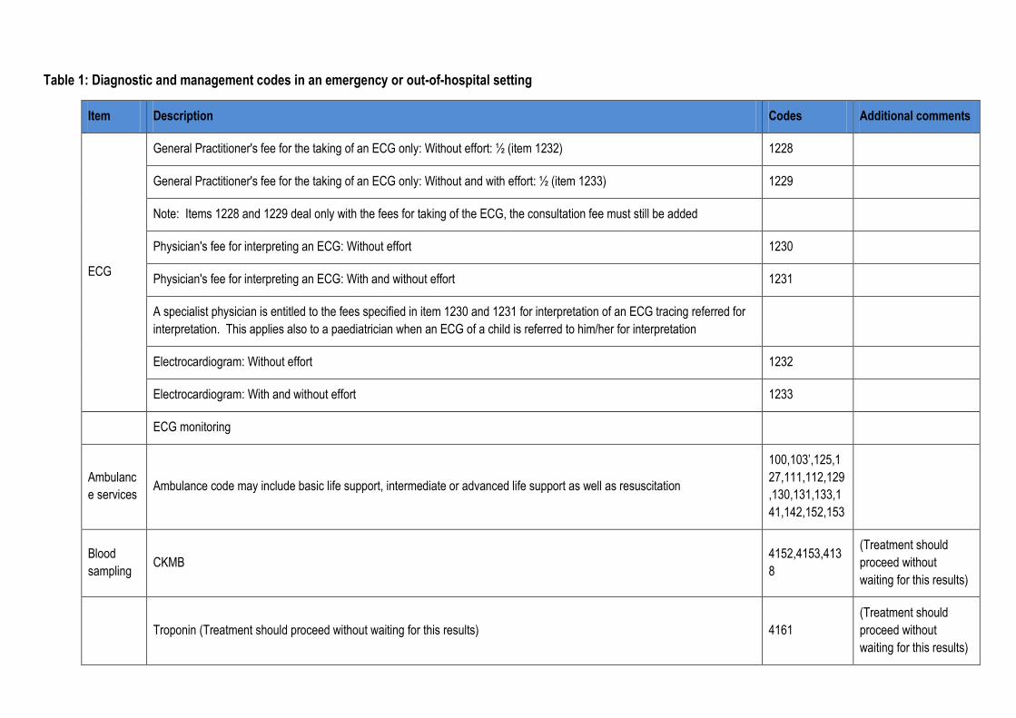
Table 1: Diagnostic and management codes in an emergency or out-of-hospital setting
Item Description Codes Additional comments
ECG
General Practitioner's fee for the taking of an ECG only: Without effort: ½ (item 1232) 1228
General Practitioner's fee for the taking of an ECG only: Without and with effort: ½ (item 1233) 1229
Note: Items 1228 and 1229 deal only with the fees for taking of the ECG, the consultation fee must still be added
Physician's fee for interpreting an ECG: Without effort 1230
Physician's fee for interpreting an ECG: With and without effort 1231
A specialist physician is entitled to the fees specified in item 1230 and 1231 for interpretation of an ECG tracing referred for
interpretation. This applies also to a paediatrician when an ECG of a child is referred to him/her for interpretation
Electrocardiogram: Without effort 1232
Electrocardiogram: With and without effort 1233
ECG monitoring
Ambulanc
e services Ambulance code may include basic life support, intermediate or advanced life support as well as resuscitation
100,103’,125,1
27,111,112,129
,130,131,133,1
41,142,152,153
Blood
sampling CKMB
4152,4153,413
8
(Treatment should
proceed without
waiting for this results)
Troponin (Treatment should proceed without waiting for this results) 4161
(Treatment should
proceed without
waiting for this results)
7
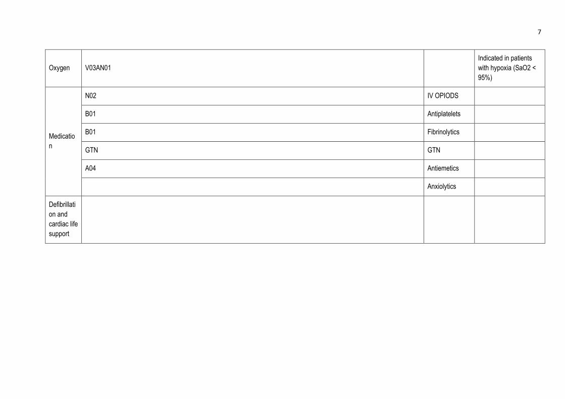
Oxygen V03AN01
Indicated in patients
with hypoxia (SaO2 <
95%)
Medicatio
n
N02 IV OPIODS
B01 Antiplatelets
B01 Fibrinolytics
GTN GTN
A04 Antiemetics
Anxiolytics
Defibrillati
on and
cardiac life
support
Table 2: Routine Investigations Management of STEMI
Type Description of the test Codes Comments
Pathology CKMB 4152,4153,4138
Troponin 4161
Full Blood Count- 3755
(Incl.
3739,3762,3783,
3785,3791)
Platelet count 3797
Glucose-Hypo and hyperglycaemia affect treatment outcomes 4057
Lipogram-Lipid profile can change within 12-24 hours 4025
CRP 3947
ESR: Markers of inflammation
U & E and Creatinine 4171
Creatinine-EGFR 4032
Pulse oximetry
Radiology Chest X-Ray: assess the patient's heart size and the presence or absence of heart failure and pulmonary oedema. This
may also assist in differential diagnosis
30110,30100
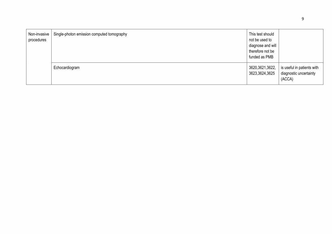
9
Non-invasive
procedures
Single-photon emission computed tomography This test should
not be used to
diagnose and will
therefore not be
funded as PMB
Echocardiogram 3620,3621,3622,
3623,3624,3625
is useful in patients with
diagnostic uncertainty
(ACCA)

5 Logistical considerations for Management of Patients with STEMI
a) Who should participate in care of patients with STEMI
All STEMI patients should undergo rapid evaluation for reperfusion and have reperfusion strategy
implemented promptly after contact with medical system.
The goal is to facilitate rapid recognition and treatment of patients such that door to Needle time for
fibrinolytic therapy is achieved within 30 minutes (door to needle) or that time for PCI can be kept
under 90 -120 minutes. This goal may not be relevant for patient with diagnostic uncertainties, or co-
morbidities such as respiratory failure.
Access to emergency treatment and prevention of delays is important in the management of STEMI.
There is an understanding that the Cardiologist coverage in the country is insufficient, even in the
private sector. There are provinces that do not have cardiologists and some small towns do not have a
specialist physician.
Due to these limitations, general practitioner, other specialist other than internal physicians or
cardiologist, nurses and paramedics play an important role in the management and facilitation
of care for patients with acute myocardial infarct.
According to Regulation 8(6), a medical scheme may not prohibit or enter into arrangements or
contracts that prohibit the initiation of an appropriate intervention by a health care provider prior to
receiving authorisation from medical scheme or any other party, in respect of an emergency medical
condition.
Therefore once a STEMI has been diagnosed, a first contact medical provider must initiate care which
should include emergency transfer with a suitable mode of transport, to a facility and provider capable
of providing treatment for acute myocardial infarct.
b) Facilities for Diagnosis and treatment of STEMI
Initial diagnosis and emergency care can take place at home, in the ambulance, or at emergency rooms
or general practitioner’s (GP) rooms depending on where the member first presented.
Whenever possible patients must be transported or transferred to the nearest PCI facility. If a PCI
facility is a non-designated service provider (DSP) these constitute involuntary use of a DSP.
If it is anticipated that delays will be longer than 2 hours due to distance, amongst other things, then the
first medical contact personnel must provide pharmacological reperfusion treatment under the remote
supervision of the cardiologist or physician if necessary in line with their registered scope of practice.
According to explanatory note to Annexure A: The objective of specifying a set of Prescribed Minimum
Benefits within these regulations is two-fold:
(i) To avoid incidents where individuals lose their medical scheme cover in the event of serious illness
and the consequent risk of unfunded utilisation of public hospitals.
(ii) To encourage improved efficiency in the allocation of Private and Public health care resources.
11
In view of point (ii), if state is the schemes DSP; and both the public and private PCI are equally
accessible to the member; it is considered a prescribed minimum benefit that a patient who belongs to
the medical scheme access the private facility as the use of the public sector will result in inaccessible
care for indigent patients. It should be noted that PCI and interventional cardiologist coverage is far
lower in the public sector as compared to private sector. Therefore, channelling patients to public sector
will defeat objective (ii).
c) Selection of Reperfusion Strategy
Primary PCI without fibrinolytic therapy is the preferred reperfusion strategy in patients with STEMI,
provided it can be performed expeditiously by an experienced team (5) (6)(7).
d) Clinical presentation of the patient
Time from Onset of symptoms:
Time from onset of symptoms to pharmacological reperfusions is an important predictor of clinical
outcomes. The beneficial effect of pharmacological reperfusion is substantially higher in patients
presenting within 2 hours after symptom onset compared to those presenting later (Boersman et al), but
the effect is even greater when accessed earlier. There is, however, some benefit when the treatment is
offered beyond this period.
Risk of bleeding
When both types of reperfusion are available, patients with high risk of bleeding with pharmacological
reperfusion should receive PCI as reperfusion strategy.
e) Availability and time required to transfer to PCI facility
The availability and location of the interventional cardiology facility is a key determinant of whether PCI
can be provided or not. If a patient presents in a PCI- capable facility or can be transferred to a PCI-
capable facility within 2 hours, PCI approach remains superior to pharmacological reperfusion.
A decision must be made when a patient presents to a non-PCI facility to refer for PCI or initiate
pharmacological reperfusion. Fibrinolytic agents can generally be provided sooner than PCI especially
in provinces and towns where there is no interventional cardiologist. Fibrinolytic agents do not require a
high skilled professional; can be provided by many health care professionals (in line with scope of
practice as per regulatory bodies) and even more appropriate in South Africa where the coverage for
PCI facilities and interventional cardiology is low.
6 Reperfusion Strategies
6.1 Percutaneous procedures
As this component of the treatment of the DTP 907E is not only specified in general terms i.e. “medical
management” or “surgery”, but also in specific terms i.e. “percutaneous procedures”, the latter
component it is not subject to the provision made in the explanatory note (2) to Annexure A in the
regulations.
Percutaneous coronary interventions (PCIs) as prescribed minimum benefits are therefore not restricted
to availability of this intervention in the Public sector. A protocol should be developed on the basis of
12
the principles stated in Regulation 15D (b) and 15H namely, evidence based medicine, taking into
account considerations of cost-effectiveness and affordability.
i. Indications
PCI is the best preferred method of treatment if it can be provided within 90-120 minutes of first
medical contact in patients with STEMI
It can also be provided if the symptoms were within 3 hours and PCI can be done within an
hour of diagnosis.
When fibrinolytic ineligible patient present within 12-24 hours
Patients with a new LBBB within 12 hours of onset of symptoms
Within 36 hours if a patient develops shock
If patients has no contraindication to DAPT and is more likely to be compliant on DAPT.
A rescue PCI is indicated in patients with failed fibrinolytic therapy as indicated by residual ST
element elevation post fibrinolysis.
In patients with multi vessel disease, only infarct related artery should be treated during initial
intervention. The only exceptions when multi vessel PCI is indicated during STEMI is when
patients are in cardiogenic shock with > 90% occlusion. (6) (5)
ii. Contraindication
Inability to take or comply with DAPT
Asymptomatic patients more than 12 hours after onset of STEMI
Door to balloon delay of > 2hours. In this instance fibrinolytic therapy offers relatively better
outcomes.
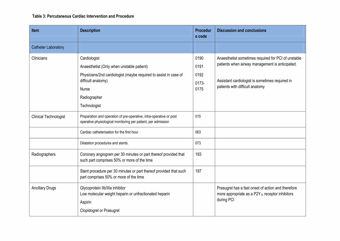
Item Description Procedur
e code
Discussion and conclusions
Catheter Laboratory
Clinicians Cardiologist
Anaesthetist (Only when unstable patient)
Physicians/2nd cardiologist (maybe required to assist in case of
difficult anatomy)
Nurse
Radiographer
Technologist
0190
0191
0192
0173-
0175
Anaesthetist sometimes required for PCI of unstable
patients when airway management is anticipated.
Assistant cardiologist is sometimes required in
patients with difficult anatomy
Clinical Technologist Preparation and operation of pre-operative, intra-operative or post
operative physiological monitoring per patient, per admission
015
Cardiac catheterisation for the first hour. 063
Dilatation procedures and stents. 073
Radiographers Coronary angiogram per 30 minutes or part thereof provided that
such part comprises 50% or more of the time
193
Stent procedure per 30 minutes or part thereof provided that such
part comprises 50% or more of the time
197
Ancillary Drugs Glycoprotein IIb/IIIa inhibitor
Low molecular weight heparin or unfractionated heparin
Aspirin
Clopidogrel or Prasugrel
Prasugrel has a fast onset of action and therefore
more appropriate as a P2Y12 receptor inhibitors
during PCI
Table 3: Percutaneous Cardiac Intervention and Procedure
Codes
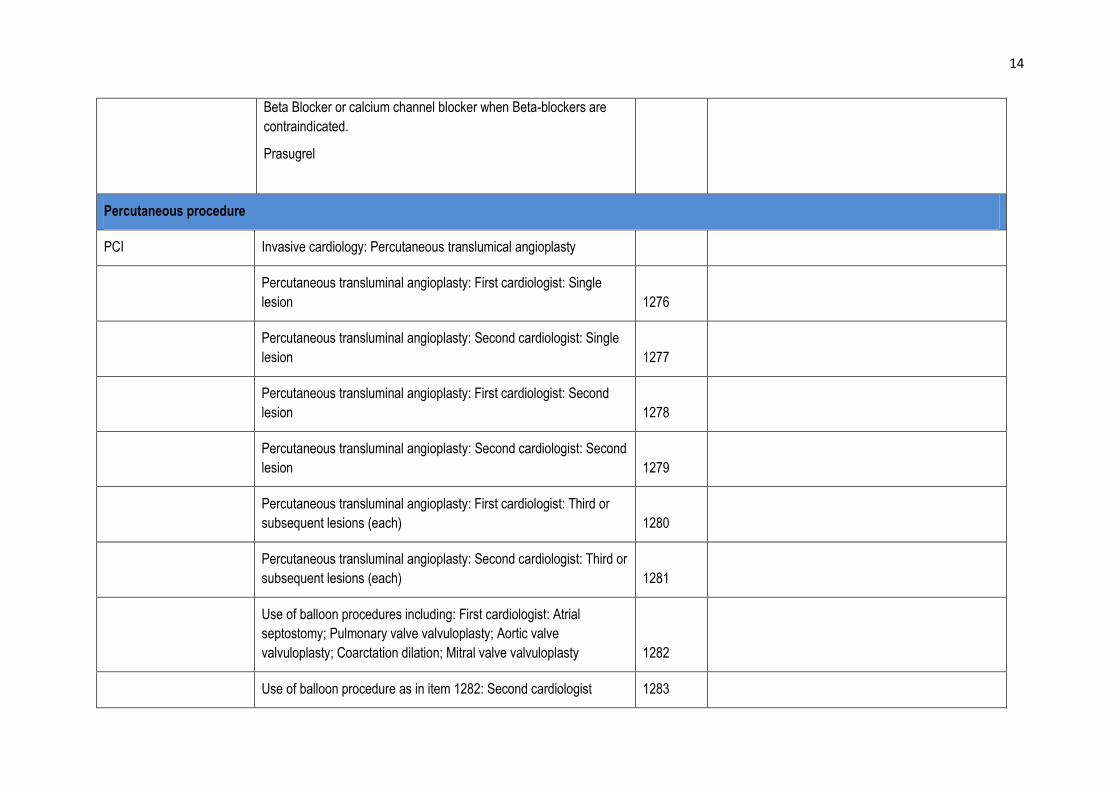
14
Beta Blocker or calcium channel blocker when Beta-blockers are
contraindicated.
Prasugrel
Percutaneous procedure
PCI Invasive cardiology: Percutaneous translumical angioplasty
Percutaneous transluminal angioplasty: First cardiologist: Single
lesion 1276
Percutaneous transluminal angioplasty: Second cardiologist: Single
lesion 1277
Percutaneous transluminal angioplasty: First cardiologist: Second
lesion 1278
Percutaneous transluminal angioplasty: Second cardiologist: Second
lesion 1279
Percutaneous transluminal angioplasty: First cardiologist: Third or
subsequent lesions (each) 1280
Percutaneous transluminal angioplasty: Second cardiologist: Third or
subsequent lesions (each) 1281
Use of balloon procedures including: First cardiologist: Atrial
septostomy; Pulmonary valve valvuloplasty; Aortic valve
valvuloplasty; Coarctation dilation; Mitral valve valvuloplasty 1282
Use of balloon procedure as in item 1282: Second cardiologist 1283
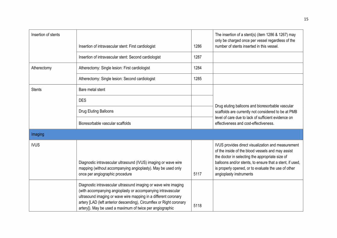
15
Insertion of stents
Insertion of intravascular stent: First cardiologist 1286
The insertion of a stent(s) (item 1286 & 1267) may
only be charged once per vessel regardless of the
number of stents inserted in this vessel.
Insertion of intravascular stent: Second cardiologist 1287
Atherectomy Atherectomy: Single lesion: First cardiologist 1284
Atherectomy: Single lesion: Second cardiologist 1285
Stents Bare metal stent
Drug eluting balloons and bioresorbable vascular
scaffolds are currently not considered to be at PMB
level of care due to lack of sufficient evidence on
effectiveness and cost-effectiveness.
DES
Drug Eluting Balloons
Bioresorbable vascular scaffolds
Imaging
IVUS
Diagnostic intravascular ultrasound (IVUS) imaging or wave wire
mapping (without accompanying angioplasty). May be used only
once per angiographic procedure 5117
IVUS provides direct visualization and measurement
of the inside of the blood vessels and may assist
the doctor in selecting the appropriate size of
balloons and/or stents, to ensure that a stent, if used,
is properly opened, or to evaluate the use of other
angioplasty instruments
Diagnostic intravascular ultrasound imaging or wave wire imaging
(with accompanying angioplasty or accompanying intravascular
ultrasound imaging or wave wire mapping in a different coronary
artery [LAD (left anterior descending), Circumflex or Right coronary
artery]). May be used a maximum of twice per angiographic 5118
16
procedure
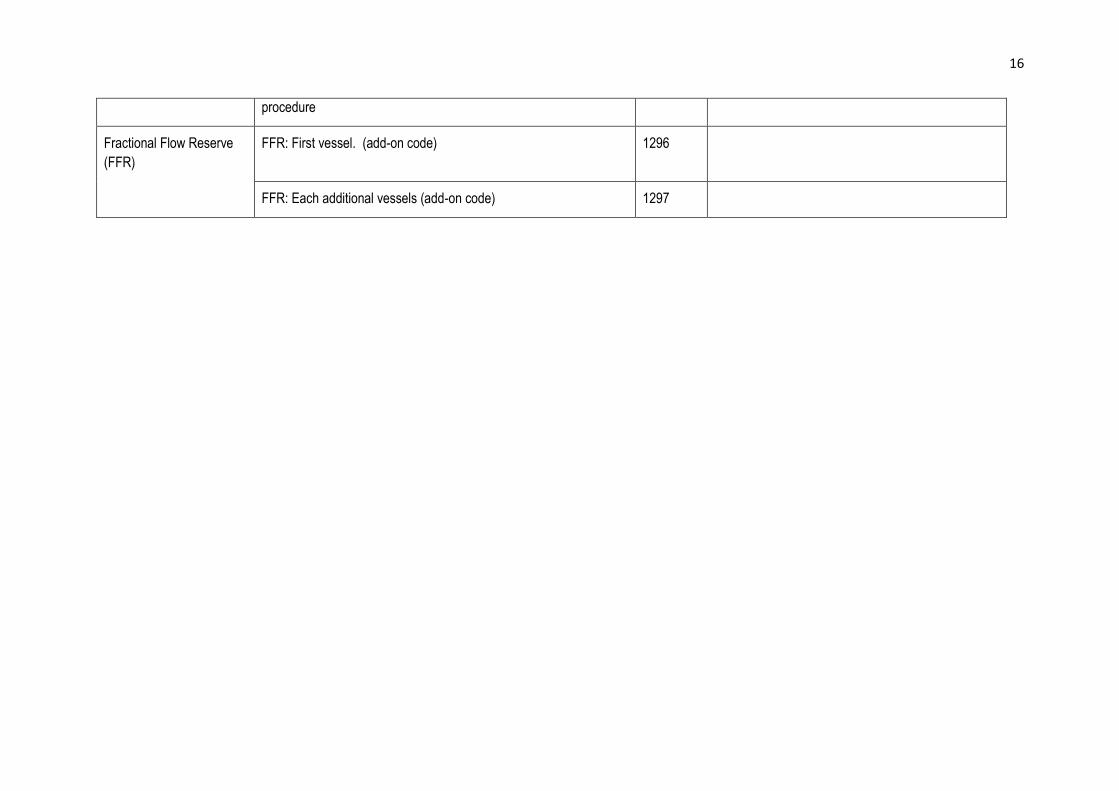
Fractional Flow Reserve
(FFR)
FFR: First vessel. (add-on code)
1296
FFR: Each additional vessels (add-on code) 1297
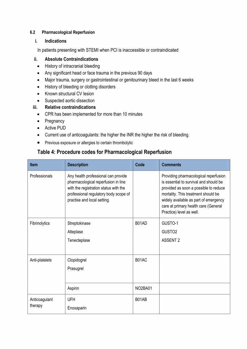
6.2 Pharmacological Reperfusion
i. Indications
In patients presenting with STEMI when PCI is inaccessible or contraindicated
ii. Absolute Contraindications
History of intracranial bleeding
Any significant head or face trauma in the previous 90 days
Major trauma, surgery or gastrointestinal or genitourinary bleed in the last 6 weeks
History of bleeding or clotting disorders
Known structural CV lesion
Suspected aortic dissection
iii. Relative contraindications
CPR has been implemented for more than 10 minutes
Pregnancy
Active PUD
Current use of anticoagulants: the higher the INR the higher the risk of bleeding.
Previous exposure or allergies to certain thrombolytic
Table 4: Procedure codes for Pharmacological Reperfusion
Item Description Code Comments
Professionals Any health professional can provide
pharmacological reperfusion in line
with the registration status with the
professional regulatory body scope of
practise and local setting.
Providing pharmacological reperfusion
is essential to survival and should be
provided as soon a possible to reduce
mortality. This treatment should be
widely available as part of emergency
care at primary health care (General
Practice) level as well.
Fibrinolytics Streptokinase
Alteplase
Tenecteplase
B01AD GUSTO-1
GUSTO2
ASSENT 2
Anti-platelets Clopidogrel
Prasugrel
B01AC
Aspirin NO2BA01
Anticoagulant
therapy
UFH
Enoxaparin
B01AB
18
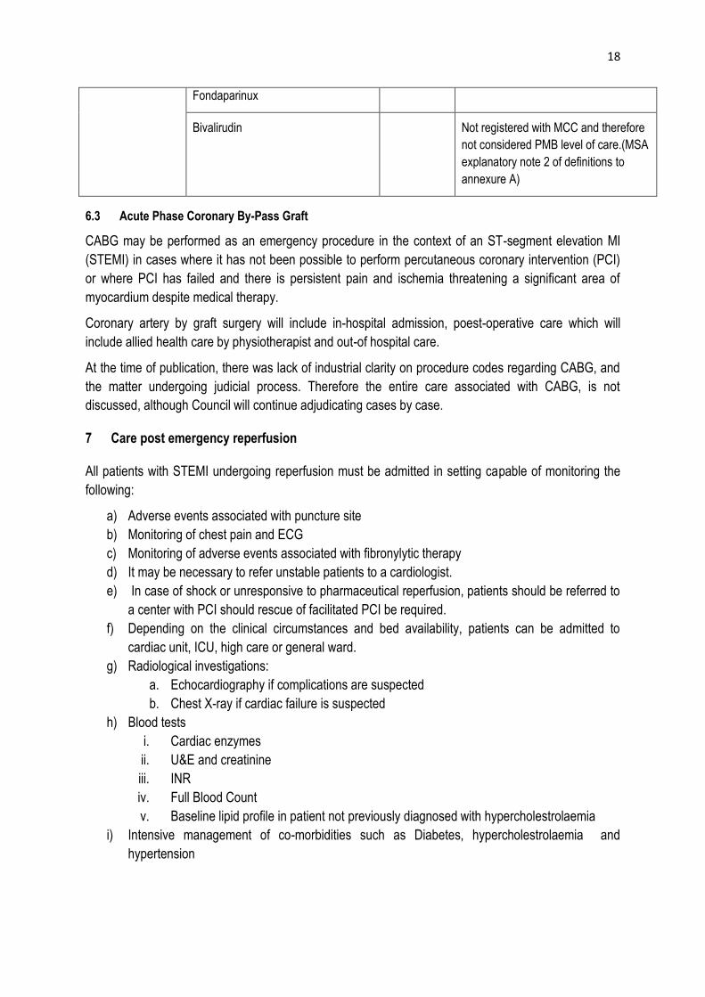
Fondaparinux
Bivalirudin Not registered with MCC and therefore
not considered PMB level of care.(MSA
explanatory note 2 of definitions to
annexure A)
6.3 Acute Phase Coronary By-Pass Graft
CABG may be performed as an emergency procedure in the context of an ST-segment elevation MI
(STEMI) in cases where it has not been possible to perform percutaneous coronary intervention (PCI)
or where PCI has failed and there is persistent pain and ischemia threatening a significant area of
myocardium despite medical therapy.
Coronary artery by graft surgery will include in-hospital admission, poest-operative care which will
include allied health care by physiotherapist and out-of hospital care.
At the time of publication, there was lack of industrial clarity on procedure codes regarding CABG, and
the matter undergoing judicial process. Therefore the entire care associated with CABG, is not
discussed, although Council will continue adjudicating cases by case.
7 Care post emergency reperfusion
All patients with STEMI undergoing reperfusion must be admitted in setting capable of monitoring the
following:
a) Adverse events associated with puncture site
b) Monitoring of chest pain and ECG
c) Monitoring of adverse events associated with fibronylytic therapy
d) It may be necessary to refer unstable patients to a cardiologist.
e) In case of shock or unresponsive to pharmaceutical reperfusion, patients should be referred to
a center with PCI should rescue of facilitated PCI be required.
f) Depending on the clinical circumstances and bed availability, patients can be admitted to
cardiac unit, ICU, high care or general ward.
g) Radiological investigations:
a. Echocardiography if complications are suspected
b. Chest X-ray if cardiac failure is suspected
h) Blood tests
i. Cardiac enzymes
ii. U&E and creatinine
iii. INR
iv. Full Blood Count
v. Baseline lipid profile in patient not previously diagnosed with hypercholestrolaemia
i) Intensive management of co-morbidities such as Diabetes, hypercholestrolaemia and
hypertension

19
8 Post discharge follow-up
Longer-term issues post-PCI are very patient-specific and variable but broadly involves detection and
treatment of recurrent ischaemia, arrhythmias and heart failure, appropriate antiplatelet therapy and
secondary prevention.
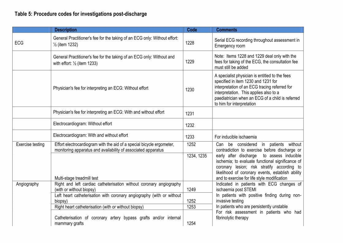
Table 5: Procedure codes for investigations post-discharge
Description Code Comments
ECG General Practitioner's fee for the taking of an ECG only: Without effort:
½ (item 1232) 1228 Serial ECG recording throughout assessment in Emergency room
General Practitioner's fee for the taking of an ECG only: Without and
with effort: ½ (item 1233) 1229 Note: Items 1228 and 1229 deal only with the fees for taking of the ECG, the consultation fee must still be added
Physician's fee for interpreting an ECG: Without effort 1230
A specialist physician is entitled to the fees specified in item 1230 and 1231 for interpretation of an ECG tracing referred for interpretation. This applies also to a paediatrician when an ECG of a child is referred to him for interpretation
Physician's fee for interpreting an ECG: With and without effort 1231
Electrocardiogram: Without effort 1232
Electrocardiogram: With and without effort 1233 For inducible ischaemia
Exercise testing Effort electrocardiogram with the aid of a special bicycle ergometer, monitoring apparatus and availability of associated apparatus
1252 Can be considered in patients without contradiction to exercise before discharge or early after discharge to assess inducible ischemia; to evaluate functional significance of coronary lesion; risk stratify according to likelihood of coronary events, establish ability and to exercise for life style modification Multi-stage treadmill test
1234, 1235
Angiography Right and left cardiac catheterisation without coronary angiography (with or without biopsy) 1249
Indicated in patients with ECG changes of ischaemia post STEMI In patients with positive finding during non-invasive testing In patients who are persistently unstable For risk assessment in patients who had fibrinolytic therapy
Left heart catheterisation with coronary angiography (with or without biopsy) 1252
Right heart catheterisation (with or without biopsy) 1253
Catheterisation of coronary artery bypass grafts and/or internal mammary grafts 1254

21
Echocardiography Cardiac examination plus Doppler colour mapping 3620 It is indicated in patients with STEMI when there is a negative change in clinical status. It is reasonable to repeat the procedure in 1 to 3 months time. It is used to assess and re-evaluate LV function and to evaluate suspected complications. It can be used in patient with suspected RV infarction and inferior STEMI.
Cardiac examination (MMode) 3621
Cardiac examination: 2 Dimensional 3622
Cardiac examination + effort 3623
Cardiac examinations + contrast 3624
Cardiac examinations + doppler 3625
Cardiac examination + phonocardiography 3626
Pharmacological stress testing

9 Secondary prevention for STEMI Patients
Secondary prevention is a prescribed minimum benefit and constitutes the following
i. Lifestyle modification (7)
All persons with risk factors for ischaemic heart disease should be encouraged to make the
following lifestyle changes as appropriate:
Smoking cessation.
Weight reduction in overweight patients, i.e. BMI > 25 kg/m2
Maintain ideal weight, i.e. BMI < 25 kg/m
Reduce alcohol intake to no more than 2 standard drinks/day
Follow a prudent eating plan i.e. Low saturated fat, high fibre and unrefined carbohydrates,
with adequate fresh fruit and vegetables.
Moderate aerobic exercise, e.g. 30 minutes brisk walking at least 3 times a week
Members must be encouraged to participate in wellness and prevention activities as offered
by the scheme in line with scheme rules.
ii. Lipid lowering agents
The 2012 Essential drug list recommends lipid lowering agents in all Ischaemic heart disease
irrespective of cholesterol and triglyceride plasma concentration. The intention is to reduce LDL
by at least 25%.
iii. Control of Diabetes
Maintain to HbA1 C < 7%.
iv. Antiplatelets agents
Post STEMI patients must receive dual antiplatelet therapy. Aspirin must be continued indefinitely.
Clopidogrel must be used for at least a month if bare metal stents were used and for 6 to 12 months if
drug eluting stents were used.
v. Blood pressure control
The main aim is to maintain BP at < 140/90 or < 130/80 in patients with chronic kidney disease and
diabetes mellitus.
Antihypertensive as per scheme’s formulary and CDL algorithm must be used however this should
include beta blockerS and angiotensin converting enzyme (ACE) inhibitors as a minimum benefit.
23
10 Bibliography
1. Risk factors associated with myocardial infarct in Africa-The INTERHEART study. Steyn, K, Silwa, K and
Hawkins, S. 2005, Circulation, pp. 3554-61.
2. Budern of non-communicable disease in South Africa. Mayosi, B, et al. s.l. : Lancet, 2009, Vol. 374.
3. Initial burden of disease estimates for South Africa. 2000. Bradshaw, D, Groenewald, P and Laubscher, R.
s.l. : S Afr Med J, 2003, Vol. 93.
4. A hidden menace: Cardiovascular disease in South Africa and the costs of an inadequate policy response. •
Maredza M, Hofman KJ, Tollman SM. A hidden menace: Cardiovascular disease in South Africa and the
costs of an inadequate policy response. SAHeart 2011, et al. s.l. : SAHeart, 20``, Vol. 8.
5. ESC Guidelines for the management of acute myocardial infarction in patients presentingwith ST-segment
elevation . The Task Force on the management of ST-segment elevation acute. s.l. : European Heart
Journal, 2012, Vol. 33. doi:10.1093.
6. Guidelines for the management of Patients with ST-Elevation Infarction-Executive summary: A reprot of the
American Cardiology/American Heart Association Task Force on Practice guidelines. American Heart
Association. s.l. : Circulation, 2004, Vol. 110. doi:10.116/01.CIR00001347891.68010.FA.
7. Department of Health. Hospital Essential Drug List. 2012.