
Dr. Tamara Alqudah
67
Physiology Lab Dr. Tamara Alqudah
Transcript of Dr. Tamara Alqudah

Physiology LabDr. Tamara Alqudah

The Experiments
• Red Blood Cell (RBC) Count
• White Blood Cell (WBC) Count
• Differential Leukocyte count (DLC)
• Reticulocyte count
• Packed cell volume (PCV)
• Hemoglobin concentration
• Erythrocyte Sedimentation rate (ESR)
• Blood Type
• Bleeding Time
• Clotting Time
• Osmotic Fragility Test

Red Blood Cells (RBCs)• Normal RBCs are biconcave discs, they have few organelles and
no nuclei.
• A major function of RBCs is to transport hemoglobin, which in
turn carries oxygen from the lungs to the tissues.
• The average number of RBCs in healthy men is 5,200,000/mm3
(±300,000) and in healthy women 4,700,000/mm3 (±300,000)
• The number of RBCS is regulated within narrow limits, so that
oxygen is transported adequately to the tissues and at the same
time the cells do not become so numerous that they impede
blood flow.

• RBC count is typically ordered as a part of complete blood count (CBC) and may be used as a part of health checkup to screen for variety of conditions.
• Causes of high RBC count (Polycythemia)
1. Living at high altitudes
2. Cardiac or pulmonary diseases
3. Erythropoietin secreting tumors
4. Smoking.
5. Polycythemia Vera
6. Dehydration
• Causes of low RBC count (Anemia)
1. Internal or external bleeding
2. Nutritional deficiencies
3. Bone marrow failure
4. Hemolysis of RBCs
5. Chronic renal failure

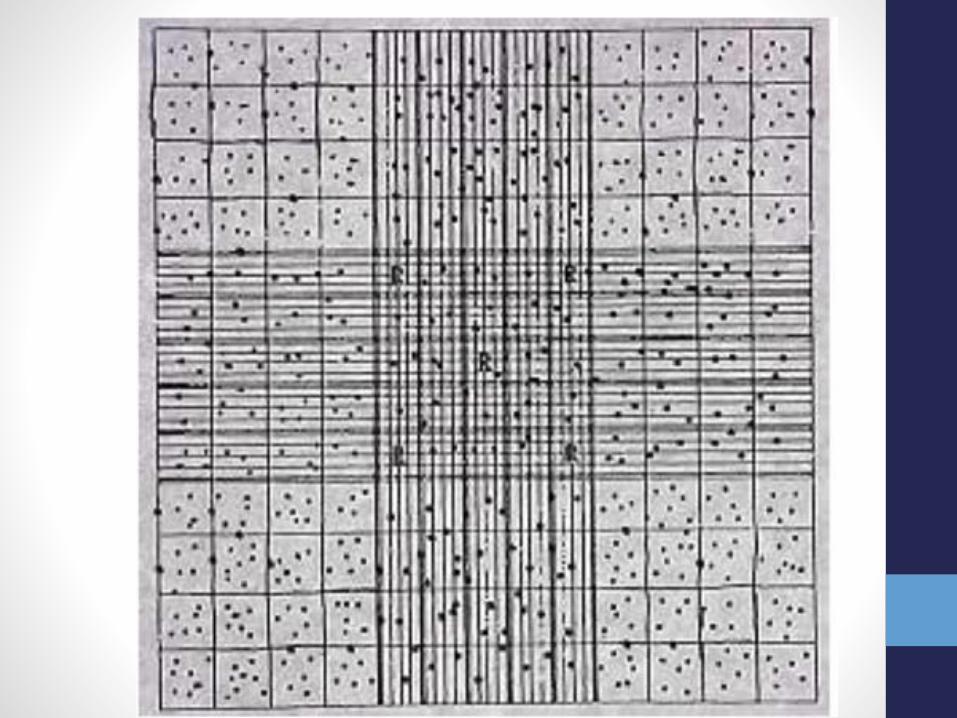
• Hemocytometer is a special microscopic slide that has specific grids
engraved on it’s counting chamber and is designed to hold a specific
volume of fluid.


1mm 0.2mm
3mm
3mm
Orientation
lines
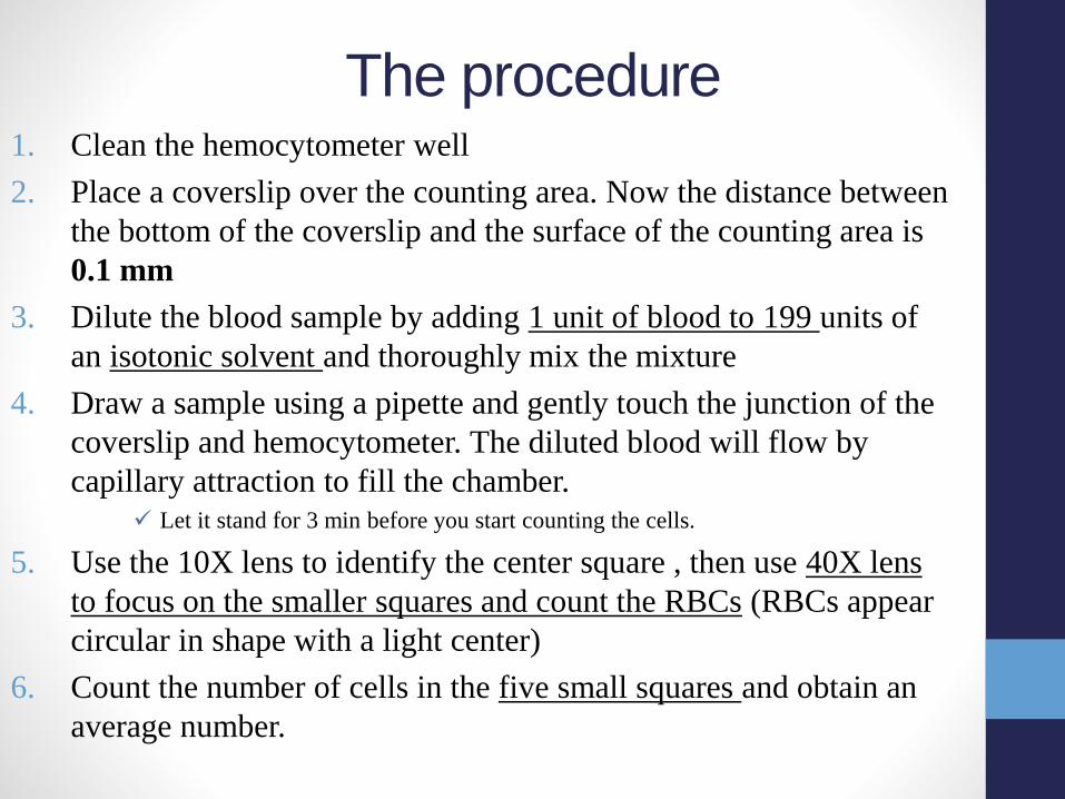
The procedure1. Clean the hemocytometer well
2. Place a coverslip over the counting area. Now the distance between
the bottom of the coverslip and the surface of the counting area is
0.1 mm
3. Dilute the blood sample by adding 1 unit of blood to 199 units of
an isotonic solvent and thoroughly mix the mixture
4. Draw a sample using a pipette and gently touch the junction of the
coverslip and hemocytometer. The diluted blood will flow by
capillary attraction to fill the chamber. ✓ Let it stand for 3 min before you start counting the cells.
5. Use the 10X lens to identify the center square , then use 40X lens
to focus on the smaller squares and count the RBCs (RBCs appear
circular in shape with a light center)
6. Count the number of cells in the five small squares and obtain an
average number.

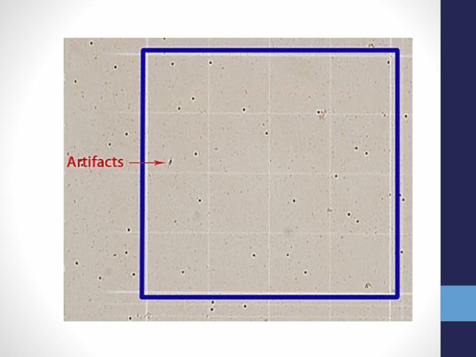
Count all the cells contained within the
square and those touching the upper and
right outer lines. The cells that touch the
left and bottom outer lines are not to be
counted. In each of the four areas, conduct
the count in a zig-zag line.

• Start counting from the left to the right and proceed in a zig-zag.
•To avoid counting the same cells twice, cells that are touching the lines at the tops and left sides of the squares are counted, but cells that are touching the bottoms and right sides of the squares are not counted.

• If the RBC count was (100, 90, 94, 96, 95). What is the RBC count
in the blood sample?
1. In this case the average RBC count is 95
2. Dilution factor (DF) = Final volume/ volume of blood
• Blood is diluted at (1:199 ) so DF= 200
3. The volume of fluid contained in one small square =
(0.2x 0.2 x 0.1)= 0.004 mm3
• Volume Correction Factor (VCF)= Desired Volume/ Actual volume
• Desired volume = 1 mm3
• VCF = 1/0.004 = 250
• The number of RBCs in blood sample= The average number
of RBCs X DF X VCF = 95 X 200 X 250 = 4,750,000
cells/mm3

Important Points
• Before you obtain the average number of RBCS make sure the
count in the five squares doesn't vary by more than 20 cells.
• If there is a big variation discard the sample from the slide and
repeat the experiment.
• DF can change based on the dilution performed during the
experiment
• VCF is always the same
WBC count
• White Blood Cells are part of the immune system
• Move to areas of severe infection or inflammation to provide a rapid and potent defense for the body
• Normal WBC count is 4000 - 11,000 cells/mm3
• This test is often included in the complete blood count (CBC), it is done to get an impression about the immune system since the Leukocytes (WBC) play an integral role, to get more informative results it is often combined with the differential count

➢Causes of High WBC count (Leukocytosis)
1. Active inflammation or infection.
2. Certain malignancies
3. Recent vigorous exercise, thermal burn, electric shock, surgery, or
trauma.
4. Certain medications e.g. glucocorticoids (neutrophilia)
5. Dehydration.
➢Causes of Low WBC count (Leukopenia)
1. Bone marrow failure due to aplastic anemia, fibrosis, metastatic
cancer, radiotherapy or chemotherapy
2. Autoimmune diseases.
3. Infections like HIV & tuberculosis.

The procedure1. Clean the hemocytometer well
2. Place a coverslip over the counting area.
3. Dilute the blood sample by adding 1 unit of blood to 19 units of
solvent and thoroughly mix the mixture. ✓ The dilution fluid contains an agent (glacial acetic acid) which lyses the red
cells. It also contains a dye that stains the nuclei of WBCs. This allows a proper
count of WBCs.
4. Draw a sample using a pipette and gently touch the junction of
the coverslip and hemocytometer . The diluted blood will flow
by capillary attraction to fill the chamber.
✓Let it stand for 3 min before you count the cells.
5. Use the 10X lens to count the WBC in the four large corner
squares .(WBCs appear as dark dots)

The calculation1. Blood is diluted at (1:19 ) so DF = 20
2. The volume of fluid in the corner square is (1 X 1 X 0.1= 0.1 mm3)
SO the VCF is 10
✓ If we counted an average of 40 cells in the 4 squares the count of
WBCs is….
40 X 20 X 10 = 8000 cells/mm3 which is a normal value
•Before you obtain the average number of WBCS make sure the count in the
four squares doesn't vary by more than 10 cells
Differential Leukocyte Count (DLC)
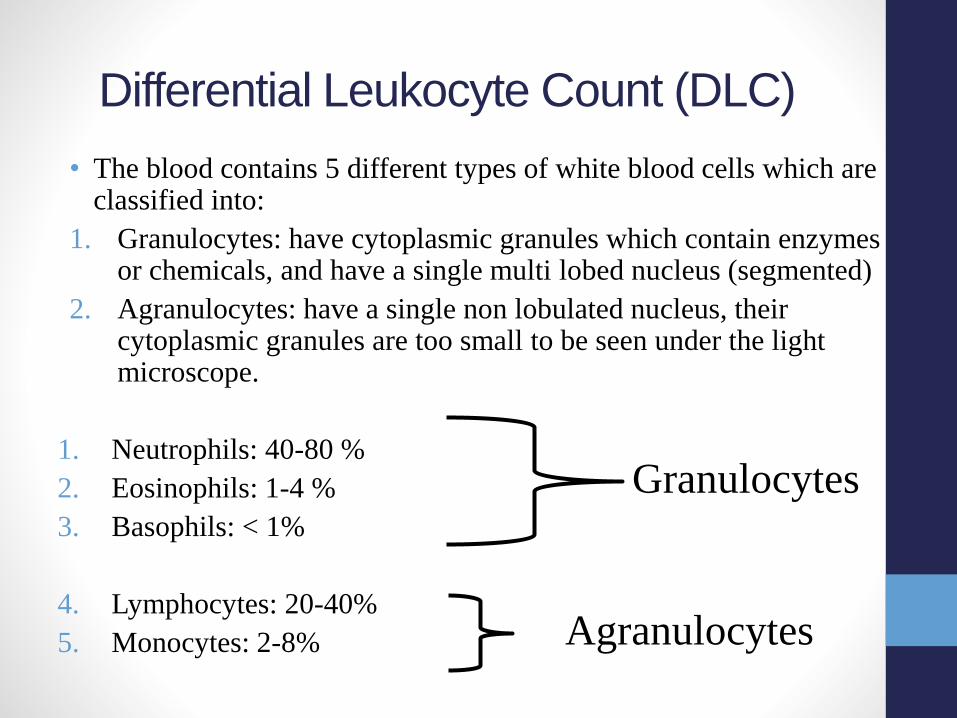
• The blood contains 5 different types of white blood cells which are classified into:
1. Granulocytes: have cytoplasmic granules which contain enzymes or chemicals, and have a single multi lobed nucleus (segmented)
2. Agranulocytes: have a single non lobulated nucleus, their cytoplasmic granules are too small to be seen under the light microscope.
1. Neutrophils: 40-80 %
2. Eosinophils: 1-4 %
3. Basophils: < 1%
4. Lymphocytes: 20-40%
5. Monocytes: 2-8%
Granulocytes
Agranulocytes
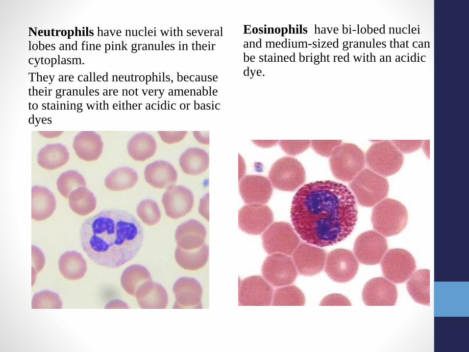
Neutrophils have nuclei with several lobes and fine pink granules in their cytoplasm.
They are called neutrophils, because their granules are not very amenable to staining with either acidic or basic dyes
Eosinophils have bi-lobed nuclei and medium-sized granules that can be stained bright red with an acidic dye.

Basophils have bi-lobed or S shaped
nuclei and large granules which stain
dark blue with basic dyes and
completely obscure the nucleus
Neutrophilic Band cells are
immature neutrophils, usually
make less than 5% of the total
WBC count, their nucleus isn’t
segmented
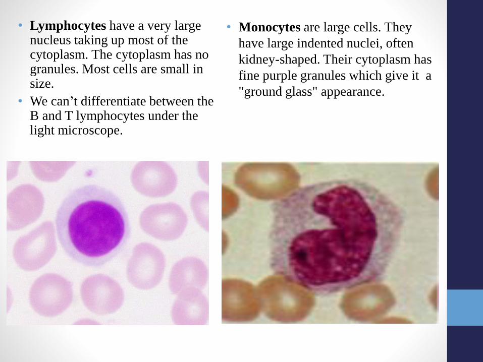
• Monocytes are large cells. They
have large indented nuclei, often
kidney-shaped. Their cytoplasm has
fine purple granules which give it a
"ground glass" appearance.
• Lymphocytes have a very large nucleus taking up most of the cytoplasm. The cytoplasm has no granules. Most cells are small in size.
• We can’t differentiate between the B and T lymphocytes under the light microscope.

The procedure1. A drop of blood is thinly spread over a glass slide, air dried,
and stained with an acidic dye (red) and a basic dye (blue-
purple).
2. The slide is examined under a microscope using an oil
immersion lens.
3. Two hundred white cells are then counted and classified.
4. The number of each type of cells is expressed as a
percentage.
• To do this one must be able to distinguish between the 5 types
of WBCs
Importance of DLC
• Gives relative percentage of each type of WBC
• Helps reveal the presence of abnormal WBCs like blasts or
lymphoma cells.
• Used along with WBC count to generate an absolute value
for each type of WBCs.
• Relative percentages can be misleading
• Absolute values are also useful for monitoring certain conditions.
• Absolute count =WBC (cells/ µL) x percent of the specific WBC
type ÷100

Absolute count calculation• If the WBC count is 6000 cells/mm3 and the lymphocytes
make 30% of the DLC, the Absolute lymphocyte count (ALC)
will be:
WBC count x (Lymphocyte%)/100 =
(6000 X 30)/100 = 1800 cells/mm3
• Absolute neutrophil count (ANC)=WBC (cells/ µL) x percent
(neutrophils + neutrophilic band cells) ÷100
1. Neutrophilic leukocytosis: is defined as a total WBC
above 11,000/µL along with an absolute neutrophil count (ANC)
greater than 7700/µL
• Bacterial infections, inflammatory conditions, stress.
2. Lymphocytic leukocytosis : is defined as a total WBC
above 11,000/µL along with an absolute lymphocyte count greater than
4500/µL
• Viral infections as infectious mononucleosis, mumps, rubella and pertussis
or in acute and chronic lymphocytic leukemias.
3. Monocytic leukocytosis:
• Acute or chronic bacterial infection and chronic inflammation
4. Eosinophilic leukocytosis :
• Parasitic infections & allergic conditions

6. Basophilic leukocytosis:
• Allergic conditions
7. Neutropenia : absolute neutrophil count is less than 1,500
cells/ mm3
• Certain infections like typhoid fever, HIV & CMV, chemotherapy,
radiotherapy, and autoimmune diseases.
8. Lymphocytopenia:
• May occur in the normal elderly or be associated with chronic
infection or malignancy.

Reticulocyte Count• Reticulocytes are the immediate precursor of RBCs, following
their release to the blood stream they mature within 1-2 days
into RBCs.
• Contain a small amount of basophilic material, mainly remnants
of the Golgi apparatus & mitochondria
• They normally make less than1-2% of all RBCs
• Used to estimate the degree of effective erythropoiesis
• Their number increases in cases of bleeding and RBC hemolysis
and decreases in cases of bone marrow failure

If supravital staining (new methelene blue) is performed on a blood smear,
the reticulocytes appear larger than RBCs and contain dark blue dots and
curved linear structures in their cytoplasm (remnants of ribosomes).

The procedure➢500-1000 RBCs should be
counted and the number of reticulocytes noted. The count is expressed as a percentage which can be used to calculate the absolute reticulocyte count (ARC) .
➢ ARC accurately reflects the degree of reticulocytosisregardless of the degree of anemia. The normal absolute reticulocyte count is between 25,000 to 75,000/mm3
➢ ARC = (RBC count X reticulocyte%)/100

Reticulocytosis and Reticulocytopenia
• Condition associated with an increase in reticulocytes:
• Hemolytic anemias: Immune hemolytic anemia, RBC membrane
defects, Sickle cell diseases,
• Following hemorrhage
• Following treatment of anemias
• Condition associated with a decrease in reticulocytes:
• Iron deficiency anemia
• Aplastic anemia
• Radiation therapy
• Tumor in bone marrow
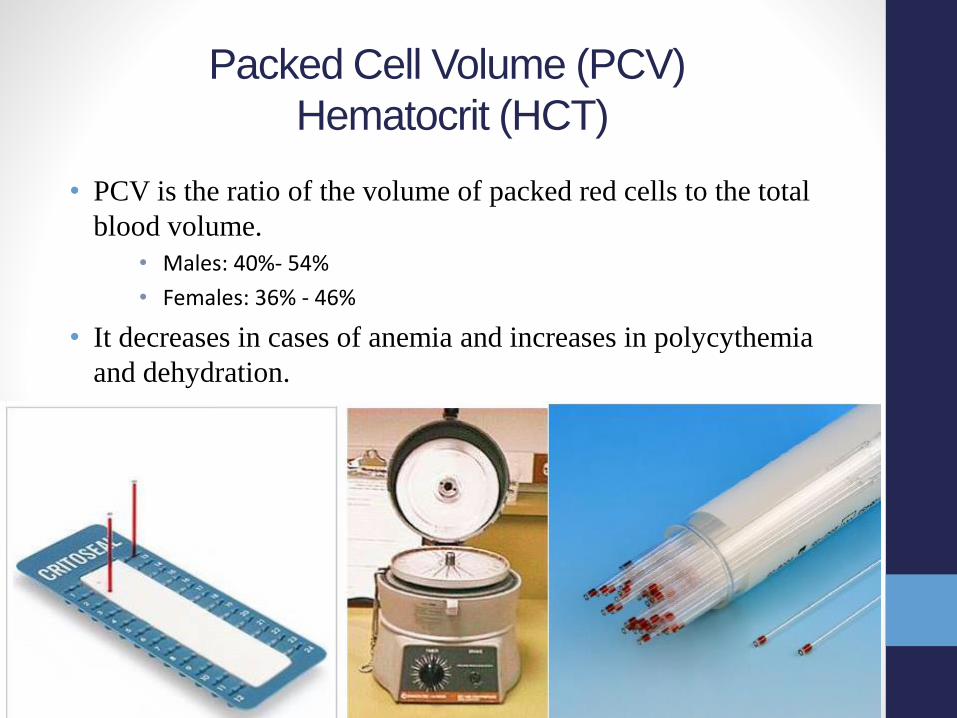
Packed Cell Volume (PCV)
Hematocrit (HCT)
• PCV is the ratio of the volume of packed red cells to the total
blood volume.
• Males: 40%- 54%
• Females: 36% - 46%
• It decreases in cases of anemia and increases in polycythemia
and dehydration.

• A blood sample is
centrifuged in a heparinized
capillary tube (red tip ),
• The RBCs become packed
at the bottom of the tube.
• The PCV is then calculated
according to the following
formula:
• PCV= RBC height x100Total height
• Beware not to include the
buffy coat
The procedure
Hemoglobin Concentration• Hemoglobin is a globular protein made up of four subunits. Each
subunit contains a heme group conjugated to a polypeptide.
Heme is an iron-containing porphyrin derivative.
• Heme has the ability to bind oxygen reversibly and carry it to
tissues.
➢Normal values of hemoglobin
• 14-17.5 g/ 100 ml in males
• 12-15 g/ 100 ml of in females
➢Different methods can be used to find the hemoglobin
concentration one of them is Sahli’s method.
➢Based on the fact that when blood is mixed with HCl,
hemoglobin is converted to acid hematin which is brown in color
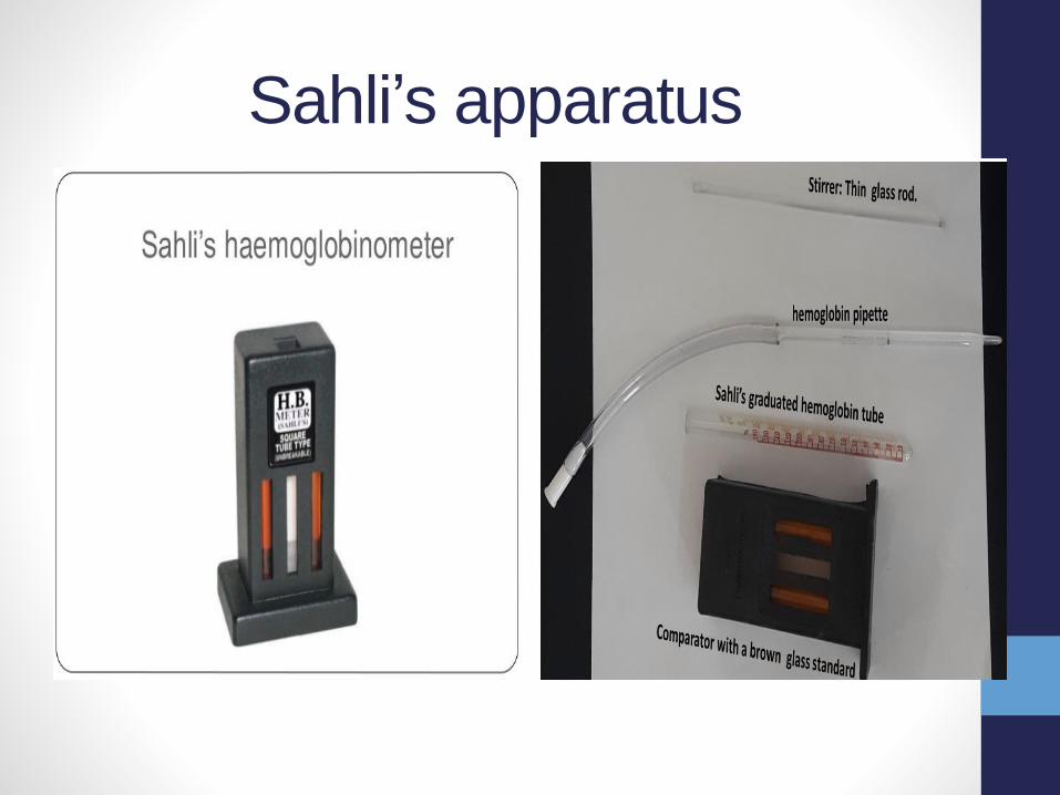
Sahli’s apparatus


The procedure1. Add HCl into the tube up to 2g% mark
2. Mix the EDTA sample gently and fill the pipette with 20 Ul blood.
3. Wipe the external surface of the pipette to remove any excess blood.
4. Add the blood into the tube containing HCl. Wash out the contents of the hemoglobin pipette by drawing in and blowing out the acid few times so that the blood is mixed with the acid thoroughly.
5. Allow to stand undisturbed for 10 min. (This is because, maximum conversion of hemoglobin to acid hematin, occurs in the first ten minutes)
6. Place the hemoglobinometer tube in the comparator and add distilled water to the solution drop by drop stirring with the glass rod until it’s colour matches that of the comparator glass.
7. Remove the stirrer and take the reading directly
➢Hemoglobin concentration is read directly from the graduated scale on the dilution tube.


Erythrocyte Sedimentation Rate
(ESR)• The rate at which RBCs sediment in a period of one hour.
• The ESR is a simple non-specific screening test that indirectly
measures the presence of inflammation in the body.
• It reflects the tendency of red blood cells to settle more rapidly
in the presence of some disease states, usually because of
increases in plasma fibrinogen, immunoglobulins, and other
acute-phase reaction proteins.
• Changes in red cell shape or numbers may also affect the ESR.

• In our lab we use the Wintrobe
tube which is 100 mm long.
• EDTA anticoagulated blood is
drawn into the Wintrobe tube till
the zero mark
• The tube is placed in its rack in a
strictly vertical position for 1
hour at room temperature
• the RBCs – under the influence of
gravity - settle out from the
plasma.
• The rate at which they settle is
measured as the number of
millimeters of clear plasma
present at the top of the column
after one hour (mm/hr).
The procedure
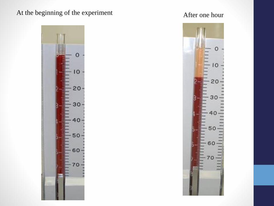
At the beginning of the experiment After one hour

RBCs sedimentation• The RBCs sediment because their density is greater
than that of plasma. The sedimentation increases
with stacking of RBCs (rouleaux formation)
• Rouleaux formation is possible because of the
discoid shape of RBCs
• Normally, RBCs have negative charges on the
outside of the cells, which cause them to repel each
other.
• Many plasma proteins have positive charges and can
neutralize the negative charges of the RBCs, which
allows for the formation of the rouleaux.
• Therefore, an increase in plasma proteins (present in
inflammatory conditions) will increase the rouleaux
formations, which settle more readily than single red
blood cells

•Normal ESR values• Men < 15mm/hr
• Women < 20mm/hr
• High ESR
➢ Inflammation
➢Anemia
➢Old age
➢Pregnancy
➢Technical factors: tilted ESR tube, high room temperature.
• Some interferences which decrease ESR:
• Abnormally shaped RBC (sickle cells and spherocytosis)
• Polycythemia
• Technical factors: low room temperature, delay in test performance
(>2 hours), clotted blood sample

RDW (Red Cell Distribution Width)
MPV Mean platelet volume

• At least 30 commonly occurring antigens and hundreds of
other rare antigens composed of glycoproteins and glycolipids
are found on the surface of RBCs.
• Each of which can at times cause antigen- antibody reactions
leading to immediate or delayed agglutination and hemolysis
of RBCs.
• Most of the antigens are weak.
• Two particular types of antigens (agglutinogens) are likely to
cause blood transfusion reactions: the ABO system of antigens
and the Rh system.
• Based on these two systems we have 8 blood groups:
• A +ve, A –ve, B +ve, B –ve, AB +ve, AB –ve, O +ve & O -ve
Blood Groups

ABO Blood Group
• The ABO blood group is based on two glycolipid antigens
called A and B.
• Blood plasma usually contains antibodies called agglutinins
that react with the A or B antigens. These are the anti-A
antibody, which reacts with antigen A, and the anti-B antibody,
which reacts with antigen B.
• Agglutinins start to appear in the blood within a few months
after birth.
• They are formed naturally. Their production is thought to be
stimulated when the immune system encounters the "missing"
ABO blood group antigens in food or in micro-organisms.

Rh blood group
• There are six common types of Rh antigens, each of which is
called an Rh factor. These types are designated C, D, E, c, d, and
e.
• The type D antigen is widely prevalent in the population and
considerably more antigenic than the other Rh antigens.
• Anyone who has this type of antigen is said to be Rh positive
(85% of population), whereas a person who doesn’t have type D
antigen is said to be Rh negative.
• In contrast to ABO system there is no preformed Anti-D in the
Rh–ve individual

Hemolytic disease of the newborn (HDN)
• Rh–ve mother is exposed to Rh +ve blood from the
fetus through the placenta during birth, abortion ,
or miscarriage.
• The mother will start to make anti-Rh antibodies.
• The firstborn baby usually is not affected.
• If the mother becomes pregnant again anti-Rh
antibodies can cross the placenta and enter the
bloodstream of the fetus. If the fetus is Rh +ve
agglutination and hemolysis occur in the fetal
blood leading to anemia and jaundice. The disease
- erythroblastosis fetalis or hemolytic disease of
the newborn- may result in fetal death.
• An injection of anti-Rh antibodies called anti-Rh
gamma globulin can be given to prevent HDN.
• Rh –ve women should receive it before delivery,
and soon after every delivery, miscarriage, or
abortion.

Determination of blood type
1. Prick the tip of a finger with a lancet and put three separate drops of blood on a clean microscopic slide.
2. Add one drop of Anti-A to the first drop, Anti-B to the second drop, and Anti-D to the third drop.
3. Mix well, using separate wooden sticks.
4. The results are read directly from the slide.
➢ If agglutination occurs in the first drop the blood type is A , if agglutination occur in the second drop the blood type is B, if it occurs in both it is AB and if it doesn't occur in any drop it is type O.
➢ If agglutination occurs in the Rh drop the blood is considered as Rh+ve. (This reaction might take some time to develop)
➢The strength of agglutination reaction is not the same in all people, so in some cases it may be necessary to examine the slide under the microscope to look for agglutination.


Type AB
Type A
Type O
Type B
Type A +ve

What is the type of blood in each
test presented below?
1.
2.
3.
4.

Hemostasis
• Hemostasis is prevention of blood loss from circulatory
system.
• Depends on the integrity of blood vessels, platelets and clotting
factors.
• The hemostatic response to vascular injury is achieved by several mechanisms:
1. Vasoconstriction 2. Formation of a platelet plug3. Formation of a blood clot
Bleeding time• A bleeding time is used to evaluate the second phase of hemostasis,
which involves adherence of the platelets to the injured vessel,
platelet activation and aggregation (formation of a plug).
✓The time measures how long it takes for a platelet plug to form.
✓ Normal range: 3-5 minutes
✓It increases when the platelets count is low (thrombocytopenia),
platelet function is abnormal or with the use of aspirin .
• Disadvantages: Insensitive, Invasive & operator dependent.

• Duke method
1. Clean the tip of the finger or the ear
lobe with alcohol.
2. Puncture the skin with a special
lancet. The wound should be 3–4 mm
deep.
3. Wipe the blood drop by a filter paper
every 30 seconds
4. Repeat until no more blood is
absorbed by the filter paper.
5. Multiply the number of blood drops
by 30 seconds
• Or divide the number of spots of blood
by 2 and that will give you the bleeding
time in minutes.

Clotting time
• It measures the time required for a blood sample to coagulate in
vitro. Clotting time depends on the availability of coagulation
factors.
• Normal value is 6-10 minutes.
• It is prolonged in conditions like hemophilia, vitamin K deficiency,
liver diseases, and warfarin overdose.

1. Clean the tip of the finger with alcohol then prick it with a lancet.
2. Draw blood into non-heparinized capillary tubes.
3. After 2 minutes, start breaking the capillary tubes to see whether a
thread of coagulated blood is formed between the two broken ends.
4. It is preferred to calculate the clotting time from the average of two
capillary tubes.
Non-heparinized Heparinized

Osmotic fragility• when RBCs reside in an isotonic medium, the intracellular and
extracellular fluids are in osmotic equilibrium across the cell
membrane, and there is no net influx or efflux of water.
• When RBCs reside in a hypotonic medium, a net influx of water
occurs so the cells swell and the integrity of their membranes is
disrupted resulting in hemolysis
• When RBCs reside in a hypertonic media , a net efflux of water
occurs so the cells lose their normal biconcave shape, undergoing
collapse.

280 mOsm/L

Osmotic fragility test
• A test designed to measures red blood cell’s resistance to
hemolysis when exposed to a series of increasingly dilute
saline solutions.
• The susceptibility of RBCs to hemolysis is a function of:
➢Surface area to volume ratio.
➢Cell membrane composition and integrity
• This test is mainly used to diagnose hereditary
spherocytosis but it is also used in some countries to
screen for thalassemia.

• The procedure:
1. Put labeled centrifuge tubes in a rack.
2. Prepare NaCl solutions of different concentrations starting from 0.9% NaCl till 0.2% NaCl.
3. Add 10 ml of each solution to a different tube then add one drop of blood to each tube.
4. Shake each tube well and allow them to stand for 20 minutes. After 20 minutes, the tubes are centrifuged for 10 minutes
5. Transfer supernatant fluid from each tube into spectrophotometer cuvettes
6. The absorbance is then measured at 540 nm and used to calculate the percentage of hemolysis for each solution.
7. The results are plotted against the NaCl concentrations, this yields an osmotic fragility curve which is then compared to a standard curve.

Hemolysis starts
Complete Hemolysis
• In this example• From 0.7% to 0.5% there is no hemolysis.• At the concentration of 0.45% hemolysis starts and the
solution becomes red in color, but there are some settled RBCs in the tube.
• At the concentration of 0.30%, the solution is bright red and there are no settled RBCs (complete hemolysis).

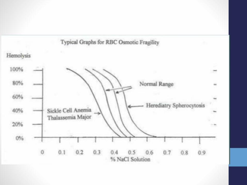

• Decreased red cell fragility (increased resistance to
hemolysis) is seen with the following conditions:➢ Thalassemia.
➢ Iron deficiency anemia.
➢ Sickle cell anemia
✓These cells have a high surface area: volume ratio
• Increased red cell fragility (increased susceptibility to hemolysis) is seen in the following conditions:
➢Hereditary spherocytosis
➢Autoimmune hemolytic anemia
➢ Toxic chemicals, poisons, infections, and some drugs.
➢ Severe burns.
✓These cells have a low surface area: volume ratio


