DR. SAIDUNNISA Professor and chairperson Department of Biochemistry Nucleic acid Structure.
36
DR. SAIDUNNISA Professor and chairperson Department of Biochemistry Nucleic acid Structure
-
Upload
adelia-francis -
Category
Documents
-
view
215 -
download
0
Transcript of DR. SAIDUNNISA Professor and chairperson Department of Biochemistry Nucleic acid Structure.
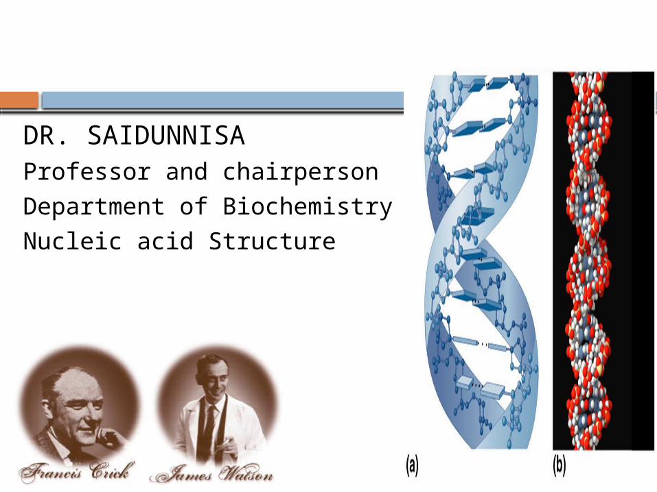
DR. SAIDUNNISAProfessor and chairperson
Department of Biochemistry
Nucleic acid Structure
Learning Objectives
At the end of the session student shall be able to:
Describe the biochemical structure of nucleosides, nucleotide and their different levels of organization into nuclear DNA, nucleosomes chromatin and chromosome.
Learn the different types of nucleosides and nucleotides and list the biologically important forms present in humans
List the synthetic analogs and their use in clinical medicine
DNA story
In 1869, Swiss biochemist Friedrich Miescher each morning, he called at the local clinic to pick up discarded surgical pus bandages. Adding alkali to burst open the nuclei of white blood cells, isolated a new substance he called it "nuclein".
On 25 April 1953, James Watson and Francis Crick, then at Cambridge University, reported the discovery of the structure of DNA (deoxyribonucleic acid) - the molecule that genes are made of, and acts as vehicles of genetic inheritance.
Friedrich Miescher in 1869
Isolated what he called nuclein from the nuclei of pus cells.
Nuclein was shown to have acidic properties, hence it became called nucleic acid
The distribution of nucleic acids in the eukaryotic cell
DNA is found in the nucleus.
Small amounts in mitochondria.
RNA is found throughout the cell.
Nucleic acids
There are two types of nucleic acids:
1. DNA (Deoxy ribonucleic acid)
2. RNA (Ribonucleic acid)
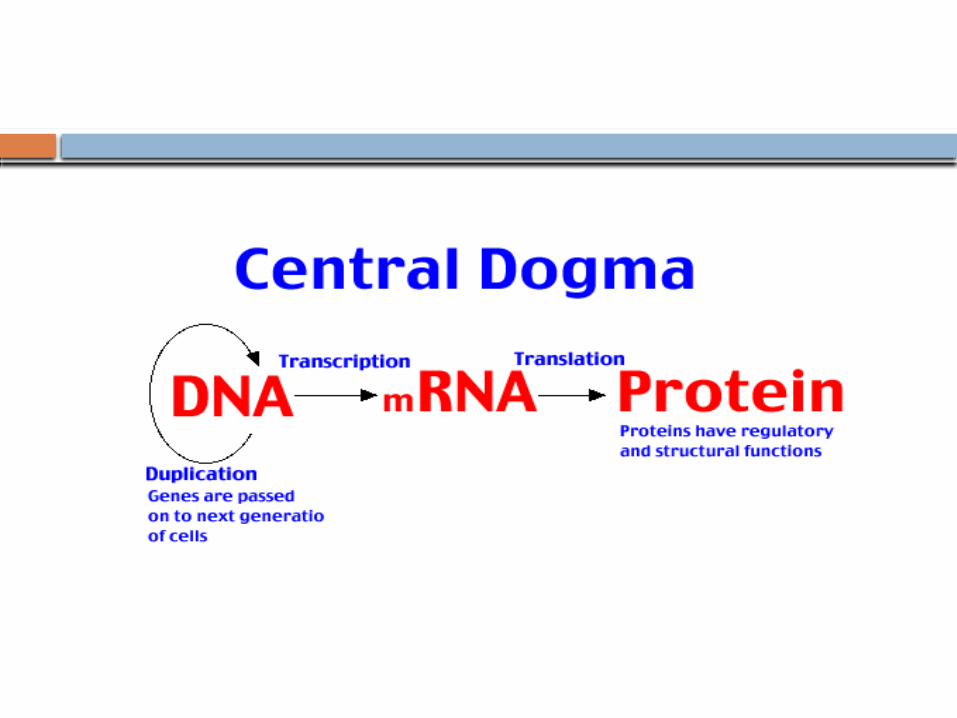
Serve as transmitters of genetic information.
Nucleic acids are the polymers of nucleotides.
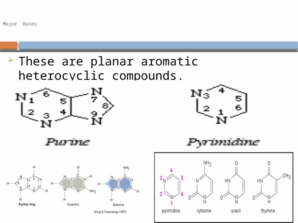
Major Bases
These are planar aromatic heterocyclic compounds.
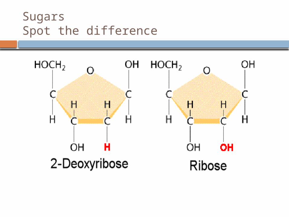
SugarsSpot the difference
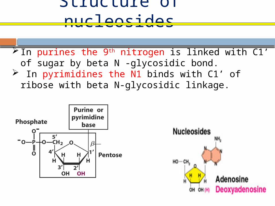
Structure of nucleosides
H
In purines the 9th nitrogen is linked with C1’ of sugar by beta N -glycosidic bond.
In pyrimidines the N1 binds with C1’ of ribose with beta N-glycosidic linkage.
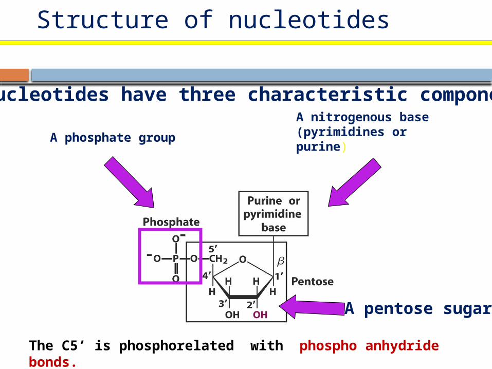
Structure of nucleotides
A phosphate group
Nucleotides have three characteristic components:A nitrogenous base(pyrimidines or purine)
A pentose sugar
The C5’ is phosphorelated with phospho anhydride bonds.
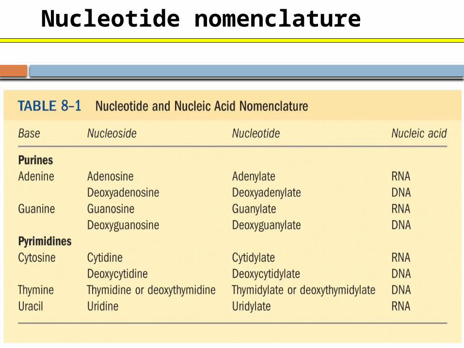
Nucleotide nomenclature
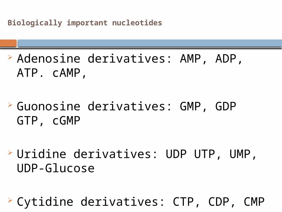
Biologically important nucleotides
Adenosine derivatives: AMP, ADP, ATP. cAMP,
Guonosine derivatives: GMP, GDP GTP, cGMP
Uridine derivatives: UDP UTP, UMP, UDP-Glucose
Cytidine derivatives: CTP, CDP, CMP
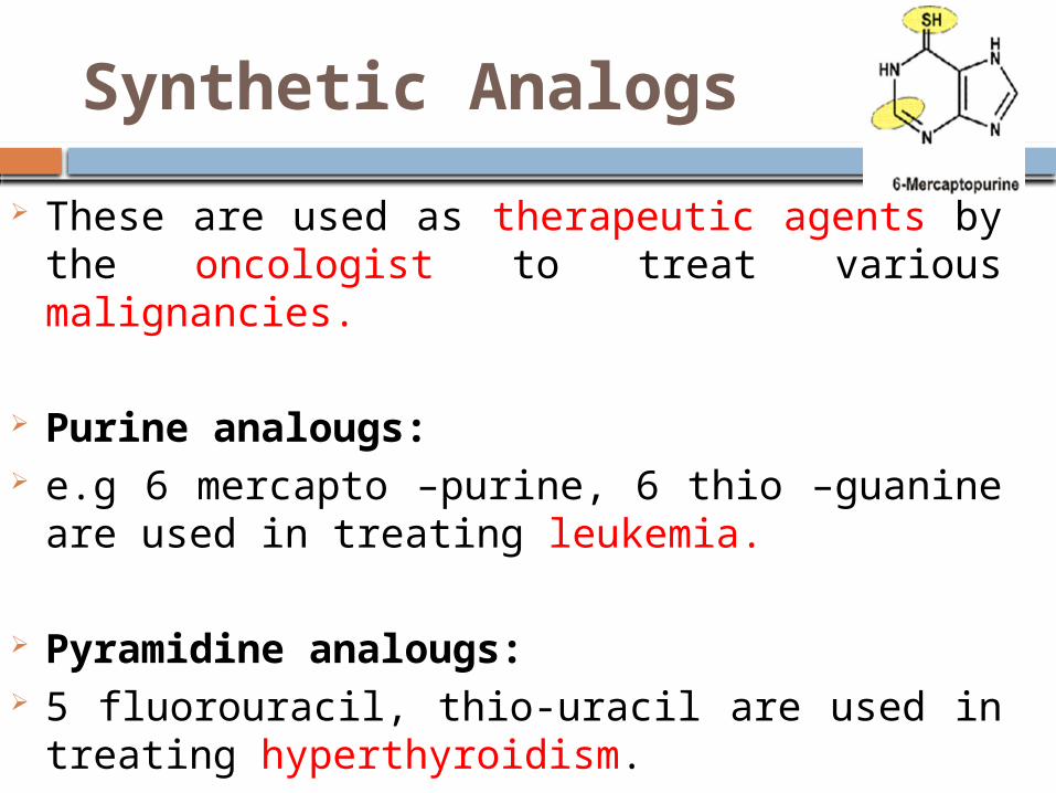
Synthetic Analogs
These are used as therapeutic agents by the oncologist to treat various malignancies.
Purine analougs: e.g 6 mercapto –purine, 6 thio –guanine are used
in treating leukemia.
Pyramidine analougs: 5 fluorouracil, thio-uracil are used in treating
hyperthyroidism.
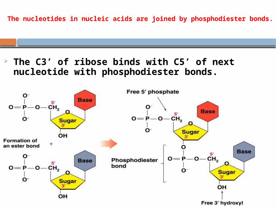
The nucleotides in nucleic acids are joined by phosphodiester bonds.
The C3’ of ribose binds with C5’ of next nucleotide with phosphodiester bonds.
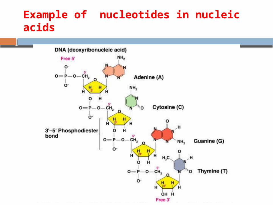
Example of nucleotides in nucleic acids
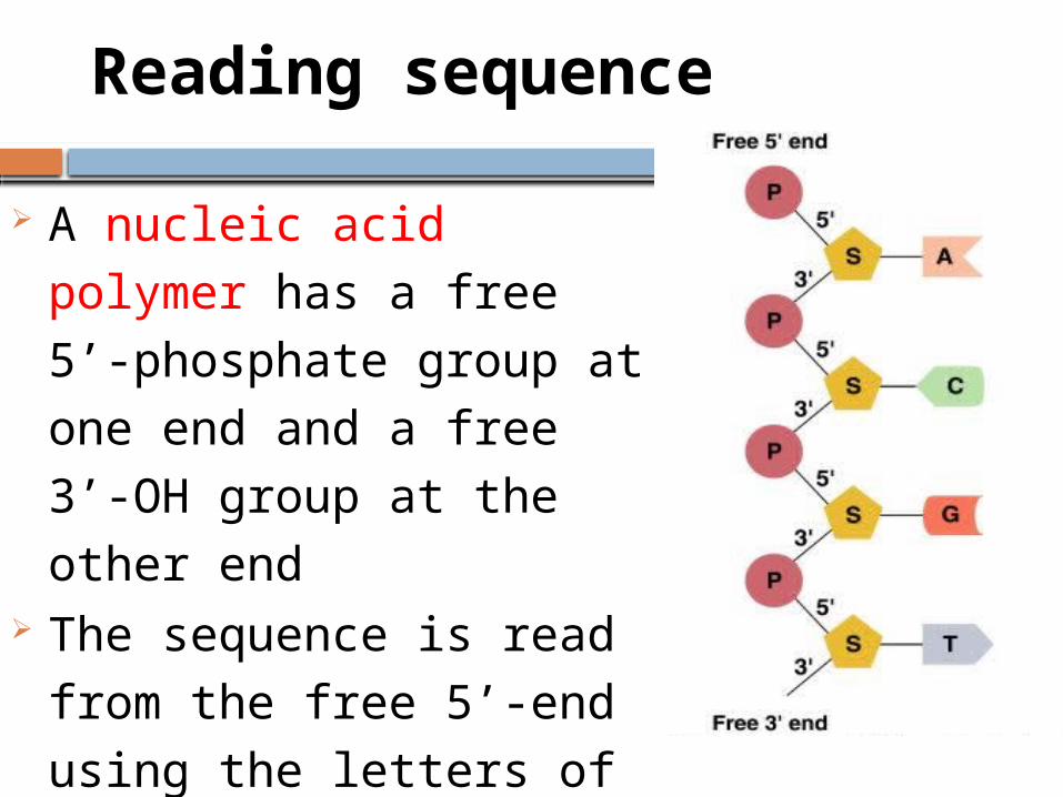
Reading sequence
A nucleic acid polymer has a
free 5’-phosphate group at
one end and a free 3’-OH
group at the other end The sequence is read from
the free 5’-end using the
letters of the bases This example reads 5’—A—C—G—T—3’
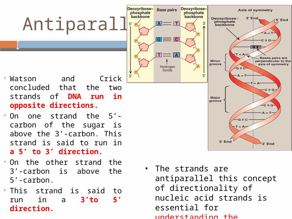
Antiparallel
Watson and Crick concluded that the two strands of DNA run in opposite directions.
On one strand the 5’-carbon of the sugar is above the 3’-carbon. This strand is said to run in a 5’ to 3’ direction.
On the other strand the 3’-carbon is above the 5’-carbon.
This strand is said to run in a 3’to 5’ direction.
• The strands are antiparallel this concept of directionality of nucleic acid strands is essential for understanding the mechanisms of replication and transcription.
P
P
P
P
P
P
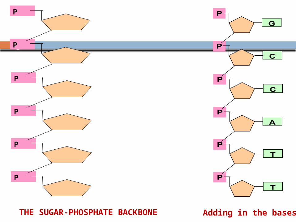
Adding in the basesTHE SUGAR-PHOSPHATE BACKBONE
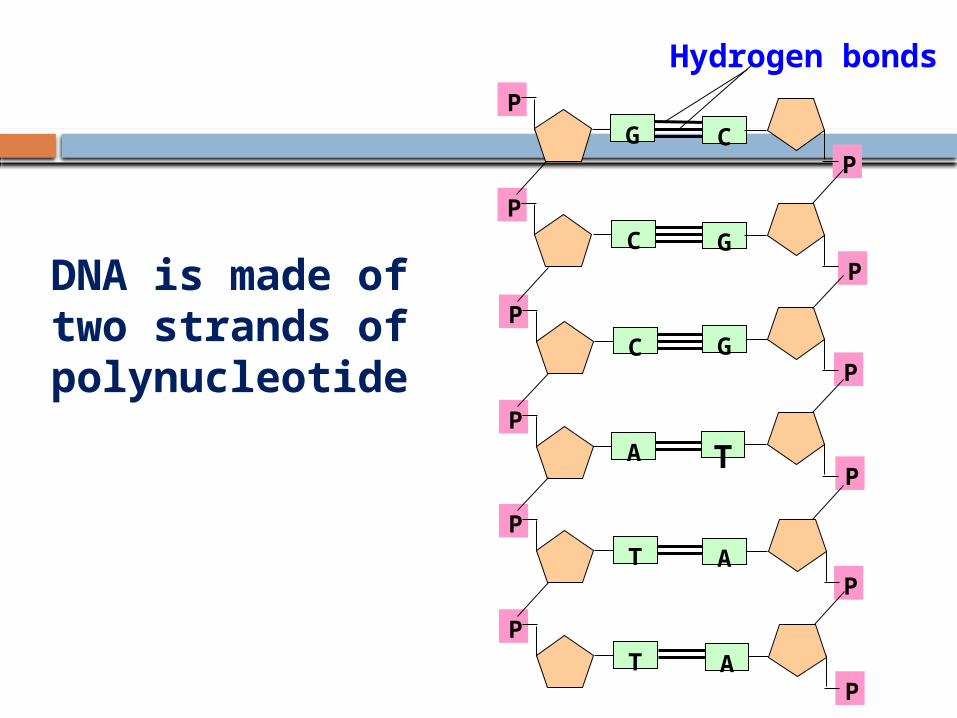
DNA is made of two strands of polynucleotide
P
P
P
P
P
P
C
G
G
T
A
A
P
P
P
P
P
P
G
C
C
A
T
T
Hydrogen bonds
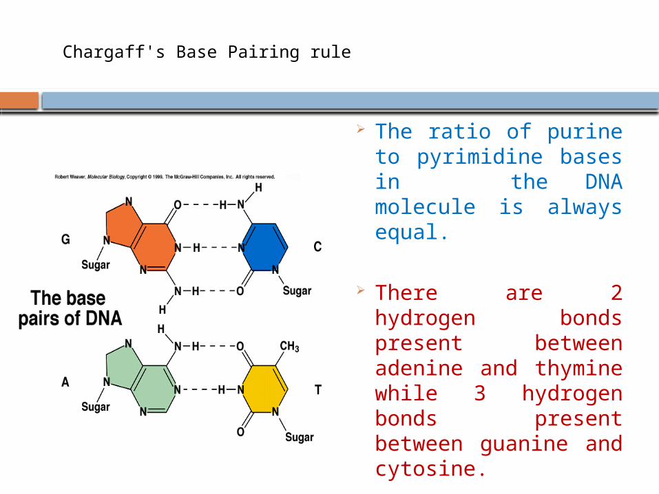
Chargaff's Base Pairing rule
The ratio of purine to pyrimidine bases in the DNA molecule is always equal.
There are 2 hydrogen bonds present between adenine and thymine while 3 hydrogen bonds present between guanine and cytosine.
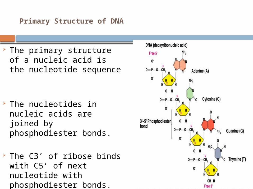
Primary Structure of DNA
The primary structure of a nucleic acid is the nucleotide sequence
The nucleotides in nucleic acids are joined by phosphodiester bonds.
The C3’ of ribose binds with C5’ of next nucleotide with phosphodiester bonds.
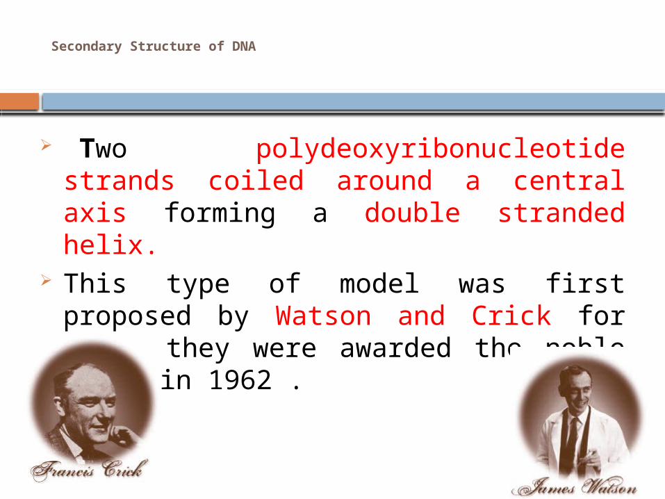
Secondary Structure of DNA
Two polydeoxyribonucleotide strands coiled around a central axis forming a double stranded helix.
This type of model was first proposed by Watson and Crick for which they were awarded the noble prize in 1962 .
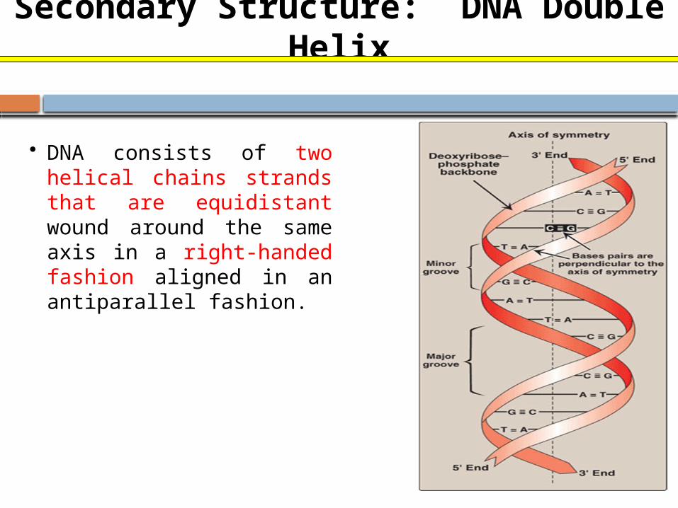
Secondary Structure: DNA Double Helix
• DNA consists of two helical chains strands that are equidistant wound around the same axis in a right-handed fashion aligned in an antiparallel fashion.
DNA-Physical characters
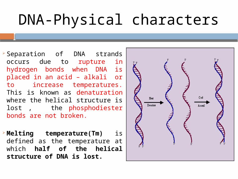
Separation of DNA strands occurs
due to rupture in hydrogen bonds when DNA is placed in an acid – alkali or to increase temperatures. This is known as denaturation where the helical structure is lost , the phosphodiester bonds are not broken.
Melting temperature(Tm) is defined as the temperature at which half of the helical structure of DNA is lost.
Renaturation
Renaturation or reannealing or hybridization is a process in which the separated complementary DNA strands can reform the double helix when an acid alkali is removed and with decrease in temperature.
Higher Organization of DNA
Supercoils In higher organisms the linear DNA is twisted
around its own axis when a supercoil is formed it can be either right handed or positive direction same as B form DNA or left handed or negative twisted in the opposite direction.
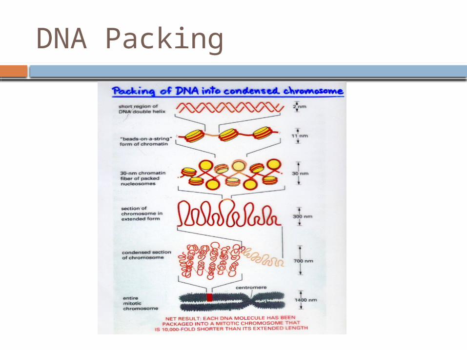
DNA packing
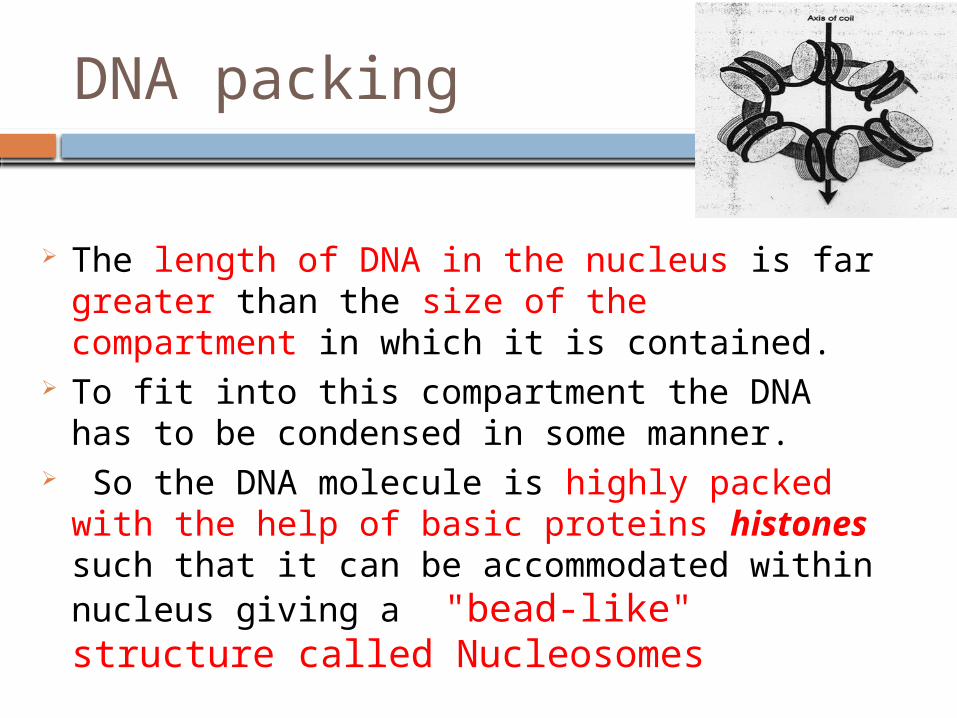
The length of DNA in the nucleus is far greater than the size of the compartment in which it is contained.
To fit into this compartment the DNA has to be condensed in some manner.
So the DNA molecule is highly packed with the help of basic proteins histones such that it can be accommodated within nucleus giving a "bead-like" structure called Nucleosomes
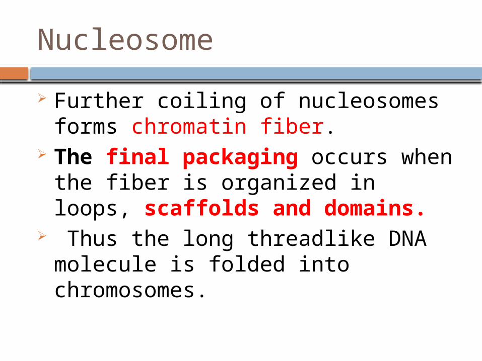
Nucleosome
Further coiling of nucleosomes forms chromatin fiber.
The final packaging occurs when the fiber is organized in loops, scaffolds and domains.
Thus the long threadlike DNA molecule is folded into chromosomes.
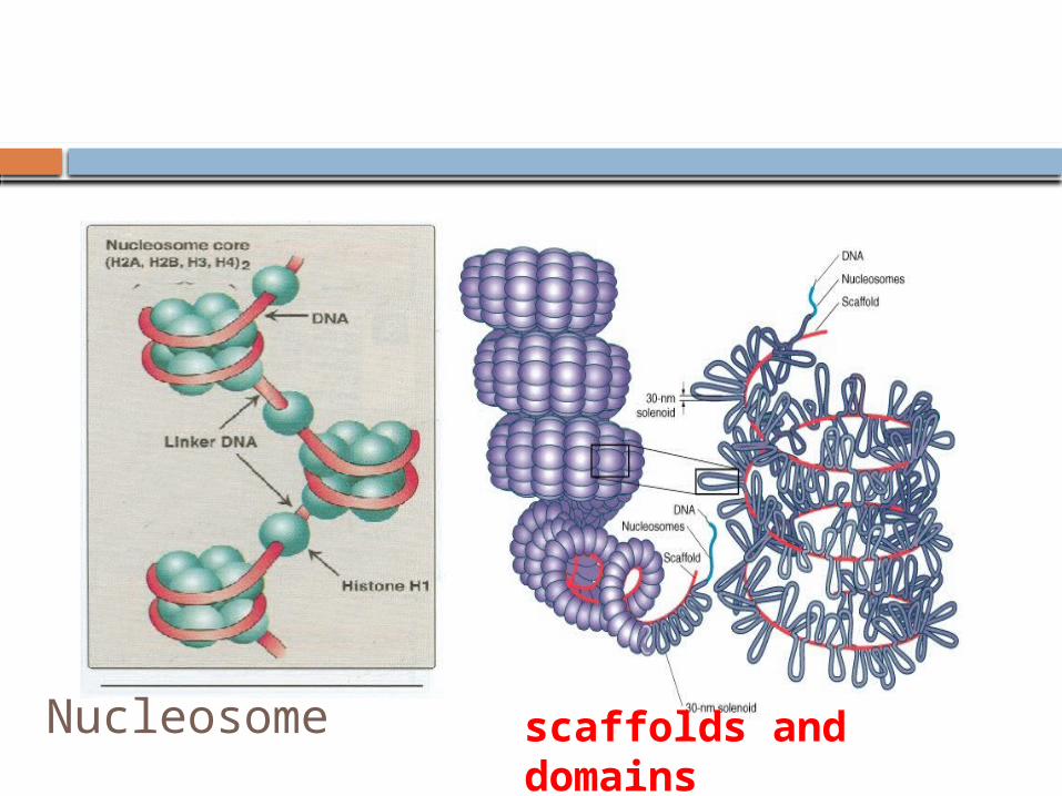
Nucleosome scaffolds and domains
DNA Packing
Motivate your Answer
What is the relationship between Nucleosomes, chromatin fibers and the scaffold structure with respect to the organization of DNA in the nucleus?
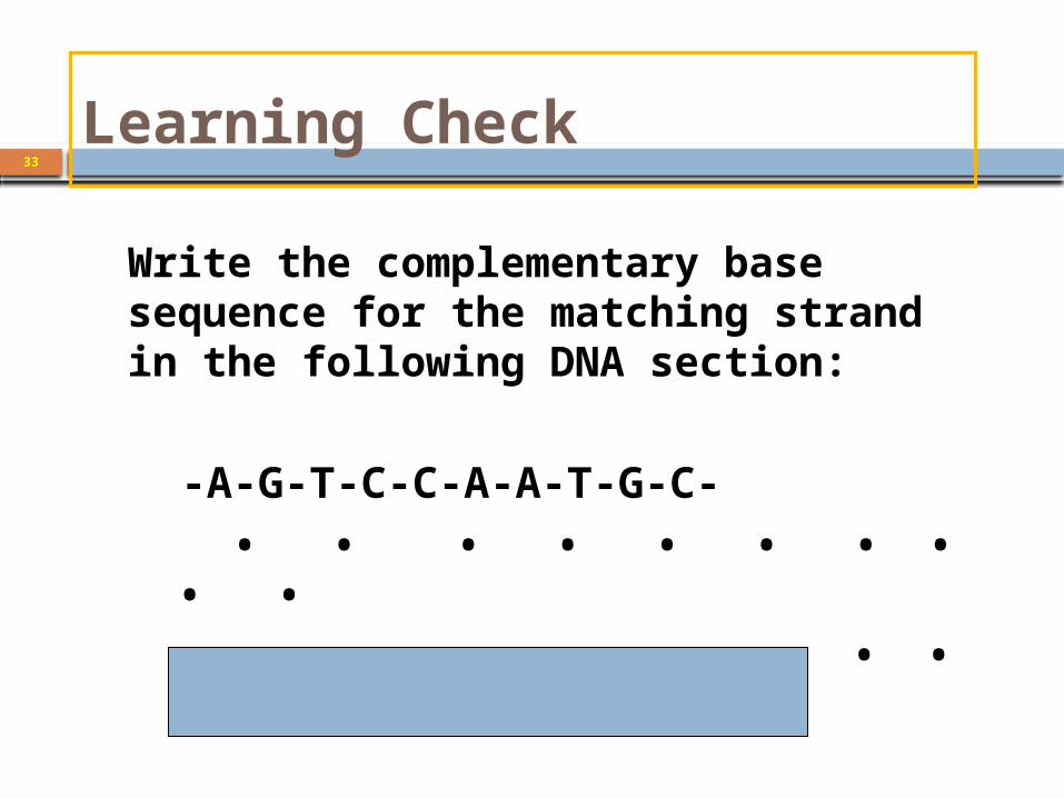
Learning Check33
Write the complementary base sequence for the matching strand in the following DNA section:
-A-G-T-C-C-A-A-T-G-C- • • • • • • • • • •
• • • • • • • • • •
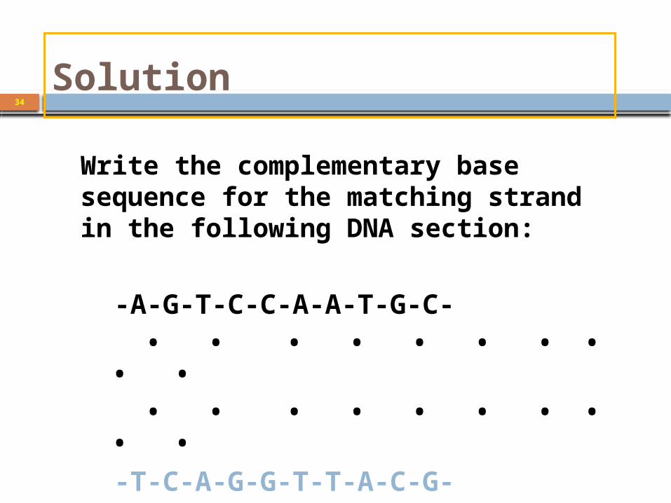
Solution34
Write the complementary base sequence for the matching strand in the following DNA section:
-A-G-T-C-C-A-A-T-G-C- • • • • • • • • • • • • • • • • • • • •
-T-C-A-G-G-T-T-A-C-G-
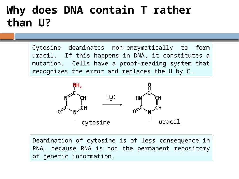
Why does DNA contain T rather than U?
N
CHC
O
HN
CHCO
C
NH2
N
N
CHCO
CH
cytosine uracil
H2O
Cytosine deaminates non-enzymatically to form uracil. If this happens in DNA, it constitutes a mutation. Cells have a proof-reading system that recognizes the error and replaces the U by C.
Cytosine deaminates non-enzymatically to form uracil. If this happens in DNA, it constitutes a mutation. Cells have a proof-reading system that recognizes the error and replaces the U by C.
Deamination of cytosine is of less consequence in RNA, because RNA is not the permanent repository of genetic information.
Deamination of cytosine is of less consequence in RNA, because RNA is not the permanent repository of genetic information.
Reference:
Lippincott's illustrated reviews Biochemistry 6th edition, Pages 395-398(DNA structure)