DR. NAITIK TRIVEDI – DR. NAITIK TRIVEDI - DR. NAITIK D. … · 2020. 4. 11. · CAUSES: The main...
32
DR. NAITIK D TRIVEDI & DR. UPAMA N. TRIVEDI 3. ANEMIA https://www.drnaitiktrivedi.com/ 1 !! Jay Ambe !! PREPARED BY DR. NAITIK D. TRIVEDI, M. PHARM, PH. D LECTURER AT GOVERNMENT AIDED, A. R. COLLEGE OF PHARMACY & G. H. PATEL INSTITUTE OF PHARMACY, VALLABH VIDYANAGAR, ANAND, GUJARAT Mobile: +91 - 9924567864 E-mail: [email protected] & DR. UPAMA N. TRIVEDI, M. PHARM, PH. D ASSOCIATE PROFESSOR & HoD (Pharm.D), INDUBHAI PATEL COLLEGE OF PHARMACY AND RESEARCH CENTRE, DHARMAJ, GUJARAT E-mail: [email protected]
Transcript of DR. NAITIK TRIVEDI – DR. NAITIK TRIVEDI - DR. NAITIK D. … · 2020. 4. 11. · CAUSES: The main...

DR. NAITIK D TRIVEDI
&
DR. UPAMA N. TRIVEDI
3. ANEMIA
https://www.drnaitiktrivedi.com/ 1
!! Jay Ambe !!
PREPARED BY
DR. NAITIK D. TRIVEDI,
M. PHARM, PH. D
LECTURER AT GOVERNMENT AIDED,
A. R. COLLEGE OF PHARMACY & G. H. PATEL INSTITUTE OF
PHARMACY, VALLABH VIDYANAGAR, ANAND, GUJARAT
Mobile: +91 - 9924567864
E-mail: [email protected]
&
DR. UPAMA N. TRIVEDI,
M. PHARM, PH. D
ASSOCIATE PROFESSOR & HoD (Pharm.D),
INDUBHAI PATEL COLLEGE OF PHARMACY AND
RESEARCH CENTRE, DHARMAJ, GUJARAT
E-mail: [email protected]

DR. NAITIK D TRIVEDI
&
DR. UPAMA N. TRIVEDI
3. ANEMIA
https://www.drnaitiktrivedi.com/ 2
ANEMIA:
Anemia is the condition in which the oxygen carrying capacity of blood is reduced. In the
anemia the total number of RBCs decreases so indirectly decreases the oxygen level so decrease
the production of ATP and energy.
GENERAL PATHOPHYSIOLOGY OF ANEMIA:
Our blood contains RBCs
RBCs contains Hb
Hb contains the iron
Iron transfer the oxygen in body
Oxygen is useful for production of ATP and Heat
ATP provide energy
Anemia is not a single disease entity but a sign of disease. Independent of the cause, anemia is
associated with a reduction in circulating Hb because of reduced number of erythrocytes or less
Hb per erythrocytes. The number of erythrocytes varies with age, sex and atmospheric pressure.
CAUSES:
The main causes of anemia are:
1. Blood loss 2. Lack of red blood cell production 3. High rates of red blood cell
destruction
The other causes for the anemia are as under:
Blood Loss
Blood loss is the most common cause of anemia, especially iron-deficiency anemia.
Blood loss can be short term or persist over time.
Heavy menstrual periods or bleeding in the digestive or urinary tract can cause blood
loss. Surgery, trauma, or cancer also can cause blood loss.
If a lot of blood is lost, the body may lose enough red blood cells to cause anemia.

DR. NAITIK D TRIVEDI
&
DR. UPAMA N. TRIVEDI
3. ANEMIA
https://www.drnaitiktrivedi.com/ 3
Lack of Red Blood Cell Production
Both acquired and inherited conditions and factors can prevent your body from making
enough red blood cells. "Acquired" means you aren't born with the condition, but you
develop it. "Inherited" means your parents passed the gene for the condition on to you.
Acquired conditions and factors that can lead to anemia include poor diet, abnormal
hormone levels, some chronic (ongoing) diseases, and pregnancy.
Aplastic anemia also can prevent your body from making enough red blood cells. This
condition can be acquired or inherited.
Diet
A diet that lacks iron, folic acid (folate), or vitamin B12 can prevent your body from
making enough red blood cells. Your body also needs small amounts of vitamin C,
riboflavin, and copper to make red blood cells.
Conditions that make it hard for your body to absorb nutrients also can prevent your
body from making enough red blood cells.
Hormones
Our body needs the hormone erythropoietin (eh-rith-ro-POY-eh-tin) to make red blood
cells. This hormone stimulates the bone marrow to make these cells. A low level of this
hormone can lead to anemia.
Diseases and disease treatments
Chronic diseases, like kidney disease and cancer, can make it hard for your body to
make enough red blood cells.
Some cancer treatments may damage the bone marrow or damage the red blood cells'
ability to carry oxygen. If the bone marrow is damaged, it can't make red blood cells
fast enough to replace the ones that die or are destroyed.
People who have HIV/AIDS may develop anemia due to infections or medicines used
to treat their diseases.
Pregnancy
Anemia can occur during pregnancy due to low levels of iron and folic acid and
changes in the blood.
During the first 6 months of pregnancy, the fluid portion of a woman's blood (the
plasma) increases faster than the number of red blood cells. This dilutes the blood and
can lead to anemia.

DR. NAITIK D TRIVEDI
&
DR. UPAMA N. TRIVEDI
3. ANEMIA
https://www.drnaitiktrivedi.com/ 4
Aplastic anemia
Some infants are born without the ability to make enough red blood cells. This condition
is called aplastic anemia. Infants and children who have aplastic anemia often
need blood transfusions to increase the number of red blood cells in their blood.
Acquired conditions or factors, such as certain medicines, toxins, and infectious
diseases, also can cause aplastic anemia.
High rates of red blood cell destruction
Both acquired and inherited conditions and factors can cause your body to destroy too
many red blood cells. One example of an acquired condition is an enlarged or diseased
spleen.
The spleen is an organ that removes worn out red blood cells from the body. If the
spleen is enlarged or diseased, it may remove more red blood cells than normal, causing
anemia.
Examples of inherited conditions that can cause your body to destroy too many red
blood cells include sickle cell anemia, thalassemias, and lack of certain enzymes. These
conditions create defects in the red blood cells that cause them to die faster than healthy
red blood cells.
Hemolytic anemia is another example of a condition in which your body destroys too
many red blood cells. Inherited or acquired conditions or factors can cause hemolytic
anemia. Examples include immune disorders, infections, certain medicines, or reactions
to blood transfusions.
SIGNS & SYMPTOMS:
Common symptoms of anemia:
fatigue
decreased energy
weakness
shortness of breath
light headedness
palpitations (feeling of the heart
racing or beating irregularly) and
looking pale
Symptoms of severe anemia may include:
chest pain, angina, or heart attack
dizziness
fainting or passing out; and
rapid heart rate.

DR. NAITIK D TRIVEDI
&
DR. UPAMA N. TRIVEDI
3. ANEMIA
https://www.drnaitiktrivedi.com/ 5
Some of the signs that may indicate anemia in an individual may include:
Change in stool color, including black and tarry stools (sticky and foul smelling),
maroon-colored, or visibly bloody stools if the anemia is due to blood loss through the
gastrointestinal tract;
rapid heart rate;
low blood pressure;
rapid breathing;
pale or cold skin;
yellow skin called jaundice if anemia is due to red blood cell breakdown;
heart murmur; and
enlargement of the spleen with certain causes of anemia.
NORMAL HEMOGLOBINE VALUE AS PER AGE AND SEX:
Age & Sex Mean Hb ( g/dl )
1-3 days 16.0 – 19.0
6 Months – 2 Yrs 12.0 – 14.5
12 – 18 Yrs
Male
Female
14.5 – 17.0
12.5 – 14.0
Adult
Male
Female
15.5 – 17.0
13.0 – 15.0
Note: Atmospheric pressure:
People at high altitudes have more erythrocytes than at low altitudes.
NORMAL HEMATOLOGIC & BIOCHEMICAL PARAMETERS:
Component Conventional
Hematologic
Hematocrit (Ratio of RBC-Blood Vol.)
Male
Female
45 – 52 %
37 – 48 %
Hemoglobin
Male
Female
14 – 18 g /dL
12 – 15 g/Dl
Erythrocyte count 4.2 -5.9 x 10⁶/mm
Reticulocyte count 0.5 – 1.5 %
MCV 80 – 90 fmol
MCH 27 – 30 pg
MCHC 32 – 36 g/dL
RDW 11.5 – 14.5 %

DR. NAITIK D TRIVEDI
&
DR. UPAMA N. TRIVEDI
3. ANEMIA
https://www.drnaitiktrivedi.com/ 6
Biochemical
Iron
Male
Female
80 – 200 mcg/dL
60 – 190 mcg/dL
Transferrin 170 – 370 mg/dL
TIBC 250 – 410 g/ml
Transferrin saturation 20 – 55 %
Transferrin receptors 2.8 – 8.5 mg/L
Ferritin 1.5 – 30 mcg/dL
Zinc protoporphyrin <70 mcg/dL red cell
Folate
Normal
Borderline
2 – 10 ng/ml
1 - 1.9 ng/ml
Vitamin B12 200 – 1000pg/ml
Methylmalonic acid 53 – 376 nmol/ L
Homocysteine 4.1 – 21.3 mcmol / L
DIAGNOSIS:
A detailed medical & medication history along with hematologic & biochemical tests is
obtained.
I. Hematologic tests:
It includes blood Hb concentration, cell counts, mean corpuscular volume (MCV), Hct,
mean corpuscular hemoglobin (MCH), mean corpuscular hemoglobin concentration
(MCHC), etc.
1. MCV cell size
Microcytic anemia - < 80fL
Macrocytic anemia - >100fL
2. MCH & MCHC cell colour
Hypochromic anemia - low MCHC
Hyperchromic anemia - high MCHC
3. Red blood cell distribution width (RDW ) variation in erythrocyte size in blood
sample
Iron deficiency anemia increased RDW

DR. NAITIK D TRIVEDI
&
DR. UPAMA N. TRIVEDI
3. ANEMIA
https://www.drnaitiktrivedi.com/ 7
4. Reticulocyte counts bone marrow activity
Iron, B12 or folic acid therapy for respective deficiency states reticulocytosis
5. Others : differential white cell count, platelet count, microscopic examination of
peripheral blood smears & bone marrow aspirates.
II. Biochemical tests:
It includes measurement of serum vitamin conc, transport proteins, saturation of protein
binding sites & storage amounts.
TYPES OF ANEMIA
CLASSIFIED ACCORDING TO THE SIZE OF THE RED BLOOD CELLS:
a) Microcytic anemia:
If the red blood cells are smaller than normal, this is called microcytic anemia. The
major causes of this type are iron deficiency (low level iron) anemia and thalassemia
(inherited disorders of hemoglobin).
b) Normocytic anemia
If the red blood cells size are normal in size (but low in number), this is
called normocytic anemia, such as anemia that accompanies chronic disease or anemia
related to kidney disease.
c) Macrocytic anemia
If red blood cells are larger than normal, then it is called macrocytic anemia. Major
causes of this type are pernicious anemia and anemia related to alcoholism.
ACCORDING TO THE CAUSE, ANEMIA ARE CLASSIFIED AS UNDER:
A) IRON DEFICIENCY ANEMIA:
It is cause by excessive loss of iron or inadequate absorption of iron.
It is most often in female than male.
Iron absorption is regulated by iron needs & body stores.
When iron stores are low, higher proportion of available iron is absorbed & vice versa.
Except, in primary hemochromatosis, thalassemia & sideroblastic anemia iron
absorption remains normal & even elevated despite increased iron stores.
It is primarily absorbed in upper duodenum.

DR. NAITIK D TRIVEDI
&
DR. UPAMA N. TRIVEDI
3. ANEMIA
https://www.drnaitiktrivedi.com/ 8
PATHOPHYSIOLOGY OF IRON DEFICIENCY ANEMIA:
Iron is an essential element for erythropoiesis, tissue respiration & several enzyme
catalyzed reactions. The average adult body contains 3 to 5 g elemental iron.
Iron is distributed in body in two forms: functional & storage.
I . Functional iron
It exists as Hb , little as myoglobin , transferring & tissue enzymes.
Hb is the oxygen binding protein that transports oxygen from lungs to tissues.
Myoglobin, a hemo protein in muscle accepts oxygen from hemoglobin in the
peripheries & acts as an oxygen store in muscle. If oxygen supply is limited, it
releases oxygen to cytochrome oxidase leading to oxidative phosphorylation.
Transferrin is a specific iron binding protein that transports iron through plasma &
extravascular spaces. Each molecule of transferrin binds 2 molecules of iron in
ferric state. The TIBC is high in iron deficiency & low in iron overload.
II. Storage iron:
It is in the form of ferritin & hemosiderin, which is located in parenchymal cells of
liver & reticuloendothelial cells of the bone marrow, spleen & liver.
Low iron stores are an early sign of iron deficiency & may help differentiate
between iron deficiency anemia & other causes of anemia.
DAILY IRON NEEDS:
Recommended daily allowances of iron:
category Age ( Yr ) Iron ( mg )
Infants 0 – 0.5 6
0.5 - 1 10
Children 1 - 10 10
Boys & men 11 - 18 12
> 19 10
Girls & women 11 - 50 15
> 51 10
Pregnant women 30
Lactating women 15
DR. NAITIK D TRIVEDI
&
DR. UPAMA N. TRIVEDI
3. ANEMIA
https://www.drnaitiktrivedi.com/ 9
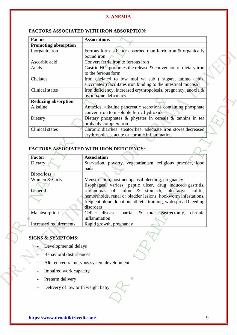
FACTORS ASSOCIATED WITH IRON ABSORPTION:
Factor Associations
Promoting absorption
Inorganic iron Ferrous form is better absorbed than ferric iron & organically
bound iron.
Ascorbic acid Convert ferric iron to ferrous iron
Acids Gastric HCl promotes the release & conversion of dietary iron
to the ferrous form
Chelates Iron chelated to low mol wt sub ( sugars, amino acids,
succinates ) facilitates iron binding to the intestinal mucosa
Clinical states Iron deficiency, increased erythropoiesis, pregnancy, anoxia &
pyridoxine deficiency
Reducing absorption
Alkaline Antacids, alkaline pancreatic secretions containing phosphate
convert iron to insoluble ferric hydroxide
Dietary Dietary phosphates & phytates in cereals & tannins in tea
probably complex iron
Clinical states Chronic diarrhea, steatorrhea, adequate iron stores,decreased
erythropoiesis, acute or chronic inflammation
FACTORS ASSOCIATED WITH IRON DEFICIENCY:
Factor Association
Dietary Starvation, poverty, vegetarianism, religious practice, food
pads
Blood loss :
Women & Girls
General
Menstruation, postmenopausal bleeding, pregnancy
Esophageal varices, peptic ulcer, drug induced gastritis,
carcinomas of colon & stomach, ulcerative colitis,
hemorrhoids, renal or bladder lesions, hookworm infestations,
frequent blood donation, athletic training, widespread bleeding
disorders
Malabsorption Celiac disease, partial & total gastrectomy, chronic
inflammation
Increased requirements Rapid growth, pregnancy
SIGNS & SYMPTOMS:
- Developmental delays
- Behavioral disturbances
- Altered central nervous system development
- Impaired work capacity
- Preterm delivery
- Delivery of low birth weight baby

DR. NAITIK D TRIVEDI
&
DR. UPAMA N. TRIVEDI
3. ANEMIA
https://www.drnaitiktrivedi.com/ 10
- Others like brittle or spoon shaped nails, angular stomatitis, atrophic tongue &
pharyngeal & esophageal webs causing dysphagia & atrophic gastric mucosa.
DIAGNOSIS:
- Medical history, full blood count & peripheral smears.
- Blood Hb concentrations & erythrocyte numbers are normal in mild cases.
- As deficiency worsens MCV & erythrocyte count & Hb decreases & RDW increases.
- Hypochromia or poikilocytosis shown when Hb conc are 7.0 g/dl or less for women &
9.0 g/dl or less for men.
- Absence of stainable iron in bone marrow aspirates is ultimate proof of deficiency but
is painful & expensive so not used routinely.
- After hemorrhage or iron therapy reticulocytes increase.
- Serum ferritin is the first parameter to change in iron deficiency. It fall ( < 15 mcg/ml)
in deficiency but increase abnormally in iron storage conditions.
- ZPP (zinc protoporphyrin ) is an early indicator of iron deficient erythropoiesis than
anemia. It represents the amount of protoporphyrin not incorporated into heme, it
increase when insufficient iron is available for Hb synthesis.
- TfR (transferring receptor ) provides information on later stages of iron deficiency,
increasing only after iron stores are depleted.
- Since ZPP & TfR are not affected by inflammatory processes are useful in
differentiating it from iron deficiency anemia.
- Serum iron is low & TIBC is high.
PREVENTION:
It can be done by identifying the underlying cause of iron deficiency & correcting it through
diet or supplementation.
Dietary manipulation :
- Food fortification is best recommended.
- When dietary iron supplementation is not possible or adequate, oral supplementation
should be initiated.
- In infants CDC recommends following guidelines :
1. breastfeeding for 4 – 6 months after birth.
2. use of 1 mg/kg/day of iron from supplemental foods or iron drops when
breastfeeding is stopped.

DR. NAITIK D TRIVEDI
&
DR. UPAMA N. TRIVEDI
3. ANEMIA
https://www.drnaitiktrivedi.com/ 11
3. use of only iron fortified infant formula as a substitute for breast milk.
4. use of 2 – 4 mg/kg/day of iron drops ( max 15mg/day ) for preterm or low birth
weight infants starting at 1 mth & continuing until 12 mths after birth.
5. introduction of iron fortified infant cereal at age 4 to 6 mths.
- The CDC recommends universal treatment with 30 mg iron/day during pregnancy to
prevent iron deficiency.but since iron can cause side effects & potentially affect
absorption of other nutrients, it is recommended only for women at risk of iron
deficiency anemia.
Screening of iron deficiency:
The CDC recommends screening of infants who are at risk ( preterm, low birth weight,
low iron diet ) at 9 to 12 mths & at 15 to 18 mths of age.
Women with risk factors (poor diet, excessive menstrual bleeding, chronic blood loss)
should be screened annually.
TREATMENT:
1. Oral iron therapy :
Generally, 30 to 40 mg elemental iron is used to treat iron deficiency states.
This can be calculated from maximum rate of Hb regeneration. 0.25 g Hb/100 ml blood/
day x 5000 ml blood x 3.4 mg Fe/1g Hb = 40 mg Fe/day
Since only 10 to 20 % of iron is absorbed, 200 to 400 mg iron would result in absorption
of 40 mg elemental iron.
Maximum absorption occurs if iron is taken before meals or between meals.
Side effects:
It includes epigastric distress, abdominal cramping, nausea, diarrhoea &
constipation caused by gastric irritation.
This can be minimized by reducing daily dosage, taking the iron with food or
changing to once a week dosing.
Use of enteric coated products to minimize the side effects is not preferred since it
prevents the dissolution & so decrease the absorption.
Iron therapy can also cause the stools to appear black about which patients should
be educated to differentiate from that of GI bleeding.

DR. NAITIK D TRIVEDI
&
DR. UPAMA N. TRIVEDI
3. ANEMIA
https://www.drnaitiktrivedi.com/ 12
Drawbacks:
Iron absorption may be reduced in patients with reduced gastric acid production or prior
GI surgeries.
When an ability to absorb iron is suspected, oral bolus dose of 325 mg ferrous sulphate
should be administered. The serum iron levels after 2 & 4 hours should be 21 to 23
mcmol/ L. otherwise it indicates decreased iron absorption.
Antacids, H 2 blockers & proton pump inhibitors may also decrease iron absorption
Fails in cases of malabsorption, non-compliance with oral iron therapy, severe
uncontrolled intolerance to iron therapy, excessive iron loss or erythropoiesis as seen in
patients on renal dialysis receiving erythropoietin.
*common oral iron preparations includes salts of iron such as ferrous sulphate, ferrous
fumarate, ferrous gluconate, etc
2. parenteral iron therapy :
To overcome the drawbacks of iron therapy, parenteral iron therapy is preferred.
The amount of parenteral iron needed to replenish iron stores & restore Hb levels in
patients with iron deficiency anemia can be obtained by formula :
- Dosage (mg) = 0.3 x body weight (lb) x [ 100- {Hb (g/dl) x 100/14.8} ]
Or
- Dosage (mg) = 0.66 x body weight (kg) x [100-(Hb{g/dl} x 100/14.8}]
The iron dose calculated is divided by 50 mg iron / ml to provide the volume of iron
dextran injection needed.
For children weighing < 15 kg, a normal mean of Hb of 12g/L is used in place of 14.8
g/dL.
To determine iron replacement dosage in patients with active blood loss, one assumes
that 1 ml of normochromic, normocytic erythrocytes contains 1 mg elemental iron :
o Dosage (mg) = 1 mg iron / ml blood x blood loss ( ml ) x Hct
It is administered by deep IM inj into the upper quadrant of the buttock or IV, eitheras
a bolus or a total dose infusion (TDI).
DR. NAITIK D TRIVEDI
&
DR. UPAMA N. TRIVEDI
3. ANEMIA
https://www.drnaitiktrivedi.com/ 13
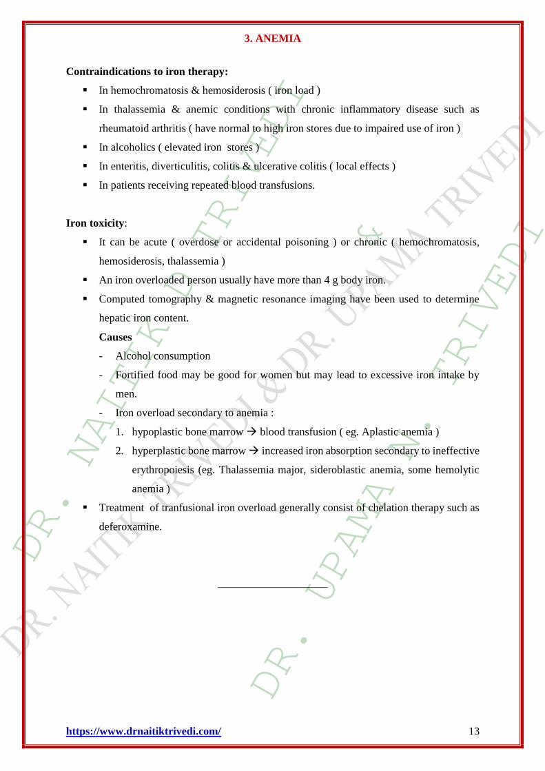
Contraindications to iron therapy:
In hemochromatosis & hemosiderosis ( iron load )
In thalassemia & anemic conditions with chronic inflammatory disease such as
rheumatoid arthritis ( have normal to high iron stores due to impaired use of iron )
In alcoholics ( elevated iron stores )
In enteritis, diverticulitis, colitis & ulcerative colitis ( local effects )
In patients receiving repeated blood transfusions.
Iron toxicity:
It can be acute ( overdose or accidental poisoning ) or chronic ( hemochromatosis,
hemosiderosis, thalassemia )
An iron overloaded person usually have more than 4 g body iron.
Computed tomography & magnetic resonance imaging have been used to determine
hepatic iron content.
Causes
- Alcohol consumption
- Fortified food may be good for women but may lead to excessive iron intake by
men.
- Iron overload secondary to anemia :
1. hypoplastic bone marrow blood transfusion ( eg. Aplastic anemia )
2. hyperplastic bone marrow increased iron absorption secondary to ineffective
erythropoiesis (eg. Thalassemia major, sideroblastic anemia, some hemolytic
anemia )
Treatment of tranfusional iron overload generally consist of chelation therapy such as
deferoxamine.
____________________

DR. NAITIK D TRIVEDI
&
DR. UPAMA N. TRIVEDI
3. ANEMIA
https://www.drnaitiktrivedi.com/ 14
B) MEGALOBLASTIC ANEMIA/PERNICIOUS ANEMIA:
It is cause by insufficient of hemopoiesis.
In this condition stomach decreases the production of intrinsic factors because they
decrease the absorption of vitamin B12.
It is a sub class of the macrocytic anemias.
It is characterized by a lowered blood Hb mass due to reduced erythropoiesis secondary
to defective DNA synthesis in the developing erythroid cells of the bone marrow.
CAUSES:
It can be mainly due to deficiencies of vitamin B12 and folate.
It can be by drug induced interferences, either direct or indirect, i.e. with DNA synthesis
or nutritional status.
1. VITAMIN B12 DEFICIENCY MEGALOBLASTIC ANEMIA:
Stages of vitamin B12 deficiency anemia:
Stage B12
concentration
MCV Hb Signs &
symptoms
Normal Normal Normal Normal None
Negative
balance
Normal Normal Normal None
Depletion of
stores
Slight decrease Normal Normal Possible
B12 deficient
erythropoiesis
Mod decrease Increased Normal Possible
B12 deficiency
anemia
Severe decrease Increased Decreased Probable
Vitamin B12 Needs:
1. Daily requirement for humans is 0.5 to 1 mcg.
2. The total body stores amount to 2-5 mg mainly into liver.
PATHOGENESIS OF MEGALOBLASTIC ANAEMIA DUE TO VIT B12:
Vit B 12 is well absorbed from GIT in a sequence by three different binding proteins
i.e. R proteins, IF, & transcobalamin II (TCII).
Extravascular R proteins also known as cobalophilins, are the first binding proteins for
B12.

DR. NAITIK D TRIVEDI
&
DR. UPAMA N. TRIVEDI
3. ANEMIA
https://www.drnaitiktrivedi.com/ 15
The cobalamin remains bound to R protein in the upper small intestine until pancreatic
proteases viz, trypsin partially degrade the complex releasing B12.
Then B12 binds to IF, a specific B12 binding glycoprotein.
The IF – B12 complex is highly resistant to proteolysis, passes down the small intestine
to the distal ileum where it attaches to specific receptors.
This attachment with receptor is not energy dependent but requires extracellular
calcium & pH higher than 5.4.
The majority of B12 in the circulation binds to intravascular R protein i.e.
transcobalamin I ( haptocorrin ).
But transcobalamin II (TCII) is a functional binding protein responsible for releasing
B12 to the tissues.
Patients with TC II deficiency may have normal serum B12 concentration as binding to
TC I compensates for it.
Features of severe B12 deficiency occur as TCI – B12 complex does not deliver the
vitamin to the tissues.
Another mechanism of absorption is diffusion, which is a potential method of providing
oral B12 therapy to people with low IF levels (pernicious anemia).
Lack of B12 allows folic acid to be trapped as non-functional methyl
tetrahydrofolate(folate trap) So deficiency of functional FH4 causes impairment of
formation of deoxy thymidine monophosphate(dTMP) which is needed for DNA
synthesis As a result large proerythroblast fails to divide rapidly to make mature RBC
rather immature precursors of erythocyte(blast cell) appear to cause megaloblastic
anaemia.
ETIOLOGY:
I Dietary inadequate intake.
II Impaired transport transcobalamin II deficiency
III Malabsorption:
Pernicious anemia: It is a B12 malabsorption caused by the loss of gastric IF secretion.
Its an auto immune reaction against gastric parietal cells.
Gastric disorders: gastrectomy (absolute deficiency of IF), atrophic gastritis,
achlorhydria, vagotomy, partial gastrectomy & the use of H 2 receptor antagonists
(prevent release of vitamin from food).

DR. NAITIK D TRIVEDI
&
DR. UPAMA N. TRIVEDI
3. ANEMIA
https://www.drnaitiktrivedi.com/ 16
Intestinal problems: Zollinger – Ellison syndrome, surgical resection or bypass of the
ileum, Crohn’s disease, celiac disease, lymphomas, Whipple’s disease, bacterial
overgrowth.
Drug induced: colchicine, PAS, neomycin, H2 blockers, proton pump inhibitors,
ethanol, Cholestyramine, etc decrease vitamin B12 absorption.
SIGNS & SYMPTOMS:
Peripheral neuropathy
Strange feeling in extremities
Loss of hand coordination
Deterioration in hand writing
Tingling of extremities
Loss of propioception
Depression
Psychosis
Spinal cord degeneration
Sore tongue or mouth, glossitis, beefy red tongue
Lateral column disruption results in weakness & spasticity, exemplified by myoclonus,
hyperreflexia, & a positive Babanski’s sign. If it remains untreated, instability of gait
& virtual paralysis results.
DIAGNOSIS:
Measurement of holo - TC to differentiate between B12 deficiency or TC II deficiency.
But clinically not used because of limited assay availability.
TC II saturation it decreases in early B12 deficiency.
Assessment of metabolite production – MMA & Hcy where MMA is more specific for
B12 deficiency. But MMA also increases in renal disease so renal function should be
assessed.
MCV > 100 fL indicating macrocytosis indicates its deficiency.
Schilling test (with or without IF)
- Stage I An oral dose of Co labeled B12 is given, followed by an IM dose of
unlabelled B12. The large IM dose saturates B12 binding proteins in the blood. So
there are few finding sites for labeled B12 & a substantial portion is excreted in urine.

DR. NAITIK D TRIVEDI
&
DR. UPAMA N. TRIVEDI
3. ANEMIA
https://www.drnaitiktrivedi.com/ 17
Urine is collected over 24 hours & the amt of labeled B12 is measured. If B12 conc is
less than 10 % absorption is impaired & if its <5% than it indicates pernicious anemia.
- Stage II Same process is repeated but now IF is given with labeled B12.If IF
deficiency is there than stage I will be abnormal & stage II will be normal.
- Stage III Patients are given antibiotics (tetracycline). So if there is bacterial overload
stage I & II will be abnormal & stage III will be normal.
- If all the stages show abnormal absorption, it indicates ileal disorder.
- Results of test :
Condition Stage I Stage II Stage III
Normal Normal
Inadequate diet Normal
Pernicious anemia Low Normal
Bacterial overgrowth Low Low Normal
Ileal defect Low Low Low
Evaluating smear for megaloblastic changes viz, neutrophil hypersegmentation &
oval shaped erythrocytes, generally differentiates a B12 or folate deficiency from other
causes.
If iron deficiency occurs with vit B12, the MCV may appear normal but the blood
smear should show both megaloblastic & microcytic cells.
Others : thymidine uptake ( deoxyuridine suppression test [DUST] ) by bone marrow
cells, food cobalamin absorption, erythropoietin measurement, folate concentrations &
gastrin & pepsinogen analysis.
TREATMENT:
Identify B12 deficiency early ( before anemia or neurologic symptoms develop )
Correct the cause of deficiency if possible
Replenish the depleted stores
If necessary administer maintenance B12 therapy
Dietary changes & supplemental B12 given orally, intranasally or parenterally.

DR. NAITIK D TRIVEDI
&
DR. UPAMA N. TRIVEDI
3. ANEMIA
https://www.drnaitiktrivedi.com/ 18
PHARMACOTHERAPY:
1. Oral vitamin B12 therapy :
- Usual oral dose of vitamin B12 supplementation is 1 to 10 mcg/day.
- In patients with malabsorption, B12 dosages should be 1000 mcg or higher to produce
favorable long term results.
- Concerns with oral therapy are the potential for erratic absorption, poor compliance &
subsequent development of neurologic symptoms.
- Patient evaluation & monitoring for compliance & therapeutic response should
whether long term oral therapy is optimal for each patient.
- Preparations containing iron in different forms are used eg. Ferrous fumarate, ferrous
gluconate, etc.
2. Parenteral vitamin B12 therapy :
- Being safe, dosages of 100 to 1000 mcg can be given.
- It is given IM or by deep Sc injection .
- Peak serum concentrations after IM are reached in about 1 hr.
- The half-life of the parenteral B12 is about 6 days & its half-life in liver is 400 days.
- Two synthetic forms of B12 are available: cyanocobalamin & hydroxocobalamin.
- Hydroxocobalamin is more protein bound & so requires less frequent dosing.
Cyanocobalamin has a side effect of optic neuropathy.
- It is well tolerated & has fewer allergic & anaphylactic reactions.
- Iron dextran is commonly used.
3. Intranasal Vitamin B12 therapy:
- Intranasal sprays & gels are available for patients who refuse or cannot tolerate
parenteral therapy & don’t respond to oral treatment.
2. FOLATE DEFICIENCY MEGALOBLASTIC ANEMIA:
- Folate deficiency occurs in stages, with depletion of stores leading to deficiency that
can result in megaloblastic anemia & other hematologic abnormalities.
- Role of folate deficiency is evaluated in pregnancy, heart disease, stroke & peripheral
arterial disease.

DR. NAITIK D TRIVEDI
&
DR. UPAMA N. TRIVEDI
3. ANEMIA
https://www.drnaitiktrivedi.com/ 19
PATHOPHYSIOLOGY OF FOLATE RELATED MEGALOBLASTIC ANEMIA:
- Dietary folate is usually in polyglutamate form, which must be converted to
monoglutamate form for absorption.
- Active absorption of dietary folate occurs mainly in the proximal part of the small
intestine.
- Synthetic folate is already in monoglutamate form & has greater stability & better
absorption.
- Folic acid from formulations is completely absorbed in the upper duodenum, even in
the presence of malabsorption.
- The principle circulating form is extensively protein bound & undergoes enterohepatic
cycling but not reabsorbed from the bile.
- Reduced form of folate ( tetrahydrofolate ) are cofactors for transformylation reactions
in the biosynthesis of purines & thymidylates of nucleic acids.
- Folate deficiency, so leads to defective DNA synthesis resulting in megaloblast
formation & bone marrow suppression.
- It is needed in Hcy metabolism or any other methylation reactions.
FOLATE NEEDS:
- The minimum daily requirement of folate is 50 to 100 mcg/day in general & in
pregnancy an additional 400 mcg/ day is recommended.
- The average amount stored in the body is 5 to 10 mg, one half of which is found in
liver.
EPIDEMIOLOGY:
- Malnutrition
- In alcoholics due to poor diet & altered absorption.
- In pregnancy folate needs increase thrice the normal requirement due to large increase
in nucleic acid synthesis associated with growth of the fetus, placenta & uterus & with
increased maternal erythrocyte mass.
- Folate needs also increase during malignancy, increased erythropoiesis, conditions
causing rapid cell turnover, chronic hemolytic anemia, exfoliative dermatitis,
generalized psoriasis or extensive burns.
- Drugs can also cause folate deficiency by either reducing absorption or by altering
metabolism.

DR. NAITIK D TRIVEDI
&
DR. UPAMA N. TRIVEDI
3. ANEMIA
https://www.drnaitiktrivedi.com/ 20
- Reduced absorption ethanol, metformin, oral contraceptives,
Sulfasalazine, sulfamethoxazole
- Altered metabolism methotrexate, trimethoprim, triamterene, alcohol
SIGNS & SYMPTOMS:
- They are similar to those of other anemias.
- In addition may cause megaloblastosis, glossitis, diarrhoea & weight loss.
DIAGNOSIS:
- Serum or erythrocyte folate concentrations are assessed where erythrocyte folate
concentration reflects tissue status & is a better indicator of depletion.
- Hcy concentration is increased in deficiency. (also increased in B12 deficiency &
decreased renal function)
- A blood smear & hematologic evaluation show macrocytosis with megaloblastosis.(
also shown by B12 deficiency ).
TREATMENT:
- Primary prevention is by dietary manipulation or oral supplementation.
- Women planning to become pregnant should take atleast 400 mcg/ day, to prevent fetal
neural tube defects.
- Folate deficiency is usually treated with oral folic acid 1 mg daily.
- Parenteral administration is indicated when oral administration is unacceptable or not
possible.
- Long term therapy is needed in chronic hemolytic states, myelofibrosis, refractory
malabsorption, postgastrectomy states, prolonged stress or infection, chronic fever &
persistent diarrhoea.
____________________

DR. NAITIK D TRIVEDI
&
DR. UPAMA N. TRIVEDI
3. ANEMIA
https://www.drnaitiktrivedi.com/ 21
C. ANEMIA OF RENAL FAILURE:
o The severity of the anemia correlates with the extent of uremia.
o Most of the patients with serum creatinine concentration higher than 310mcmol/L &
97% of those on maintenance dialysis are affected.
o The cells are normochromic & normocytic but often are irregular in shape.
PATHOPHYSIOLOGY:
It is a multifactorial process but primarily due to reduced secretion of erythropoietin by
the diseased kidneys.
Accumulation of the inhibitors of erythropoiesis, reduced RBC life span & chronic
blood loss.
Parathyroid hormone & the polyamine spermine have been implicated in reducing
marrow responsiveness to erythropoietin.
Erythrocyte survival also decreases to an average of one-half of normal uremia due to
mild, chronic hemolysis.
Chronic blood loss both from a GI source & during hemodialysis may also contribute
to it.
Others: folic acid deficiency caused by losses to the dialysate, the accumulation of fat
sol vit A, aluminum toxicity caused by long term hemodialysis & the use of aluminum
containing phosphate binders & osteitis fibrosa, a complication of hyperparathyroidism
in which myelofibrosis reduces viable erythroid cellular mass.
TREATMENT:
Iron & folate supplementation should be provided as necessary, & blood loss & use of
aluminum containing antacids should be minimized whenever possible.
For treatment of acute symptoms of hypoxia transfusions of RBCs is done.
Risks of transfusions : Hs reactions, transmission of viral hepatitis, bone marrow
suppression, iron overload, etc.
Drugs : androgens, recombinant human erythropoietin, etc.
___________________

DR. NAITIK D TRIVEDI
&
DR. UPAMA N. TRIVEDI
3. ANEMIA
https://www.drnaitiktrivedi.com/ 22
D. APLASTIC ANEMIA
Aplastic anemia is distinguished by hypocellularity of the bone marrow & subsequent
pancytopenia that is unrelated to malignancy or myeloproliferative disease.
The characteristic anemia , neutropenia & thrombocytopenia result from failure of the
pluripotent stem cells due to congenital or acquired process.
ETIOLOGY:
It is mostly idiopathic.
Myelosuppression is a component of several congenital disease but is more commonly
acquired after exposure to :
- drugs ( chloramphenicol, anticonvulsants, acetazolamide, etc)
- chemicals ( arsenic, benzene, ethanol, etc)
- viral ( HIV, influenza, rubella etc )
- others ( ionizing radiation, pregnancy, mycobacterial infection, etc.)
PATHOPHYSIOLOGY :
It develops when hematopoiesis is interrupted because of deficient or defective stem
cells.
Others : immune mediated suppression of stem cell function, disturbances in the bone
marrow microenvironment, & alterations in the cellular or humoral interactions that
normally sustain hematopoiesis.
CLINICAL PRESENTATION:
Pallor & fatigue initially mild but more pronounced when accompanied by bleeding
due to thrombocytopenia.
Ecchymoses & petechiae
Fever due to infection caused by underlying neutropenia.
DIAGNOSIS:
Peripheral blood - number of morphologically normal cells.
Erythrocytes – normochromic, normocytic or slightly macrocytic
Reticulocyte count, absolute granulocyte count -
Bone marrow biopsy – extensive areas of hypocellularity interspersed with small
patches of hematopoietic cells.

DR. NAITIK D TRIVEDI
&
DR. UPAMA N. TRIVEDI
3. ANEMIA
https://www.drnaitiktrivedi.com/ 23
TREATMENT:
Removal of potential causative agents, supportive care & restoration with normal
hematopoiesis with pharmacologic therapy or BMT.
Blood transfusions, preferably with leukocyte depleted products, antiplatelet
antibodies, broad spectrum antibiotics, cyclophosphamide, androgens, hematopoietic
growth factors.
PRECAUTIONS FOR TREATMENT:
Blood products from family members should not be used in candidates for marrow
transplantation.
Due to risk of hematoma, IM inj should be avoided in patients with thrombocytopenia
& so should be avoided aspirin, NSAIDS & other agents with antiplatelet properties.
____________
E. SICKLE CELL ANEMIA:
The term sickle cell disease encompasses a variety of hemoglobinopathies, including
sickle cell anemia, sickle Hb C (SC) disease, & sickle cell thalassemia.
Although the clinical presentations of all are often similar, the manifestations of sickle
cell are more severe & so mainly considered.
PATHOPHYSIOLOGY:
Hb is distinguished as Hb A1, HbA2, Hb C, Hb F & Hb S of which Hb A1, Hb A2 &
Hb F are normal.
Hb A1 – a tetramer consist 2 pairs of globin chains α & β
Substitution of valine for glutamic acid in both the β chains.
Each parent contributes a single β chain gene, the heterozygous genotype AS is also
possible & is expressed as the sickle cell trait phenotype.
Deoxygenation in capillaries induces rapid polymerization of the sickling Hb, Hb S &
results in formation of helical strands of parallel fibres.
The elongated, crescent shaped cells characteristic of sickle cell anemia are so
produced.
The affected erythrocytes are rigid & unable to pass through the microvasculature.
Vasooclusion with subsequent painful ischemia & chronic organ damage.

DR. NAITIK D TRIVEDI
&
DR. UPAMA N. TRIVEDI
3. ANEMIA
https://www.drnaitiktrivedi.com/ 24
Sickling is reversible upon reexposure to oxygen, however repeated sickling episodes
eventually damage the cell membrane.
The rate of Hb polymerization depends on its concentration in the erythrocyte.
The co polymerization of Hb S with Hb F inhibits further polymer growth ; intracellular
Hb F concentrations are inversely correlated with severity of disease.
CLINICAL PRESENTATIONS
Constitutional
- Impaired growth & development
- Increased risk of infection viz meningitis, pneumonia, septicemia
Hematologic
- Hemolytic anemia
- Aplastic crises
- Splenic sequestration crises
Vasoocclusive :
1. Cardiovascular:
- Cardiac enlargement
- Systolic murmur
2. GI:
- Autosplenecetomy
- Gallstones / cholecystitis
- Hepatic insufficiency
- Intrahepatic cholelithiais
3. Genitourinary:
- Hematuria
- Impotence
- Priapism
- Renal insufficiency
4. Neurologic :
- Cerebral thrombosis
- Intracerebral hemorrhage
- Seizures
- Subarachnoid haemorrhage
5. Ocular:
- Retinopathy
- Secondary glaucoma
6. Painful crises
7. Pulmonary:
- Acute chest syndrome
- Chronic obstructive disease
- Infarction
8. Skin & skeletal :
- Arthropathy
- Aseptic necrosis
- Leg ulcers
DIAGNOSIS:
Hb electrophoresis types & proportion of Hb present.
Is rapid & inexpensive screening test

DR. NAITIK D TRIVEDI
&
DR. UPAMA N. TRIVEDI
3. ANEMIA
https://www.drnaitiktrivedi.com/ 25
It establishes the patients genotype.
If both parents have the AS genotype there is a 1 in 4 chance that their child will have
homozygous SS disease.
Prenatal diagnosis also possible
TREATMENT:
1. Management of major complications:
a. Anemia
- Blood transfusions
- Folate supplementations
b. Infection
- Cefuroxime for Pneumonia & erythromycin & azithromycin for Mycoplasma
pneumonia treatment. Prophylactic penicillin for pneumococcal septicemias.
- Ampicillin & cephalosporins for salmonella infections.
c. Painful crisis
- Vigorous hydration is initiated & oxygen administered if hypoxia is present.
- Ketorolac is given if codeine or oxycodones singly or in combination with
acetaminophen are not effective.
2. Management of the sickle cell disease:
a. Transfusion therapy
b. Pharmacologic management : clotrimazole. Pentoxiphylline, antineoplastics,
hydroxyurea.
c. Bone marrow transplantation

DR. NAITIK D TRIVEDI
&
DR. UPAMA N. TRIVEDI
3. ANEMIA
https://www.drnaitiktrivedi.com/ 26
F. THALASSEMIAS:
It is an inherited disease.
Thalassemia is also the hemolytic anemia in which hemoglobin production is decreased.
The RBCs are small, pale and short lived.
It required the blood transfusion for life.
Hemolytic disease of the newborn Rh+ antibodies of a sensitized Rh– mother cross the
placenta and attack and destroy the RBCs of an Rh+ baby.
Rh– mother becomes sensitized when exposure to Rh+ blood causes her body to
synthesize Rh+ antibodies.
The drug RhoGAM can prevent the Rh– mother from becoming sensitized
Thalassemias are a group of hereditary disorders of Hb synthesis characterized by
impaired production of one or more of the normal polypeptide chains of globin.
The most prevalent thalassemia syndromes are those that involve diminished or absent
synthesis of the α or β globin chains of HbA1.
PATHOPHYSIOLOGY:
The thalassemia syndromes are collectively one of the most common genetic disorders
of the human.
Reduced production of normal α2 β2 tetramer of HbA1 results in the production of
smaller erythrocytes with a low Hb content.
The synthesis & accumulation of excess normal globin chains within the red cells lead
to the formation of unstable aggregates, which may precipitate & cause cell membrane
damage
These deformed cells undergo premature destruction either in the bone marrow
(Extravascular hemolysis) or the peripheral circulation ( intravascular hemolysis).
Chronic hemolysis is a primary complication of the clinically significant α & β
thalassemia syndromes.(Hb H disease & β thalassemia major ).
The ineffective erythropoiesis & microcytic, hypochromic anemia described earlier are
associated with a compensatory in the absorption of dietary iron.
CLINICAL PRESENTATION:
This may contribute to the iron overload due to blood transfusion therapy.
in erythropoietic activity in the bone marrow& in extramedullary sites.

DR. NAITIK D TRIVEDI
&
DR. UPAMA N. TRIVEDI
3. ANEMIA
https://www.drnaitiktrivedi.com/ 27
In severe form, excessive erythropoiesis causes significant bone marrow hypertrophy,
growth retardation, lymphadenopathy & hepatosplenomegaly.
Bone marrow expansion in untreated patients leads to skeletal deformities & fragility
i. α – THALASSEMIA :
Four genes are involved in the production of α globin chains, with one pair occurring
on each DNA strand (αα/αα).
The most common form of it result from deletion of one or more of these genes.
Excess production of β & chains result in the formation of unstable & nonfunctional
4 (Hb Bart’s) & β4 (Hb H) tetramers.
Comparison of α-thalassemia syndromes :
Syndrome Genotypes Hb conc
(g/L)
RBC
morphology
Clinical manifestation
Silent carrier -α/αα 150 Normal None
α-thalassemia
trait
-α/-α or -/ αα 120-130 Microcytic Mild anemia
Hb H disease -/-α 60-100 Microcytic,
deformed
Chronic hemolysis,
splenomegaly
Hydrops fetalis -/- - Nucleated
RBC
Intrauterine or neonatal
death
ii. β-THALASSEMIA:
It results from faulty mRNA transcription of the β gene.
Excess of α-chain accumulate & cause membrane damage in RBC precursors.
So premature destruction of the cells in the bone marrow or peripheral blood.
α- & - chain production is usually unaffected & so levels of Hb A2 (α22).
Comparison of β-thalassemia syndromes:
Syndrome Hb conc
(g/L)
Clinical manifestation Conventional treatment
Heterozygous
Minima Normal None None
Minor (trait) >100 Mild anemia Genetic/medical counseling
Homozygous
Intermedia 70 - 100 Mod – sev anemia, impaired
growth & splenomegaly
Intermittent blood transfusion
& chelation therapy
Major 20 - 70 Sev anemia, abnormal
skeletal growth,
splenomegaly, iron load
complications
Chronic blood transfusion &
chelation therapy

DR. NAITIK D TRIVEDI
&
DR. UPAMA N. TRIVEDI
3. ANEMIA
https://www.drnaitiktrivedi.com/ 28
G. HEMOLYTIC ANEMIAS:
It is caused by an increased rate of RBC destruction.
It can be because of the production of the defective or damaged RBC’s (megaloblastic
anemias, thalassemias, sickle cell anemias) or drug induced.
CLASSIFICATION:
1. Inherited:
a. Globin synthesis defect
- Sickle cell anemia
- Thalassemia
- Unstable Hb disease
b. Erythrocyte membrane defect
- Hereditary spherocytosis
- Hereditary elliptocytosis
- Hereditary stomatocytosis
c. Erythrocyte enzyme defect
- HMP shunt defect
- Glycolytic enzyme defect
- Other enzyme defect ( adenylate kinase )
2. Acquired
A. Immune mediated
a. Warm reacting Ab ( IgG )
- Primary ( idiopathic )
- Secondary ( collagen vascular
disease, lymphoproliferative
disorders )
- Drug induced
b. Cold agglutinin disease ( IgM )
- Acute (mycoplasma pneumonia,
infectious mononucleosis )
- Chronic ( lymphoid neoplasms, idiopathic )
c. Paroxysmal nocturnal hemoglobinuria
d. Transfusions reactions
e. Hemolytic disease of newborns

DR. NAITIK D TRIVEDI
&
DR. UPAMA N. TRIVEDI
3. ANEMIA
https://www.drnaitiktrivedi.com/ 29
B. Microangiopathic & traumatic
- Disseminated intravascular coagulation
- Hemolytic uremic syndrome
- Thrombotic thrombocytopenic purpura
- Prosthetic or diseased heart valves
C. Infection
D. Exogenous substances
E. Others
- Liver disease
- Hypophosphatemia
PATHOPHYSIOLOGY:
It involves hemolysis of RBC within the circulation (intravascular haemolysis) or
taken up by the RES & destroyed ( extravascular hemolysis ).
Intravascular hemolysis may be caused by trauma to the RBC, complement fixation
to the RBC (immune mediated), or exposure to exogenous substances.
During hemolysis, if haptoglobin binding capacity is exceeded, unbound Hb levels ,
resulting in hemoglobinemia.

DR. NAITIK D TRIVEDI
&
DR. UPAMA N. TRIVEDI
3. ANEMIA
https://www.drnaitiktrivedi.com/ 30
Free Hb is filtered through the glomerulus & usually is reabsorbed by the proximal
tubules.
In severe intravascular hemolysis, the reabsorptive capacity is exceeded, causing
hemoglobinuria.
Some Hb molecules are transferred from hemopexin to albumin forming
methemalbumin.
CLINICAL PRESENTATION & DIAGNOSIS:
Moderate
hemolysis
Severe
hemolysis
Physical findings
Jaundice + +
Hemoglobinuria 0 +
Laboratory indices: urine
Hemoglobin 0 +
Hemosiderin 0 +
Laboratory indices: plasma/ serum
Reticulocytosis + ++
Plasma Hb + ++
RBC Hb Low Low
Hematocrit Low Low
Bilirubin (unconjugated ) + ++
Haptoglobin Low Low / -
Hemopexin Normal / low Low / -
Methemalbumin 0 +
Lactate dehydrogenase 0 +
I Inherited hemolytic anemia : G6PD deficiency :
It is the most prevalent inherited RBC enzyme defect, a sex linked ( X chromosome )
disorder.
The G6PD enzyme, with glutathione & NADP acts as a protective antioxidant for RBCs
against external oxidative stresses.
In G6PD deficiency, oxidative stresses on the RBC viz drugs, infection or acidosis can
lead to denaturation of the globin chains which ppts intracellularly onto the cell
membrane as Heinz bodies & premature hemolysis occurs oxidative hemolysis.
DR. NAITIK D TRIVEDI
&
DR. UPAMA N. TRIVEDI
3. ANEMIA
https://www.drnaitiktrivedi.com/ 31
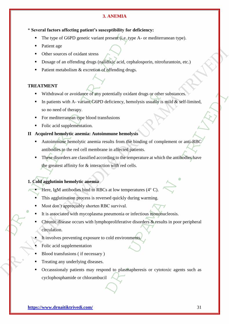
* Several factors affecting patient’s susceptibility for deficiency:
The type of G6PD genetic variant present (i.e. type A- or mediterranean type).
Patient age
Other sources of oxidant stress
Dosage of an offending drugs (nalidixic acid, cephalosporin, nitrofurantoin, etc.)
Patient metabolism & excretion of offending drugs.
TREATMENT
Withdrawal or avoidance of any potentially oxidant drugs or other substances.
In patients with A- variant G6PD deficiency, hemolysis usually is mild & self-limited,
so no need of therapy.
For mediterranean type blood transfusions
Folic acid supplementation.
II Acquired hemolytic anemia: Autoimmune hemolysis
Autoimmune hemolytic anemia results from the binding of complement or anti-RBC
antibodies to the red cell membrane in affected patients.
These disorders are classified according to the temperature at which the antibodies have
the greatest affinity for & interaction with red cells.
1. Cold agglutinin hemolytic anemia
Here, IgM antibodies bind to RBCs at low temperatures (4 C).
This agglutination process is reversed quickly during warming.
Most don’t appreciably shorten RBC survival.
It is associated with mycoplasma pneumonia or infectious mononucleosis.
Chronic disease occurs with lymphoproliferative disorders & results in poor peripheral
circulation.
It involves preventing exposure to cold environments.
Folic acid supplementation
Blood transfusions ( if necessary )
Treating any underlying diseases.
Occasssionaly patients may respond to plasmapheresis or cytotoxic agents such as
cyclophosphamide or chlorambucil

DR. NAITIK D TRIVEDI
&
DR. UPAMA N. TRIVEDI
3. ANEMIA
https://www.drnaitiktrivedi.com/ 32
2. Warm autoimmune hemolytic anemia :
IgG or occasionally IgA have greatest affinity for red cells at room temperature (37C).
Hemolysis involves the attachment & subsequent destruction of IgG coated
erythrocytes to receptors on macrophages in the RES.
It may be idiopathic, secondary to an underlying disease that affects immune system
(chronic lymphocytic leukemia, non Hodgkins lymphoma, or systemic lupus
erythematosus), or secondary to certain drugs.
When hemolysis is clinically significant, corticosteroid therapy is effective & blood
transfusions may be needed.
Splenectomy
Alternative therapies include immunosuppressive agents, danazol, IVIG &
cyclosporine