Download the Protease Science Brief - Enzyme Essentials
20
Systemic Proteolytic Enzymes A Review of Mechanisms of Action and the Resulting Health Benefits Copyright © 2013 Transformation Enzyme Corp. 1 ST EM ssful Nutritional Practice
Transcript of Download the Protease Science Brief - Enzyme Essentials
Systemic Proteolytic Enzymes
A Review of Mechanisms of Action and the Resulting Health Benefits
Copyright © 2013 Transformation Enzyme Corp.
11ATA LY S T
T E M
The Successful Nutritional Practice
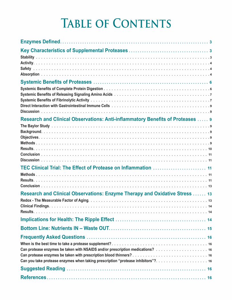
Enzymes Defined. . . . . . . . . . . . . . . . . . . . . . . . . . . . . . . . . . . . . . . . . . . . . . . . . . . . . . . . . . . . . . . . . . . . 3
Key.Characteristics.of.Supplemental.Proteases. . . . . . . . . . . . . . . . . . . . . . . . . . . . . . . . . . . . . 3Stability.. . . . . . . . . . . . . . . . . . . . . . . . . . . . . . . . . . . . . . . . . . . . . . . . . . . . . . . . . . . . . . . . . . . . . . . . . . . . . . . . . . . . . . . . . . . . . . . . . . . . . . . . . . . . . . . . . . . . . . . . . . . . . . . . . . . . . . 3
Activity. .. .. .. .. .. .. .. .. .. .. .. .. .. .. .. .. .. .. .. .. .. .. .. .. .. .. .. .. .. .. .. .. .. .. .. .. .. .. .. .. .. .. .. .. .. .. .. .. .. .. .. .. .. .. .. .. .. .. .. .. .. .. .. .. .. .. .. . 4
Safety .. . . . . . . . . . . . . . . . . . . . . . . . . . . . . . . . . . . . . . . . . . . . . . . . . . . . . . . . . . . . . . . . . . . . . . . . . . . . . . . . . . . . . . . . . . . . . . . . . . . . . . . . . . . . . . . . . . . . . . . . . . . . . . . . . . . . . . . . 4
Absorption.. . . . . . . . . . . . . . . . . . . . . . . . . . . . . . . . . . . . . . . . . . . . . . . . . . . . . . . . . . . . . . . . . . . . . . . . . . . . . . . . . . . . . . . . . . . . . . . . . . . . . . . . . . . . . . . . . . . . . . . . . . . . . . . . . . 4
Systemic Benefits of Proteases . . . . . . . . . . . . . . . . . . . . . . . . . . . . . . . . . . . . . . . . . . . . . . . . . . . . . 6Systemic Benefits of Complete Protein Digestion. .. .. .. .. .. .. .. .. .. .. .. .. .. .. .. .. .. .. .. .. .. .. .. .. .. .. .. .. .. .. .. .. .. .. .. .. .. .. .. .. . 6
Systemic Benefits of Releasing Signaling Amino Acids.. . . . . . . . . . . . . . . . . . . . . . . . . . . . . . . . . . . . . . . . . . . . . . . . . . . . . . . . . . . . . . . . . . . . . . . . . . 7
Systemic Benefits of Fibrinolytic Activity.. . . . . . . . . . . . . . . . . . . . . . . . . . . . . . . . . . . . . . . . . . . . . . . . . . . . . . . . . . . . . . . . . . . . . . . . . . . . . . . . . . . . . . . . . . . . 7
Direct.Interaction.with.Gastrointestinal.Immune.Cells.. . . . . . . . . . . . . . . . . . . . . . . . . . . . . . . . . . . . . . . . . . . . . . . . . . . . . . . . . . . . . . . . . . . . . . . . . . . . 9
Discussion.. . . . . . . . . . . . . . . . . . . . . . . . . . . . . . . . . . . . . . . . . . . . . . . . . . . . . . . . . . . . . . . . . . . . . . . . . . . . . . . . . . . . . . . . . . . . . . . . . . . . . . . . . . . . . . . . . . . . . . . . . . . . . . . . . . 9
Research and Clinical Observations: Anti-inflammatory Benefits of Proteases . . . . . . 9The Baylor Study .. . . . . . . . . . . . . . . . . . . . . . . . . . . . . . . . . . . . . . . . . . . . . . . . . . . . . . . . . . . . . . . . . . . . . . . . . . . . . . . . . . . . . . . . . . . . . . . . . . . . . . . . . . . . . . . . . . . . . . . . . . 9
Background. .. .. .. .. .. .. .. .. .. .. .. .. .. .. .. .. .. .. .. .. .. .. .. .. .. .. .. .. .. .. .. .. .. .. .. .. .. .. .. .. .. .. .. .. .. .. .. .. .. .. .. .. .. .. .. .. .. .. .. .. .. .. .. .. . 9
Objectives. .. .. .. .. .. .. .. .. .. .. .. .. .. .. .. .. .. .. .. .. .. .. .. .. .. .. .. .. .. .. .. .. .. .. .. .. .. .. .. .. .. .. .. .. .. .. .. .. .. .. .. .. .. .. .. .. .. .. .. .. .. .. .. .. .. . 9
Methods. .. .. .. .. .. .. .. .. .. .. .. .. .. .. .. .. .. .. .. .. .. .. .. .. .. .. .. .. .. .. .. .. .. .. .. .. .. .. .. .. .. .. .. .. .. .. .. .. .. .. .. .. .. .. .. .. .. .. .. .. .. .. .. .. .. .. . 9
Results. .. .. .. .. .. .. .. .. .. .. .. .. .. .. .. .. .. .. .. .. .. .. .. .. .. .. .. .. .. .. .. .. .. .. .. .. .. .. .. .. .. .. .. .. .. .. .. .. .. .. .. .. .. .. .. .. .. .. .. .. .. .. .. .. .. .. .. 10
Conclusion . .. .. .. .. .. .. .. .. .. .. .. .. .. .. .. .. .. .. .. .. .. .. .. .. .. .. .. .. .. .. .. .. .. .. .. .. .. .. .. .. .. .. .. .. .. .. .. .. .. .. .. .. .. .. .. .. .. .. .. .. .. .. .. .. 11
Discussion.. . . . . . . . . . . . . . . . . . . . . . . . . . . . . . . . . . . . . . . . . . . . . . . . . . . . . . . . . . . . . . . . . . . . . . . . . . . . . . . . . . . . . . . . . . . . . . . . . . . . . . . . . . . . . . . . . . . . . . . . . . . . . . . . .11
TEC Clinical Trial: The Effect of Protease on Inflammation . . . . . . . . . . . . . . . . . . . . . . . . . 11Methods. .. .. .. .. .. .. .. .. .. .. .. .. .. .. .. .. .. .. .. .. .. .. .. .. .. .. .. .. .. .. .. .. .. .. .. .. .. .. .. .. .. .. .. .. .. .. .. .. .. .. .. .. .. .. .. .. .. .. .. .. .. .. .. .. .. .. 11
Results. .. .. .. .. .. .. .. .. .. .. .. .. .. .. .. .. .. .. .. .. .. .. .. .. .. .. .. .. .. .. .. .. .. .. .. .. .. .. .. .. .. .. .. .. .. .. .. .. .. .. .. .. .. .. .. .. .. .. .. .. .. .. .. .. .. .. .. 11
Conclusion . .. .. .. .. .. .. .. .. .. .. .. .. .. .. .. .. .. .. .. .. .. .. .. .. .. .. .. .. .. .. .. .. .. .. .. .. .. .. .. .. .. .. .. .. .. .. .. .. .. .. .. .. .. .. .. .. .. .. .. .. .. .. .. .. 13
Research and Clinical Observations: Enzyme Therapy and Oxidative Stress . . . . . . . 13Redox - The Measurable Factor of Aging ... ... ... ... ... ... ... ... ... ... ... ... ... ... ... ... ... ... ... ... ... ... ... ... ... ... ... ... ... ... ... ... ... ... ... ... ... ... ... ... ... ... ... ... ... .. 13
Clinical Findings. .. .. .. .. .. .. .. .. .. .. .. .. .. .. .. .. .. .. .. .. .. .. .. .. .. .. .. .. .. .. .. .. .. .. .. .. .. .. .. .. .. .. .. .. .. .. .. .. .. .. .. .. .. .. .. .. .. .. .. .. .. 14
Results. .. .. .. .. .. .. .. .. .. .. .. .. .. .. .. .. .. .. .. .. .. .. .. .. .. .. .. .. .. .. .. .. .. .. .. .. .. .. .. .. .. .. .. .. .. .. .. .. .. .. .. .. .. .. .. .. .. .. .. .. .. .. .. .. .. .. .. 14
Implications for Health: The Ripple Effect . . . . . . . . . . . . . . . . . . . . . . . . . . . . . . . . . . . . . . . . . . 14
Bottom Line: Nutrients IN – Waste OUT. . . . . . . . . . . . . . . . . . . . . . . . . . . . . . . . . . . . . . . . . . . . . 15
Frequently Asked Questions . . . . . . . . . . . . . . . . . . . . . . . . . . . . . . . . . . . . . . . . . . . . . . . . . . . . . . . 16When is the best time to take a protease supplement?. .. .. .. .. .. .. .. .. .. .. .. .. .. .. .. .. .. .. .. .. .. .. .. .. .. .. .. .. .. .. .. .. .. .. .. .. .. 16
Can protease enzymes be taken with NSAIDS and/or prescription medications? .. . . . . . . . . . . . . . . . . . . . . . . . . . . . . . . . . . . . . . . . 16
Can protease enzymes be taken with prescription blood thinners?. .. .. .. .. .. .. .. .. .. .. .. .. .. .. .. .. .. .. .. .. .. .. .. .. .. .. .. .. .. 16
Can you take protease enzymes when taking prescription “protease inhibitors”?. .. .. .. .. .. .. .. .. .. .. .. .. .. .. .. .. .. .. .. .. 16
Suggested Reading . . . . . . . . . . . . . . . . . . . . . . . . . . . . . . . . . . . . . . . . . . . . . . . . . . . . . . . . . . . . . . . . 16
References. . . . . . . . . . . . . . . . . . . . . . . . . . . . . . . . . . . . . . . . . . . . . . . . . . . . . . . . . . . . . . . . . . . . . . . . . 16
Table of Contents
Systemic Proteolytic Enzymes Transformation Enzyme Corp. • page �
Transformation Enzyme Corporation (TEC) is a nutritional supplement company specializing in enzyme therapy since 1991. Our goal is to educate health care professionals and the public on the fundamental health benefits of enzyme and probiotic supplementation. We believe a healthy diet and lifestyle, along with optimal digestion and a strong immune system, is the foundation of wellness and therefore enzyme therapy is the Genesis of Good Health™.
Enzymes DefinedEnzymes are protein molecules that catalyze chemical reactions. In the human body, digestive enzymes catalyze or facilitate the breakdown of food molecules within the gastrointestinal tract. Metabolic enzymes are those found in the cells and tissues that are responsible for all chemical reactions in the body. Supplemental proteolytic enzymes or “proteases” are digestive enzymes that specifically digest proteins into small peptides or amino acids.
As digestive aids, when taken with meals they facilitate complete digestion of animal and plant proteins. When taken between meals, proteolytic enzymes are absorbed into the blood stream. In this sense, they are known as “systemic enzymes” as they impact the entire body in a positive way. This literature review will look at the key characteristics and research available on the proteolytic enzymes used by TEC and discuss the mechanisms of action and systemic health benefits they have on the immune system, circulatory system, and ultimately the entire body.
Systemic Proteolytic Enzymes:A Review of Mechanisms of Action and the Resulting Health Benefits
Key.Characteristics.of.Supplemental.ProteasesThere are several sources for supplemental enzymes available on the market today. They are animal (pancre-atic, trypsin, chymotrypsin), plant (bromelain, papain), and microbial (fungal and bacterial). TEC uses microbial and plant enzymes for their long history of safety, quality, purity and efficacy.
StabilityGastric stability is very important when selecting supple-mental enzymes. As protein molecules, it is logical to think they would be denatured in the harsh environment of the stomach. For example, endogenous pancreatic enzymes are secreted into the small intestines, bypassing the stom-ach all together. So when pancreatic enzymes are taken orally as supplements they must have an enteric coating in order to survive the acid in the stomach. Microbial and plant enzymes on the other hand are not as susceptible to the acid (Mamadou 2005).
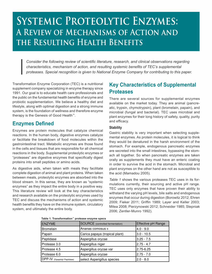
Table 1 shows the various proteases TEC uses in its for-mulations currently, their sourcing and active pH range. TEC uses only enzymes that have proven their ability to withstand the varying pH levels, bile salts and endogenous enzymes that occur during digestion (Borowitz 2012; Ehren 2009; Fieker 2011; Griffin 1989; Layer and Keller 2003; Mitea 2008; Pierzynowski 2012; Schneider 1985; Stepniak 2006; Zentler-Munro 1992).
Consider the following review of scientific literature, research, and clinical observations regarding characteristics, mechanism of action, and resulting systemic benefits of TEC’s supplemental proteases. Special recognition is given to National Enzyme Company for contributing to this paper.
ENZYME SOURCE (controlled fermentation) Effective pH Range
Bromelain Ananas comosus 6 4.0 - 9.0
Papain Carica papaya (tropical plant) 3.0 - 10.5
Peptidase Aspergillus oryzae 3.25 - 7.5
Protease 3.0 Aspergillus niger 2.75 - 4.7
Protease 4.5 Aspergillus oryzae var 2.75-6.25
Protease 6.0 Aspergillus oryzae 2.75 - 7.0
DPP-IV (Dipeptidyl Peptidase) select Asperigillus species 2.0 - 8.0
Chart 2. Transformation™ protease enzyme specs
Table.1 ..Transformation™.protease.enzyme.specs
Systemic Proteolytic Enzymes Transformation Enzyme Corp. • page �
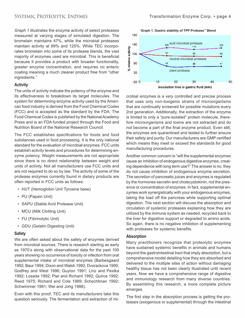
Graph 1 illustrates the enzyme activity of select proteases measured at varying stages of simulated digestion. The bromelain maintains �7%, while the microbial proteases maintain activity at 89% and 125%. While TEC incorpo-rates bromelain into some of its protease blends, the vast majority of enzymes used are microbial. This is beneficial because it provides a product with broader functionality, greater enzyme concentration, and requires no enteric coating meaning a much cleaner product free from “other ingredients.”
ActivityThe units of activity indicate the potency of the enzyme and its effectiveness to breakdown its target molecules. The system for determining enzyme activity used by the Ameri-can food industry is derived from the Food Chemical Codex (FCC) and is accepted as the standard by the FDA. The Food Chemical Codex is published by the National Academy Press and is an FDA funded project through the Food and Nutrition Board of the National Research Council.
The FCC establishes specifications for foods and food substances used in food processing. It is the only national standard for the evaluation of microbial enzymes. FCC units establish activity levels and procedures for determining en-zyme potency. Weight measurements are not appropriate since there is no direct relationship between weight and units of activity. Not all manufacturers use FCC units and are not required to do so by law. The activity of some of the protease enzymes currently found in dietary products are often reported in FCC units as follows:
HUT (Hemoglobin Unit Tyrosine base)
PU (Papain Unit)
SAPU (Stable Acid Protease Unit)
MCU (Milk Clotting Unit)
FU (Fibrinolytic Unit)
GDU (Gelatin Digesting Unit)
SafetyWe are often asked about the safety of enzymes derived from microbial sources. There is research starting as early as 1970’s along with observational data for the past 100 years showing no occurrence of toxicity or infection from oral supplemental intake of microbial enzymes (Barbesgaard 1992; Baur 199�; Dixon and Walsh 1992; Dvorackova 1990; Godfrey and West 1996; Guyton 1991; Linz and Pestka 1992; Losada 1992; Pier and Richard 1992; Quirce 1992; Reed 1975; Richard and Cole 1989; Schechtman 1992; Schwimmer 1981; Wei and Johg 1986).
Even with this proof, TEC and its manufacturers take this question seriously. The fermentation and extraction of mi-
•
•
•
•
•
•
crobial enzymes is a very controlled and precise process that uses only non-toxigenic strains of microorganisms that are continually screened for possible mutations every 2nd generation. Additionally, the extraction of the enzyme is limited to only a “pure-isolated” protein molecule, there-fore microorganisms and toxins are not extracted and do not become a part of the final enzyme product. Even still, the enzymes are quarantined and tested to further ensure their safety and purity. Our manufacturers are GMP certified which means they meet or exceed the standards for good manufacturing procedures.
Another common concern is “will the supplemental enzymes cause an inhibition of endogenous digestive enzymes, creat-ing dependence with long term use?” The answer is no, they do not cause inhibition of endogenous enzyme secretion. The secretion of pancreatic juices and enzymes is regulated by the hormones secretin and cholecystokinin, not the pres-ence or concentration of enzymes. In fact, supplemental en-zymes work synergistically with your endogenous enzymes, taking the load off the pancreas while supporting optimal digestion. The next section will discuss the absorption and circulation of systemic proteases explaining how they are utilized by the immune system as needed, recycled back to the liver for digestive support or degraded to amino acids. So again, there is no negative inhibition of supplementing with proteases for systemic benefits.
AbsorptionMany practitioners recognize that proteolytic enzymes have sustained systemic benefits in animals and humans beyond the gastrointestinal tract that imply absorption, but a comprehensive model detailing how they are absorbed and delivered to the multiple sites of action without damaging healthy tissue has not been clearly illustrated until recent years. Now we have a comprehensive range of digestive and immunology research from many diverse countries. By assembling this research, a more complete picture emerges.
The first step in the absorption process is getting the pro-teases (exogenous or supplemental) through the intestinal
Graph.1 ..Gastric.stability.of.TPP.Protease™ Blend
neutral microbial protease
plant protease
acid microbial protease
Systemic Proteolytic Enzymes Transformation Enzyme Corp. • page 5
mucous and across the intestinal epithelium. Mucous pene-tration for many proteases is relatively simple because many of them thin mucous by digesting the proteins in it (Chang 2012; Braga 1993, 1990; Majima 1986). Once through the mucous, there are at least four ways for proteases to get across the epithelium (Lorkowski 2012).
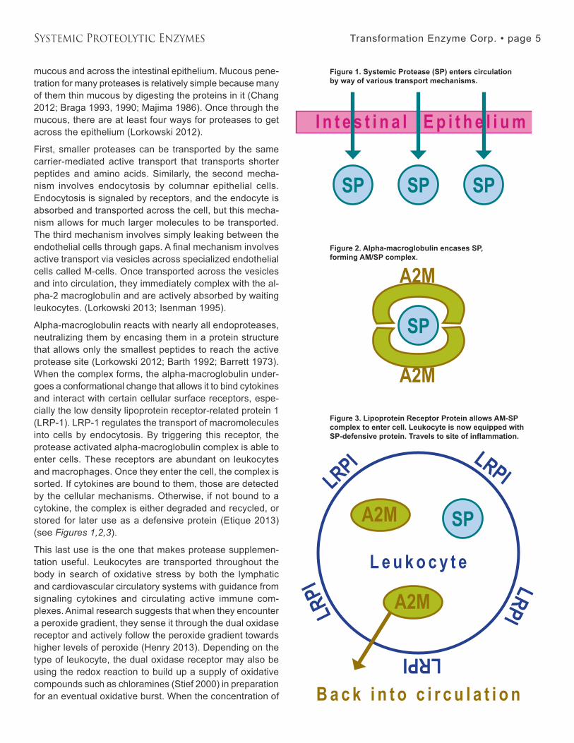
First, smaller proteases can be transported by the same carrier-mediated active transport that transports shorter peptides and amino acids. Similarly, the second mecha-nism involves endocytosis by columnar epithelial cells. Endocytosis is signaled by receptors, and the endocyte is absorbed and transported across the cell, but this mecha-nism allows for much larger molecules to be transported. The third mechanism involves simply leaking between the endothelial cells through gaps. A final mechanism involves active transport via vesicles across specialized endothelial cells called M-cells. Once transported across the vesicles and into circulation, they immediately complex with the al-pha-2 macroglobulin and are actively absorbed by waiting leukocytes. (Lorkowski 2013; Isenman 1995).
Alpha-macroglobulin reacts with nearly all endoproteases, neutralizing them by encasing them in a protein structure that allows only the smallest peptides to reach the active protease site (Lorkowski 2012; Barth 1992; Barrett 1973). When the complex forms, the alpha-macroglobulin under-goes a conformational change that allows it to bind cytokines and interact with certain cellular surface receptors, espe-cially the low density lipoprotein receptor-related protein 1 (LRP-1). LRP-1 regulates the transport of macromolecules into cells by endocytosis. By triggering this receptor, the protease activated alpha-macroglobulin complex is able to enter cells. These receptors are abundant on leukocytes and macrophages. Once they enter the cell, the complex is sorted. If cytokines are bound to them, those are detected by the cellular mechanisms. Otherwise, if not bound to a cytokine, the complex is either degraded and recycled, or stored for later use as a defensive protein (Etique 201�) (see Figures 1,2,3).
This last use is the one that makes protease supplemen-tation useful. Leukocytes are transported throughout the body in search of oxidative stress by both the lymphatic and cardiovascular circulatory systems with guidance from signaling cytokines and circulating active immune com-plexes. Animal research suggests that when they encounter a peroxide gradient, they sense it through the dual oxidase receptor and actively follow the peroxide gradient towards higher levels of peroxide (Henry 201�). Depending on the type of leukocyte, the dual oxidase receptor may also be using the redox reaction to build up a supply of oxidative compounds such as chloramines (Stief 2000) in preparation for an eventual oxidative burst. When the concentration of
Figure 1. Systemic Protease (SP) enters circulation .by.way.of.various.transport.mechanisms .
Figure 2. Alpha-macroglobulin encases SP, .forming AM/SP complex.
Figure 3. Lipoprotein Receptor Protein allows AM-SP complex to enter cell. Leukocyte is now equipped with SP-defensive protein. Travels to site of inflammation.
Systemic Proteolytic Enzymes Transformation Enzyme Corp. • page 6
peroxide reaches a sufficient level, it triggers the anticipated oxidative burst, releasing both the oxidative compounds and the stored defensive proteases complexed with alpha macroglobulin (Henry 2012; Niethammer, 2009; Stief 2000). The oxidative compounds attack whatever molecules are contributing to the inflammation and also neutralize the al-pha macroglobulin complexes, releasing and activating the stored proteases. These then act on whatever proteins are present. By oxidizing and digesting the molecules present, the immune response neutralizes most sources of inflam-mation and any immune complexes and cytokines in the local area. When the oxidative potential subsides, fresh alpha macroglobulin arrives and re-encases the protease if it is still active, and the cycle starts over.
To summarize thus far, we have addressed the most com-mon questions regarding the use of supplemental microbial enzymes. The facts are:
Microbial enzymes are GI stable and are not de-stroyed in the harsh environment of the stomach.
They are safe and pose no toxic or infection threat, and do not cause inhibition of endogenous enzymes.
They are absorbed intact as functional systemic enzymes into circulation, escorted by alpha 2 macro-globulins and utilized by the leukocytes.
As a defensive protein, protease is directed to the site of inflammation by the immune system, where the immune system then uses it as a tool to protect cells, signal other cells, or to degrade unwanted or invasive proteins. When used in this manner they have antioxidant, immune-stimu-latory, immunosuppressive, and anti-inflammatory effects depending on how your immune system is directing them. Basically, they are a tool the immune system uses to inter-act with cells and proteins to signal or degrade them. Now, let’s explore more closely the functions and mechanisms of action that gives both endogenous and supplemental proteases their systemic benefit.
Systemic Benefits of ProteasesIt is important to note that most of what we know about proteases’ interaction with the immune system has been learned from the study of endogenous protease re-absorp-tion and systemic use. In an ideal world and healthy body, supply meets demand and all is well. So why does the body need supplemental proteases? The body is a very complex organism that also strives to be very efficient. It is known that many compounds that our body is able to synthesize are absorbed intact from the digestive system to save energy or because our body is not making enough. Proteases are proteins that are very complex molecules that require a lot of energy and amino acids to synthesize. Consequently, the
1.
2.
�.
body has a limited capacity to synthesize proteases quickly. Plants, animals and microbial organisms contain a lot of proteases that are very similar to human proteases. When the body has a need, and recognizes these proteases, it will absorb them from the digestive tract in the same manner as it reabsorbs human trypsin, and uses them for the same purposes. In our fast paced, high stress, processed food society, the need for supplemental proteases to assist the body in maintaining health has never been greater.
The important thing to remember when considering supple-mental proteases is they are more like supplemental vita-mins for your immune system. They are not like drugs which interfere with some aspect of either your body’s function or of a pathogen’s function. Supplemental proteases save the body the energy of synthesizing a tool it already makes al-lowing that energy to be used elsewhere. They also provide an additional supply when the body cannot keep up with the demand. And lastly, because they are so similar to naturally occurring molecules there are no negative side effects as commonly seen with medications.
There are numerous ways in which proteases can impart systemic benefits. Some assign the systemic effects com-pletely to the nutritional benefits of more rapid and complete digestion of proteins, making more amino acids available not only for building but also for signaling. Another benefit related to digestion is proteases ability to clear the blood stream of short chain peptides. Others make the point that proteases can eliminate innate immune system triggering proteins before they reach the intestinal lining. Still others suggest that the proteases interact directly with the ex-tensive portion of the immune system that extends along the gastrointestinal tract, suppressing the immune system rapidly via a variety of mechanisms at the surface of the cells. Finally, there are the proponents of action via intestinal absorption and delivery discussed in the previous section. The benefits of protease supplementation are diverse, and it follows that the mechanisms of action are likely to be di-verse and multifaceted as well. Recently, scientific research has become available to construct scientific models that coherently explain how proteases might provide systemic benefits via each of these mechanisms.
Systemic Benefits of Complete Protein DigestionIt is well established that proteases digest proteins, deliver-ing amino acids to the body to build and repair cells. The faster and more complete this nutritional process occurs, the greater the overall benefit to the body. The fact that amino acids are a necessary component for virtually every cell in our body (enzymes, hormones, neurotransmitters, immune cells, blood cells, muscles, bones, etc.) is enough argument alone to support complete protein digestion with supplemental proteases. Moreover, there are some proteins
Systemic Proteolytic Enzymes Transformation Enzyme Corp. • page 7
more resistant to digestion than others. When these proteins are not completely digested the result is not only a lack of amino acids for building, but worse, an increased potential for “trigger proteins” that initiate oxidative stress, inflamma-tion, and over whelm the immune system.
A key factor in this resistance can be the number and distribution of proline amino acids. Proline bonds create a tight bend in an amino acid chain, causing a protein to fold. This creates the opportunity for cross-linkages to form and numerous folds with cross-linkages make it difficult for endogenous proteases such as pepsin, trypsin and chy-motrypsin to digest proteins or portions of proteins. A good example of this is the protein gluten which resists digestion because it is rich in proline bonds. In addition, the most re-sistant degradation peptides are rich in cross-linking amino acids such as glutamine. When gluten and related peptides reach the intestinal epithelium, the innate immune system recognizes them as threats through the toll-like receptor systems, triggering immune responses (Palová-Jelinková 2013; Junker 2012; Volta 2012; Sapone 2011). In sensitive individuals, repeated exposures to undigested proteins can cause local inflammation in the gastrointestinal tract as well as systemic effects throughout the body (Volta 2012).
The good news is, there are certain microbial proteases, also known as “endo-peptidases,” that are quite effective at digesting proline bonds, working with the body to digest these proteins and eliminate immune triggering proteins. Since there are many other resistant proteins in food capable of triggering similar responses, more complete protein digestion is capable of improving systemic health in sensitive individuals.
Systemic Benefits of Releasing Signaling Amino AcidsAnother way proteases may provide systemic benefit via complete protein digestion is by releasing signaling amino acids. By making specific signaling amino acids readily available you are supporting the health of many tissues of the body. Repeated stimulation of the immune system by undigested proteins and other components of the typical western diet may result in loosening of the tight junctions between the cells lining the intestines, allowing some bac-terial toxins to “leak” past the epithelial barrier and trigger more intense immune responses (Muniandy 2009). The amino acids glutamine and arginine are known to stimu-late the tightening of these junctions and the amino acid leucine stimulates the replication of the intestinal lining cells (Beutheu 2013; Rapin 2010). Properly formulated proteases consumed with protein may support more rapid and complete release of these amino acids, indirectly and rapidly supporting intestinal integrity through better nutrition (Rhoads 2009). Beyond the intestinal epithelium, glutamine is the most important fuel for intestinal immune cells, and
is an important factor in T-cell differentiation (Ruth 201�; Waickman 2012).
Furthermore, research has shown that amino acids from pro-tein support muscle development and retention best when the amino acids are released more rapidly from protein. The branched chain amino acid leucine is especially important as increased concentrations of free leucine in mammalian muscle cells signal to the cell that adequate amino acid supplies are available. This initiates increased growth and cell division via stimulation of the mammalian target of rapamycin (mTOR) enzyme. It also inhibits apoptosis (cell death) by inhibiting the 5’ adenosine monophosphate-ac-tivated protein kinase (AMPK) enzyme (Li 2011; Kim2009; Kimura 2003). Other amino acids such as isoleucine, valine and glutamine have an additional upstream effect on mTOR and muscle growth, but their roles are not yet as clearly defined by research (You 2012; Nicklin 2009; Wu 2009; Yao 2008). mTOR is also present in many other cell types, regulating many systems. An example is the cells of the hypothalamus, where stimulation of mTOR and suppression of AMPK is being investigated as playing a central role in suppressing hunger (Guzmán-Quevedo 2013; Journel 2012; Cota 2006). Glutamine and tryptophan have been identified as signaling molecules in the neurological system, and the amino acids arginine and glutamine have been identified as having cardiovascular benefits (Albrecht 2010; Dong, 2011; Wu 2009; Stone 2007; Kakoki 2006). Many other signaling amino acid benefits have been recently identified, clarify-ing the nutritional aspect of proteases as releasing not just the building blocks of the body, but the fuel for the building and the signals that regulate the building (Sousa 2012; Wu 2009; Ropelle 2008).
Systemic Benefits of Fibrinolytic ActivityAn area in which supplemental proteases have far reaching systemic benefit is their support of healthy blood flow. We have discussed the importance of optimal protein digestion so now let’s review the importance of optimal circulation and the role proteases can play. Red blood cells are responsible for carrying nutrients and oxygen to the cells and carrying waste from the cells. Better blood flow will naturally enhance this process and support cellular nutrition, function, and detoxification.
Earlier in this paper we discussed the fact that endogenous proteases bound to alpha 2 macroglobulin have the ability to break down short chain peptides circulating in the blood. Relative to cardiovascular health, proteases also have the ability to break down excess fibrin and thrombocytes in blood. Blood clots are formed by thrombocytes (small frag-mented “platelet” cells) and fibrin (a fibrous protein mesh) that holds the thrombocytes together. Fibrin is activated and controlled by the presence of fibrinogen. During times of
Systemic Proteolytic Enzymes Transformation Enzyme Corp. • page 8
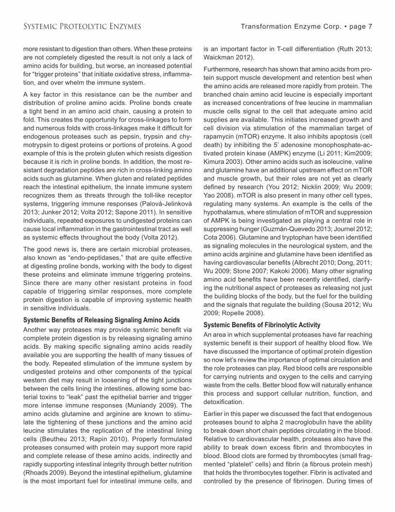
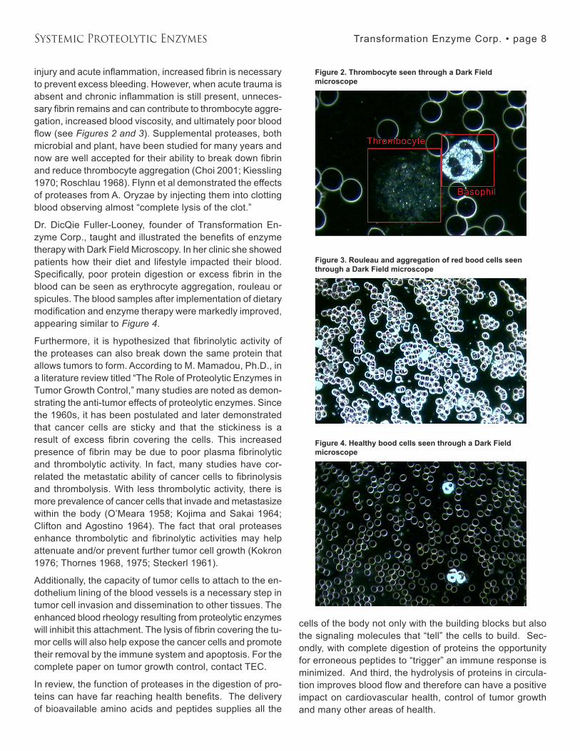
injury and acute inflammation, increased fibrin is necessary to prevent excess bleeding. However, when acute trauma is absent and chronic inflammation is still present, unneces-sary fibrin remains and can contribute to thrombocyte aggre-gation, increased blood viscosity, and ultimately poor blood flow (see Figures 2 and 3). Supplemental proteases, both microbial and plant, have been studied for many years and now are well accepted for their ability to break down fibrin and reduce thrombocyte aggregation (Choi 2001; Kiessling 1970; Roschlau 1968). Flynn et al demonstrated the effects of proteases from A. Oryzae by injecting them into clotting blood observing almost “complete lysis of the clot.”
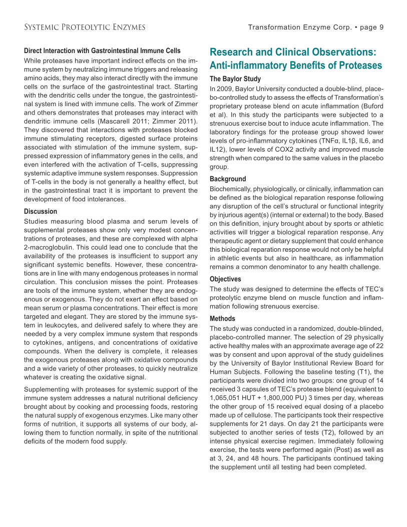
Dr. DicQie Fuller-Looney, founder of Transformation En-zyme Corp., taught and illustrated the benefits of enzyme therapy with Dark Field Microscopy. In her clinic she showed patients how their diet and lifestyle impacted their blood. Specifically, poor protein digestion or excess fibrin in the blood can be seen as erythrocyte aggregation, rouleau or spicules. The blood samples after implementation of dietary modification and enzyme therapy were markedly improved, appearing similar to Figure 4.
Furthermore, it is hypothesized that fibrinolytic activity of the proteases can also break down the same protein that allows tumors to form. According to M. Mamadou, Ph.D., in a literature review titled “The Role of Proteolytic Enzymes in Tumor Growth Control,” many studies are noted as demon-strating the anti-tumor effects of proteolytic enzymes. Since the 1960s, it has been postulated and later demonstrated that cancer cells are sticky and that the stickiness is a result of excess fibrin covering the cells. This increased presence of fibrin may be due to poor plasma fibrinolytic and thrombolytic activity. In fact, many studies have cor-related the metastatic ability of cancer cells to fibrinolysis and thrombolysis. With less thrombolytic activity, there is more prevalence of cancer cells that invade and metastasize within the body (O’Meara 1958; Kojima and Sakai 1964; Clifton and Agostino 196�). The fact that oral proteases enhance thrombolytic and fibrinolytic activities may help attenuate and/or prevent further tumor cell growth (Kokron 1976; Thornes 1968, 1975; Steckerl 1961).
Additionally, the capacity of tumor cells to attach to the en-dothelium lining of the blood vessels is a necessary step in tumor cell invasion and dissemination to other tissues. The enhanced blood rheology resulting from proteolytic enzymes will inhibit this attachment. The lysis of fibrin covering the tu-mor cells will also help expose the cancer cells and promote their removal by the immune system and apoptosis. For the complete paper on tumor growth control, contact TEC.
In review, the function of proteases in the digestion of pro-teins can have far reaching health benefits. The delivery of bioavailable amino acids and peptides supplies all the
cells of the body not only with the building blocks but also the signaling molecules that “tell” the cells to build. Sec-ondly, with complete digestion of proteins the opportunity for erroneous peptides to “trigger” an immune response is minimized. And third, the hydrolysis of proteins in circula-tion improves blood flow and therefore can have a positive impact on cardiovascular health, control of tumor growth and many other areas of health.
Figure 2. Thrombocyte seen through a Dark Field microscope
Figure 3. Rouleau and aggregation of red bood cells seen through a Dark Field microscope
Figure 4. Healthy bood cells seen through a Dark Field microscope
Systemic Proteolytic Enzymes Transformation Enzyme Corp. • page 9
Direct.Interaction.with.Gastrointestinal.Immune.CellsWhile proteases have important indirect effects on the im-mune system by neutralizing immune triggers and releasing amino acids, they may also interact directly with the immune cells on the surface of the gastrointestinal tract. Starting with the dendritic cells under the tongue, the gastrointesti-nal system is lined with immune cells. The work of Zimmer and others demonstrates that proteases may interact with dendritic immune cells (Mascarell 2011; Zimmer 2011). They discovered that interactions with proteases blocked immune stimulating receptors, digested surface proteins associated with stimulation of the immune system, sup-pressed expression of inflammatory genes in the cells, and even interfered with the activation of T-cells, suppressing systemic adaptive immune system responses. Suppression of T-cells in the body is not generally a healthy effect, but in the gastrointestinal tract it is important to prevent the development of food intolerances.
DiscussionStudies measuring blood plasma and serum levels of supplemental proteases show only very modest concen-trations of proteases, and these are complexed with alpha 2-macroglobulin. This could lead one to conclude that the availability of the proteases is insufficient to support any significant systemic benefits. However, these concentra-tions are in line with many endogenous proteases in normal circulation. This conclusion misses the point. Proteases are tools of the immune system, whether they are endog-enous or exogenous. They do not exert an effect based on mean serum or plasma concentrations. Their effect is more targeted and elegant. They are stored by the immune sys-tem in leukocytes, and delivered safely to where they are needed by a very complex immune system that responds to cytokines, antigens, and concentrations of oxidative compounds. When the delivery is complete, it releases the exogenous proteases along with oxidative compounds and a wide variety of other proteases, to quickly neutralize whatever is creating the oxidative signal.
Supplementing with proteases for systemic support of the immune system addresses a natural nutritional deficiency brought about by cooking and processing foods, restoring the natural supply of exogenous enzymes. Like many other forms of nutrition, it supports all systems of our body, al-lowing them to function normally, in spite of the nutritional deficits of the modern food supply.
Research and Clinical Observations: Anti-inflammatory Benefits of ProteasesThe Baylor StudyIn 2009, Baylor University conducted a double-blind, place-bo-controlled study to assess the effects of Transformation’s proprietary protease blend on acute inflammation (Buford et al). In this study the participants were subjected to a strenuous exercise bout to induce acute inflammation. The laboratory findings for the protease group showed lower levels of pro-inflammatory cytokines (TNFα, IL1β, IL6, and IL12), lower levels of COX2 activity and improved muscle strength when compared to the same values in the placebo group.
BackgroundBiochemically, physiologically, or clinically, inflammation can be defined as the biological reparation response following any disruption of the cell’s structural or functional integrity by injurious agent(s) (internal or external) to the body. Based on this definition, injury brought about by sports or athletic activities will trigger a biological reparation response. Any therapeutic agent or dietary supplement that could enhance this biological reparation response would not only be helpful in athletic events but also in healthcare, as inflammation remains a common denominator to any health challenge.
ObjectivesThe study was designed to determine the effects of TEC’s proteolytic enzyme blend on muscle function and inflam-mation following strenuous exercise.
MethodsThe study was conducted in a randomized, double-blinded, placebo-controlled manner. The selection of 29 physically active healthy males with an approximate average age of 22 was by consent and upon approval of the study guidelines by the University of Baylor Institutional Review Board for Human Subjects. Following the baseline testing (T1), the participants were divided into two groups: one group of 1� received � capsules of TEC’s protease blend (equivalent to 1,065,051 HUT + 1,800,000 PU) 3 times per day, whereas the other group of 15 received equal dosing of a placebo made up of cellulose. The participants took their respective supplements for 21 days. On day 21 the participants were subjected to another series of tests (T2), followed by an intense physical exercise regimen. Immediately following exercise, the tests were performed again (Post) as well as at 3, 24, and 48 hours. The participants continued taking the supplement until all testing had been completed.
Systemic Proteolytic Enzymes Transformation Enzyme Corp. • page 10
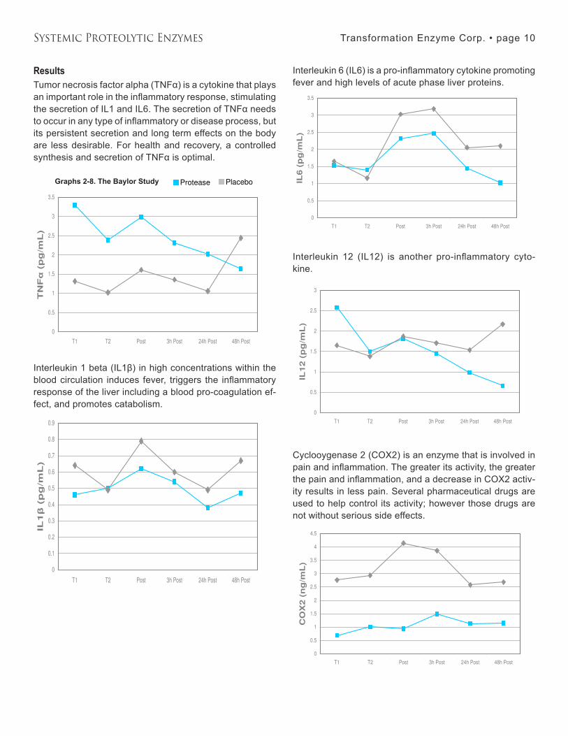
ResultsTumor necrosis factor alpha (TNFα) is a cytokine that plays an important role in the inflammatory response, stimulating the secretion of IL1 and IL6. The secretion of TNFα needs to occur in any type of inflammatory or disease process, but its persistent secretion and long term effects on the body are less desirable. For health and recovery, a controlled synthesis and secretion of TNFα is optimal.
Interleukin 1 beta (IL1β) in high concentrations within the blood circulation induces fever, triggers the inflammatory response of the liver including a blood pro-coagulation ef-fect, and promotes catabolism.
Interleukin 6 (IL6) is a pro-inflammatory cytokine promoting fever and high levels of acute phase liver proteins.
Interleukin 12 (IL12) is another pro-inflammatory cyto-kine.
Cyclooygenase 2 (COX2) is an enzyme that is involved in pain and inflammation. The greater its activity, the greater the pain and inflammation, and a decrease in COX2 activ-ity results in less pain. Several pharmaceutical drugs are used to help control its activity; however those drugs are not without serious side effects.
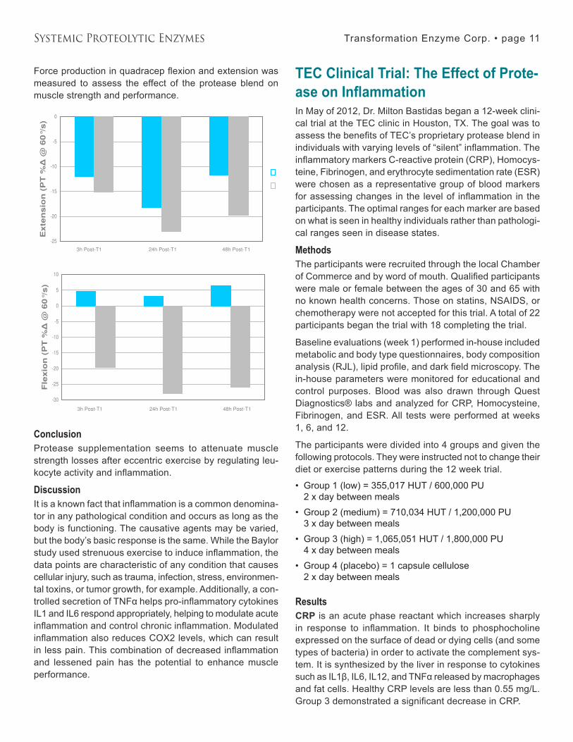
Dominant Leg Flexion PT %3h Post-T1 24h Post-T148h Post-T1
Protease 4.73 3.01 6.44
Placebo -19.54 -27.94 -25.87
Dominant Leg Flexion
-30
-25
-20
-15
-10
-5
0
5
10
Time
Fle
xio
n (
PT
%
@ 6
0°/
s)
Protease
Placebo
Dominant Leg Flexion PT %3h Post-T1 24h Post-T148h Post-T1
Protease 4.73 3.01 6.44
Placebo -19.54 -27.94 -25.87
Dominant Leg Flexion
-30
-25
-20
-15
-10
-5
0
5
10
Time
Fle
xio
n (
PT
%
@ 6
0°/
s)
Protease
PlaceboGraphs 2-8. The Baylor Study
TNF(pg/mL) T1 T2 Post 3h Post 24h Post 48h Post
Protease 3.29 2.39 2.98 2.32 2.03 1.64
Placebo 1.32 1.02 1.61 1.36 1.06 2.44
TNF
0
0.5
1
1.5
2
2.5
3
3.5
T1 T2 Post 3h Post 24h Post 48h Post
Time
TN
F
(p
g/m
L)
Protease
Placebo
IL1(pg/mL) T1 T2 Post 3h Post 24h Post 48h Post
Protease 0.46 0.5 0.62 0.54 0.38 0.47
Placebo 0.64 0.49 0.79 0.6 0.49 0.67
IL1
0
0.1
0.2
0.3
0.4
0.5
0.6
0.7
0.8
0.9
T1 T2 Post 3h Post 24h Post 48h Post
Time
IL1
(p
g/m
L)
Protease
Placebo
IL6(pg/mL) T1 T2 Post 3h Post 24h Post 48h Post
Protease 1.53 1.4 2.32 2.47 1.45 1.02
Placebo 1.65 1.16 3.03 3.19 2.05 2.1
IL6
0
0.5
1
1.5
2
2.5
3
3.5
T1 T2 Post 3h Post 24h Post 48h Post
Time
IL6 (
pg
/mL
)
Protease
Placebo
IL12(pg/mL) T1 T2 Post 3h Post 24h Post 48h Post
Protease 2.58 1.5 1.81 1.45 0.98 0.66
Placebo 1.65 1.38 1.87 1.71 1.54 2.17
IL12
0
0.5
1
1.5
2
2.5
3
T1 T2 Post 3h Post 24h Post 48h Post
Time
IL12 (
pg
/mL
)
Protease
Placebo
COX2(ng/mL) T1 T2 Post 3h Post 24h Post 48h Post
Protease 0.69 1.01 0.94 1.49 1.13 1.15
Placebo 2.76 2.93 4.14 3.86 2.58 2.69
COX2
0
0.5
1
1.5
2
2.5
3
3.5
4
4.5
T1 T2 Post 3h Post 24h Post 48h Post
Time
CO
X2 (
ng
/mL
)
Protease
Placebo
Systemic Proteolytic Enzymes Transformation Enzyme Corp. • page 11
Force production in quadracep flexion and extension was measured to assess the effect of the protease blend on muscle strength and performance.
ConclusionProtease supplementation seems to attenuate muscle strength losses after eccentric exercise by regulating leu-kocyte activity and inflammation.
DiscussionIt is a known fact that inflammation is a common denomina-tor in any pathological condition and occurs as long as the body is functioning. The causative agents may be varied, but the body’s basic response is the same. While the Baylor study used strenuous exercise to induce inflammation, the data points are characteristic of any condition that causes cellular injury, such as trauma, infection, stress, environmen-tal toxins, or tumor growth, for example. Additionally, a con-trolled secretion of TNFα helps pro-inflammatory cytokines IL1 and IL6 respond appropriately, helping to modulate acute inflammation and control chronic inflammation. Modulated inflammation also reduces COX2 levels, which can result in less pain. This combination of decreased inflammation and lessened pain has the potential to enhance muscle performance.
TEC Clinical Trial: The Effect of Prote-ase on InflammationIn May of 2012, Dr. Milton Bastidas began a 12-week clini-cal trial at the TEC clinic in Houston, TX. The goal was to assess the benefits of TEC’s proprietary protease blend in individuals with varying levels of “silent” inflammation. The inflammatory markers C-reactive protein (CRP), Homocys-teine, Fibrinogen, and erythrocyte sedimentation rate (ESR) were chosen as a representative group of blood markers for assessing changes in the level of inflammation in the participants. The optimal ranges for each marker are based on what is seen in healthy individuals rather than pathologi-cal ranges seen in disease states.
MethodsThe participants were recruited through the local Chamber of Commerce and by word of mouth. Qualified participants were male or female between the ages of �0 and 65 with no known health concerns. Those on statins, NSAIDS, or chemotherapy were not accepted for this trial. A total of 22 participants began the trial with 18 completing the trial.
Baseline evaluations (week 1) performed in-house included metabolic and body type questionnaires, body composition analysis (RJL), lipid profile, and dark field microscopy. The in-house parameters were monitored for educational and control purposes. Blood was also drawn through Quest Diagnostics® labs and analyzed for CRP, Homocysteine, Fibrinogen, and ESR. All tests were performed at weeks 1, 6, and 12.
The participants were divided into � groups and given the following protocols. They were instructed not to change their diet or exercise patterns during the 12 week trial.
Group 1 (low) = 355,017 HUT / 600,000 PU 2 x day between mealsGroup 2 (medium) = 710,034 HUT / 1,200,000 PU � x day between mealsGroup 3 (high) = 1,065,051 HUT / 1,800,000 PU � x day between meals Group � (placebo) = 1 capsule cellulose 2 x day between meals
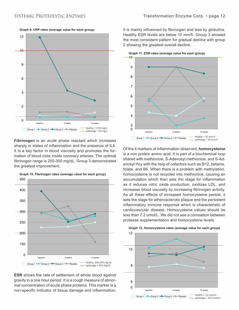
ResultsCRP is an acute phase reactant which increases sharply in response to inflammation. It binds to phosphocholine expressed on the surface of dead or dying cells (and some types of bacteria) in order to activate the complement sys-tem. It is synthesized by the liver in response to cytokines such as IL1β, IL6, IL12, and TNFα released by macrophages and fat cells. Healthy CRP levels are less than 0.55 mg/L. Group 3 demonstrated a significant decrease in CRP.
•
•
•
•
Dominant Leg Extension PT %3h Post-T1 24h Post-T148h Post-T1
Protease -12 -18.17 -11.74
Placebo -15.15 -23 -19.73
Dominant Leg Extension
-25
-20
-15
-10
-5
0
Time
Exte
nsio
n (
PT
%
@ 6
0°/s
)
Protease
Placebo
Dominant Leg Extension PT %3h Post-T1 24h Post-T148h Post-T1
Protease -12 -18.17 -11.74
Placebo -15.15 -23 -19.73
Dominant Leg Extension
-25
-20
-15
-10
-5
0
3h Post-T1 24h Post-T1 48h Post-T1
Time
Exte
nsio
n (
PT
%
@ 6
0°/
s)
Protease
Placebo
Dominant Leg Flexion PT %3h Post-T1 24h Post-T148h Post-T1
Protease 4.73 3.01 6.44
Placebo -19.54 -27.94 -25.87
Dominant Leg Flexion
-30
-25
-20
-15
-10
-5
0
5
10
Time
Fle
xio
n (
PT
%
@ 6
0°/s
)
Protease
Placebo
Dominant Leg Extension PT %3h Post-T1 24h Post-T148h Post-T1
Protease -12 -18.17 -11.74
Placebo -15.15 -23 -19.73
Dominant Leg Extension
-25
-20
-15
-10
-5
0
3h Post-T1 24h Post-T1 48h Post-T1
Time
Exte
nsio
n (
PT
%
@ 6
0°/
s)
Protease
Placebo
Systemic Proteolytic Enzymes Transformation Enzyme Corp. • page 12
Fibrinogen is an acute phase reactant which increases sharply in states of inflammation and the presence of IL6. It is a key factor in blood viscosity and promotes the for-mation of blood clots inside coronary arteries. The optimal fibrinogen range is 200-300 mg/dL. Group 3 demonstrated the greatest improvement.
ESR shows the rate of settlement of whole blood against gravity in a one hour period. It is a rough measure of abnor-mal concentration of acute phase proteins. This marker is a non-specific indicator of tissue damage and inflammation.
It is mainly influenced by fibrinogen and less by globulins. Healthy ESR levels are below 10 mm/h. Group � showed the most consistent pattern for gradual decline with group 2 showing the greatest overall decline.
Of the 4 markers of inflammation observed, homocysteine is a non protein amino acid. It is part of a biochemical loop shared with methionine, S-Adenosyl methionine, and S-Ad-enosyl Hcy with the help of cofactors such as B12, betaine, folate, and B6. When there is a problem with methylation, homocysteine is not recycled into methionine, causing an accumulation which then sets the stage for inflammation as it reduces nitric oxide production, oxidizes LDL, and increases blood viscosity by increasing fibrinogen activity. As all these effects of increased homocysteine persist, it sets the stage for atherosclerotic plaque and the persistent inflammatory immune response which is characteristic of cardiovascular disease. Homocysteine values should be less than 7.2 umol/L. We did not see a correlation between protease supplementation and homocysteine levels.
healthy < 0.55 mg/Lpathology > 10 mg/L
Graph 9. CRP rates (average value for each group)
healthy 200-300 mg/dLpathology > 425 mg/dL
Graph 10. Fibrinogen rates (average value for each group)
healthy < 10 mm/hpathology > 20 mm/h
Graph 11. ESR rates (average value for each group)
healthy 200-300 mg/dLpathology > 425 mg/dL
healthy < 10 mm/hpathology > 20 mm/h
healthy < 7.2 umol/Lpathology > 10.4 umol/L
Graph 12. Homocysteine rates (average value for each group)
healthy < 7.2 umol/Lpathology > 10.4 umol/L
Systemic Proteolytic Enzymes Transformation Enzyme Corp. • page 1�
ConclusionElevated levels of CRP and Fibrinogen have been corre-lated with a significant increased risk of future heart attacks although they have not been proven to be a causal risk factor of Coronary Heart Disease. Both are acute phase reactants which rise sharply in states of inflammation or tissue damage. The participants in group � had the highest CRP and fibrinogen levels at baseline. They were given the high dose protease and demonstrated significant decreases in CRP and fibrinogen. This finding is consistent with the study performed at Baylor University where TEC protease blend reduced inflammatory cytokines such as IL1β, IL6, TNFα, and IL12.
In reviewing the medical literature, IL6R Genetics Consor-tium and Emerging Risk Factors Collaboration has shown that a genetic variant (Asp358Ala) in the IL6R gene can dampen the inflammatory effect of IL6 receptor, reduce CRP and Fibrinogen, and decrease Coronary Heart Disease. This finding demonstrates a cause and effect relationship between a specific inflammatory protein and the develop-ment of Coronary Heart Disease. If we were able to dampen inflammation like the genetic variant then we can also pos-sibly affect cardiovascular consequences.
Inflammation is a common denominator in almost all chronic conditions that drive patients to your office, from cardiovas-cular disease to auto-immune conditions. As stated by Dr. Bastidas, “Transformation’s protease blend has demon-strated its strength in impacting markers of inflammation and gives us a healthy and effective alternative in supporting diet and lifestyle changes when dealing with these chronic conditions that we see on a day to day basis.”
Research and Clinical Observations: Enzyme Therapy and Oxidative StressThis report shows the overall benefits that diet and lifestyle modification along with enzyme therapy, including protease supplementation, has on oxidative stress. Fast-paced life-style, unhealthy diet, poor digestion, and constant exposure to the environment are just a few of the variables that lead to increased inflammation and oxidation. This condition is known as “oxidative stress” and involves the release of free radicals. Free radicals are un-paired electrons that stray from chemical solutions and damage important cellular components such as DNA or the cell membrane. Cells may function poorly or even die if this occurs, which initiates inflammation and promotes premature aging.
As you get older, several physical changes occur that affect your health. Muscle mass usually decreases, metabolism slows down, and your body is not able to repair damaged DNA as efficiently as it once did. If such irreparable damage
occurs, one of three things usually happens. First, the cell might die on its own or the “T” cells of the immune system do their job and eliminate the damaged cell. Secondly, the cell might replicate without restriction and transform into either a benign or a malignant tumor. Third, and very often, the cell will simply not be able to perform the functions for which it was designed. All these factors act as pre-cursors to aging.
So what happens when your cells are not functioning prop-erly? One possibility is that the DNA of the cell is unable to produce the necessary enzymes for the body, or perhaps that the enzymes that it does produce are unusable. Modi-fying our lifestyle in an effort to reduce the stress on our body and cells is essential. As the cells in our body become less productive, does it not also make sense to supplement our diets with tools that can help it? When the cells in our body are unable to work the way they need to, it is actually possible to supplement our body with digestive enzymes, vitamins, minerals, and antioxidants to support the body in fighting oxidative stress. The digestive system is a vast network of organs and glands that begins at the mouth and ends at the anus. It is one of the first systems in the body to become compromised due to stress. When imbalanced, it often begins a cascade of chronic health challenges in other systems. Repairing, rebuilding, or simply supporting a healthy digestive system is essential to improving the absorption of nutrients, enhancing immune response, and reducing oxidative stress. Thus, healthy digestion equals healthy aging and increased quality of life.
Redox - The Measurable Factor of AgingYour biological clock is always ticking, but did you know that your chronological age is not necessarily a reflection of your body’s biological age? Instead, there is a measurable biochemical process called “oxidative-reduction.” Redox (rH2) is a measurement of oxidation-reduction potential (free radicals) under a specific pH measurement. This calculable factor mirrors your body’s actual aging process.
Oxidation-reduction reactions create high cellular energy in the form of ATP and also oxidize (“burn up”) invading pollut-ants, xenobiotics (foreign materials that get into the body), and microorganisms that lead to degenerative diseases and contribute to premature aging. Measuring the rH2 of body fluids yields information about the body’s ability to produce energy (mitochondrial function), the degree to which excess free radicals are being produced, and ultimately cellular integrity and function.
According to Dr. Robert Greenberg, an rH2 value below 28 is considered “reduced” and has more electron donors than acceptors. A reduced solution is desirable, as increased electron donors in the cell and mitochondria means the TCA cycle is producing electron-rich intermediates that the
Systemic Proteolytic Enzymes Transformation Enzyme Corp. • page 1�
mitochondria can readily convert into energy. An rH2 value greater than 28 is considered “oxidized” and has a greater number of electron acceptors which produce unstable mol-ecules (free radicals) in excess.
This is why doctors emphasize the benefits of eating foods rich in antioxidants. The principle micronutrient (vitamin) antioxidants are vitamin E, beta-carotene, vitamin C, se-lenium, and zinc. Other antioxidants that are rapidly gain-ing notoriety are lutein and zeaxanthin. The body cannot manufacture these micronutrients, so they must be supplied through the diet via proper digestion and absorption.
An oxidized solution also indicates a decreased ability to produce high energy cellular fuel. Ultimately, as your rH2 decreases, so does your body’s “potential” biological age with respect to oxidation reduction reactions. Oxidation is not always bad. In fact, it is essential for life. The key is to find that healthy balance where the oxidation meets the body’s needs but is not in excess.
Clinical FindingsTransformation Enzyme Therapy Clinic utilized a Qualitative Fluid Analyzer (QFA) to monitor the biological terrain of our clients. The body fluids used are blood, saliva, and urine. The param-eters we analyze are pH, redox, and re-sistivity. While all the parameters of biologi-cal terrain analysis are equally important, the focus of these clinical observations is to look at oxidation reduction (rH2) results.
A total of 106 patient files containing at least two QFA tests were reviewed for compari-son of rH2 readings. All clients had been given dietary recommendations, an enzyme protocol, and lifestyle suggestions as per the individual biochemical needs. When we speak of “showing improvement,” it means the redox value moved closer to or into the ideal range for that body fluid. The ideal values are as follows:
Blood 21.5 - 2�.5Saliva 21.5 - 2�.5Urine 22.5 - 2�.5
•••
ResultsUrine rH2 showed the greatest improvement, with 5�.7% of the records reflecting improved values. This indicates greater utilization of electrons for energy production.In 51.8% of the records, Saliva rH2 showed improve-ment. This represents better digestion, absorption, metabolism, and elimination of waste.In �6.2% of the records, Blood rH2 showed improve-ment. This means the blood has a greater number of electrons and is primed with potential energy. The blood is able to create a chemical reaction whether to destroy a pollutant or to provide more energy.Another point of interest is the correlation between im-proved saliva pH and improved saliva rH2. We saw this positive correlation in 62% of our records. Each time, it indicated both better digestion and lowered oxidative stress.
For the complete report please contact TEC and request our Clinical Observation – Nutrition and Aging.
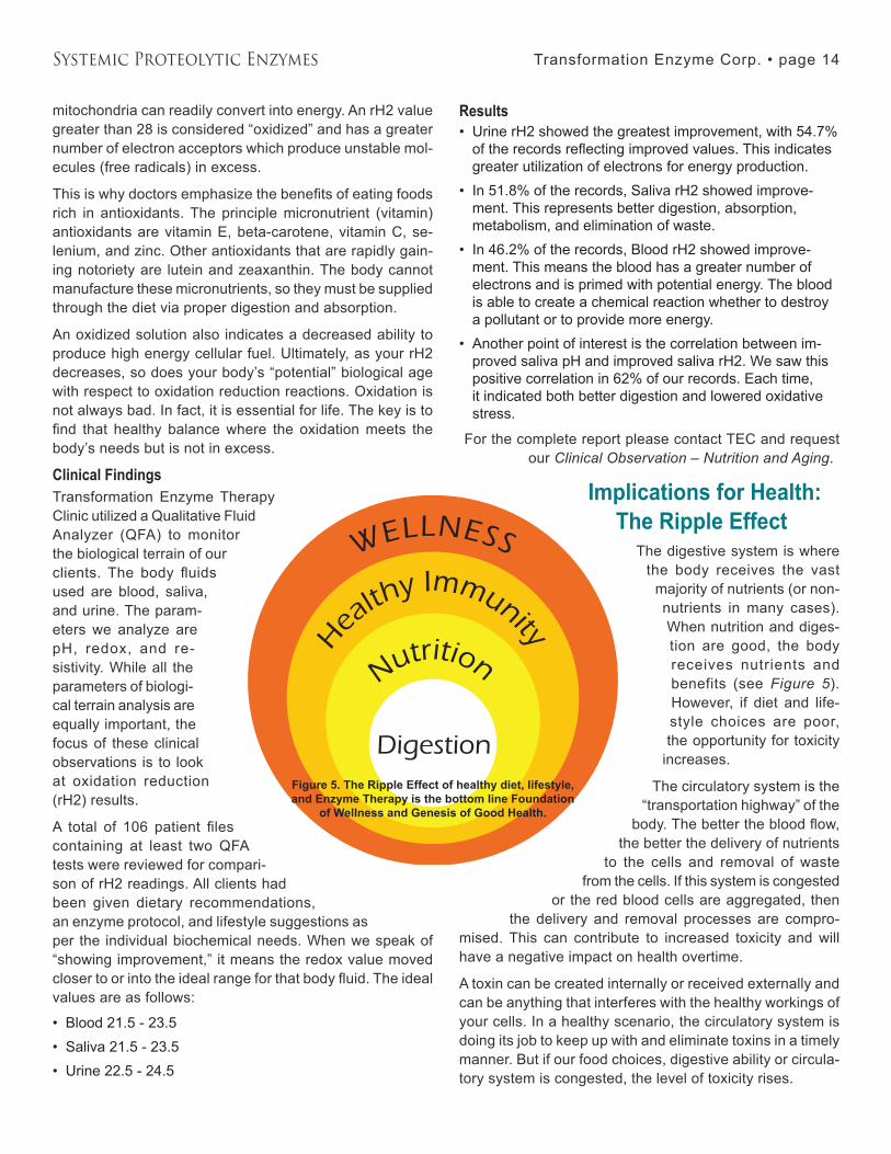
Implications for Health: The Ripple Effect
The digestive system is where the body receives the vast
majority of nutrients (or non-nutrients in many cases). When nutrition and diges-tion are good, the body receives nutrients and benefits (see Figure 5). However, if diet and life-style choices are poor, the opportunity for toxicity
increases.
The circulatory system is the “transportation highway” of the
body. The better the blood flow, the better the delivery of nutrients
to the cells and removal of waste from the cells. If this system is congested
or the red blood cells are aggregated, then the delivery and removal processes are compro-
mised. This can contribute to increased toxicity and will have a negative impact on health overtime.
A toxin can be created internally or received externally and can be anything that interferes with the healthy workings of your cells. In a healthy scenario, the circulatory system is doing its job to keep up with and eliminate toxins in a timely manner. But if our food choices, digestive ability or circula-tory system is congested, the level of toxicity rises.
•
•
•
•
Wellness
H
ealthy Immunity
nutrition
DigestionFigure 5. The Ripple Effect of healthy diet, lifestyle, and Enzyme Therapy is the bottom line Foundation
of Wellness and Genesis of Good Health.
Systemic Proteolytic Enzymes Transformation Enzyme Corp. • page 15
If poor blood flow and increased toxicity persists over time, cellular damage can occur. The toxicity leads to inflamma-tion or oxidative stress and the lack of nutrients impedes timely repair. This is why holistic and integrative practitioners focus on nutrition and chronic inflammation as the root of degenerative diseases.
When toxicity and cellular damage persist at a greater rate than does nutrition and elimination, then the result is what we see as a disease in your patient.
If you believe fundamentally that “nutrition is the foundation of wellness” then you agree proteases play a key role in the nutrition process. Whether it is your own endogenous proteases or with the help of supplemental proteases, healthy blood flow supports delivery of the nutrients and removal of waste for optimal cellular function and integrity. It is this basic concept that drives the clinical application of supplemental proteases in every practice regardless of modality or focus.
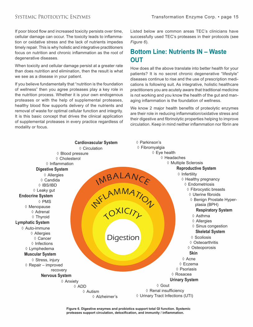
Listed below are common areas TEC’s clinicians have successfully used TEC’s proteases in their protocols (see Figure 6).
Bottom Line: Nutrients IN – Waste OUTHow does all the above translate into better health for your patients? It is no secret chronic degenerative “lifestyle” diseases continue to rise and the use of prescription medi-cations is following suit. As integrative, holistic healthcare practitioners you are acutely aware that traditional medicine is not working and you know the health of the gut and man-aging inflammation is the foundation of wellness.
We know 2 major health benefits of proteolytic enzymes are their role in reducing inflammation/oxidative stress and their digestive and fibrinolytic properties helping to improve circulation. Keep in mind neither inflammation nor fibrin are
Cardiovascular System Circulation
Blood pressureCholesterol
Inflammation Digestive System
AllergiesCandida
IBS/IBD Leaky gut
Endocrine System PMS
MenopauseAdrenal Thyroid
Lymphatic.SystemAuto-immune
AllergiesCancer
InfectionsLymphedema
Muscular.System.Stress, injury
Repair – improved recovery
Nervous System Anxiety
ADDAutism
Alzheimer’s
◊◊◊
◊
◊◊
◊◊
◊◊
◊◊
◊◊
◊◊
◊
◊◊
◊◊
◊◊
Parkinson’sFibromyalgia
Eye healthHeadaches
Multiple Sclerosis Reproductive System
Infertility Healthy pregnancy
EndometriosisFibrocystic breastsUterine fibroidsBenign Prostate Hyper-plasia (BPH) Respiratory System AsthmaAllergiesSinus congestion Skeletal.System.Scoliosis
OsteoarthritisOsteoporosis
SkinAcne
EczemaPsoriasis
Rosacea Urinary System
GoutRenal insufficiency
Urinary Tract Infections (UTI)
◊◊
◊◊
◊
◊◊
◊◊◊◊
◊◊◊
◊◊
◊
◊◊
◊◊
◊◊
◊
Imbalance
In
flammatIon
toxIcIty
Digestion
Figure 6. Digestive enzymes and probiotics support total GI function. Systemic proteases support circulation, detoxification, and immunity / inflammation.
Systemic Proteolytic Enzymes Transformation Enzyme Corp. • page 16
bad. It is obvious both the clotting of blood and inflammation are necessary for life. It is when the response is chronic or excessive that we have a problem.
The Ripple Effect of poor choices can easily be reversed by making healthy choices and implementing enzyme therapy. Through better digestion and a healthy gut, nutrients are delivered to the cells rather than toxins. Through improved circulation, toxins are removed in a timely manner and inflammation is reduced. The end result of nutrients in and waste out is health and balance to the entire body (see Figure 5).
While our first priority is to promote a healthy diet and life-style, we are all exposed to injurious agents on a regular basis. So our second priority is to support the body in a healthy response that allows it to return to balance. This extensive review of literature highlights the mechanisms of action, showing how the proteases work and how they can be used to complement all modalities and treatment protocols.
Frequently Asked QuestionsWhen is the best time to take a protease supplement? For maximum systemic benefit, it is best to take proteases between meals as this allows for faster absorption into circulation. However, if this is not realistic for your patient, then it is ok to take proteases with meals knowing that some of the protease enzymes may be used to digest food proteins. Also, it is better to take small doses several times throughout the day rather than one or two large doses in a day. Common dosing times are first thing in the morning,
mid-morning, mid-afternoon, and bedtime.
Can protease enzymes be taken with NSAIDS and/or pre-scription medications?Digestive enzymes function by breaking down specific chemical bonds in foods. In most cases digestive enzymes can therefore safely be taken with medications. However, it is of course always recommended to let your health care provider know what you are taking.
Can protease enzymes be taken with prescription blood thinners? One area of caution is with prescription blood thinning agents. These types of prescription drugs interfere with the natural blood clotting mechanisms, while proteases break down fibrin allowing for better blood flow. They can be taken in conjunction, but it is recommended to dose them about four hours apart and monitor lab work closely. We recom-mend notifying and working with the doctor prescribing the medications.
Can you take protease enzymes when taking prescription “protease inhibitors”?More often than not, the term “protease” that describes proteolytic enzymes is used in very general terms. There are many metabolic proteases in our body, each with many different functions. The medications that are designed as protease inhibitors are targeting a very specific viral prote-ase. The supplemental digestive proteases are very differ-ent and will not interfere with the medication. In fact, oral supplemental digestive enzymes can be very supportive to those patients with auto-immune disorders.
Suggested ReadingScience Brief – Improved Digestion Supplementing with Oral Digestive Enzymes for more info on TPP Digest™
Science Brief – Improved Gluten Digestion with Innovative Enzyme Solutions for more info on TPP Carbo-G™
Science Brief – Probiotics: Their Benefit on Human Health for more info on TPP Probiotic™
ReferencesENZYME SAFETY AND STABILITYBarbesgaard P, Heldt-Hansen HP, Diderichsen B. “On the safety of Aspergillus oryzae: a review.” Appl Microbiol Biotechnol. �6(5):569-72 (1992).Baur X, Chen Z, Sander I. “Isolation and denomination of an important allergen in baking additives: amylase from Aspergillus oryzae (Asp o II).” Clin Exp Allergy 2�:�65-70 (199�).Dixon DM, Walsh TJ. “Human Pathogenesis.” in Aspergillus: Biol-ogy and Industrial Applications. Bennet JW and Klich MA, eds. pp
2�9-65. Boston: Butterworth-Heinemann, 1992.Dvorackova I. Aflatoxins and Human Health. Boca Raton: CRC Press, 1990.Godfrey T, West S. Industrial Enzymology, 2nd edition. New York: Stockton Press, 1996.Guyton AC. Textbook of Medical Physiology, 8th edition. Philadel-phia: W.B. Saunders Company, 1991.Linz JE, Pestka JJ. “Mycotoxins: Molecular Strategies for Control” in Aspergillus: Biology and Industrial Applications. Bennet JW and Klich MA, eds. pp 219-29. Boston: Butterworth-Heinemann, 1992.Losada E, Hinojosa M, Quirce S, Sanchez-Cano M, Moneo I. “Occupational asthma caused by alpha-amylase inhalation: clini-cal and immunologic findings and bronchial response patterns. J Allergy Clin Immunol. 89(1):118-25 (1992).Mamadou M, Marr S, Paydon K, Medhektar R. “Stability and Activity of Supplemental Digestive Enzymes in Simulated Gastric Fluid” presented at the Scripps Conference in San Diego, January 7-9, 2005.Pier AC, Richard JL. “Mycoses and Mycotoxicoses of Animals Caused by Aspergilli.” in Aspergillus: Biology and Industrial Ap-
Systemic Proteolytic Enzymes Transformation Enzyme Corp. • page 17
plications. Bennet JW and Klich MA, eds. pp 233-47. Boston: Butterworth-Heinemann, 1992.Quirce S, Cueva M, Diez-Gomez M, Fernandez-Rivas M, Hino-josa M, Gonzalez R, Losada E. “Respiratory allergy to Aspergil-lus-derived enzymes in bakers’ asthma.” J Allergy Clin Immunol. 90(6):970-8 (1992).Reed G. Enzymes in Food Processing, 2nd edition. New York: Academic Press, 1975.Richard J, Cole R. Mycotoxins: Economic and Health Risks Report No. 116. Ames, IA: Council for Agricultural Science and Technology Task Force Report, 1989.Schechtman MG. “United States Government Regulations Af-fecting Aspergilli and their Products” in Aspergillus: Biology and Industrial Applications. Bennet JW and Klich MA, eds. pp 271-96. Boston: Butterworth-Heinemann, 1992.Schwimmer S. Source Book of Food Enzymology. Westport, CT: The AVI Publishing Company, Inc. 1981.Wei DL, Johg SC. “Production of aflatoxins by strains of the Aspergillus flavus group maintained in ATCC.” Mycopathologia. 93(1):19-24 (1986).ENZYME STABILITYBorowitz D, Stevens C, Brettman LR, Campion M, Wilschanski M, Thompson H. “Liprotamase long-term safety and support of nu-tritional status in pancreatic-insufficient cystic fibrosis.” J Pediatr Gastroenterol Nutr. 2012 Feb;54(2):248-57.Ehren J, Morón B, Martin E, Bethune MT, Gray GM, Khosla C. “A Food-Grade Enzyme Preparation with Modest Gluten Detoxifica-tion Properties.” PLoS One. 2009;4(7):e6313.Fieker A, Philpott J, Armand M. “Enzyme replacement therapy for pancreatic insufficiency: present and future.” Clin Exp Gastroen-terol. 2011;�:55–7�.Griffin SM, Alderson D, Farndon JR. “Acid resistant lipase as replacement therapy in chronic pancreatic exocrine insufficiency: a study in dogs.” Gut. 1989,30,1012-1015.Layer P, Keller J. “Lipase supplementation therapy: standards, alternatives, and perspectives.” Pancreas. 2003 Jan;26(1):1-7.Mitea C, Havenaar R, Drijfhout JW, Edens L, Dekking L, Kon-ing F. “Efficient degradation of gluten by a prolyl endoprotease in a gastrointestinal model: implications for coeliac disease.” Gut. 2008 Jan;57(1):25-32.Pierzynowski S, Szwiec K, Valverde Piedra JL, Gruijc D, Szy-manczyk S, Swieboda P, Prykhodko O, Fedkiv O, Kruszewska D, Filip R, Botermans J, Svendsen J, Ushakova G, Kovalenko T, Osadchenko I, Goncharova K, Skibo G, Weström B. “Exogenous pancreatic-like enzymes are recovered in the gut and improve growth of exocrine pancreatic insufficient pigs.” Jour of Animal Sci. 2012;90 Suppl �:�2�-�26.Schneider MU, Knoll-Ruzicka ML, Domschke S, Heptner G, Domschke W. “Pancreatic enzyme replacement therapy: com-parative effects of conventional and enteric-coated microspheric pancreatin and acid-stable fungal enzyme preparations on ste-atorrhoea in chronic pancreatitis.” Hepatogastroenterology. 1985 Apr;�2(2):97-102.Stepniak D, Spaenij-Dekking L, Mitea C, Moester M, de Ru A, Baak-Pablo R, van Veelen P, Edens L, Koning F. “Highly efficient gluten degradation with a newly identified prolyl endoprotease: implications for celiac disease.” Am Jour of Physiology - Gastroin-testinal and Liver Physiology. 2006 October 1;291(G621-G629).
Zentler-Munro PL, Assoufi BA, Balasubramanian K, Cornell S, Benoliel D, Northfield TC, Hodson ME. “Therapeutic potential and clinical efficacy of acid-resistant fungal lipase in the treat-ment of pancreatic steatorrhoea due to cystic fibrosis.” Pancreas. 1992;7(�):�11-9.ABSORPTIONGonzalez Regimen NCI. http://www.cancer.gov/cancertopics/pdq/cam/gonzalez/HealthProfessional/page1Heyman M, Desjeux JF. “Significance of intestinal food protein transport.” J Pediatr Gastroenterol Nutr. 1992;15:48-57.Isenman L, Liebow C, Rothman S. “Transport of proteins across membranes - a paradigm in transition.” Biochimica et Biophysics Acta. 1995;12�1:��1-�70.Lorkowski G. “Gastrointestinal absorption and biological activities of serine and cysteine proteases of animal and plant origin: review on absorption of serine and cysteine proteases.” Int J Physiol Pathophysiol Pharmacol. 2012;4(1):10–27.Moriya H, Mirowaki C, Akimoto S, Yamaguchi K, Iwadare M. “Intestinal absorption of serratia protease ‘TSP.’” Folia Pharmaco-logica Japonica. 1971;67(1):40-48.Rothman S, Liebow C, Isenman L. “Conservation of Digestive Enzymes.” Physiol Rev. 2002;82:1–18.Sakuma N. “Studies on Intestinal Absorption of Semi-Alkali Pro-teinase Produced by Aspergillus melleus.” 1981;10(4):319-324.Yamaguchi K, Moriwaki C, Sugiura M. “Intestinal Absoprtion of the Semialkaline Proteinase from Aspergillus melleus.” Studies on Aspergillus Proteinase VIII. 1974;94:1404-1406.ANTI-INFLAMMATORY EFFECTSArai K, Lee F, et al. “Cytokines: coordination of immunological and inflammatory responses.” Ann. Rev., Biochem. 59:783 (1990). Bastidas M, Helffrich L. “Transformation Clinical Trial: The Effect of TPP Protease on Inflammation.” Oct 2012.Boyne PS, Medhurst H. “Oral anti-inflammatory enzyme therapy in injuries in professional footballers.” The Practitioner 198:543-46 (1967).Buford TW, Cooke MB, Redd LL, Hudson GM, Shelmadine BD, Willoughby DS. “Protease supplementation improves muscle function after eccentric exercise.” Med Sci Sports Exerc. 2009 Oct;41(10):1908.Emele JF, Shanaman J, Winbury MM. “The Analgesic Anti-Inflam-matory Activity of Papain.” Warner Lambert Research Institute, Department of Pharmacology; Morris Plains, NJ.The Emerging Risk Factors Collaboration. “C-reactive protein concentration and risk of coronary heart disease, stroke, and mortality: an individual participant meta-analysis.” Lancet. 2010 Jan 9;375(9709):132-140.“IL6R Genetics Consortium Emerging Risk Factors Collabora-tion. Interleukin-6 receptor pathways in coronary heart disease: a collaborative meta-analysis of 82 studies.” Lancet. 2012 Mar 31;379(9822):1205-13.Kumakura S, Yamashita M, Tsurufuji S. “Effect of bromelain on kaolin-induced inflammation in rats.” Eur J Pharmacol. 1988 Jun 10;150(�):295-�01.Netti C, Bandi CL, et al. “Anti-inflammatory action of proteolytic enzymes of animal, vegetable, or bacterial origin administered orally compared with that of known antiphlogistic compounds.” II Farmaco 27:�5� (1972).
Systemic Proteolytic Enzymes Transformation Enzyme Corp. • page 18
Pirotta F, De Giuli-Morghen C. “Bromelain: a deeper pharma-cological study. Note 1- anti-inflammatory and serum fibrinolytic activity after oral administration in the rat.” Drugs exp. Clin. Res. 4:1 (1978).Ryan JW. “Renin-like enzyme in the adrenal gland.” Science. 1967 Dec 22;158(808):1589-90.Ryan JW, Ferris TF. “Release of a renin-like enzyme from the pregnant uterus of the rabbit.” Biochem J. 1967 Oct;105(1):16C-17C.Sandle GI, Warhurst G, Butterfield I, Higgs NB, Lomax RB. “Somatostatin peptides inhibit basolateral potassium channels in human colonic crypts.” Am J Physiol. 1999 Nov;277(5 Pt 1):G967-75.Shaw PC. “The use of a trypsin-chymotrypsin formulation in frac-tures of the hand.” Br J Clin Pract. 1969 Jan 1;23(1):25-6.Smyth DS, Utsumi S. “Structure at the hinge region in rabbit im-munoglobulin-G.” Nature. 1967 Oct 28;216(5113):332-5.Smyth RD, Moss JN, Brennan R, Harris JC, Martin GJ. “Biochemi-cal studies on the resolution of experimental inflammations in ani-mals treated with bromelain.” Exp Med Surg. 1967;25(1):229-�5.Tarayre JP, Lauressergues H. “Advantages of a combination of proteolytic enzymes, flavonoids and ascorbic acid in comparison with non-steroid anti-inflammatory agents.” Arzneim-forsch./Drug. Res. 27(I):11��-�9 (no. 6, 1997)BLOOD FLOWChoi HS, Sa YS. “Fibrinolytic and antithrombotic protease from Spirodela polyrhiza.” Biosci Biotechnol Biochem. 2001 Apr;65(4):781-6.Felton GE. “Fibrinolytic and antithrombotic action of bromelain may eliminate thrombosis in heart patients.” Med Hypotheses. 1980 Nov;6(11):1123-33.Kiessling H, Svensson R. “Influence of an enzyme from Aspergil-lus oryzae, Protease I, on some components of the fibrinolytic system. Acta Chemica Scandinavica.” 1970;2�:569-579.Larsson LJ, Frisch EP, Törneke K, Lindblom T, Björk I. “Proper-ties of the complex between alpha 2-macroglobulin and brinase, a proteinase from Aspergillus oryzae with thrombolytic effect.” Thromb Res. 1988 Jan 1;49(1):55-68.Maurer HR. “Bromelain: biochemistry, pharmacology and medical use.” Cell Mol Life Sci. 2001 Aug;58(9):1234-45.Roschlau WHE. “Thrombolytic therapy with CA-7, a fibrinolytic enzyme from Aspergillus oryzae: a report of two representative cases.” Canad Med Ass J. 1968 Apr;98(16):757-761.Taussig SJ, Batkin S. “Bromelain, the enzyme complex of pine-apple (Ananas comosus) and its clinical application: an update.” J Ethnopharmacol. 1988 Feb-Mar;22(2):191-203.TUMOR GROWTH CONTROLAhumada R. Fallbericht. 199�.Alth G, et al. “Aspects of the immunologic treatment of lung can-cer.” Cancer Chemother. Rep. 197�;�:271.Argyropoulos G, Tritthart H. “Lecture at the chemotherapy Con-gress, Zurich, Switzerland.” Cyanamide papers 75. 1977.Batkin S, et al. “Antimetastatic effect of bromelain with or without its proteolytic and anticoagulant activity.” J. Cancer Res. Clin. Oncol. 1988b;114:507.Batkin S, et al. “Modulation of pulmonary metastasis (Lewis lung carcinoma) by bromelain, an extract of the pineapple stem
(Ananas comosus).” Cancer Invest. 1988a;6:241.Beard J. “On the occurrence of dextrorotatory albumins in organic nature.” Biol. Central blatt. 1913;XXXIII:150.Beard J. “The enzyme treatment of cancer.” Loc. Cit. Pp. 1911;185.Beard J. “The interlude of cancer.” Medical record, 1907.Bessing WD. Adjuvante biologische Krebstherapie eines Kolonkarzinoms, heilkunst - extra 3:1. 1994.Castell JV. “Intestinal absorption of undegraded bromelain in humans” in Absorption of Orally administered enzymes, Eds. Gardner MLG and Steffens K-J. Springer-Verlag. 1995.Clifton E, Agostino D. “Effects of inhibiotors of fibrinolytic enzymes on development of pulmonary metastases.” Journal Nat. Cancer Inst. 196�;��:75�.Garbin, et al. “Bromelain proteinase F9 augments human lym-phocyte-mediated growth inhibition of various tumor cells invitro.” International J. of Oncology 1994;5:197.Gerard G. “Anti-cancer treatment and bromelain.” Agressology 1972;1�:261.Kesztele V, et al. Wochenschrift 1976;25:412.Kim D, et al. “A study of cell mediated immunity in cancer patients. II. Natural killer cell mediated cytotoxicity.” Korean J. Int. Med. 1981;24:461.Kim H, et al. “The alteration of cellular immunity following the enzymes therapy.” Dept. of Internal Medicine, Catholic University Hospital, Seoul, Korea. 1982.Kim JP, et al. “Study on ADCC of cancer patients.” 7th Korean cancer Res. Soc., Seoul National Univ. Hospital. 1980 Oct 4,5:12.Kojima K, Sakai I. “On the role of stickiness of tumor cells in the formation of metastases.” Cancer Res. 1964;24:1887.Kokron O, et al. “Proteolytic enzymes in the treatment of malig-nant pleural effusions and solid metastases” in Chemotherapy, eds. Hellmann K and Connors TA. 1976;8:517.Maurer HR, Hozumi M, et al. “Bromelain induces the differentia-tion of leukemic cells in vitro: an explanation for its cytostatic effect?” Planta Med. 1988;377.Moser K. “Trypsin: value as a thrombolytic and antiinflammatory agent.” New Engl. J. Med. 1957;256:258.O’Meara RAQ. “Coagulative properties of cancer.” Ir. J. Med. Sci. 1958;394:474.Pirotta F and De Giuli-Morghen C. “Bromelain: a deeper pharma-cological study. Note 1 - anti-inflammatory and serum fibrinolytic activity after oral administration in the rat.” Drugs exp. Clin. Res. 1978;4:1.Sakalova A. “Multiple myeloma and paraproteinemias.” Slovak Academic Press, Bratislava, Slovak Republic. 1995.Sakalova, et al. “Immunomodulatory effects of proteolytic en-zymes combined with thymic factor in multiple myeloma, para proteinemic vasculitis and solid tumors.” Annals of Hematology 67, Suppl. II, A166. 199�.Seifert, et al. “The quantitative absorption of orally administered proteins and histological evidence of enzymes in the wound” in Absorption of Orally Administered Enzymes, eds. Gardner and Steffens, Springer Verlag, Stuttgart, Germany. 1995.Steckerl F, et al. “Effect of alpha amylase on the Ehrlich mouse ascites tumor.” Nature 1961;�79�:111�.
Systemic Proteolytic Enzymes Transformation Enzyme Corp. • page 19
Steffen C and Menzel J. “In vivo degradation of immune com-plexes in the kidneys via orally administered enzymes.” Wien Klin. Wochenschr 1987;99:525.Taussig SJ, Batkin S, et al. “Bromelain, the enzyme complex of pineapple (Ananas comosus) and its clinical application, an up-date.” J. ethnopharmacol 1988;22:191.Taussig SJ, Yokoyama MM, et al. “Bromelain: a proteolytic en-zyme and its clinical application.” Hir. J. Med Sci. 1975;24:185.Thornes RD. “Adjuvant therapy of cancer via the cellular immune mechanisms of fibrin by fibrinolysis and oral anticoagulants.” Cancer 1975;�5:91.Thornes RD. “Inhibition of antiplasmin, and effect of protease1 in patients with leukemia.” Lancet 1968;1220.Wald M, Zavadova E, et al. “Polyenzyme preparation Wobe-Mugos inhibits growth of solid tumors and development of experimental metastases in mice.” Life Sciences (Pharmacology Letters) 1998;62:43.Wessenbacher ER. “Oral enzymes as biological modifiers.” Inter. J. of Feto-maternal Med. 1993;6(3):7.Wolf M, Ransberger K, eds. Enzymtherapie. Maudrich Verlag, Vienna, Austria. 1970.Wrba H. Kombinierte Tumortherapie. Hippokrates Verlag Stutt-gart, Germany, 2nd edition. 1995.IMMUNE MODULATIONAlbrecht J, Sidoryk-Węgrzynowicz M, Zielińska M, Aschner M. “Roles of glutamine in neurotransmission.” Neuron Glia Biology 2010 Nov;6(�):26�-276.Barrett AJ, Starkey PM. “The interaction of alpha 2-macroglobulin with proteinases: characteristics and specificity of the reaction, and a hypothesis concerning its molecular mechanism.” The Bio-chemical Journal 1973 Aug;133(4):709-724.Beutheu S, Ghouzali I, Galas L, Déchelotte P, Coëffier M. “Gluta-mine and arginine improve permeability and tight junction protein expression in methotrexate-treated caco-2 cells.” Clin Nutr (Edin-burgh, Scotland) 2013 Oct;32(5):863-869.Borth W. “Alpha 2-macroglobulin, a multifunctional binding protein with targeting characteristics.” The FASEB Jour 1992 Dec;6(15):���5-��5�.Braga PC, Moretti M, Piacenza A, Montoli CC, Guffanti EE. “Ef-fects of Seaprose on the rheology of bronchial mucus in patients with chronic bronchitis: a double-blind study vs placebo.” Int Jour of Clin Pharma Res 1993;13(3):179-185.Braga PC, Rampoldi C, Ornaghi A, Caminiti G, Beghi G, Allegra L. “In vitro rheological assessment of mucolytic activity induced by Seaprose.” Pharmacological Research: The Official Journal of the Italian Pharmacological Society 1990 Oct;22(5):611-617.Chang M, Alsaigh T, Kistler EB, Schmid-Schönbein BW. “Break-down of mucin as barrier to digestive enzymes in the ischemic rat small intestine.” PloS One 2012;7(6):e40087.Cota D, Proulx K, Blake Smith KA, Kozma SC, Thomas G, Woods SC, Seeley RJ. “Hypothalamic mTOR signaling regulates food intake.” Science 2006 May;�12(5775):927-9�0.Dong JY, Qin LQ, Zhang Z, Zhao Y, Wang J, Arigoni F, Zhang W. “Effect of oral L-arginine supplementation on blood pressure: a meta-analysis of randomized, double-blind, placebo controlled trials.” Amer Heart Jour 2011 Dec;162(6):959-965.Etique N, Verzeaux L, Dedieu S, Emonard H. “LRP-1: a check-
point for the extracellular matrix proteolysis.” BioMed Research International 201�.Guzmán-Quevedo O, Da Silva Aragão R, Pérez García G, Matos RKB, de Sa Braga Oliveira A, de Castro RM, Bolaños-Jiménez F. “Impaired hypothalamic mTOR activation in the adult rat offspring born to mothers fed a low-protein diet.” PloS One 2013;8(9):e7�990.Henry KM, Loynes CA, Whyte MKB, Renshaw SA. “Zebrafish as a model for the study of neutrophil biology.” Journal of Leukocyte Biology 201� Mar;9�(�):6��-6�2.Isenman L, Liebow C, Rothman S. “Transport of proteins across membranes: a paradigm in transition.” Biochimica et Biophysica Acta 1995 Dec;12�1(�):��1-�70.Journel M, Chaumontet C, Darcel N, Fromentin G, Tomé D. “Brain responses to high-protein diets.” Advances in Nutrition (Bethesda, Md.) 2012 May;�(�):�22-�29.Junker Y, Zeissig S, Kim SJ, Barisani D, Wieser H, Leffler DA, Zevallos V, et al. “Wheat amylase trypsin inhibitors drive intestinal inflammation via activation of toll-like receptor 4.” Jour Experimen-tal Med 2012 Dec;209(13):2395-2408.Kakoki M, Kim HS, Edgell CJS, Maeda N, Smithies O, Mattson DL. “Amino acids as modulators of endothelium-derived nitric ox-ide.” Amer Jour Physio. Renal Physio 2006 Aug;291(2):F297-304.Kim, H W, B P Chew, T S Wong, J S Park, B B Weng, K M Byrne, M G Hayek, and G A Reinhart. “Dietary lutein stimulates immune response in the canine.” Veterinary Immunology and Immunopa-thology 2000 May;7�(�-�):�15-�27.Kimura N, Tokunaga C, Dalal S, Richardson C, Yoshino K, Hara K, Kemp BE, Witters LA, Mimura O, Yonezawa K. “A possible linkage between AMP-activated protein kinase (AMPK) and mam-malian target of rapamycin (mTOR) signalling pathway.” Genes to Cells: Devoted to Molecular & Cellular Mechanisms 200� Jan;8(1):65-79.Li F, Yin Y, Tan B, Kong X, Wu G. “Leucine nutrition in animals and humans: mTOR signaling and beyond.” Amino Acids 2011 Jul;41(5):1185-1193.Lorkowski G. “Gastrointestinal absorption and biological activi-ties of serine and cysteine proteases of animal and plant origin: review on absorption of serine and cysteine proteases.” Interna-tional Journal of Physiology, Pathophysiology and Pharmacology 2012;�(1):10-27.Majima Y, Inagaki M, Hirata K, Saida S, Takeuchi K, Morishita A, Sakakura Y. “Effect of serratiopeptidase on viscoelasticity of nasal mucus in patients with chronic sinusitis.” Practica Oto-Rhino-Lar-yngologica 1986;79(9):1535-1540.Rhoads MJ, Wu G. “Glutamine, arginine, and leucine signaling in the intestine.” Amino Acids 2009 Jan;37(1):111-122.Mascarell L, Zimmer A, Van Overtvelt L, Tourdot S, Moingeon P. “Induction of allergen-specific tolerance via mucosal routes.” Cur-rent Topics in Microbiology and Immunol 2011;352:85-105.Muniandy S, Qvist R, Yan GOS, Bee CJ, Chu YK, Rayappan AV. “The oxidative stress of hyperglycemia and the inflammatory process in endothelial cells.” The Journal of Medical Investigation: JMI 2009 Feb;56(1-2):6-10.Nicklin P, Bergman P, Zhang B, Triantafellow E, Wang H, Nyfeler B, Yang H, et al. “Bidirectional transport of amino acids regulates mTOR and autophagy.” Cell 2009 Feb;1�6(�):521-5��.Niethammer P, Grabher C, Look AT, Mitchison TJ. “A tissue-scale
Systemic Proteolytic Enzymes Transformation Enzyme Corp. • page 20
gradient of hydrogen peroxide mediates rapid wound detection in zebrafish.” Nature 2009 Jun;459(7249):996-999.Palová-Jelínková L, Dáňová K, Drašarová H, Dvořák M, Funda DP, Fundová P, Kotrbová-Kozak A, et al. “Pepsin digest of wheat gliadin fraction increases production of IL-1β via TLR4/MyD88/TRIF/MAPK/NF-κB signaling pathway and an NLRP3 inflamma-some activation.” PloS One 2013;8(4):e62426.Rapin JR, Wiernsperger N. “Possible links between intestinal permeablity and food processing: a potential therapeutic niche for glutamine.” Clinics 2010 Jun;65(6):635-643.Ropelle ER, Pauli JR, Fernandes MFA, Rocco SA, Marin RM, Morari J, Souza KK, et al. “A central role for neuronal AMP-acti-vated protein kinase (AMPK) and mammalian target of rapamycin (mTOR) in high-protein diet-induced weight loss.” Diabetes 2008 Mar;57(�):59�-605.Ruth MR, Field CJ. “The immune modifying effects of amino acids on gut-associated lymphoid tissue.” Journal of Animal Science and Biotechnology 201�;�(1):27.Sapone A, Lammers KM, Casolaro V, Cammarota M, Giuliano MT, De Rosa M, Stefanile R, et al. “Divergence of gut permeability and mucosal immune gene expression in two gluten-associated condi-tions: celiac disease and gluten sensitivity.” BMC Med 2011;9:2�.Sousa, Gabriela TD, Lira FS, Rosa JC, de Oliveira EP, Oyama LM, Santos RV, Pimentel GD. “Dietary whey protein lessens sev-eral risk factors for metabolic diseases: a review.” Lipids in Health and Disease 2012;11:67.Stief TW, Kropf J, Kretschmer V, Doss MO, Fareed J. “Singlet oxygen ((1)O2) inactivates plasmatic free and complexed alpha 2-macroglobulin.” Thrombosis Res 2000 Jun;98(6):541-547.Stone TW, Forrest CM, Mackay GM, Stoy N, Darlington LG. “Tryp-
tophan, adenosine, neurodegeneration, and neuroprotection.” Metabolic Brain Disease 2007 Dec;22(�-�):��7-�52.Volta U, De Giorgio R. “New understanding of gluten sensitiv-ity.” Nature Reviews. Gastroenterology & Hepatology 2012 May;9(5):295-299.Waickman AT, Powell JD. “mTOR, metabolism, and the regula-tion of T-cell differentiation and function.” Immunological Reviews 2012 Sep;249(1):43-58.Wu G. “Amino acids: metabolism, functions, and nutrition.” Amino Acids 2009 May;�7(1):1-17.Yao K, Yin YL, Chu W, Liu Z, Deng D, Li T, Huang R, et al. “Di-etary arginine supplementation increases mTOR signaling activity in skeletal muscle of neonatal pigs.” The Journal of Nutrition 2008 May;138(5):867-872.You JS, Frey JW, Hornberger TA. “Mechanical stimulation induces mTOR signaling via an ERK-independent mechanism: implica-tions for a direct activation of mTOR by phosphatidic acid.” PloS One 2012;7(10):e47258.Zimmer A, Bouley J, Le Mignon M, Pliquet E, Horiot S, Turfkruyer M, Baron-Bodo V, et al. “A regulatory dendritic cell signature correlates with the clinical efficacy of allergen-specific sublingual immunotherapy.” Jour Allerg Clin Immun 2012 Apr;129(4):1020-10�0.Zimmer A, Luce S, Gaignier F, Nony E, Naveau M, Biola-Vidam-ment A, Pallardy M, Van Overtvelt L, Mascarell L, Moingeon P. “Identification of a new phenotype of tolerogenic human dendritic cells induced by fungal proteases from aspergillus oryzae.” Jour of Immunol (Baltimore, MD: 1950) 2011 Apr;186(7):3966-3976.
©201� Transformation
Questions? 1-800-777-1474email [email protected]
*These statements have not been evaluated by the Food and Drug Administration. This product is not intended to diagnose, treat, cure, or prevent any disease.
Lisa Helffrich, RDN, is a graduate of the University of Texas-Austin and is Director of Education and Technical Services for Transformation Enzyme Corp.
Dr. DicQie Fuller is the author of The Healing Power of Enzymes and is the Founder of and Chief Science Officer for Transformation Enzyme Corp.