Down-regulation of ABCC2 protein expression in HepG2 cells ...
30
1 Down-regulation of ABCC2 protein expression in HepG2 cells after rifampicin treatment is mediated by microRNA-379. Sierk Haenisch, Sandra Laechelt, Henrike Bruckmueller, Anneke Werk, Andreas Noack, Oliver Bruhn, Cornelia Remmler, Ingolf Cascorbi Institute for Experimental and Clinical Pharmacology (S.H., S.L., H.B., A. W., O.B., C.R., I.C.) and Division of Molecular Oncology, Institute of Experimental Cancer Research, Comprehensive Cancer Center North (A.N.), University Hospital Schleswig-Holstein, Kiel; Germany MOL #70714 Molecular Pharmacology Fast Forward. Published on May 3, 2011 as doi:10.1124/mol.110.070714 Copyright 2011 by the American Society for Pharmacology and Experimental Therapeutics. This article has not been copyedited and formatted. The final version may differ from this version. Molecular Pharmacology Fast Forward. Published on May 3, 2011 as DOI: 10.1124/mol.110.070714 at ASPET Journals on February 20, 2022 molpharm.aspetjournals.org Downloaded from
Transcript of Down-regulation of ABCC2 protein expression in HepG2 cells ...

1
Down-regulation of ABCC2 protein expression in HepG2 cells after rifampicin
treatment is mediated by microRNA-379.
Sierk Haenisch, Sandra Laechelt, Henrike Bruckmueller, Anneke Werk, Andreas
Noack, Oliver Bruhn, Cornelia Remmler, Ingolf Cascorbi
Institute for Experimental and Clinical Pharmacology (S.H., S.L., H.B., A. W., O.B.,
C.R., I.C.) and Division of Molecular Oncology, Institute of Experimental Cancer
Research, Comprehensive Cancer Center North (A.N.), University Hospital
Schleswig-Holstein, Kiel; Germany
MOL #70714 Molecular Pharmacology Fast Forward. Published on May 3, 2011 as doi:10.1124/mol.110.070714
Copyright 2011 by the American Society for Pharmacology and Experimental Therapeutics.
This article has not been copyedited and formatted. The final version may differ from this version.Molecular Pharmacology Fast Forward. Published on May 3, 2011 as DOI: 10.1124/mol.110.070714
at ASPE
T Journals on February 20, 2022
molpharm
.aspetjournals.orgD
ownloaded from

2
Running title:
miRNA-379 regulates ABCC2 protein expression
Correspondence address:
Ingolf Cascorbi, MD, PhD
Institute for Experimental and Clinical Pharmacology
University Hospital Schleswig-Holstein
Bldg. 30 Arnold-Heller-Str. 3 D-24105 Kiel Germany Phone: +49-431-597-3520 Fax: +49-431-597-3522 [email protected]
Text pages: 15 Tables: 0 Figures: 5 References: 23 Abstract: 231 words Introduction: 249 words Discussion: 1482 words Abbreviations: ABC, ATP-binding cassette BCRP, breast cancer resistance protein DMSO, dimethyl sulfoxide MDR, multidrug resistance protein miRNA or miR, microRNA MRP, multidrug resistance associated protein P-gp, P-glycoprotein PXR, pregnane X receptor RXR, retinoid X receptor TBS, Tris Buffered Saline UTR, untranslated region
MOL #70714This article has not been copyedited and formatted. The final version may differ from this version.
Molecular Pharmacology Fast Forward. Published on May 3, 2011 as DOI: 10.1124/mol.110.070714 at A
SPET
Journals on February 20, 2022m
olpharm.aspetjournals.org
Dow
nloaded from

3
Abstract
Background: microRNAs, which contribute to the post-transcriptional processing
through 3’-UTR-interference, have been recently shown to be involved in the
regulation of ABC membrane transporters. The aim of this study was to investigate if
ABCC2, an important efflux transporter for various endogenous and exogenous
compounds at several compartment barriers, is subject to miRNA mediated post-
transcriptional gene regulation. Methods: We screened the expression of 377 human
miRNAs in HepG2 cells after 48 hours of treatment with 5 µM of the PXR ligand
rifampicin or vehicle using RT-PCR based low-density arrays. Specific miRNA,
ABCC2 mRNA and protein expression was monitored in HepG2 cells under
treatment of rifampicin for 72 hours. Loss and gain of function experiments, as well
as reporter gene assays, were performed for further confirmation. Results: Highly
deregulated miRNAs compared with in-silico data revealed miR-379 as candidate
miRNA targeting ABCC2 mRNA. Under rifampicin treatment, ABCC2 mRNA
increased significantly with a maximum fold change of 1.56+/-0.43 after 24 hours.
Also miR-379 increased (maximally 4.10+/-1.33-fold after 48 h), whereas ABCC2
protein decreased with a maximum fold change of 0.47+/-0.08 after 72 hours. In
contrast, transfection of miR-379 inhibitor led to an elevation of ABCC2 protein
expression after rifampicin incubation for 48 hours. Conclusion: We identified a
miRNA negatively regulating ABCC2 on the post-transcriptional level and provide
evidence that this miRNA impedes over-expression of ABCC2 protein after a PXR-
mediated external transcriptional stimulus in HepG2 cells.
MOL #70714This article has not been copyedited and formatted. The final version may differ from this version.
Molecular Pharmacology Fast Forward. Published on May 3, 2011 as DOI: 10.1124/mol.110.070714 at A
SPET
Journals on February 20, 2022m
olpharm.aspetjournals.org
Dow
nloaded from

4
Introduction
Recently it has been demonstrated that microRNAs, which contribute to post-
transcriptional processing through 3’-UTR-interference, are involved in the regulation
of ABC membrane transporters. In cancer cell lines, miR-451 (Kovalchuk et al., 2008)
and miR-326 (Liang et al., 2010) negatively regulate ABCB1 (MDR1, P-gp) and
ABCC1 (MRP1), respectively. miR-328 (Pan et al., 2009) and miR-519c (To et al.,
2008) were shown to affect post-transcriptionally the expression of ABCG2 (BCRP).
An impaired expression of these miRNAs might be one of the underlying
mechanisms of drug resistance against anticancer agents in neoplastic cells. So far,
no information is available concerning the role of microRNAs in the post-
transcriptional regulation of ABCC2 (MRP2), another important efflux transporter.
This drug efflux transporter not only eliminates exogenous compounds, but is also
involved in the elimination of conjugated and un-conjugated endogenous substances
(Jemnitz et al., 2010). The aim of this study was to identify miRNAs targeting the
ABCC2 3’-UTR and to determine their role in ABCC2 protein expression.
Based on the hypothesis that ABCC2 transcriptional processing could be
accompanied by an altered expression of miRNAs targeting ABCC2, we performed
an array-based miRNA screen of HepG2 cells pre-incubated with either the PXR-
ligand rifampicin or vehicle. After in-silico analysis of differentially regulated miRNAs
and subsequent loss and gain-of-function experiments as well as reporter gene
assays, we identified a miRNA which negatively regulates ABCC2 protein expression
by direct RNA interference. The identified miRNA seems to counteract over-
expression of ABCC2 protein in HepG2 cells in presence of the transcriptional
stimulus rifampicin.
MOL #70714This article has not been copyedited and formatted. The final version may differ from this version.
Molecular Pharmacology Fast Forward. Published on May 3, 2011 as DOI: 10.1124/mol.110.070714 at A
SPET
Journals on February 20, 2022m
olpharm.aspetjournals.org
Dow
nloaded from
5
Material and Methods
Cell culture
Human hepatoblastoma HepG2 cells were cultured in RPMI 1640 medium
supplemented with 10% fetal bovine serum (FBS) and 100 units/ml penicillin and 100
µg/ml streptomycin at 37 °C in a 5% CO2 atmosphere. Cell culture medium and
supplements were purchased from PAA (Pasching, Austria). HepG2 cells were
obtained from DSMZ (Braunschweig, Germany).
miRNA expression profiling
HepG2 cells were seeded in 10 cm dishes (105 cells /ml). After 24 hours, medium
was replaced by medium supplemented with 5 µM rifampicin or vehicle (DMSO,
1:6000), both purchased from Sigma (Steinheim, Germany). Cells were incubated for
48 hours. In all experiments, fresh medium supplemented with rifampicin or vehicle
was replaced every 24 hours simulating a daily dosage. RNA was isolated using the
miRvana™ miRNA isolation kit (Applied Biosystems / Ambion, Darmstadt, Germany)
and miRNAs contained in 800 ng of total RNA were reverse transcribed using
Megaplex™ Primer Pools, Human Pools A v2.1 and the TaqMan® microRNA
Reverse Transcription Kit (Applied Biosystems). cDNAs and Universal Master Mix
(Applied Biosystems) were loaded on TaqMan® Array Human MicroRNA A cards v2.0
(Applied Biosystems) in a volume of 100 µl per port, according to the manufacturer’s
recommendation. Quantitative real-time PCR (rtPCR) reactions were conducted
using an ABI Prism 7900HT (Applied Biosystems). Data were analyzed by performing
the 2-Δct method. Mammalian U6 snRNA present in quadruplicate on the array was
used as endogenous control. MicroRNAs were considered to have a strong impact if
they exhibited a more than 8-fold altered expression level under rifampicin treatment
compared to vehicle. For in-silico identification of miRNAs predicted to target ABCC2,
MOL #70714This article has not been copyedited and formatted. The final version may differ from this version.
Molecular Pharmacology Fast Forward. Published on May 3, 2011 as DOI: 10.1124/mol.110.070714 at A
SPET
Journals on February 20, 2022m
olpharm.aspetjournals.org
Dow
nloaded from
6
a search using the microCosm miRNA target data base (http://www.ebi.ac.uk/enright-
srv/microcosm/htdocs/targets/v5/) was performed.
miR-379, mRNA and protein quantification
Effects of the PXR-ligand rifampicin were determined after 24 hours incubation in
normal medium followed by 72 hours of incubation in medium supplemented with 5
µM rifampicin or vehicle (DMSO). Total RNA and protein were isolated after 0, 8, 24,
48 and 72 hours of incubation. Two-hundred ng of total RNA were reverse
transcribed in a duplex approach using specific RT-primers for miR-379 and RNU6B.
RNU6B, one of the least variable small nuclear RNAs in human cell lines, was used
as internal control. cDNA quantification was performed with TaqMan® microRNA
assays (miR379 ID 001138 and RNU6B ID 001093) in singleplex PCR reactions
using Universal Master Mix (Applied Biosystems).
ABCC2 mRNA quantification was performed after reverse transcription of 200 ng
total RNA (Transcriptor First Strand cDNA Synthesis kit, Roche, Mannheim,
Germany) using Universal Master Mix and the TaqMan® assay (Hs00166123_m1).
18SrRNA was measured as internal reference using TaqMan® assay
Hs99999901_s1.
All rtPCR reactions were performed on an ABI Prism 7900HT (Applied Biosystems) in
duplicates. Data was calculated using the 2-Δct method.
Proteins were isolated using the Qproteome Mammalian Protein Prep Kit (Qiagen,
Hilden, Germany) and quantified with the Bio-Rad Protein Assay (Bio-Rad, Munich,
Germany). For Western blotting, proteins were separated on Nu-PAGE Novex 10%
bis-Tris Gels (Invitrogen, Karlsruhe, Germany) and transferred to polyvinylidene
fluoride microporous membranes (Millipore, Schwalbach, Germany) using a tank
blotting system (Bio-Rad). Membranes were blocked in 5% (w/v) non-fat dried milk in
MOL #70714This article has not been copyedited and formatted. The final version may differ from this version.
Molecular Pharmacology Fast Forward. Published on May 3, 2011 as DOI: 10.1124/mol.110.070714 at A
SPET
Journals on February 20, 2022m
olpharm.aspetjournals.org
Dow
nloaded from

7
TBS-T buffer (0.27mM KCl, 13.7mM NaCl, 5mM Tris–HCl, pH 7.4, 0.1% (w/v) Tween
20) and incubated overnight with the primary antibodies MRP2 M2III-6 (dilution
1:500; Alexis, San Diego, CA, USA) or anti-P-glycoprotein (ABCB1) (dilution 1:500;
Alexis, San Diego, CA, USA) and anti-β-actin as internal standard (dilution: 1:10 000;
Sigma). Horseradish peroxidase-labeled rabbit anti-mouse IgG (dilution: 1:10 000;
Sigma) was used as secondary antibody. Detection was performed by
chemiluminescence with ECL Western Blotting Detection Reagents (GE Healthcare,
Munich, Germany). Blots were quantified by densitometry using Quantity One 1-D
Analysis Software 4.6.3 (Bio-Rad).
miR-379/RNU6B, ABCC2mRNA/18SrRNA, as well as ABCC2 protein/β-actin protein
ratios, were normalized to values under respective vehicle stimulated conditions.
Data were obtained from three identical experiments.
Transient transfection with miR-379
In order to investigate the post-transcriptional influence of selected miR-379 on
ABCC2 mRNA and protein expression, HepG2 cells were reverse transfected in six-
well plates by covering 600 µl of transfection complex with 2.4 ml of cell suspension
(105 cells/ml) per well. siPORT™ NeoFX™ Transfection Agent (Applied Biosystems /
Ambion) was diluted in Opti-MEM I Medium (Invitrogen) to a final dilution of 0.3%
(v/v). Additionally, dilutions of miR-379 miRNA precursor (PM 10316) in final
concentrations of 5 nM, 20 nM, 50 nM and 100 nM, miR-379 miRNA inhibitor (AM
10316; 50 nM, 100 nM), as well as miRNA precursor (AM 17110; 20 nM) and miRNA
inhibitor (AM 17010; 50 nM) negative controls were prepared from the respective
stock solutions (6.25 µM) in Opti-MEM I medium. All were purchased from Applied
Biosystems / Ambion. After 10 minutes of incubation, transfection complexes were
obtained by mixing equal parts of the diluted transfection agent and the respective
MOL #70714This article has not been copyedited and formatted. The final version may differ from this version.
Molecular Pharmacology Fast Forward. Published on May 3, 2011 as DOI: 10.1124/mol.110.070714 at A
SPET
Journals on February 20, 2022m
olpharm.aspetjournals.org
Dow
nloaded from

8
diluted miRNA precursors or miRNA inhibitors. Optimization experiments were
performed according to the manufacturer’s recommendations using miR-1 precursor
(AM17150, down-regulating PTK9 mRNA) and let-7c inhibitor (AM10436, up-
regulating HMG2A mRNA) as positive controls. Minimal effective transfection
concentrations were 5 to 20 nM for miRNA precursor and 50 nM for miRNA inhibitor
molecules, respectively (data not shown). Moreover, no evidence of cytotoxicity was
observed for miRNA precursor and miRNA inhibitor negative controls at
concentrations from 5 to 100 nM. The cytotoxicity factor, expressed as ratio of Ct-
value of 18S rRNA of transfected cells to Ct-value of 18S rRNA of non-transfected
HepG2 cells, was 1.0 for all tested concentrations. Therefore, all further transfection
experiments used 20 nM of miRNA precursor negative control and 50 nM of miRNA
inhibitor negative control.
Twenty-four hours after transfection, medium was replaced by fresh normal growth
medium. After additional 24 hours of incubation, protein and total RNA were isolated.
ABCC2 protein was quantified from three identical experiments and ABCC2 mRNA
from two, as described above.
In-vitro investigation of the ABCC2 3’-UTR as a target of miR-379
To validate the ABCC2 3’-UTR as a target of miR-379, in-vitro assays using the
miTarget™ microRNA 3'-UTR target sequence expression clones (Genecopoeia,
Rockville, MD, USA) were performed. These miRNA target clones consist of the
pEZX-MT01 vector containing the coding sequences of firefly luciferase and renilla
luciferase under the control of the SV40 enhancer and CMV promoter, respectively.
The miRNA ABCC2 3'-UTR target clone (HmiT002260-MT01) exhibits the ABCC2 3’-
UTR (accession number: NM_000392.2) inserted downstream of the firefly luciferase
sequence. According to the Sanger miRBase (Kozomara and Griffiths-Jones 2010),
MOL #70714This article has not been copyedited and formatted. The final version may differ from this version.
Molecular Pharmacology Fast Forward. Published on May 3, 2011 as DOI: 10.1124/mol.110.070714 at A
SPET
Journals on February 20, 2022m
olpharm.aspetjournals.org
Dow
nloaded from

9
the binding site of miR-379 (5’-UGGUAGACUAUGGAACGUAGG; nucleotides of
seed sequence are marked in bold) is predicted at positions 180-200. The vector
pEZX-MT01 (CmiT000001-MT01) exhibiting no 3’-UTR insert downstream of the
firefly luciferase was used as positive control. After heat shock transformation in
competent E. coli cells (One Shot® TOP 10 competent cells, Invitrogen), plasmids
were amplified in LB-Medium supplemented with 50 µg/ml kanamycin (Sigma).
Plasmid DNA was purified by QIAprep® spin miniprep kit (Quiagen). Correctness of
amplified plasmids was confirmed by capillary sequencing (GATC Biotech, Konstanz,
Germany), using the sequencing primers 5'-GATCCGCGAGATCCTGAT (forward) or
5'-TTGGCGTTACTATGGGAACAT (reverse).
Co-transfection of 100 ng/well of plasmid DNA and precursor miRNA or miRNA
inhibitor molecules was performed essentially as described above in a 96-well plate.
The activities of both firefly and the internal control renilla luciferases were
determined 48 hours after transfection with the Dual-Luciferase® reporter assay
system (Promega, Mannheim, Germany) and analyzed on a Veritas™ microplate
luminometer (Tuner Biosystems, Sunnyvale, CA, USA). Seven identical co-
transfections were conducted.
Mutations within the potential miR-379 binding site were introduced by site-directed
mutagenesis of the ABCC2 3’-UTR using appropriate primer pairs (Sigma-Aldrich,
Munich, Germany), PfuUltra Hotstart DNA Polymerase (Agilent Technologies,
Waldbronn, Germany), QIAquick PCR Purification Kit (Qiagen, Hilden, Germany),
DpnI (New England Biolabs, Frankfurt a.M., Germany) and DH5α competent cells.
The first, third and fifth position within the seed sequence of the predicted miRNA
binding site was changed: wild type: 5’-GAGAAACCCCTCGATT
GTCTACCTCGATCGTACTTCCTTGC-3’, mutant: 5’-GAGAAACCCCTCGATTCT
MOL #70714This article has not been copyedited and formatted. The final version may differ from this version.
Molecular Pharmacology Fast Forward. Published on May 3, 2011 as DOI: 10.1124/mol.110.070714 at A
SPET
Journals on February 20, 2022m
olpharm.aspetjournals.org
Dow
nloaded from

10
GTTCCTCGATCGTACTTCCTTGC-3’. All reporter gene assays with mutated target
clones were performed in quadruplicate.
Statistical analysis
Mann-Whitney U-tests were performed to compare the various effects of rifampicin
on miR-379, ABCC2 mRNA, ABCC2 and ABCB1 protein expression and to test for
effects of miR-379 precursor or inhibitor on ABCC2 mRNA and protein expression as
well as on reporter gene activities using SPSS 17.0. Values are given as means with
standard deviations. A p-value ≤ 0.05 was considered to be statistically significant.
Results
Screen of deregulated miRNA expression after rifampicin treatment
Screening of HepG2 cells for differentially expressed microRNAs after rifampicjn
incubation revealed that 111 out of 377 human miRNAs were not expressed at all or
showed only expression in one of the different treatment conditions. After log2
transformation of fold changes, eleven miRNAs were identified having a more than
threefold altered expression (Fig. 1). miR-31 was 5.4-fold down-regulated, whereas
all others, namely miR-193a-5p, miR-296-5p, miR-324-5p, miR-379, miR-411, miR-
489, miR505, miR-519a, miR-545 and miR-548b-5p, exhibited up to 5.8-fold higher
expression levels. Out of these deregulated miRNAs, only miR-379 matched with in-
silico predicted miRNAs (microCosm miRNA target database) potentially targeting
ABCC2.
[Fig. 1]
MOL #70714This article has not been copyedited and formatted. The final version may differ from this version.
Molecular Pharmacology Fast Forward. Published on May 3, 2011 as DOI: 10.1124/mol.110.070714 at A
SPET
Journals on February 20, 2022m
olpharm.aspetjournals.org
Dow
nloaded from
11
Time dependent impact of rifampicin on ABCC2 and miR-379 expression
In order to further explore the co-regulation of miR-379 with ABCC2 under rifampicin
treatment, we simultaneously investigated the time dependent expression of miR-
379, ABCC2 mRNA and ABCC2 protein under rifampicin (5 µM) treatment up to 72
hours in HepG2 cells. After normalizing the data to respective values of DMSO-
treated cells compared to time point 0 hour, the relative miR-379 expression showed
significant changes after 48 hours (4.10+/-1.33-fold, p=0.04). The relative ABCC2
mRNA was maximally up-regulated after 24 hours (1.56+/-0.43-fold, p=0.04),
whereas the relative protein content was down-regulated with a minimum after 72
hours (0.47+/-0.08-fold, p=0.04) (Fig. 2).
[Fig. 2]
Induction of ABCB1 protein expression by rifampicin
As proof of principle the influence of rifampicin on ABCB1 expression in HepG2 cells
at protein level was determined after stimulation with 5 µM rifampicin or DMSO as
vehicle at different time points. After 72 h ABCB1 protein was maximally induced and
3.4 +/- 0.1 times higher compared to the unstimulated control (supplementary figure).
Impact of miR-379 on ABCC2 mRNA and protein expression
The influence of miRNA-379 on ABCC2 gene expression at mRNA and protein level
was determined after transfection of increasing concentrations of miR-379 precursor
and inhibitor, as well as their respective negative controls. The mean relative ABCC2
mRNA expression decreased slightly to a minimal level of 72.0% at 50 nM of miR-
379 precursor. The densitometrical analysis of Western blots revealed significantly
decreased mean relative ABCC2 protein amounts with a minimum of (46.2+/-12.1%,
p=0.04), also at 50 nM of miR-379 precursor. Transfection of 50 nM or 100 nM miR-
379 inhibitor did not affect ABCC2 mRNA expression, but 100 nM led to a significant
MOL #70714This article has not been copyedited and formatted. The final version may differ from this version.
Molecular Pharmacology Fast Forward. Published on May 3, 2011 as DOI: 10.1124/mol.110.070714 at A
SPET
Journals on February 20, 2022m
olpharm.aspetjournals.org
Dow
nloaded from

12
up-regulation (143.7+/-34.8%; p=0.04) of ABCC2 protein as compared to cells
transfected with miRNA inhibitor negative control (Fig. 3).
[Fig. 3]
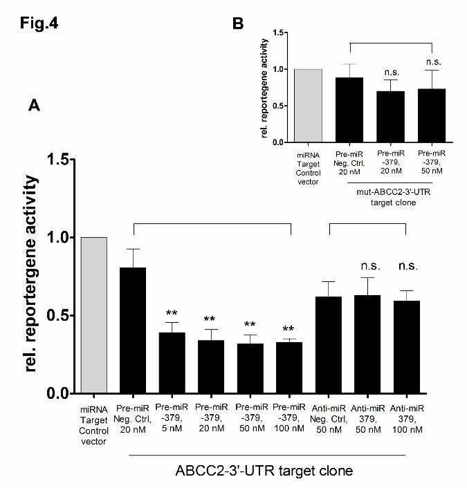
ABCC2 3’-UTR as target of miR-379
For confirmation that ABCC2 3’-UTR sequence represents a target site for miR-379,
we additionally determined the reporter gene activities of the miRNA ABCC2 3'-UTR
target clone after co-transfection with increasing concentrations of miR-379 precursor
and inhibitor, as well as their respective negative controls in HepG2 cells.
Transfection of miR-379 precursor led to a highly significant decrease of relative
luciferase activities with a minimum of 39.6+/-5.1% (p=0.001) at a concentration of 50
nM as compared to cells co-transfected with miRNA precursor negative control. In
contrast, cells co-transfected with the target clone and 50 nM or 100 nM miR-379
inhibitor did not show any changes in luciferase activities compared to cells co-
transfected with miRNA inhibitor negative control (Fig. 4A). A further proof of principle
was performed by transfecting an ABCC2 3’-UTR target clone exhibiting a mutated
seed sequence. In this case co-transfection of miR-379 precursor did not lead to
significantly lowered reporter gene activities as compared to cells co-transfected with
miRNA precursor negative control (Fig. 4B), indicating the significance of the miR-
379 binding site at position 180-200 in the 3’-UTR.
[Fig. 4]
Interplay of miR-379 and rifampicin in ABCC2 protein expression
Since rifampicin led to a down-regulation of ABCC2 protein, we investigated if
inhibition of miR-379 expression could diminish this effect. Therefore we transfected
HepG2 cells with miR-379 inhibitor (50 nM) or miRNA inhibitor negative control (50
nM). Subsequently, cells were either treated with 5 µM rifampicin or vehicle for 48
hours. As expected, cells transfected with the negative control vector exhibited a
MOL #70714This article has not been copyedited and formatted. The final version may differ from this version.
Molecular Pharmacology Fast Forward. Published on May 3, 2011 as DOI: 10.1124/mol.110.070714 at A
SPET
Journals on February 20, 2022m
olpharm.aspetjournals.org
Dow
nloaded from

13
significantly lower ABCC2 protein expression after rifampicin incubation (78.6%+/-
17.9%, p=0.04, n=3). In contrast, cells transfected with miR-379 inhibitor showed a
significantly upregulated ABCC2 expression after rifampicin treatment (187%+/-
32.1%, p=0.04, n=3) (Fig. 5).
[Fig. 5]
Discussion
The aim of our study was to investigate if ABCC2 is a target of miRNA mediated
post-transcriptional gene regulation. Our major results disclose that ABCC2 is
targeted by miR-379 in a concentration dependent manner slightly on mRNA but
more pronounced and significant on protein level. However, we can’t exclude a
contribution of further miRNAs, putatively by an indirect mechanism, to ABCC2
expression. Treatment with the PXR ligand and ABCC2 inducer rifampicin results in a
concurrent increase in both ABCC2 mRNA and miR-379 levels and a significant
down-regulation of ABCC2 protein.
Due to the fact that there was no microRNA that had been shown to affect ABCC2
mRNA at the beginning of our study, we attempted to confine those miRNAs
potentially targeting ABCC2 mRNA. We assumed that a stimulus inducing the
transcription of ABCC2 could be accompanied by altered expression of miRNAs
controlling ABCC2 gene expression on the post-transcriptional level. We therefore
treated HepG2 cells, which provide readily detectable ABCC2 expression under
normal growth conditions, with the PXR ligand and inducer of ABCC2 transcription
rifampicin (Gerk and Vore, 2002; Kast et al., 2002; Oscarson et al., 2007). As proof of
concept, we could detect a significant up-regulation of ABCB1 protein (Martin et al.
2008). After applying an rtPCR based low density array which was comprised of 377
of the most common human microRNAs, we could identify eleven more than 8-fold
deregulated miRNAs, from which only miR-379 was predicted to target ABCC2
MOL #70714This article has not been copyedited and formatted. The final version may differ from this version.
Molecular Pharmacology Fast Forward. Published on May 3, 2011 as DOI: 10.1124/mol.110.070714 at A
SPET
Journals on February 20, 2022m
olpharm.aspetjournals.org
Dow
nloaded from

14
mRNA in an in-silico alignment. miR-379 was then confirmed to down-regulate
ABCC2 in HepG2 cells, only slightly on mRNA level but significantly on ABCC2
protein level, with a maximal decrease of 54% at 50 nM of transfected miRNA
precursor. These observations suggest that miR-379 is predominantly inhibiting the
translation of ABCC2 mRNA as a result of imperfect matching of the miRNA to the 3’-
UTR, rather than the degradation. Using a vector construct exhibiting the ABCC2 3’-
UTR target sequence downstream of the firefly luciferase sequence revealed an even
more pronounced decrease of reporter gene activity, with a maximal decrease of
60% at a concentration of 50 nM of the co-transfected miRNA precursor. Moreover,
after mutagenesis of the seed sequence in the predicted miR-379 binding site (Pos.
180-200) the performed reporter gene experiments support not only the finding of
direct interference of miR-379 and ABCC2 3’-UTR, they also provide evidence about
the exact location of the miRNA binding site.
In contrast, transfection experiments with 50 nM miR-379 inhibitor showed no
increasing effect on ABCC2 mRNA or reporter gene activity of the ABCC2 3’UTR
vector construct. However, miR-379 inhibitor concentrations of 100 nM led to a
significant increase of ABCC2 protein expression, again giving evidence for the
important role of miR-379 for the regulation of ABCC2. One reason for the necessity
to apply relatively high concentrations of the miRNA inhibitor in order to diminish the
basal miR-379 expression and to obtain increasing ABCCC2 protein expression
might be the relatively weak inhibiting effect of the miRNA inhibitor on the mature
miR-379. More likely, the observation could also be explained by a relatively low
basal miRNA-379 expression under normal growth conditions in HepG2 cells.
There are certain ways to identify miRNAs targeting genes of interest. Wang and
coworkers (Wang et al., 2009) determined the correlation between miRNA and
genomewide mRNA expression in 90 lymphoblastoid cell lines. This approach
MOL #70714This article has not been copyedited and formatted. The final version may differ from this version.
Molecular Pharmacology Fast Forward. Published on May 3, 2011 as DOI: 10.1124/mol.110.070714 at A
SPET
Journals on February 20, 2022m
olpharm.aspetjournals.org
Dow
nloaded from

15
revealed 7207 inversely or co-regulated miRNA-mRNA pairs, presuming a false
discovery rate of q=0.01. The drawback of this approach is that an inverse correlation
of mRNA and miRNA depends on the mechanistic assumption that miRNAs degrade
their target mRNAs. However, due to imperfect matching of many miRNAs with the
3’-UTR, impairment of protein translation is more likely than destabilization of the
targeted mRNA. Moreover, miRNA-mRNA pairs need to be confirmed by loss and
gain of function experiments. A further approach would be to systematically screen all
in-silico predicted miRNAs for effects on the gene of interest in-vitro. Our method of
using a stimulus to alter the target gene transcription could demonstrate a more
simplified and potentially time-saving approach to identify miRNAs.
Rifampicin incubation for 72 hours caused an up-regulation of both ABCC2 mRNA
and its targeting miRNA, indicating concurrent transcription. However, at the same
time, a continuous decrease in total ABCC2 protein content could be observed.
These findings suggest a miRNA-mediated negative feedback mechanism impeding
ABCC2 over-expression by the transcriptional stimulus rifampicin. This hypothesis
was substantiated by the observation that the presence of miR-379 inhibitor caused
an increase in ABCC2 protein expression in HepG2 cells after rifampicin treatment.
Therefore, the question arises whether ABCC2 and miRNA-379 are both regulated
by PXR. A RXR/PXR consensus sequence has been identified in the rat MRP2
promoter (Kast et al., 2002). While human ABCC2 is located on chromosome 10,
such a consensus sequence could also be located on chromosome 14 upstream of
the predicted DNA coding region of the miR-379 precursor at position 101488403-
101488469 (Sanger miRBase). Interestingly, another miRNA, namely miR-411, was
found to be highly up-regulated after rifampicin treatment in our study. miR-411
precursor is encoded by a sequence located less than 1200 bp downstream of the
miR-379 precursor at position 101489662-101489757 (Sanger miRBase). Therefore,
MOL #70714This article has not been copyedited and formatted. The final version may differ from this version.
Molecular Pharmacology Fast Forward. Published on May 3, 2011 as DOI: 10.1124/mol.110.070714 at A
SPET
Journals on February 20, 2022m
olpharm.aspetjournals.org
Dow
nloaded from

16
it may be speculated that both miRNAs could originate from a single polycistronic
primary transcript (pri-miRNA). Furthermore we found also one microRNA, namely
miR-31, to be highly down regulated. Reportedly, the expression of this microRNA is
inversely associated with the metastatic capacity of 15 different breast epithelial cell
lines, as well as human primary breast tumors (Valastyan et al., 2009a; Valastyan et
al., 2009b). If rifampicin treatment thereby bears tumor promoting effects, however,
remains currently open. The results of our study contribute to the explanation of the
frequently observed weak correlation between mRNA and protein expression (Maier
et al., 2009), especially for ABC transporter proteins exhibiting long turnover times,
such as ABCB1, which has a half life of 3.7 to 5 days (Kipp et al., 2001; Petriz et al.,
2004). In contrast, the half-life of ABCC2 mRNA is 11-15 hours, as we showed in an
earlier study (Laechelt et al., 2010).
As reviewed by Kipp and Arias, ABC-transporters are subject to a cycling between
intracellular pools and the cell membrane (Kipp and Arias, 2002). Under physiological
conditions, the intracellular stored fraction of total transporter protein amount is
several times higher than the membrane expressed fraction. Triggered by an external
stimulus, transporter proteins can rapidly traffic from these reservoirs into the
membrane without necessarily increasing transcription or translation of the protein
(‘transporter on demand’). In this context, the negatively regulating effect of miR-379
on ABCC2 protein expression probably does not affect the final membranal
transporter function, but may be considered as an attenuator between transcription,
translation and function of the transporter.
It is currently unclear whether our results on miR-379 in the human hepatoblastoma
cell line HepG2 can be extrapolated to healthy tissues. Fromm et al. (Fromm et al.,
2000) showed an increase of ABCC2 mRNA and protein after rifampicin treatment in
duodenal epithelium, an observation which was recently confirmed by our group
MOL #70714This article has not been copyedited and formatted. The final version may differ from this version.
Molecular Pharmacology Fast Forward. Published on May 3, 2011 as DOI: 10.1124/mol.110.070714 at A
SPET
Journals on February 20, 2022m
olpharm.aspetjournals.org
Dow
nloaded from

17
(Haenisch et al., 2008). The length of 3’-UTRs can vary in different tissue, possibly
enabling rapidly proliferating tissues to exhibit a higher gene expression (Zhang et
al., 2005; Sandberg et al., 2008), As a consequence, the regulating effects of
miRNAs in various tissues may be impaired or disrupted. Additionally, some in vitro
studies have reported an up-regulation of ABCC2 protein in HepG2 cells 48 hours
after rifampicin treatment (Kauffmann et al., 2002; Martin et al., 2008; Schrenk et al.,
2001). Although we cannot clarify this discrepancy, high numbers of cell passages
may also cause a selection of a cell population with truncated 3’-UTRs additionally
contributing to varying results in rifampicin mediated ABCC2 induction by loss of
miRNA binding sites. Such an effect was described for the occurrence of drug
resistance in S1 colon cancer cells as a result of ABCG2 over-expression.
Investigations of the ABCG2 3’-UTR revealed a truncated 3’-UTR and loss of the
binding site of the ABCG2 inhibiting microRNA miR-519c (To et al., 2008).
While miRNA encoding regions seem to be highly conserved (Saunders et al., 2007),
a further parameter contributing to alteration of miRNA function may be genetic
variants in the 3’-UTR or further variants that alter the secondary structure of the
mRNA transcript. In a recent study, we identified different ABCC2 mRNA secondary
structures for different ABCC2 haplotypes (Laechelt et al., 2011). These different
structures may cause different folding of the 3’-UTR and consequently miRNA
binding sites, possibly contributing to the observation of marked inter-individual
differences of ABCC2 expression and non-correlation of duodenal mRNA and protein
expression after rifampicin treatment (Haenisch et al., 2008).
In conclusion we could identify - to our knowledge, for the first time - a microRNA that
negatively regulates the translation of the ABCC2 efflux transporter. Moreover, miR-
MOL #70714This article has not been copyedited and formatted. The final version may differ from this version.
Molecular Pharmacology Fast Forward. Published on May 3, 2011 as DOI: 10.1124/mol.110.070714 at A
SPET
Journals on February 20, 2022m
olpharm.aspetjournals.org
Dow
nloaded from

18
379 is co-induced by rifampicin, impeding the over-expression of ABCC2 protein after
treatment with this PXR ligand in HepG2 cells.
Acknowledgement
The technical assistance of Britta Schwarten and Micheline Neubert is gratefully
acknowledged.
Authors Contributions
Participated in research design: Haenisch, Laechelt, Noack, Bruhn, Remmler,
Cascorbi
Conducted experiments: Haenisch, Laechelt, Bruckmueller, Werk, Remmler
Contributed new reagents or analytic tools: Haenisch, Noack
Performed data analysis: Haenisch, Bruckmueller, Bruhn, Werk
Wrote or contributed to the writing of the manuscript: Haenisch, Laechelt, Remmler,
Cascorbi
MOL #70714This article has not been copyedited and formatted. The final version may differ from this version.
Molecular Pharmacology Fast Forward. Published on May 3, 2011 as DOI: 10.1124/mol.110.070714 at A
SPET
Journals on February 20, 2022m
olpharm.aspetjournals.org
Dow
nloaded from

19
References:
Fromm MF, Kauffmann HM, Fritz P, Burk O, Kroemer HK, Warzok RW, Eichelbaum
M, Siegmund W and Schrenk D (2000) The effect of rifampin treatment on
intestinal expression of human MRP transporters. Am J Pathol 157:1575-80.
Gerk PM and Vore M (2002) Regulation of expression of the multidrug resistance-
associated protein 2 (MRP2) and its role in drug disposition. J Pharmacol Exp
Ther 302:407-15.
Haenisch S, May K, Wegner D, Caliebe A, Cascorbi I and Siegmund W (2008)
Influence of genetic polymorphisms on intestinal expression and rifampicin-type
induction of ABCC2 and on bioavailability of talinolol. Pharmacogenet Genomics
18:357-65.
Jemnitz K, Heredi-Szabo K, Janossy J, Ioja E, Vereczkey L and Krajcsi P (2010)
ABCC2/Abcc2: a multispecific transporter with dominant excretory functions. Drug
Metab Rev 42:402-36.
Kast HR, Goodwin B, Tarr PT, Jones SA, Anisfeld AM, Stoltz CM, Tontonoz P,
Kliewer S, Willson TM and Edwards PA (2002) Regulation of multidrug resistance-
associated protein 2 (ABCC2) by the nuclear receptors pregnane X receptor,
farnesoid X-activated receptor, and constitutive androstane receptor. J Biol Chem
277:2908-15.
Kauffmann HM, Pfannschmidt S, Zoller H, Benz A, Vorderstemann B, Webster JI and
Schrenk D (2002) Influence of redox-active compounds and PXR-activators on
human MRP1 and MRP2 gene expression. Toxicology 171:137-46.
Kipp H and Arias IM (2002) Trafficking of canalicular ABC transporters in
hepatocytes. Annu Rev Physiol 64:595-608.
MOL #70714This article has not been copyedited and formatted. The final version may differ from this version.
Molecular Pharmacology Fast Forward. Published on May 3, 2011 as DOI: 10.1124/mol.110.070714 at A
SPET
Journals on February 20, 2022m
olpharm.aspetjournals.org
Dow
nloaded from

20
Kipp H, Pichetshote N and Arias IM (2001) Transporters on demand: intrahepatic
pools of canalicular ATP binding cassette transporters in rat liver. J Biol Chem
276:7218-24.
Kovalchuk O, Filkowski J, Meservy J, Ilnytskyy Y, Tryndyak VP, Chekhun VF and
Pogribny IP (2008) Involvement of microRNA-451 in resistance of the MCF-7
breast cancer cells to chemotherapeutic drug doxorubicin. Mol Cancer Ther
7:2152-9.
Kozomara A and Griffiths-Jones S (2010) miRBase: integrating microRNA annotation
and deep-sequencing data. Nucleic Acids Res. 2010 Oct 30. [Epub ahead of print].
Laechelt S, Turrini E, Ruehmkorf A, Siegmund W, Cascorbi I and Haenisch S (2011)
Impact of ABCC2 haplotypes on transcriptional and posttranscriptional gene
regulation and function. Pharmacogenomics J. 11:25-34.
Liang Z, Wu H, Xia J, Li Y, Zhang Y, Huang K, Wagar N, Yoon Y, Cho HT, Scala S
and Shim H (2010) Involvement of miR-326 in chemotherapy resistance of breast
cancer through modulating expression of multidrug resistance-associated protein
1. Biochem Pharmacol 79:817-24.
Maier T, Guell M and Serrano L (2009) Correlation of mRNA and protein in complex
biological samples. FEBS Lett 583:3966-73.
Martin P, Riley R, Back DJ and Owen A (2008) Comparison of the induction profile
for drug disposition proteins by typical nuclear receptor activators in human
hepatic and intestinal cells. Br J Pharmacol 153:805-19.
Oscarson M, Burk O, Winter S, Schwab M, Wolbold R, Dippon J, Eichelbaum M and
Meyer UA (2007) Effects of rifampicin on global gene expression in human small
intestine. Pharmacogenet Genomics 17:907-18.
MOL #70714This article has not been copyedited and formatted. The final version may differ from this version.
Molecular Pharmacology Fast Forward. Published on May 3, 2011 as DOI: 10.1124/mol.110.070714 at A
SPET
Journals on February 20, 2022m
olpharm.aspetjournals.org
Dow
nloaded from

21
Pan YZ, Morris ME and Yu AM (2009) MicroRNA-328 negatively regulates the
expression of breast cancer resistance protein (BCRP/ABCG2) in human cancer
cells. Mol Pharmacol 75:1374-9.
Petriz J, Gottesman MM and Aran JM (2004) An MDR-EGFP gene fusion allows for
direct cellular localization, function and stability assessment of P-glycoprotein.
Curr Drug Deliv 1:43-56.
Sandberg R, Neilson JR, Sarma A, Sharp PA and Burge CB (2008) Proliferating cells
express mRNAs with shortened 3' untranslated regions and fewer microRNA
target sites. Science 320, 1643-1647.
Saunders MA, Liang H and Li WH (2007) Human polymorphism at microRNAs and
microRNA target sites. Proc Natl Acad Sci U S A 104:3300-5.
Schrenk D, Baus PR, Ermel N, Klein C, Vorderstemann B and Kauffmann HM (2001)
Up-regulation of transporters of the MRP family by drugs and toxins. Toxicol Lett
120:51-7.
To KK, Zhan Z, Litman T and Bates SE (2008) Regulation of ABCG2 expression at
the 3' untranslated region of its mRNA through modulation of transcript stability
and protein translation by a putative microRNA in the S1 colon cancer cell line.
Mol Cell Biol 28:5147-61.
Valastyan S, Benaich N, Chang A, Reinhardt F and Weinberg RA (2009a)
Concomitant suppression of three target genes can explain the impact of a
microRNA on metastasis. Genes Dev 23:2592-7.
Valastyan S, Reinhardt F, Benaich N, Calogrias D, Szasz AM, Wang ZC, Brock JE,
Richardson AL and Weinberg RA (2009b) A pleiotropically acting microRNA, miR-
31, inhibits breast cancer metastasis. Cell 137:1032-46.
Wang L, Oberg AL, Asmann YW, Sicotte H, McDonnell SK, Riska SM, Liu W, Steer
CJ, Subramanian S, Cunningham JM, Cerhan JR and Thibodeau SN (2009)
MOL #70714This article has not been copyedited and formatted. The final version may differ from this version.
Molecular Pharmacology Fast Forward. Published on May 3, 2011 as DOI: 10.1124/mol.110.070714 at A
SPET
Journals on February 20, 2022m
olpharm.aspetjournals.org
Dow
nloaded from

22
Genome-wide transcriptional profiling reveals microRNA-correlated genes and
biological processes in human lymphoblastoid cell lines. PLoS One 4:e5878.
Zhang H, Lee JY and Tian B (2005) Biased alternative polyadenylation in human
tissues. Genome Biol 6, R100.
MOL #70714This article has not been copyedited and formatted. The final version may differ from this version.
Molecular Pharmacology Fast Forward. Published on May 3, 2011 as DOI: 10.1124/mol.110.070714 at A
SPET
Journals on February 20, 2022m
olpharm.aspetjournals.org
Dow
nloaded from

23
Footnotes The authors declare no conflict of interest.
MOL #70714This article has not been copyedited and formatted. The final version may differ from this version.
Molecular Pharmacology Fast Forward. Published on May 3, 2011 as DOI: 10.1124/mol.110.070714 at A
SPET
Journals on February 20, 2022m
olpharm.aspetjournals.org
Dow
nloaded from
24
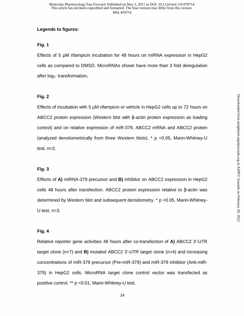
Legends to figures:
Fig. 1
Effects of 5 µM rifampicin incubation for 48 hours on miRNA expression in HepG2
cells as compared to DMSO. MicroRNAs shown have more than 3 fold deregulation
after log2- transformation.
Fig. 2
Effects of incubation with 5 µM rifampicin or vehicle in HepG2 cells up to 72 hours on
ABCC2 protein expression (Western blot with β-actin protein expression as loading
control) and on relative expression of miR-379, ABCC2 mRNA and ABCC2 protein
(analyzed densitometrically from three Western blots). * p <0.05, Mann-Whitney-U
test, n=3.
Fig. 3
Effects of A) miRNA-379 precursor and B) inhibitor on ABCC2 expression in HepG2
cells 48 hours after transfection. ABCC2 protein expression relative to β-actin was
determined by Western blot and subsequent densitometry. * p <0.05, Mann-Whitney-
U test, n=3.
Fig. 4
Relative reporter gene activities 48 hours after co-transfection of A) ABCC2 3’-UTR
target clone (n=7) and B) mutated ABCC2 3’-UTR target clone (n=4) and increasing
concentrations of miR-379 precursor (Pre-miR-379) and miR-379 inhibitor (Anti-miR-
379) in HepG2 cells. MicroRNA target clone control vector was transfected as
positive control. ** p <0.01, Mann-Whitney-U test.
MOL #70714This article has not been copyedited and formatted. The final version may differ from this version.
Molecular Pharmacology Fast Forward. Published on May 3, 2011 as DOI: 10.1124/mol.110.070714 at A
SPET
Journals on February 20, 2022m
olpharm.aspetjournals.org
Dow
nloaded from

25
Fig. 5
Relative ABCC2 protein content (mean optical density of three Western blots) in
HepG2 cells after 48 hours of treatment with 5 µM rifampicin or vehicle and 72 hours
after transfection of A) miRNA inhibitor (anti-miR) negative control and B) miR-379
inhibitor (anti-miR-379), respectively. β-actin protein expression was used as internal
standard. * p <0.05, Mann-Whitney-U test, n=3.
MOL #70714This article has not been copyedited and formatted. The final version may differ from this version.
Molecular Pharmacology Fast Forward. Published on May 3, 2011 as DOI: 10.1124/mol.110.070714 at A
SPET
Journals on February 20, 2022m
olpharm.aspetjournals.org
Dow
nloaded from

This article has not been copyedited and formatted. The final version may differ from this version.Molecular Pharmacology Fast Forward. Published on May 3, 2011 as DOI: 10.1124/mol.110.070714
at ASPE
T Journals on February 20, 2022
molpharm
.aspetjournals.orgD
ownloaded from

This article has not been copyedited and formatted. The final version may differ from this version.Molecular Pharmacology Fast Forward. Published on May 3, 2011 as DOI: 10.1124/mol.110.070714
at ASPE
T Journals on February 20, 2022
molpharm
.aspetjournals.orgD
ownloaded from

This article has not been copyedited and formatted. The final version may differ from this version.Molecular Pharmacology Fast Forward. Published on May 3, 2011 as DOI: 10.1124/mol.110.070714
at ASPE
T Journals on February 20, 2022
molpharm
.aspetjournals.orgD
ownloaded from
This article has not been copyedited and formatted. The final version may differ from this version.Molecular Pharmacology Fast Forward. Published on May 3, 2011 as DOI: 10.1124/mol.110.070714
at ASPE
T Journals on February 20, 2022
molpharm
.aspetjournals.orgD
ownloaded from

This article has not been copyedited and formatted. The final version may differ from this version.Molecular Pharmacology Fast Forward. Published on May 3, 2011 as DOI: 10.1124/mol.110.070714
at ASPE
T Journals on February 20, 2022
molpharm
.aspetjournals.orgD
ownloaded from