Double-stranded Ribonucleic Acid from Cytoplasmic ...jvi.asm.org/content/2/10/1211.full.pdf ·...
12
JOURNAL OF VIROLOGY, Feb. 1968, p. 1211-1222 Vol. 2, No. 10 Copyright © 1968 American Society for Microbiology Printed in U.S.A. Double-stranded Ribonucleic Acid from Cytoplasmic Polyhedrosis Virus of the Silkworm K. MIURA, I. FUJII, T. SAKAKI, M. FUKE, AND S. KAWASE Nagoya University, Chikusa, Nagoya, anid Uniiversity of Tokyo, Honlgo, Tokyo, Japaln Received for publication 5 January 1968 Ribonucleic acid (RNA) was extracted by phenol treatment from cytoplasmic polyhedrosis virus isolated from the midgut of infected silkworms. This RNA ap- pears as threads when precipitated in alcohol. Two components having different sedimentation constants were observed. The molecular weight of the RNA prepara- tion obtained by sedimentation coefficient (weight-averaged) and intrinsic viscosity was about 2 X 10⁶ to 3 X 10⁶. It was one-half to one-third the size of the calculated molecular weight for an entire RNA molecule in a virion. Electron micrographs of this RNA preparation showed two peaks in the distribution of contour length, at 0.4 and 1.3 Am, which would correspond to molecular weights of 106 and 3 X 10⁶, re- spectively. The extracted RNA seemed to split into segments at a preferential break- ing point. This RNA was soluble in concentrated salt solution, differing from single stranded high-molecular-weight RNA. The base composition of this RNA was complementary in the ratios of adenosine to uridine and guanosine to cytosine. It contained 43 %- guanosine plus cytosine. Based on its filamentous appearance by elec- tron microscopy, typical pattern of optical rotatory dispersion and circular dichro- ism, sharp transition of the optical properties on heating, great hyperchromicity on degradation, nonreactivity with formaldehyde, and resistance to ribonucleases, it is concluded that this RNA is double-stranded and has regular base pairings of guanosine-cytosine and adenosine-uridine. Cytoplasmic polyhedrosis disease in insects results in inclusion bodies, polyhedra, which con- tain many virus particles. When the nucleic acid extracted from the cytoplasmic polyhedra formed in the midgut epithelium of the infected silkworm was precipitated in ethyl alcohol, fibrous precipi- tates were obtained in addition to flocculent precipitates (14), both of which were proven to be ribonucleic acid (RNA) by color reactions for sugar components and by digestion with nucle- ases. The fibrous precipitate fraction, however, was different from the ordinary cellular RNA species in its elution profile on a methylated al- bumin column and in its complementary base composition (15). Recently, virus particles were liberated from cytoplasmic polyhedra with the use of a carbonate buffer (pH 10.8) and were purified by ultracen- trifugation (16). The nucleic acid extracted by phenol treatment from this virus preparation pro- duced only a fibrous precipitate in alcohol, and no flocculent precipitate. The properties of this fibrous nucleic acid have now been studied, and it has been concluded that this nucleic acid is a double-stranded RNA like the RNA from reo- virus (11, 22), from wound tumor virus (2, 43), and from rice dwarf virus (29, 36). MATERIALS AND METHODS Purification of virus. On the 1st day of 5th instar, silkworms, Bombyx moni (L.), were injected in the posterior region with virus suspension, which was ob- tained by dissolving cytoplasmic polyhedra in 0.05 M Na2CO3-0.05 M NaCl (S. Kawase and S. Miyajima, J. Invert. Pathol., in press). A few days later, midguts of the infected silkworm were removed and immedi- ately put in dry ice. About 100 g of the frozen material was thawed, added to 350 ml of cold water, and ho- mogenized in a Waring Blendor (2 min at high speed). The homogenate was filtered through gauze, and the sap filtrate centrifuged at 8,000 X g for 10 min to ob- tain a residue of polyhedra. Precipitated polyhedra were then washed several times with cold water (until the supernatant fluid became clear). Finally, the wet pellet was suspended in 40 ml of a carbonate buffer (pH 10.8, 20 C; 9:1 mixture of 0.2 M Na2CO3 and 0.2 M NaHCO3). After 1 hr at room temperature, the solu- tion was diluted with three volumes of distilled water and centrifuged at 8,000 X g for 30 min. When a white polyhedra precipitate still remained, the precipitate 1211 on July 5, 2018 by guest http://jvi.asm.org/ Downloaded from
Transcript of Double-stranded Ribonucleic Acid from Cytoplasmic ...jvi.asm.org/content/2/10/1211.full.pdf ·...
JOURNAL OF VIROLOGY, Feb. 1968, p. 1211-1222 Vol. 2, No. 10Copyright © 1968 American Society for Microbiology Printed in U.S.A.
Double-stranded Ribonucleic Acid fromCytoplasmic Polyhedrosis Virus
of the SilkwormK. MIURA, I. FUJII, T. SAKAKI, M. FUKE, AND S. KAWASE
Nagoya University, Chikusa, Nagoya, anid Uniiversity of Tokyo, Honlgo, Tokyo, Japaln
Received for publication 5 January 1968
Ribonucleic acid (RNA) was extracted by phenol treatment from cytoplasmicpolyhedrosis virus isolated from the midgut of infected silkworms. This RNA ap-pears as threads when precipitated in alcohol. Two components having differentsedimentation constants were observed. The molecular weight of the RNA prepara-tion obtained by sedimentation coefficient (weight-averaged) and intrinsic viscositywas about 2 X 10⁶ to 3 X 10⁶. It was one-half to one-third the size of the calculatedmolecular weight for an entire RNA molecule in a virion. Electron micrographsof this RNA preparation showed two peaks in the distribution of contour length, at0.4 and 1.3 Am, which would correspond to molecular weights of 106 and 3 X 10⁶, re-spectively. The extracted RNA seemed to split into segments at a preferential break-ing point. This RNA was soluble in concentrated salt solution, differing fromsingle stranded high-molecular-weight RNA. The base composition of this RNA wascomplementary in the ratios of adenosine to uridine and guanosine to cytosine. Itcontained 43 %- guanosine plus cytosine. Based on its filamentous appearance by elec-tron microscopy, typical pattern of optical rotatory dispersion and circular dichro-ism, sharp transition of the optical properties on heating, great hyperchromicity ondegradation, nonreactivity with formaldehyde, and resistance to ribonucleases, itis concluded that this RNA is double-stranded and has regular base pairings ofguanosine-cytosine and adenosine-uridine.
Cytoplasmic polyhedrosis disease in insectsresults in inclusion bodies, polyhedra, which con-tain many virus particles. When the nucleic acidextracted from the cytoplasmic polyhedra formedin the midgut epithelium of the infected silkwormwas precipitated in ethyl alcohol, fibrous precipi-tates were obtained in addition to flocculentprecipitates (14), both of which were proven tobe ribonucleic acid (RNA) by color reactions forsugar components and by digestion with nucle-ases. The fibrous precipitate fraction, however,was different from the ordinary cellular RNAspecies in its elution profile on a methylated al-bumin column and in its complementary basecomposition (15).
Recently, virus particles were liberated fromcytoplasmic polyhedra with the use of a carbonatebuffer (pH 10.8) and were purified by ultracen-trifugation (16). The nucleic acid extracted byphenol treatment from this virus preparation pro-duced only a fibrous precipitate in alcohol, andno flocculent precipitate. The properties of thisfibrous nucleic acid have now been studied, andit has been concluded that this nucleic acid is a
double-stranded RNA like the RNA from reo-virus (11, 22), from wound tumor virus (2, 43),and from rice dwarf virus (29, 36).
MATERIALS AND METHODS
Purification of virus. On the 1st day of 5th instar,silkworms, Bombyx moni (L.), were injected in theposterior region with virus suspension, which was ob-tained by dissolving cytoplasmic polyhedra in 0.05 MNa2CO3-0.05 M NaCl (S. Kawase and S. Miyajima,J. Invert. Pathol., in press). A few days later, midgutsof the infected silkworm were removed and immedi-ately put in dry ice. About 100 g of the frozen materialwas thawed, added to 350 ml of cold water, and ho-mogenized in a Waring Blendor (2 min at high speed).The homogenate was filtered through gauze, and thesap filtrate centrifuged at 8,000 X g for 10 min to ob-tain a residue of polyhedra. Precipitated polyhedrawere then washed several times with cold water (untilthe supernatant fluid became clear). Finally, the wetpellet was suspended in 40 ml of a carbonate buffer(pH 10.8, 20 C; 9:1 mixture of 0.2 M Na2CO3 and 0.2M NaHCO3). After 1 hr at room temperature, the solu-tion was diluted with three volumes of distilled waterand centrifuged at 8,000 X g for 30 min. When a whitepolyhedra precipitate still remained, the precipitate
1211
on July 5, 2018 by guesthttp://jvi.asm
.org/D
ownloaded from
MIURA ET AL.
was treated again with 20 ml of carbonate buffer, di-luted with distilled water, and centrifuged. The com-bined supernatant extract, in which cytoplasmic poly-hedrosis viurs (CPV) was suspended, was centrifugedat 65,000 X g for 1 hr.The virus pellet was suspended in 40 ml of distilled
water and centrifuged at 2,000 X g for 10 min to re-move insoluble materials. A representative picture ofthe virus is shown in F:g. 1. The virus preparation wasalmost homogeneous, as shown by many electronmicrographs and sedimentation patterns, althoughthere were some empty virus particles. The infectivityof this preparation was proved by injecting silkwormlarvae (Kawase and Miyajima, in press).
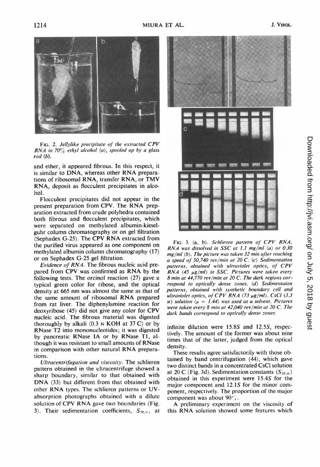
Extraction of nucleic acid. The phenol treatmentused for extraction of nucleic acid was essentially thesame as that described by Gierer and Schramm (12)for purifying RNA from tobacco mosaic virus (TMV).The CPV suspension was added to an equal volume of90% phenol, and the mixture was shaken vigorouslyfor 10 min in the cold. Centrifugation at 1,000 X gfor 10 min resulted in three separate layers. The toplayer was removed with a pipette, and the phenoltreatment was repeated for 2 min. The top water layer,with one drop of 1 M NaCI added, was poured intothree volumes of cold ethyl alcohol. The jellylikeprecipitate was spooled up with a glass rod (Fig. 2).It was then immersed in 70% ethyl alcohol and storedin a freezer until use. For physical measurements, theprecipitate was dissolved in a suitable solvent anddialyzed in the cold against that solvent. For analysis,the precipitate was dipped in ethyl alcohol, ether-ethyl alcohol (1:1), and ether, and it was then air-dried.
Analysis of nucleotide composition and analysis ofphosphorus. Three methods were used for nucleotidecomposition analysis. (i) By the acid hydrolysismethod, RNA was hydrolyzed with 1 N HCl at 100 Cfor 1 hr. The composition of the resulting pyrimidinenucleotides and purines was determined by one-di-mensional paper chromatography (24) as in a previouspaper (28). (ii) By the alkaline hydrolysis method,RNA was digested with 0.3 N KOH at 37 C for 18 hr.The composition of the resulting mixture of nucleo-tides was determined by two-dimensional paper chro-matography (4) as in previous papers (29, 31). (iii)Instead of alkaline hydrolysis, ribonuclease (RNase)T2 digestion was achieved (20); 1 mg of RNA wasdissolved in 0.1 ml of 0.05 M acetate buffer (pH 4.5)and incubated with 5 units of RNase T2 at 37 C over-night. In all cases, Whatman no. 1 analytical filterpaper was used for the separation of nucleotides orbases.
Analysis of phosphorus was performed by themethod of Chen, Toribara, and Warner (7).
Spectrophotometry. Ultraviolet (UV) spectra wereobtained with a Zeiss spectrophotometer (PMQ-1 1)and Nihon Bunko spectrophotometer (JASCO modelORD/UV-5).
Optical rotatory dispersion and circular dichroismwere measured with a Nihon Bunko spectrophotome-ter. On raising the temperature continuously by circu-lating water from a water bath into the jacket surround-
ing a cuvette, the UV absorption at 258 nm, opticalrotatory dispersion at 282 nm, and circular dichroismat 260 nm were recorded. The solvent for RNA was0.01 X SSC (0.15 M NaCl plus 0.015 M sodium cit-rate). From the experiences of Marmur and Doty(25, 26) with deoxyribonucleic acid (DNA), and fromthe present use of CPV RNA, it was concluded thatone can observe thermal transition at a low tempera-ture if the salt concentration of the nucleic acid solu-tion is low, as it is in 0.01 X SSC. The RNA was at aconcentration of approximately 40 ,.lg/ml and wascontained in a 4-ml quartz cuvette, which had a 1-cmlight path and contained a small tipped thermister.
Reactioni with formaldehyde. The conditions used forthe reaction with formaldehyde were as described byFraenkel-Conrat (9). To 3 ml of RNA solution (ap-proximately 30 Ag of RNA/ml in 0.1 M NaCI) wasadded 0.15 ml of 37% formaldehyde, to bring the finalformaldehyde concentration to 1.8%. The control(time zero) was prepared with 0.15 ml of water in-stead of formaldehyde. The mixtures were kept instoppered test tubes at 37 C, and the UV absorptionwas measured at intervals.
Digestion with RNase. A solution of pancreaticRNase IA (5 pAliters, chromatographically purified;Worthington Biochemical Corp., Freehold, N.J.) wasadded to a solution containing approximately 50 Ag ofRNA in 2 ml of 0.05 M tris(hydroxymethyl)amino-methane (Tris)-chloride buffer (pH 7.6). (In the con-trol, 5 ,uliters of water was added.) RNase Tl (1Ag, chromatographically purified; Sankyo, Ltd.) wasadded to a solution containing about 1 mg of RNAdissolved in 0.1 ml of 0.05 M Tris buffer (pH 7.5) plus2 X 10-3 M ethylenediaminetetraacetic acid (EDTA).After incubation of the mixture at 25 C, the increasein optical density at 260 nm was measured.
Determination ofsedimentation constant anzd initrinsicviscosity. Ultracentrifugal analysis of nucleic acid inSSC was performed by using a Spinco model E ana-lytical ultracentrifuge with schlieren optics and UVoptics at 20 C. The sedimentation constant at infinitedilution was obtained by extrapolation of several val-ues, measured at different concentrations of RNA.
Viscosity was measured with a Couette-type vis-cometer at an extremely low velocity gradient (0.07per sec) under conditions corresponding to those forsedimentation, because the viscosity of CPV RNAsolution was markedly influenced by the shear rate.Intrinsic viscosity was found by extrapolating the re-duced viscosity values to zero concentration.
Electron microscopy of nucleic acid. Electron mi-croscopy of nucleic acid was performed by the proteinmonolayer method (19). The suspension containingnucleic acid (5 ,ug/ml), 2 M ammonium acetate, and0.01%70 cytochrome c was spread on a surface of double-distilled water. The protein monolayer was transferredto the carbon supporting film by touching the grid tothe surface of the water. The drop of water was driedby dipping the surface of the grid into ethyl alcohol.The contrast was enhanced by rotatory shadowing withan alloy of 80%o platinum and 20% palladium. Photo-graphs were taken in electron microscope (Hitachi-HS-7).
1212 J. VIROL.
on July 5, 2018 by guesthttp://jvi.asm
.org/D
ownloaded from
DOUBLE-STRANDED RNA FROM CPV
RESULTS AND DISCUSSION when its aqueous solution was poured into more
Characteristics and column chromatography of than two volumes of ethyl alcohol. It could benucleic acid. The nucleic acid extracted from CPV spooled onto a glass rod (Fig. 2). When it wasby phenol treatment was precipitated as a jelly dehydrated by successive dipping in ethyl alcohol
FIG. 1. Piurified cytoplasmic polvhedrosis virus particles. Negative staiuiiug with phosphotustgstate. X 600,000.
VOL. 2, 1 968 1213
on July 5, 2018 by guesthttp://jvi.asm
.org/D
ownloaded from
MIURA ET AL.
FIG. 2. Jellylike precipitate of the extracted CPVRNA in 70%/cl ethyl alcohol (a), spooled itp by a glassrod (b).
and ether, it appeared fibrous. In this respect, itis similar to DNA, whereas other RNA prepara-tions of ribosomal RNA, transfer RNA, or TMVRNA, deposit as flocculent precipitates in alco-hol.
Flocculent precipitates did not appear in thepresent preparation from CPV. The RNA prep-aration extracted from crude polyhedra containedboth fibrous and flocculent precipitates, whichwere separated on methylated albumin-kiesel-guhr column chromatography or on gel filtration(Sephadex G-25). The CPV RNA extracted fromthe purified virus appeared as one component onmethylated albumin column chromatography (17)or on Sephadex G-25 gel filtration.
Evidence ofRNA. The fibrous nucleic acid pre-pared from CPV was confirmed as RNA by thefollowing tests. The orcinol reaction (27) gave atypical green color for ribose, and the opticaldensity at 665 nm was almost the same as that ofthe same amount of ribosomal RNA preparedfrom rat liver. The diphenylamine reaction fordeoxyribose (45) did not give any color for CPVnucleic acid. The fibrous material was digestedthoroughly by alkali (0.3 N KOH at 37 C) or byRNase T2 into mononucleotides; it was digestedby pancreatic RNase IA or by RNase Tl, al-though it was resistant to small amounts of RNasein comparison with other natural RNA prepara-tions.
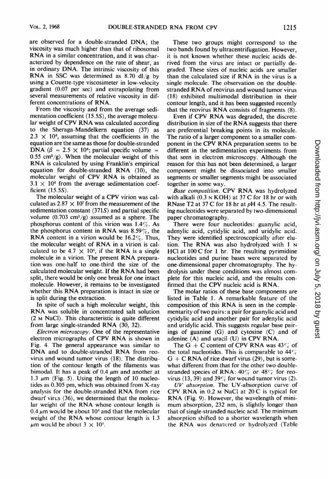
Ultracentrifligation and viscosity. The schlierenpattern obtained in the ultracentrifuge showed asharp boundary, similar to that obtained withDNA (33) but different from that obtained withother RNA types. The schlieren patterns or UV-absorption photographs obtained with a dilutesolution of CPV RNA gave two boundaries (Fig.3). Their sedimentation coefficients, S20o,,-, at
FIG. 3. (a, b). Schliere,z pattern of CPV RNA.RNA was dissolved in SSC at 1.1 mg/ml (a) or 0.30mg/ml (b). The pictlure was takeiz 32 miii after reachiiiga speed of 50,740 rev/miiz at 20 C. (c) Sedime,ztationpatterns, obtaiiied with uiltraviolet optics, of CPVRNA (45 ug/ml) in SSC. Pictures were takeir every8 mini at 44,770 rev/mihz at 20 C. The dark regioizs cor-respond to optically dezise zotzes. (d) Sedime,ztaionpatterns, obtained with syizthetic boundary cell aiidultraviolet optics, of CPV RNA (73 jig/ml). CsCI (3.3M) solutioIz (p = 1.44) wvas used as a solve,zt. Pictureswere takeii every 8 mih at 42,040 rev/mihz at 20 C. Thedark bands correspoizd to optically dense zonies.
infinite dilution were 15.8S and 12.5S, respec-tively. The amount of the former was about ninetimes that of the latter, judged from the opticaldensity.These results agree satisfactorily with those ob-
tained by band centrifugation (44), which gavetwo distinct bands in a concentrated CsCl solutionat 20 C (Fig. 3d). Sedimentation constants (S20,.)obtained in this experiment were 15.4S for themajor component and 12.1S for the minor com-ponent, respectively. The proportion of the majorcomponent was about 90-.A preliminary experiment on the viscosity of
this RNA solution showed some features which
. .. ......................... .... ...........
I... ....- .1
L-MLMALIM; .1. Al
1214 J. VIROL.
on July 5, 2018 by guesthttp://jvi.asm
.org/D
ownloaded from
DOUBLE-STRANDED RNA FROM CPV
are observed for a double-stranded DNA; theviscosity was much higher than that of ribosomalRNA in a similar concentration, and it was char-acterized by dependence on the rate of shear, asin ordinary DNA. The intrinsic viscosity of thisRNA in SSC was determined as 8.70 dl/g byusing a Couette-type viscosimeter in low-velocitygradient (0.07 per sec) and extrapolating fromseveral measurements of relative viscosity in dif-ferent concentrations of RNA.From the viscosity and from the average sedi-
mentation coefficient (15.5S), the average molecu-lar weight of CPV RNA was calculated accordingto the Sheraga-Mandelkern equation (37) as2.3 x 106, assuming that the coefficients in theequation are the same as those for double-strandedDNA (,B = 2.5 X 10⁶; partial specific volume =0.55 cm3/g). When the molecular weight of thisRNA is calculated by using Franklin's empiricalequation for double-stranded RNA (10), themolecular weight of CPV RNA is obtained as3.1 x 106 from the average sedimentation coef-ficient (15.5S).The molecular weight of a CPV virion was cal-
culated as 2.87 x 10' from the measurement of thesedimentation constant (371S) and partial specificvolume (0.703 cm3/g) assumed as a sphere. Thephosphorus content of this virion was 1.4%. Asthe phosphorus content in RNA was 8.59%, theRNA content in a virion would be 16.2%. Thus,the molecular weight of RNA in a virion is cal-culated to be 4.7 X 10⁶, if the RNA is a singlemolecule in a virion. The present RNA prepara-tion was one-half to one-third the size of thecalculated molecular weight. If the RNA had beensplit, there would be only one break for one intactmolecule. However, it remains to be investigatedwhether this RNA preparation is intact in size oris split during the extraction.
In spite of such a high molecular weight, thisRNA was soluble in concentrated salt solution(2 M NaCl). This characteristic is quite differentfrom large single-stranded RNA (30, 32).
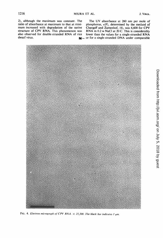
Electron microscopy. One of the representativeelectron micrographs of CPV RNA is shown inFig. 4. The general appearance was similar toDNA and to double-stranded RNA from reo-virus and wound tumor virus (18). The distribu-tion of the contour length of the filaments wasbimodal. It has a peak of 0.4 ,um and another at1.3 gm (Fig. 5). Using the length of 10 nucleo-tides as 0.305 pm, which was obtained from X-rayanalysis for the double-stranded RNA from ricedwarf virus (36), we determined that the molecu-lar weight of the RNA whose contour length is0.4,um would be about 106 and that the molecularweight of the RNA whose contour length is 1.3,um would be about 3 X 10⁶.
These two groups might correspond to thetwo bands found by ultracentrifugation. However,it is not known whether these nucleic acids de-rived from the virus are intact or partially de-graded. These sizes of nucleic acids are smallerthan the calculated size if RNA in the virus is asingle molecule. The observation on the double-stranded RNA of reovirus and wound tumor virus(18) exhibited multimodal distribution in theircontour length, and it has been suggested recentlythat the reovirus RNA consists of fragments (8).Even if CPV RNA was degraded, the discrete
distribution in size of the RNA suggests that thereare preferential breaking points in its molecule.The ratio of a larger component to a smaller com-ponent in the CPV RNA preparation seems to bedifferent in the sedimentation experiments fromthat seen in electron microscopy. Although thereason for this has not been determined, a largercomponent might be dissociated into smallersegments or smaller segments might be associatedtogether in some way.Base composition. CPV RNA was hydrolyzed
with alkali (0.3 N KOH) at 37 C for 18 hr or withRNase T2 at 37 C for 18 hr at pH 4.5. The result-ing nucleotides were separated by two-dimensionalpaper chromatography.There were four nucleotides: guanylic acid,
adenylic acid, cytidylic acid, and uridylic acid.They were identified spectroscopically after elu-tion. The RNA was also hydrolyzed with I NHCl at 100 C for 1 hr. The resulting pyrimidinenucleotides and purine bases were separated byone-dimensional paper chromatography. The hy-drolysis under these conditions was almost com-plete for this nucleic acid, and the results con-firmed that the CPV nucleic acid is RNA.The molar ratios of these base components are
listed in Table 1. A remarkable feature of thecomposition of this RNA is seen in the comple-mentarity of two pairs: a pair for guanylic acid andcytidylic acid and another pair for adenylic acidand uridylic acid. This suggests regular base pair-ings of guanine (G) and cytosine (C) and ofadenine (A) and uracil (U) in CPV RNA.The G + C content of CPV RNA was 43 ' of
the total nucleotides. This is comparable to 44%-G + C RNA of rice dwarf virus (29), but is some-what different from that for the other two double-stranded species of RNA: 40%c or 48% for reo-virus (13, 39) and 39%c for wound tumor virus (2).UV absorption. The UV-absorption curve of
CPV RNA in 0.2 M NaCl at 20 C is typical forRNA (Fig. 9). However, the wavelength of mini-mum absorption, 232 nm, is slightly longer thanthat of single-stranded nucleic acid. The minimumabsorption shifted to a shorter wavelength whenthe RNA was denatuired or hydrolyzed (Table
VOL. 2, 1968 1215
on July 5, 2018 by guesthttp://jvi.asm
.org/D
ownloaded from
2), although the maximum was constant. The The UV absorbance at 260 nm per mole ofratio of absorbance at maximum to that at mini- phosphorus, E(P), determined by the method ofmum increased with degradation of the native Chargaff and Zamenhof, (6), was 6,600 for CPVstructure of CPV RNA. This phenomenon was RNA in 0.2 M NaCl at 20 C. This is considerablyalso observed for double-stranded RNA of rice lower than the values for a single-stranded RNAdwarf virus. =- or for a single-stranded DNA under comparable
FIG. 4. Electrol, micrograph of CPV RNA. X 25,500. Tlte black bar ilndicates I ,um.
1216 MIURA ET AL. J. VIROL.
on July 5, 2018 by guesthttp://jvi.asm
.org/D
ownloaded from
DOUBLE-STRANDED RNA FROM CPV
conditions. This value for CPV RNA is nearer tothe e(P) of natural DNA; it was similar to that ofdouble-stranded rice dwarf virus RNA. Heat-de-natured CPV RNA gave a higher e(P) value un-
der the same conditions. Hydrolysis with alkali or
with RNase T2 split the RNA into mononucleo-tides, and E(P) of the hydrolysate increased fur-ther. The hyperchromicity of CPV RNA observedin alkaline digestion was considerably greater
20
u
0
0
I10
E
O L
0.5 1.0 1.5
length (p)
FIG. 5. Length distribution of CPV RNA in elec-tron micrographs, expressed as percentages of 450fibers.
TABLE 1. Nucleotide compositiolt of CPV RNAa
Hydrolysis
Nucleotide Acid Alkali RNase T2(1 N. HCl, (0.3 x (37 C,lOOC, KOH,37 C, hr1 hr) 18 h 8hr)
Guanylic acid 21.8 21.3 20.3Adenylic acid 27.8 29.1 27.8Cytidylic acid 21.2 21.2 21.1Uridylic acid 29.2 28.4 29.8
G/C 1.03 1.01 0.96A/U 0.95 1.03 0.94Pu/Pyt 0.98 1.02 0.93
(A + C)/(G + U) 0.96 1.01 0.97
G + C (%) 43.0 42.5 41.4
a Expressed as moles per 100 moles of guanylic,adenylic, cytidylic, and uridylic acid, adjusted to100% recovery. The actual recovery of bases, com-pared to total phosphorus, was 98%7o for acid hy-drolysis, 92% for alkaline hydrolysis, and 90% forRNase T2 hydrolysis. Three to five analyses were
averaged.6PU, purne;Py, pyrimidine.
than that of ordinary single-stranded RNA. Thissuggests that CPV RNA has a more orderedstructure than single-stranded RNA.
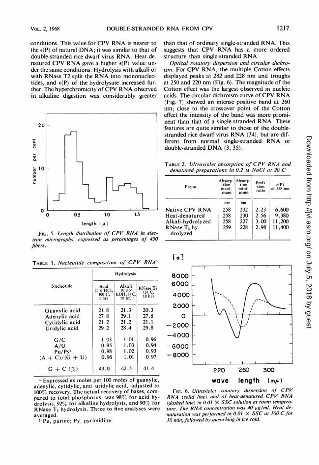
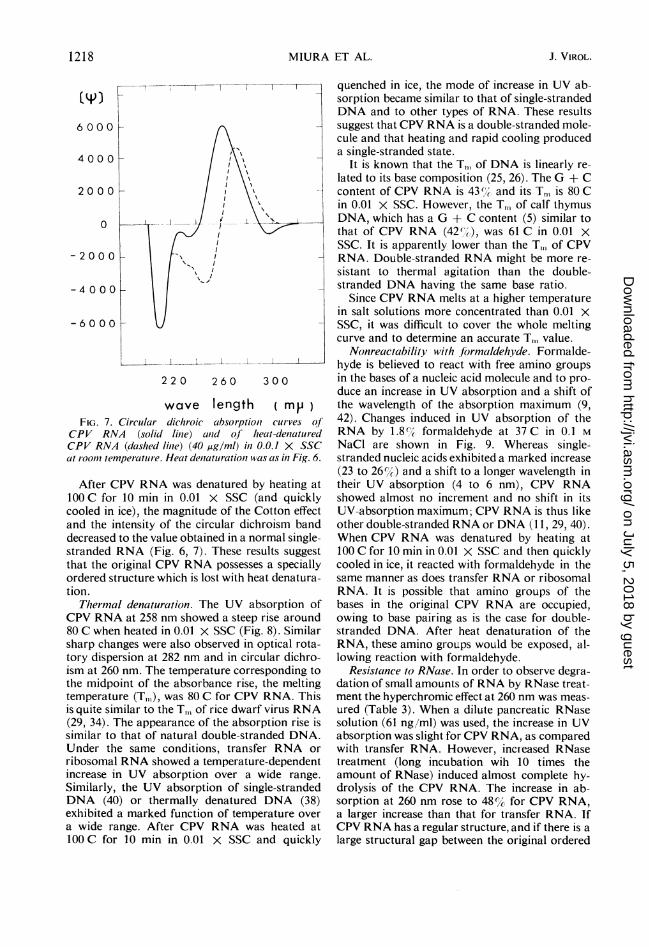
Optical rotatory dispersion and circular dichro-ism. For CPV RNA, the multiple Cotton effectsdisplayed peaks at 282 and 228 nm and troughsat 250 and 220 nm (Fig. 6). The magnitude of theCotton effect was the largest observed in nucleicacids. The circular dichroism curve of CPV RNA(Fig. 7) showed an intense positive band at 260nm; close to the crossover point of the Cottoneffect the intensity of the band was more promi-nent than that of a single-stranded RNA. Thesefeatures are quite similar to those of the double-stranded rice dwarf virus RNA (34), but are dif-ferent from normal single-stranded RNA ordouble-stranded DNA (3, 35).
TABLE2. Ultraviolet absorption of CPV RNA anddenatured preparations in 0.2 M NaCl at 20 C
Absorp- AbsorP- Emis- eP
Prepn tion tion sio a 2PnPrepn ~maxi- mini-ratio
at 2958 mnmum mum rai
n,n nnt
Native CPV RNA 258 232 2.23 6,600Heat-denatured 258 230 2.56 9,380Alkali-hydrolyzed 258 227 3.00 11,200RNase T2-hy- 259 228 2.98 11,400drolyzed
(a&
80006000
4000
2000
0
-2000
-4000
-6000-8000
220 260 300
wave length (me)
FIG. 6. Ultraviolet rotatory dispersion of CPV
RNA (solid line) and of heat-denatured CPV RNA
(dashed line) in 0.01 X SSC solutioii at room tempera-ture. The RNA concentrationi was 40 uAg/ml. Heat de-
naturation was performed in 0.01 X SSC at 100 C for10 min, followed by quenching in ice cold.
VOL. 2,1968 1217
on July 5, 2018 by guesthttp://jvi.asm
.org/D
ownloaded from
MIURA ET AL.
6000
I A
4000 2 _
CPVRNA (dashedliCie) (40 mglml) in0.0.1X SSC
-2000 _ -eat/ __00 _II_
-2000 U X, .
22 0 260 300
wave length ( mj
FIG. 7. Cireslar dticheoic albsorptioi curves of
CPV RNA (solid line) and ofhseatu-denturedCPV RNA (dashed linPe) (40 ssg/ml) ins 0.0.1 SSC
at rooder temperature.wHeatdeaatarationwas as in Fig. 6.
After CPV RNA was denatured by heating at
00 C for 10 min in 0.01 X SSC (and quicklycooled in ice), the magnitude of the Cotton effect
and the intensity of the circular dichroism band
decreased to the value obtained in a normal single-stranded RNA (Fig. 6, 7). These results suggest
that the original CPV RNA possesses a specially
ordered structure which is lost with heat denatura-
tion.
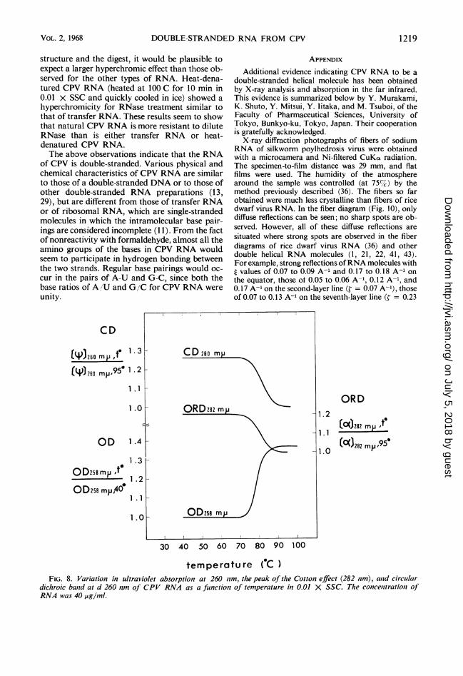
Therm34al denaturation. The UV absorption of
CPV RNA at 258 nm showed a steep rise around
80 C when heated in 0.01 X SSC (Fig. 8). Similar
sharp changes were also observed in optical rota-
tory dispersion at 282 nm and in circular dichro-
ism at 260 nm. The temperature corresponding to
the midpoint of the absorbance rise, the melting
temperature (T,), was 80 C for CPV RNA. Thisis quite similar to theTCn of rice dwarf virus RNA
(29, 34). The appearance of the absorption rise is
similar to that of natural double-stranded DNA.
Under the same conditions, transfer RNA or
ribosomal RNA showed a temperature-dependent
increase in UV absorption over a wide range.
Similarly, the UV absorption of single-stranded
DNA (40) or thermally denatured DNA (38)
exhibited a marked function of temperature over
a wide range. After CPV RNA was heated at
100OC for 10 min in 0.01 X SSC and quickly
quenched in ice, the mode of increase in UV ab-sorption became similar to that of single-strandedDNA and to other types of RNA. These resultssuggest that CPV RNA is a double-stranded mole-cule and that heating and rapid cooling produceda single-stranded state.
It is known that the T,,, of DNA is linearly re-lated to its base composition (25, 26). The G + Ccontent of CPV RNA is 43% and its T,,, is 80 Cin 0.01 X SSC. However, the TM, of calf thymusDNA, which has a G + C content (5) similar tothat of CPV RNA (42%(), was 61 C in 0.01 XSSC. It is apparently lower than the T,, of CPVRNA. Double-stranded RNA might be more re-sistant to thermal agitation than the double-stranded DNA having the same base ratio.
Since CPV RNA melts at a higher temperaturein salt solutions more concentrated than 0.01 XSSC, it was difficult to cover the whole meltingcurve and to determine an accurate T, value.
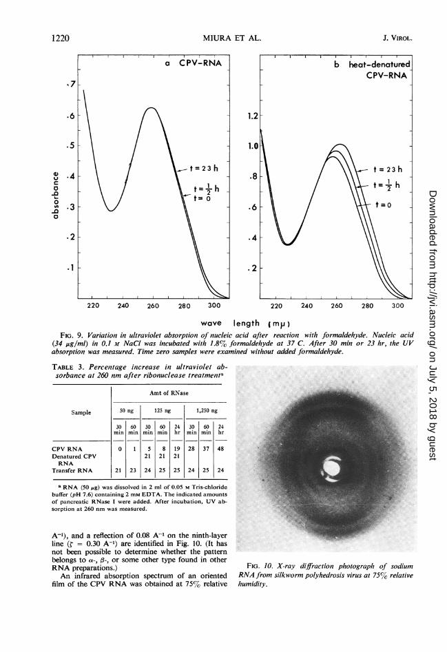
Nonreactability with fjrmnaldehyde. Formalde-hyde is believed to react with free amino groupsin the bases of a nucleic acid molecule and to pro-duce an increase in UV absorption and a shift ofthe wavelength of the absorption maximum (9,42). Changes induced in UV absorption of theRNA by 1.8%f formaldehyde at 37 C in 0.1 MNaCl are shown in Fig. 9. Whereas single-stranded nucleic acids exhibited a marked increase(23 to 26%/() and a shift to a longer wavelength intheir UV absorption (4 to 6 nm), CPV RNAshowed almost no increment and no shift in itsUV-absorption maximum; CPV RNA is thus likeother double-stranded RNA or DNA (1 1, 29, 40).When CPV RNA was denatured by heating at100 C for 10 min in 0.01 X SSC and then quicklycooled in ice, it reacted with formaldehyde in thesame manner as does transfer RNA or ribosomalRNA. It is possible that amino groups of thebases in the original CPV RNA are occupied,owing to base pairing as is the case for double-stranded DNA. After heat denaturation of theRNA, these amino groups would be exposed, al-lowing reaction with formaldehyde.
Resistance to RNase. In order to observe degra-dation of small amounts of RNA by RNase treat-ment the hyperchromic effect at 260 nm was meas-ured (Table 3). When a dilute pancreatic RNasesolution (61 ng/ml) was used, the increase in UVabsorption was slight for CPV RNA, as comparedwith transfer RNA. However, incteased RNasetreatment (long incubation wih 10 times theamount of RNase) induced almost complete hy-drolysis of the CPV RNA. The increase in ab-sorption at 260 nm rose to 48%/C, for CPV RNA,a larger increase than that for transfer RNA. IfCPV RNA has a regular structure, and if there is alarge structural gap between the original ordered
1218 J. VIROL.
on July 5, 2018 by guesthttp://jvi.asm
.org/D
ownloaded from
DOUBLE-STRANDED RNA FROM CPV
structure and the digest, it would be plausible toexpect a larger hyperchromic effect than those ob-served for the other types of RNA. Heat-dena-tured CPV RNA (heated at 100 C for 10 min in0.01 X SSC and quickly cooled in ice) showed ahyperchromicity for RNase treatment similar tothat of transfer RNA. These results seem to showthat natural CPV RNA is more resistant to diluteRNase than is either transfer RNA or heat-denatured CPV RNA.The above observations indicate that the RNA
of CPV is double-stranded. Various physical andchemical characteristics of CPV RNA are similarto those of a double-stranded DNA or to those ofother double-stranded RNA preparations (13,29), but are different from those of transfer RNAor of ribosomal RNA, which are single-strandedmolecules in which the intramolecular base pair-ings are considered incomplete (11). From the factof nonreactivity with formaldehyde, almost all theamino groups of the bases in CPV RNA wouldseem to participate in hydrogen bonding betweenthe two strands. Regular base pairings would oc-cur in the pairs of A-U and G-C, since both thebase ratios of A /U and G/C for CPV RNA wereunity.
CD
(W)260 mp
(4))260 mp.,950
OD
OD25smy ,t
OD2s5 mpA,4
1 .3k
1 .2k
1.1 _
1.0 _
1.4 h
1 .3
1 .2k
1.1
1.01
APPENDIXAdditional evidence indicating CPV RNA to be a
double-stranded helical molecule has been obtainedby X-ray analysis and absorption in the far infrared.This evidence is summarized below by Y. Murakami,K. Shuto, Y. Mitsui, Y. litaka, and M. Tsuboi, of theFaculty of Pharmaceutical Sciences, University ofTokyo, Bunkyo-ku, Tokyo, Japan. Their cooperationis gratefully acknowledged.
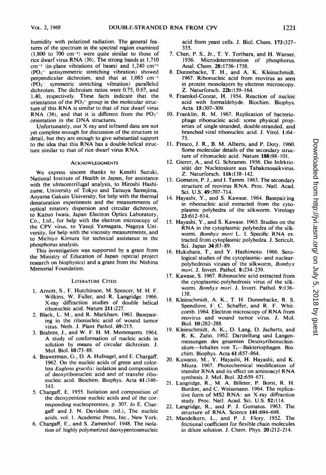
X-ray diffraction photographs of fibers of sodiumRNA of silkworm poylhedrosis virus were obtainedwith a microcamera and Ni-filtered CuKa radiation.The specimen-to-film distance was 29 mm, and flatfilms were used. The humidity of the atmospherearound the sample was controlled (at 75c%) by themethod previously described (36). The fibers so farobtained were much less crystalline than fibers of ricedwarf virus RNA. In the fiber diagram (Fig. 10), onlydiffuse reflections can be seen; no sharp spots are ob-served. However, all of these diffuse reflections aresituated where strong spots are observed in the fiberdiagrams of rice dwarf virus RNA (36) and otherdouble helical RNA molecules (1, 21, 22, 41, 43).For example, strong reflections ofRNA molecules witht values of 0.07 to 0.09 A-' and 0.17 to 0.18 A-' onthe equator, those ot 0.05 to 0.06 A-', 0.12 A-', and0.17 A-' on the second-layer line (t = 0.07 A-'), thoseof 0.07 to 0.13 A-' on the seventh-layer line (¢ = 0.23
ORD1.2
(0()282 my ,t1.1
1.0282 mp95
30 40 50 60 70 80 90 100
temperature ( C )FIG. 8. Variation in ultraviolet absorption at 260 nm, the peak of the Cotton effect (282 rnm), and circular
dichroic band at d 260 nm of CPV RNA as a function of temperature in 0.01 X SSC. The concentration ofRNA was 40 Ag/ml.
VOL. 2, 1968 1219
CD260
r-
on July 5, 2018 by guesthttp://jvi.asm
.org/D
ownloaded from
MIURA ET AL.
220 240 260 280 300 220 240 260 280 300
wave length (mp)
FIG. 9. Variation in ultraviolet absorption of nucleic acid after reaction with formaldehyde. Nucleic acid(34 ,ug/ml) in 0.1 M NaCI was incubated with 1.8% formaldehyde at 37 C. After 30 min or 23 hr, the UVabsorption was measured. Time zero samples were examined without added formaldehyde.
TABLE 3. Percentage increase in ultraviolet ab-sorbance at 260 nm after ribonuclease treatmenta
Amt of RNase
Sample 50 ng 125 ng 1,250 ng
30 60 30 60 24 30 60 24min min min min hr min min hr
CPV RNA 0 1 5 8 19 28 37 48Denatured CPV 21 21 21RNA
Transfer RNA 21 23 24 25 25 24 25 24
a RNA (50 MAg) was dissolved in 2 ml of 0.05 M Tris-chloridebuffer (pH 7.6) containing 2 mm EDTA. The indicated amountsof pancreatic RNase I were added. After incubation, UV ab-sorption at 260 nm was measured.
A-'), and a reflection of 0.08 A-' on the ninth-layerline (¢ = 0.30 A-') are identified in Fig. 10. (It hasnot been possible to determine whether the patternbelongs to a-, ,3-, or some other type found in otherRNA preparations.)An infrared absorption spectrum of an oriented
film of the CPV RNA was obtained at 7570 relative
FIG. 10. X-ray diffraction photograph of sodiumRNA from silkworm polyhedrosis virus at 75% relativehumidity.
1220
.6
.5
.4
.3
4)uc0
-0-o-0
*2
.1
J. VIROL.
on July 5, 2018 by guesthttp://jvi.asm
.org/D
ownloaded from
DOUBLE-STRANDED RNA FROM CPV
humidity with polarized radiation. The general fea-tures of the spectrum in the spectral region examined(1,800 to 700 cm-') were quite similar to those ofrice dwarf virus RNA (36). The strong bands at 1,710cm-' (in-plane vibrations of bases) and 1,240 cm-'(POr- antisymmetric stretching vibration) showedperpendicular dichroism, and that at 1,085 cm-'(PO2- symmetric stretching vibration) paralleleddichroism. The dichroism ratios were 0.75, 0.67, and1.40, respectively. These facts indicate that theorientation of the PO2- group in the molecular struc-ture of this RNA is similar to that of rice dwarf virusRNA (36), and that it is different from the PO,-orientation in the DNA structures.
Unfortunately, our X-ray and infrared data are notyet complete enough for discussion of the structure indetail, but they are enough to give substantial supportto the idea that this RNA has a double-helical struc-ture similar to that of rice dwarf virus RNA.
ACKNOWLEDGMENTS
We express sincere thanks to Kenshi Suzuki,National Institute of Health in Japan, for assistancewith the ultracentrifugal analysis, to Hiroshi Hashi-zume, University of Tokyo and Tatsuya Samejima,Aoyama Gakuin University, for help with the thermaldenaturation experiments and the measurements ofoptical rotatory dispersion and circular dichroism,to Kazuo Iwata, Japan Electron Optics Laboratory,Co., Ltd., for help with the electron microscopy ofthe CPV virus, to Yasuji Yamagata, Nagoya Uni-versity, for help with the viscosity measurements, andto Michiyo Kimura for technical assistance in thephosphorus analysis.
This investigation was supported by a grant fromthe Ministry of Education of Japan (special projectresearch on biophysics) and a grant from the NishinaMemorial Foundation.
LITERATURE CITED
1. Arnott, S., F. Hutchinson, M. Spencer, M. H. F.Wilkins, W. Fuller, and R. Langridge. 1966.X-ray diffraction studies of double helicalribonucleic acid. Nature 211:227.
2. Black, L. M., and R. Markham. 1963. Basepair-ing in the ribonucleic acid of wound tumorvirus. Neth. J. Plant Pathol. 69:215.
3. Brahms, J., and W. F. H. M. Mommaerts. 1964.A study of conformation of nucleic acids insolution by means of circular dichroism. J.Mol. Biol. 10:73-88.
4. Brawerman, G., D. A. Hufnagel, and E. Chargaff.1962. On the nucleic acids of green and color-less Euiglenia gracilis: isolation and compositionof deoxyribonucleic acid and of transfer ribo-nucleic acid. Biochim. Biophys. Acta 61:340-345.
5. Chargaff, E. 1955. Isolation and composition ofthe deoxypentose nucleic acids and of the cor-responding nucleoproteins, p. 307. In E. Char-gaff and J. N. Davidson (ed.), The nucleicacids. vol. 1. Academic Press, Inc., New York.
6. Chargaff, E., and S. Zamenhof. 1948. The isola-tion of highly polymerized deoxypentosenucleic
acid from yeast cells. J. Biol. Chem. 173:327-335.
7. Chen, P. S., Jr., T. Y. Toribara, and H. Warner.1956. Microdetermination of phosphorus.Anal. Chem. 28:1756-1758.
8. Dunnebacke, T. H., and A. K. Kleinschmidt.1967. Ribonucleic acid from reovirus as seenin protein monolayers by electron microscopy.Z. Naturforsch. 22b :159-164.
9. Fraenkel-Conrat, H. 1954. Reaction of nucleicacid with formaldehyde. Biochim. Biophys.Acta 15:307-309.
10. Franklin, R. M. 1967. Replication of bacterio-phage ribonucleic acid: some physical prop-erties of single-stranded, double-stranded, andbranched viral ribonucleic acid. J. Virol. 1:64-75.
11. Fresco, J. R., B. M. Alberts, and P. Doty. 1960.Some molecular details of the secondary struc-ture of ribonucleic acid. Nature 188:98-101.
12. Gierer, A., and G. Schramm. 1956. Die Infektio-sitat der Nucleinsaure aus Tabakmosaikvirus.Z. Naturforsch. llb:138-142.
13. Gomatos, P. J., and I. Tamm. 1963. The secondarystructure of reovirus RNA. Proc. Natl. Acad.Sci. U.S. 49:707-714.
14. Hayashi, Y., and S. Kawase. 1964. Basepairingin ribonucleic acid extracted from the cyto-plasmic polyhedra of the silkworm. Virology23:612-614.
15. Hayashi, Y., and S. Kawase. 1965. Studies on theRNA in the cytoplasmic polyhedra of the silk-worm. Bombyx mori L. I. Specific RNA ex-tracted from cytoplasmic polyhedra. J. Sericult.Sci. Japan 34:83-89.
16. Hukuhara, T., and Y. Hashimoto. 1966. Sero-logical studies of the cytoplasmic- and nuclear-polyhedrosis viruses of the silkworm, Bombyxmori. J. Invert. Pathol. 8:234-239.
17. Kawase, S. 1967. Ribonucleic acid extracted fromthe cytoplasmic-polyhedrosis virus of the silk-worm. Bombyx mori. J. Invert. Pathol. 9:136-138.
18. Kleinschmidt, A. K., T. H. Dunnebacke, R. S.Spendlove, F. C. Schaffer, and R. F. Whit-comb. 1964. Electron microscopy of RNA fromreovirus and wound tumor virus. J. Mol.Biol. 10:282-288.
19. Kleinschmidt, A. K., D. Lang, D. Jacherts, andR. K. Zahn. 1962. Darstellung und Langen-messungen des gesamten Desoxyribonuclein-saure-Inhaltes von T2 Bakteriophagen. Bio-chim. Biophys. Acta 61:857-864.
20. Kuwano, M., Y. Hayashi, H. Hayashi, and K.Miura. 1967. Photochemical modification oftransfer RNA and its effect on aminoacyl RNAsynthesis. J. Mol. Biol. 32:659-671.
21. Langridge, R., M. A. Billeter, P. Borst, R. H.Burdon, and C. Weissmann. 1964. The replica-tive form of MS2 RNA: an X-ray diffractionstudy. Proc. Natl. Acad. Sci. U.S. 52:114.
22. Langridge, R., and P. J. Gomatos. 1963. Thestructure of RNA. Science 141:694-698.
23. Mandelkern, L., and P. J. Flory. 1952. Thefrictional coefficient for flexible chain moleculesin dilute solution. J. Chem. Phys. 20:212-214.
VOL. 2, 1968 1221
on July 5, 2018 by guesthttp://jvi.asm
.org/D
ownloaded from
MIURA ET AL.
24. Markham, R., and J. D. Smith. 1951. Chromato-graphic studies of nucleic acids. IV. The nucleicacid of the turnip yellow mosaic virus, includinga note on the nucleic acid of the tomato bushystunt virus. Biochem. J. 49:401-406.
25. Marmur, J., and P. Doty. 1959. Heterogeneity indeoxyribonucleic acid. I. Dependence oncomposition of the configurational stability ofdeoxyribonucleic acids. Nature 183:1427-1429.
26. Marmur, J., and P. Doty. 1962. Determination ofthe base composition of deoxyribonucleic acidfrom its thermal denaturation temperature. J.Mol. Biol. 5:109-118.
27. Mejbaum, W. 1939. Uber die Bestimmungkleiner Pentosemengen, insbesondere in Deri-vaten der Adenylsaure. Z. Physiol. Chem.258:117-120.
28. Miura, K. 1962. The nucleotide composition ofribonucleic acids of soluble and particle frac-tions in several species of bacteria. Biochim.Biophys. Acta 55:62-70.
29. Miura, K., I. Kimura, and N. Suzuki. 1966.Double-stranded ribonucleic acid from ricedwarf virus. Virology 28:571-579.
30. Miura, K., T. Kitamura, and Y. Kawade. 1958.Fractionation of ribonucleic acid by precipita-tion with neutral salts. Biochim. Biophys.Acta 27:420-421.
31. Miura, K., and K. Matsuzaki. 1964. Nucleotidecomposition and arrangement of soluble RNAin posterior silkgland. Biochim. Biophys.Acta 91:427-432.
32. Miura, K., T. Miura, C. Hiruki, Z. Hidaka, and1. Watanabe. 1963. Fractionation of infectiousribonucleic acid isolated from tobacco mosaicvirus. Virology 19:140-146.
33. Saito, H., and K. Miura. 1963. Preparation oftransforming deoxyribonucleic acid by phenoltreatment. Biochim. Biophys. Acta 72:619-629.
34. Samejima, T., H. Hashizume, K. Imahori, I.Fujii, and K. Miura. 1968. Optical rotatory
dispersion and circular dichroism of rice dwarfvirus ribonucleic acid. J.'Mol. Biol. 34:39-48.
35. Samejima, T., and J. T. Yang. 1965. Opticalrotatory dispersion and conformation of deoxy-ribonucleic and ribonucleic acids from varioussources. J. Biol. Chem. 240:2094-2100.
36. Sato, T., Y. Kyogoku, S. Higuchi, Y. Mitsui,Y. litaka, M. ITsuboi, and K. Miura. 1966. Apreliminary investigation on the molecularstructure of rice dwarf virus RNA. J. Mol.Biol. 16:180-190.
37. Scheraga, H. A., and L. Mandelkern. 1953. Con-sideration of the hydrodynamic properties ofproteins. J. Am. Chem. Soc. 75:179-184.
38. Shack, J. 1958. On the recognition and estima-tion of denatured deoxyribonucleate. J. Biol.Chem. 233:677-680.
39. Shatkin, A. J., and B. Rada. 1967. Reovirus-directed ribonucleic acid synthesis in infectedL cells. J. Virol. 1:24-35.
40. Sinsheimer, R. L. 1959. A single-stranded deoxy-ribonucleic acid from bacteriophage 4DX174.J. Mol. Biol. 1:43-53.
41. Spencer, M., W. Fuller, M. H. F. Wilkins, andG. L. Brown. 1962. Determination of the helicalconfiguration of ribonucleic acid molecules byX-ray diffraction study of crystalline amino-acid-transfer ribonucleic acid. Nature 194:1014.
42. Staehelin, M. 1958. Reaction of tobacco mosaicvirus nucleic acid with formaldehyde. Biochim.Biophys. Acta 29:410-417.
43. Tomita, K., and A. Rich. 1964. X-ray diffractioninvestigations of complementary RNA. Nature201:1160-1163.
44. Vinograd, J., R. Bruner, R. Kent, and J. Weigle.1963. Band-centrifugation of macromoleculesand viruses in self-generating density gradients.Proc. Natl. Acad. Sci. U.S. 49:902-910.
45. Volkin, E., and W. E. Cohn. 1954. Estimation ofnucleic acids. Methods Biochem. Analy. 1:287-305.
1222 J. VIROL.
on July 5, 2018 by guesthttp://jvi.asm
.org/D
ownloaded from