Doppler in gyneacology Dr. Muhammad Bin Zulfiqar
54
Doppler Imaging in Gynecology An Introduction DR. Muhammad Bin Zulfiqar PGR IV New Radiology Department SHL/SIMS [email protected]
-
Upload
dr-muhammad-bin-zulfiqar -
Category
Health & Medicine
-
view
7.738 -
download
0
Transcript of Doppler in gyneacology Dr. Muhammad Bin Zulfiqar

Doppler Imaging in Gynecology An IntroductionDR. Muhammad Bin Zulfiqar
PGR IV New Radiology DepartmentSHL/SIMS

AIMS• UTILITY IN DIFFERENT PELVIC PATHOLOGIES• ROLE IN INFERTILITY• ROLE IN FERTILIZATION

Introduction
• Physiological Changes– Ovarian follicle– Follicular Rupture– Corpus Luteum– Changes in endometrium with respect to
Menstrual cycle• Pathological Changes
Alcazar, et al. Intratumoral blood flow in cervical cancer as assessed by transvaginal CDS: Correlation with tumor characteristics. Intern J of Gynec Cancer. Jul 2003 Vol 13 (4).8. Suren A, et al. 3D Color Power Angio imaging. Ultrasound Obs Gynecol. 1998 Feb; 11(2):133.

Techniques
• Abdominal Ultrasound • Transvaginal Ultrasound (more important)– Both these techniques are collaborated with
Doppler

Color Doppler in Cervical Cancer
• Controversial role in cervical cancer– Neovascularity in the mass (not definitive). – Response to chemotherapy via transvaginal color Doppler. – Combined clinical and radiological staging can identify the
patients needing • post surgical radiotherapy and/or chemotherapy and • help in the treatment planning versus clinical exam alone.
• Local recurrences following treatment are well assessed radiologically by ultrasound. In cases of doubt, MRI may be performed.
Alcazar, et al. Intratumoral blood flow in cervical cancer as assessed by transvaginal CDS: Correlation with tumor characteristics. Intern J of Gynec Cancer. Jul 2003 Vol 13 (4).8. Suren A, et al. 3D Color Power Angio imaging. Ultrasound Obs Gynecol. 1998 Feb; 11(2):133.

• Coronal color Doppler view of the cervix. • Coronal scan demonstrating areas of
vascularity within the hypoechoic mass. No obvious parametrial invasion was noted.

• Spectral color Doppler study. • A mixed arterial and venous waveform is
demonstrated through the region of neovascularity in the mass.

Neovascularization
• A study based on 'Folkman's theory of neovascularisation' , was done according to which malignant neoplasms elaborate a factor named Tumor Angiogenesis factor (TAF), which stimulates rapid formation of new capillaries.
Dastur Adi E. Ultrasound and Doppler in Gynecological Cancers. J Obstet Gynecol India Vol. 60, No. 1 : January/February 2010 pg 21-22

Neovascularization
• The study aimed at evaluating the efficacy of Color and Spectral Doppler in diagnosing the ovarian malignancy.
• In all, 121 patients with adnexal masses were examined over a period of 2 years, out of which 60 patients with neoplastic ovarian tumors were retained as the study subject.
• Color Doppler showed blood flow in 92.59 percent of malignant tumors in contrast to only 42.24 per cent of benign tumors.
Dastur Adi E. Ultrasound and Doppler in Gynecological Cancers. J Obstet Gynecol India Vol. 60, No. 1 : January/February 2010 pg 21-22

Neovascularization
• Absent blood flow in a solid tumor almost always ruled out the possibility of malignancy. Spectral Doppler helped to assess the nature of the blood vessels picked up on Color Doppler.
Dastur Adi E. Ultrasound and Doppler in Gynecological Cancers. J Obstet Gynecol India Vol. 60, No. 1 : January/February 2010 pg 21-22

Neovascularization
• In the present study. 96.29 per cent of malignant tumors had PI less than 0.8 in contrast to only 6.06 per cent of benign tumors.
• Similarly, 92.59 percent of malignant tumors showed RI less than 0.6 in contrast to only 9.09 per cent of benign tumors.
• Thus, Color Doppler and Spectral Doppler tremendously increased the reliability in diagnosing a malignant ovarian tumor.
Dastur Adi E. Ultrasound and Doppler in Gynecological Cancers. J Obstet Gynecol India Vol. 60, No. 1 : January/February 2010 pg 21-22

Neovascularization
• Color Doppler served as an important tool to rule out malignancy in solid tumors if they failed to show any intratumoral vascularity.
• B-Mode USG in combination with Color Doppler and Spectral Doppler is proposed as the first and foremost diagnostic modality in patients with ovarian tumor
• To establish the definite diagnosis of malignancy early in the course of the disease.
Dastur Adi E. Ultrasound and Doppler in Gynecological Cancers. J Obstet Gynecol India Vol. 60, No. 1 : January/February 2010 pg 21-22
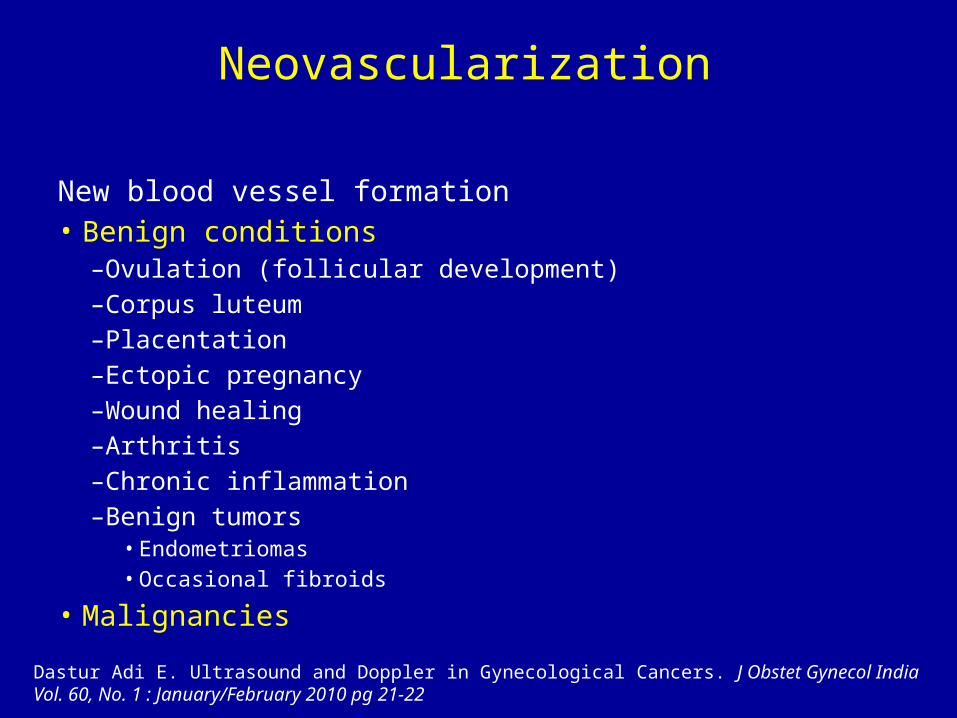
Neovascularization
New blood vessel formation • Benign conditions
–Ovulation (follicular development)–Corpus luteum–Placentation–Ectopic pregnancy–Wound healing–Arthritis–Chronic inflammation–Benign tumors
• Endometriomas• Occasional fibroids
• Malignancies
Dastur Adi E. Ultrasound and Doppler in Gynecological Cancers. J Obstet Gynecol India Vol. 60, No. 1 : January/February 2010 pg 21-22

Resistance Patterns in Ovarian Masses
• High resistance (RI <1 >0.6) –Cystadenomas, hemorrhagic cysts,
dermoid tumors, endometrioma• Intermediate resistance (RI <0.6 >0.4) –Dermoid tumors, endometriomas• Low resistance (RI <0.4 >0) –Ovarian cancer, inflammatory masses,
endometriomas, dermoids, corpus luteumDastur Adi E. Ultrasound and Doppler in Gynecological Cancers. J Obstet Gynecol India Vol. 60, No. 1 : January/February 2010 pg 21-22

Neoplastic Risk in Adnexal Masses Risk is age and state dependent
Dastur Adi E. Ultrasound and Doppler in Gynecological Cancers. J Obstet Gynecol India Vol. 60, No. 1 : January/February 2010 pg 21-22

Ovarian Masses• The ultrasound features of a malignant growth have been
described as – bilaterality, – large size (> 5 cm), – multiple locules, – papillary excrescences or solid areas, – presence of ascites or metastasis.
• The addition of Doppler and serum marker such as CA-125 has been used as a multimodality screening strategy.
• The sensitivity (89.5%) and specificity (99.8%) of the multimodality approach were superior to the other groups in detecting early ovarian cancer.Dastur Adi E. Ultrasound and Doppler in Gynecological Cancers. J Obstet Gynecol India Vol. 60, No. 1 : January/February 2010 pg 21-22

Color Doppler
• Doppler has been studied in its role in detecting – vascular flow patterns and – characterizing the resistance to blood flow in ovarian tumors.
• Malignant growths are characterized by neoangiogenesis and blood vessels with a poorly developed muscularis. The blood flow in these vessels is marked by low impedance and correspondingly, the resistance index is low (RI < 0.3)
• In contrast, benign ovarian tumors and normal ovarian blood flow is characterized by a high RIDastur Adi E. Ultrasound and Doppler in Gynecological Cancers. J Obstet Gynecol India Vol. 60, No. 1 : January/February 2010 pg 21-22

Benign / Malignant Pelvic Masses
• In experienced hands, subjective evaluation of the gray-scale ultrasound image is the best ultrasound method for discriminating between benign and malignant adnexal masses.
• The main advantage of adding Doppler examination to subjective evaluation of the gray-scale image is an increase in the confidence with which a correct diagnosis is made.
Valentin L. Prospective cross-validation of Doppler ultrasound examination and gray-scale ultrasound imaging for discrimination of benign and malignant pelvic masses. Ultrasound Obstet Gynecol. 1999 Oct;14(4):273-83.

Endometrial Cancers
• Detectable flow is unusual in–Normal endometrium–Atrophic endometrium–Most endometrial hyperplasias
• Flow is usually detectable (91%) of endometrial Ca
• Flow pattern shows low resistance (average RI ~ 0.42)

Endometrial Cancers
• Postmenopausal bleeding is the prime indicator for the risk of endometrial cancer and transvaginal ultrasound is the first step in the triage for these women.
• Metanalysis have shown that when the endometrial thickness is less than 5 mm, the risk of
• endometrial malignancy is about 1 in 1000. In such cases, endometrial sampling and histopathology can be avoided. Dastur Adi E. Ultrasound and Doppler in Gynecological Cancers. J Obstet Gynecol India Vol. 60, No. 1 : January/February 2010 pg 21-22

Endometrial Cancers
• Ultrasound markers of malignancy are• the disturbance of the interface between the
endometrium and myometrium • presence of irregular, vascular mass lesions
inside endometrial cavity.

Endometrial Cancers
• Color Doppler is useful as an adjunct in diagnosing endometrial cancer. The subendometrial blood flow and the blood flow in thickened and polypoidal endometrium shows low resistance patterns in endometrial malignancy and the RI is usually less than 0.3.
• Saline infusion sonography is useful in detecting polyps and intracavitary masses. Preoperative assessement with ultrasound results matches the result of a CT or MRI assessment.Dastur Adi E. Ultrasound and Doppler in Gynecological Cancers. J Obstet Gynecol India Vol. 60, No. 1 : January/February 2010 pg 21-22

Luteal Cyst• Detected during the secretory phase (D
15-28) of the menstrual cycle• Size : 2-7 cm• Polymorphism :
– Heterogeneous content with fibrin septa: « fish net »
– Clot simulating vegetation– Pseudo-solid cyst
Prof Soha Talaat

Luteal Cyst• Color Doppler :– Non vascular septa– Vascularized thick wall– May be misdiagnosed as a cystadenocarcinoma US Follow-up 2 months later (1 month is too early !!!)
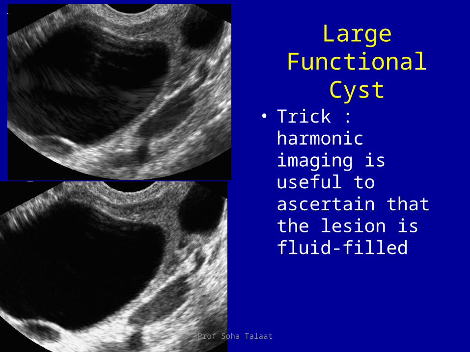
LargeFunctional
Cyst• Trick : harmonic
imaging is useful to ascertain that the lesion is fluid-filled
Prof Soha Talaat

Color Doppler?• Color Döppler is not
accurate :– In 30 % of functional
ovarian cyst walls, arteries are detected
– Presenting with a low resistive index
• Do not take it for malignancy !!!
Prof Soha Talaat
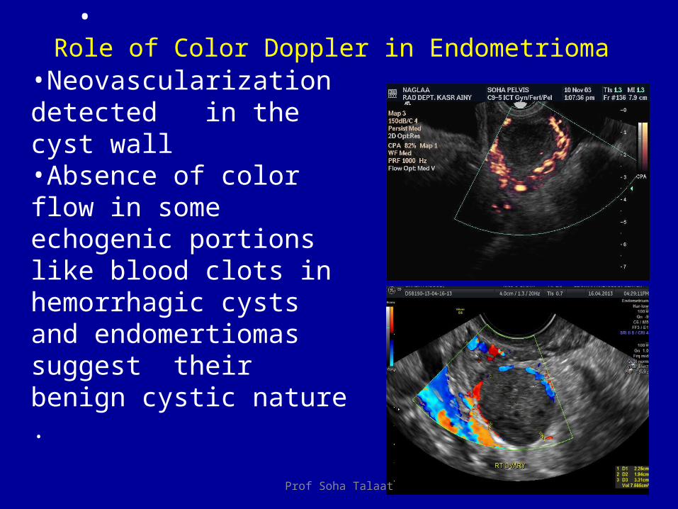
•
•Neovascularization detected in the cyst wall •Absence of color flow in some echogenic portions like blood clots in hemorrhagic cysts and endomertiomas suggest their benign cystic nature .
Role of Color Doppler in Endometrioma
Prof Soha Talaat

Benign diffuse enlargement Torsion( edema)
• Ovarian torsion (adnexal torsion) is an infrequent but significant cause of acute lower abdominal pain in women.
• This condition is usually associated with reduced venous return from the ovary as a result of stromal edema, internal hemorrhage, hyperstimulation, or a mass.
Prof Soha Talaat
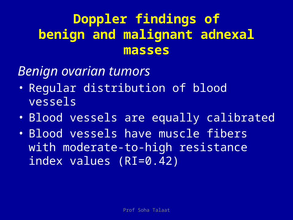
Doppler findings ofbenign and malignant adnexal masses
Benign ovarian tumors• Regular distribution of blood vessels• Blood vessels are equally calibrated• Blood vessels have muscle fibers with moderate-to-
high resistance index values (RI=0.42)
Prof Soha Talaat

Doppler findings of benign and malignant adnexal masses
Malignant ovarian tumors• Irregular distribution of blood vessels• Blood vessels have irregular diameter• Low resistance index values (RI<0.42)• Display of tumoral lakes and arterio-venous
shunts

Ultrasound
• An enlarged ovary (>5 cm)• Prominent peripheral nonovulatory follicles .• Small amount of free fluid• May depict the cyst (or, less commonly, the mass) that
predisposed the ovary to torsion.Prof Soha Talaat

Doppler
• Imaging modality of choice • An absence of arterial waveforms or high resistance to
arterial flow with absent venous flow are highly suggestive. • Particularly when those findings are accompanied by ovarian
enlargement. • However normal arterial waveforms do not rule out torsion.
Prof Soha Talaat

Acute PID
• Early on, there may be no US signs• Free fluid in cul-de-sac +/-behind adnexae• Fluid in endometrial cavity• Fluid in lumen of fallopian tubes• No typical changes in doppler patterns or
indices• Follicles (infected), with fuzzy margins

Tubovarian Abscess
• Complex, hypoechogenic, septated mass• Acoustic enhancement• Absent color doppler blood flow in mass• Ill-defined margins• Fluid in cul-de-sac• Loss of anatomical landmarks as disease
becomes chronic
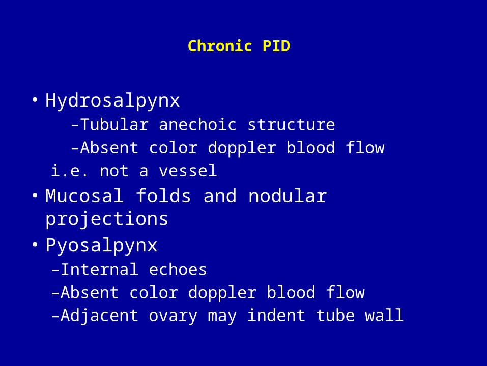
Chronic PID
• Hydrosalpynx –Tubular anechoic structure –Absent color doppler blood flowi.e. not a vessel
• Mucosal folds and nodular projections• Pyosalpynx
–Internal echoes–Absent color doppler blood flow–Adjacent ovary may indent tube wall

Ectopic Pregnancy
➤ Exclusion of an intrauterine chorion-type structure ➤ Presence of an extrauterine and extraovarian chorion-type structure ➤ Demonstrable embryonic heart activity and movements within the structure (5% of cases) ➤ Enlarged uterus with a thickened endometrium of high echogenicity ➤ Free fluid in the cul-de-sac and paracolic gutters with clot formation and fibrin strands (hemoperitoneum)

Role in Ectopic Pregnancy• On color Doppler "ring of fire” is visualized, owing to the low-
impedance high diastolic flow seen in pregnancy that can surround the tubal ring of an ectopic pregnancy.
• However, a hypervascular ring (Figs 13, 15) around a mass in the pelvis is more likely to be visualized around the corpus luteum than an ectopic pregnancy. This is because both corpus luteum cysts and ectopic pregnancies can be very vascular with low-impedance flow; however, corpus luteum cysts are much more common than ectopic pregnancies. Color Doppler imaging is most helpful when an ectopic pregnancy is not seen but is highly suspected. In that case color Doppler imaging can be used to help find a mass surrounded by bowel loops.Deborah Levine,MD. Ectopic Pregnancy. Radiology: Volume 245: Number 2—November 2007

Role in Infertility• Uterine flow velocity has a resistance index of 0.88 +/- 0.04 (2 SE) in the
proliferative phase and starts to decrease the day before ovulation. • A nadir of 0.84 +/- 0.04 is reached on day 18 and remains at that level
for the rest of the cycle. • In anovulatory cycles, these changes do not occur. A subgroup of 12
women who lacked end diastolic flow in the uterine arteries during the secretory phase were identified. Eleven of these women were infertile, 8 of whom with primary infertility.
• Ovarian artery flow velocity is usually detected when the dominant follicle reaches 12 to 15 mm. The resistance index is 0.54 +/- 0.04 and also declines on the day before ovulation.
• A nadir of 0.44 +/- 0.04 is reached 4 to 5 days later and slowly rises to 0.050 +/- 0.04 before menstruation.
Kurjak A, Kupesic-Urek S, Schulman H, Zalud. Transvaginal color flow Doppler in the assessment of ovarian and uterine blood flow in infertile women. Fertil Steril. 1991 Nov;56(5):870-3.

Role in Infertility
• There are changes in the flow velocity patterns of the uterine and ovarian arteries during the normal ovulatory menstrual cycle. Because these changes in flow velocity begin before ovulation, it can be suspected that they may involve angiogenesis as well as hormonal factors. The changes noted in these studies are statistically significant but may be too small to be used as a diagnostic tool in the study of infertility problems.
Kurjak A, Kupesic-Urek S, Schulman H, Zalud. Transvaginal color flow Doppler in the assessment of ovarian and uterine blood flow in infertile women. Fertil Steril. 1991 Nov;56(5):870-3.

Role in Fertilization
• Steer et al.23 calculated the likelihood of pregnancy on the day of embryo transfer based on pulsatility values in the uterine arteries.
• According to their findings, the likelihood of pregnancy was greatest when medium PI values were measured in the uterine artery. In 35% of cases, pregnancy did not occur when the mean PI value before the embryo transfer was greater than 3.0.
Steer CV, Mills CV, Campbell S: Vaginal color Doppler assessment on the day of embryo transfer (ET) accurately predicts patients in an invitro fertilization programme with suboptimal uterine perfusion who fail to become pregnant. Ultrasound Obstet. Gynecol. 1 (1991) 79–82

Role in Assisted Fertilization• Tsai et al. investigated the prognostic value of uterine perfusion
on the day of human chorionic gonadotropin (hCG) administration in patients who were undergoing intrauterine insemination.
• They calculated the PI of the ascending branch of the uterine artery on the day of hCG administration and compared the vascular resistance in the uterine artery with the outcome of intrauterine insemination.
• No pregnancy occurred when the PI was greater than 3. The fertilization rate was 18% when the PI was less than 2 and 19.8% when the PI was between 2 and 3.
• These data indicate that the measurement of uterine perfusion on the day of hCG administration may be of value in predicting the success of intrauterine insemination.
Tsai YC, Chang JC, Tai MJ, Kung FT, Yang LC, Chang SY: Relationship of uterine perfusion to outcome of intrauterine insemination. J. Ultrasound Med. 15 (1996) 633–636
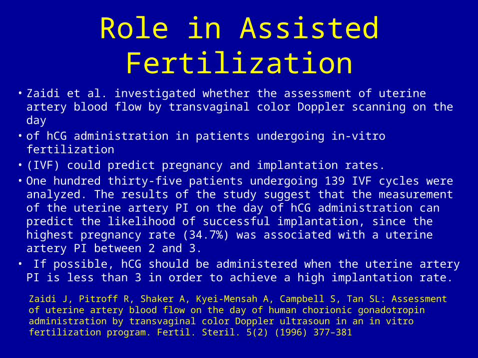
Role in Assisted Fertilization• Zaidi et al. investigated whether the assessment of uterine artery
blood flow by transvaginal color Doppler scanning on the day• of hCG administration in patients undergoing in-vitro fertilization• (IVF) could predict pregnancy and implantation rates. • One hundred thirty-five patients undergoing 139 IVF cycles were
analyzed. The results of the study suggest that the measurement of the uterine artery PI on the day of hCG administration can predict the likelihood of successful implantation, since the highest pregnancy rate (34.7%) was associated with a uterine artery PI between 2 and 3.
• If possible, hCG should be administered when the uterine artery PI is less than 3 in order to achieve a high implantation rate.
Zaidi J, Pitroff R, Shaker A, Kyei-Mensah A, Campbell S, Tan SL: Assessment of uterine artery blood flow on the day of human chorionic gonadotropin administration by transvaginal color Doppler ultrasoun in an in vitro fertilization program. Fertil. Steril. 5(2) (1996) 377–381

Uterine Blood Flow in the Normal Cycle and during Ovarian Stimulation with Confirmed
Ovulation
• Kupesic and Kurjak measured blood flow velocities in the uterine, radial, and spiral arteries during the periovulatory period of normal cycles and in stimulated cycles with confirmed ovulation.
• In normal cycles, the uterine artery PI was 3.16 two days before ovulation and decreased to 2.22 on the day before ovulation.
Kupesic S, Kurjak A: Uterine and ovarian perfusion during the periovulator period assessed by transvaginal color Doppler. Fertil. Steril. 60 (1993) 439–443

Uterine Blood Flow in the Normal Cycle and during Ovarian Stimulation with Confirmed Ovulation
• This difference was not observed in stimulated cycles. The mean PI was 3.06 and remained unchanged during the periovulatory period.
• Distinct waveforms could be recorded from the endometrium and myometrium during this period. The PI in the radial and spiral arteries showed higher values in stimulated cycles than in normal cycles.
Kupesic S, Kurjak A: Uterine and ovarian perfusion during the periovulator period assessed by transvaginal color Doppler. Fertil. Steril. 60 (1993) 439–443
Conclusion
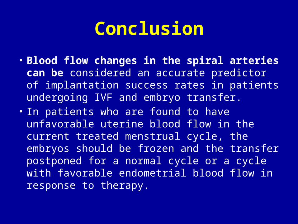
• Blood flow changes in the spiral arteries can be considered an accurate predictor of implantation success rates in patients undergoing IVF and embryo transfer.
• In patients who are found to have unfavorable uterine blood flow in the current treated menstrual cycle, the embryos should be frozen and the transfer postponed for a normal cycle or a cycle with favorable endometrial blood flow in response to therapy.

Doppler in Menopause—Ovarian Artery• Premenopausal– The RI is 0.54 shortly before ovulation, starts to
decline two days before ovulation, and reaches its low point of 0.44 at the time of ovulation.
– The mature corpus luteum normally has a diameter of 1–3cm and shows low impedance values (mean RI 0.43).
– On just the 23rd day after menstruation, the corpus luteum begins to undergo regressive changes.
– Decreased blood flow velocities and an increased RI (mean value 0.49) are the typical signals of these changes Zalud I, Kurjak A: The assessment of luteal blood flow in pregnant and non-pregnant
women by transvaginal color Doppler. J. Perinat. Med. 18 (1990) 215–221
Doppler in Menopause—Ovarian Artery
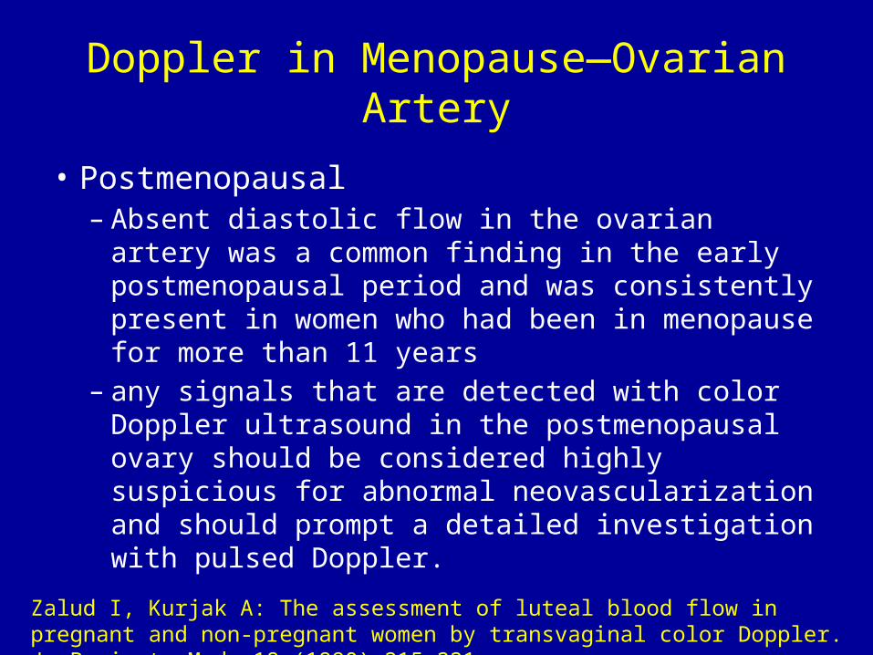
• Postmenopausal– Absent diastolic flow in the ovarian artery was a
common finding in the early postmenopausal period and was consistently present in women who had been in menopause for more than 11 years
– any signals that are detected with color Doppler ultrasound in the postmenopausal ovary should be considered highly suspicious for abnormal neovascularization and should prompt a detailed investigation with pulsed Doppler.
Zalud I, Kurjak A: The assessment of luteal blood flow in pregnant and non-pregnant women by transvaginal color Doppler. J. Perinat. Med. 18 (1990) 215–221
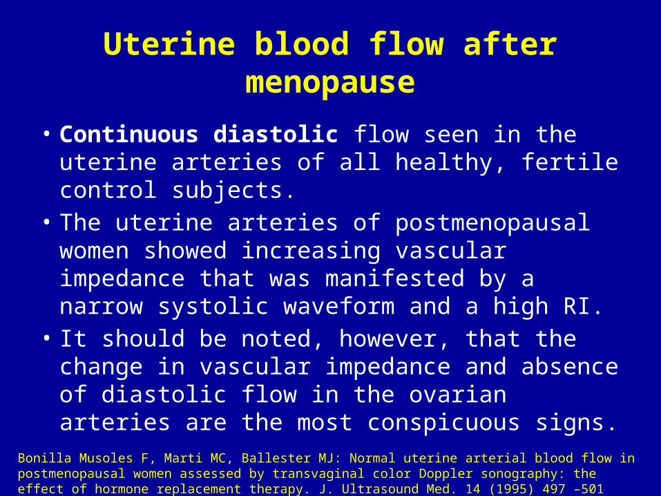
Uterine blood flow after menopause
• Continuous diastolic flow seen in the uterine arteries of all healthy, fertile control subjects.
• The uterine arteries of postmenopausal women showed increasing vascular impedance that was manifested by a narrow systolic waveform and a high RI.
• It should be noted, however, that the change in vascular impedance and absence of diastolic flow in the ovarian arteries are the most conspicuous signs.
Bonilla Musoles F, Marti MC, Ballester MJ: Normal uterine arterial blood flow in postmenopausal women assessed by transvaginal color Doppler sonography: the effect of hormone replacement therapy. J. Ultrasound Med. 14 (1995) 497 –501

Color Doppler Sonography for the Optimization ofAssisted Reproduction
• The implantation rate declines with increasing pulsatility index (Fig. 6.19).
• No pregnancies were achieved above a pulsatility index of 3.5 or above a resistance index of 0.95.
• A pulsatility index over 3.5 or a resistance index over 0.95 signified a nonreceptive endometrium with a specificity of 100% and a sensitivity of 14%. The positive predictive value for these cutoff limits is 100%.
Zaidi J, Campbell S, Pittrof R, Tan SL: Endometrial thickness, morphology vascular penetration and velocimetry in predicting implantation in an in vitro fertilization program. Ultrasound Obstet. Gynecol. 6 (1995) 191–198
B. Hüneke, A. Kleinkauf-Houcken, C. Lindner, and W. Braendle. Color Doppler Sonography in Gynecology and Obstetrics

Color Doppler Sonography for the Optimization ofAssisted Reproduction
B. Hüneke, A. Kleinkauf-Houcken, C. Lindner, and W. Braendle. Color Doppler Sonography in Gynecology and Obstetrics
Endometrial receptivity.
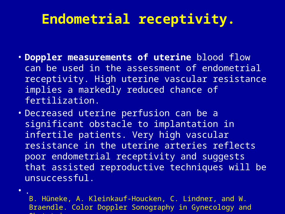
• Doppler measurements of uterine blood flow can be used in the assessment of endometrial receptivity. High uterine vascular resistance implies a markedly reduced chance of fertilization.
• Decreased uterine perfusion can be a significant obstacle to implantation in infertile patients. Very high vascular resistance in the uterine arteries reflects poor endometrial receptivity and suggests that assisted reproductive techniques will be unsuccessful.
• .
B. Hüneke, A. Kleinkauf-Houcken, C. Lindner, and W. Braendle. Color Doppler Sonography in Gynecology and Obstetrics

Endometrial receptivity.
• Although the sensitivity of uterine artery impedance measurements is only 14% owing to the numerous factors that affect receptivity, resistance values above the 90th percentile predict a nonreceptive endometrium with 100% specificity and a positive predictive value of 100%.
• With a severe decrease in uterine perfusion, the likelihood of implantation is so low that assisted reproduction therapies should be discontinued without appropriate preliminary treatment
B. Hüneke, A. Kleinkauf-Houcken, C. Lindner, and W. Braendle. Color Doppler Sonography in Gynecology and Obstetrics
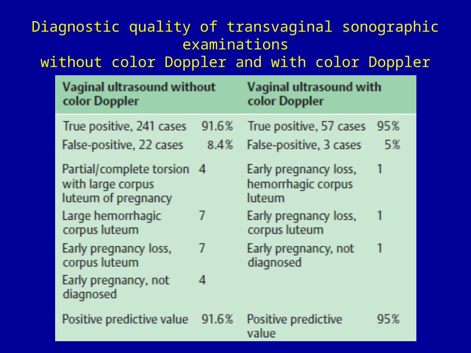
Diagnostic quality of transvaginal sonographic examinationswithout color Doppler and with color Doppler

THANK YOU