Donald School Journal of Ultrasound in Obstetrics and ...for ventriculomegaly. However, although the...
12
100 Fetal Cerebral Ventriculomegaly: Sonographic Diagnostic Workup Vincenzo D’Addario, Cristina A Rossi Fetal Medicine Unit Department of Obstetrics and Gynecology University Medical School, Bari, Italy Corresponding address: Prof. Vincenzo D’Addario, Fetal Medicine Unit, Dept. of Obstetrics and Gynecology, Ospedale Policlinico, Piazza G. Cesare, 70124 Bari, Italy, E-mail: [email protected] Donald School Journal of Ultrasound in Obstetrics and Gynecology, July-September 2008;2(3):100-111 Abstract: Dilatation of the fetal cerebral ventricles (ventriculomegaly) is a generic sonographic sign, which is common to several pathological entities carrying different prognosis. Ventriculomegaly is easily recognized by ultrasound by measuring the atrial width. This simple measure allows the recognition of even mild forms of ventricular dilatation and is used as a screening method for ventriculomegaly. However, although the diagnosis of ventriculomegaly is easy, the prenatal identification of the cause of ventricular dilatation is a more difficult task. The recognition of associated brain anomalies is a crucial point. The research of the cause of ventriculomegaly is clinically useful, since the prognosis mainly depends on the etiology and on the presence of associated abnormalities. In this article the role of prenatal sonography in recognizing the cause of the ventriculomegaly is reviewed. Keywords: Aqueductal stenosis, Chiari II malformation, corpus callosum agenesis, Dandy-Walker complex, hydrocephalus, prenatal diagnosis, ultrasound, ventriculomegaly Fetal cerebral ventriculomegaly is a sonographic sign common to different pathological entities. The incidence of congenital ventriculomegaly ranges from 0.3 to 1.5 in 1000 births in different series. 1 The term “hydrocephalus” is frequently used as synonymous; however the term “ventriculomegaly” should be preferred since it identifies the abnormal sonographic finding, independently from the etiology. The ventricular dilatation may be a consequence of the increased pressure of the cerebrospinal fluid (true hydrocephalus) due to obstruction of cerebrospinal flow (obstructive hydrocephalus) or, less frequently, to hypersecretion or defective filtration of cerebrospinal fluid. Ventriculomegaly may also be the consequence of an altered development of the intracranial architecture without increase of the fluid pressure: a typical example of this condition is the agenesis of the corpus callosum, where the posterior expansion of the lateral ventricles derives from the distorted array of the white matter tracts in the occipital lobes. 2 The main causes of ventricular dilatation in the fetus are aqueductal stenosis (33-43% of the cases), Chiari II malformation (25-30%), Dandy-Walker complex (7-10%), dysgenesis of the corpus callosum (20-30%). Other brain anomalies less frequently associated with ventriculomegaly are: occupying space lesions (tumors and cysts), haemorrhage, neuronal proliferation and differentiation disorders. Finally isolated mild ventriculomegaly may be found, without any clinical sequelae. Aqueductal stenosis is the most common cause of fetal ventriculomegaly. It can be caused by infections (toxoplasmosis, cytomegalovirus), bleeding or other pathologies that cause gliosis and subsequent obliteration of the aqueduct. Sometimes the aqueduct can be congenitally atresic or few rudimentary ependymal-lined tubules can be seen in place of it (aqueductal forking); in these cases a multifactorial pattern of inheritance is probably the cause, but in 5% of all cases an X-linked transmission is present (recurrence risk 50% of males). X-linked hydrocephalus is due to mutations of a gene on Xq28 that encodes for L1, a neural cell adhesion molecule. 3 Chiari II malformation is a complex abnormality consisting in various combinations of brainstem and cerebellar malformations, associated with ventriculomegaly and spinal defect. In Chiari II malformation the ventricular dilatation is the final result of a cascade of events on the developing brain, starting from the primitive spinal defect: the leakage of the cerebrospinal fluid through the defect causes a decompression and collapse of the ventricular cavities; failure of the rombencephalic cavity to distend causes the development of a small posterior fossa that is inadequate to house the developing cerebellum which herniates caudally into the foramen magnum, causing obstruction of the outlets of the fourth ventricle,
Transcript of Donald School Journal of Ultrasound in Obstetrics and ...for ventriculomegaly. However, although the...

Vincenzo D’Addario, Cristina A Rossi
100
Fetal Cerebral Ventriculomegaly: SonographicDiagnostic WorkupVincenzo D’Addario, Cristina A Rossi
Fetal Medicine UnitDepartment of Obstetrics and GynecologyUniversity Medical School, Bari, Italy
Corresponding address: Prof. Vincenzo D’Addario, Fetal Medicine Unit, Dept. of Obstetrics and Gynecology, OspedalePoliclinico, Piazza G. Cesare, 70124 Bari, Italy,E-mail: [email protected]
Donald School Journal of Ultrasound in Obstetrics and Gynecology, July-September 2008;2(3):100-111
Abstract: Dilatation of the fetal cerebral ventricles (ventriculomegaly)is a generic sonographic sign, which is common to several pathologicalentities carrying different prognosis.
Ventriculomegaly is easily recognized by ultrasound by measuringthe atrial width. This simple measure allows the recognition of evenmild forms of ventricular dilatation and is used as a screening methodfor ventriculomegaly. However, although the diagnosis ofventriculomegaly is easy, the prenatal identification of the cause ofventricular dilatation is a more difficult task. The recognition ofassociated brain anomalies is a crucial point. The research of the causeof ventriculomegaly is clinically useful, since the prognosis mainlydepends on the etiology and on the presence of associatedabnormalities.
In this article the role of prenatal sonography in recognizing thecause of the ventriculomegaly is reviewed.
Keywords: Aqueductal stenosis, Chiari II malformation, corpuscallosum agenesis, Dandy-Walker complex, hydrocephalus, prenataldiagnosis, ultrasound, ventriculomegaly
Fetal cerebral ventriculomegaly is a sonographic sign commonto different pathological entities. The incidence of congenitalventriculomegaly ranges from 0.3 to 1.5 in 1000 births indifferent series.1 The term “hydrocephalus” is frequently usedas synonymous; however the term “ventriculomegaly” shouldbe preferred since it identifies the abnormal sonographic finding,independently from the etiology.
The ventricular dilatation may be a consequence of theincreased pressure of the cerebrospinal fluid (true hydrocephalus)due to obstruction of cerebrospinal flow (obstructivehydrocephalus) or, less frequently, to hypersecretion or defectivefiltration of cerebrospinal fluid. Ventriculomegaly may also bethe consequence of an altered development of the intracranialarchitecture without increase of the fluid pressure: a typicalexample of this condition is the agenesis of the corpus callosum,where the posterior expansion of the lateral ventricles derives
from the distorted array of the white matter tracts in the occipitallobes.2
The main causes of ventricular dilatation in the fetus areaqueductal stenosis (33-43% of the cases), Chiari IImalformation (25-30%), Dandy-Walker complex (7-10%),dysgenesis of the corpus callosum (20-30%). Other brainanomalies less frequently associated with ventriculomegaly are:occupying space lesions (tumors and cysts), haemorrhage,neuronal proliferation and differentiation disorders. Finallyisolated mild ventriculomegaly may be found, without anyclinical sequelae.
Aqueductal stenosis is the most common cause of fetalventriculomegaly. It can be caused by infections (toxoplasmosis,cytomegalovirus), bleeding or other pathologies that causegliosis and subsequent obliteration of the aqueduct. Sometimesthe aqueduct can be congenitally atresic or few rudimentaryependymal-lined tubules can be seen in place of it (aqueductalforking); in these cases a multifactorial pattern of inheritanceis probably the cause, but in 5% of all cases an X-linkedtransmission is present (recurrence risk 50% of males). X-linkedhydrocephalus is due to mutations of a gene on Xq28 thatencodes for L1, a neural cell adhesion molecule.3
Chiari II malformation is a complex abnormality consistingin various combinations of brainstem and cerebellarmalformations, associated with ventriculomegaly and spinaldefect. In Chiari II malformation the ventricular dilatation isthe final result of a cascade of events on the developing brain,starting from the primitive spinal defect: the leakage of thecerebrospinal fluid through the defect causes a decompressionand collapse of the ventricular cavities; failure of therombencephalic cavity to distend causes the development of asmall posterior fossa that is inadequate to house the developingcerebellum which herniates caudally into the foramen magnum,causing obstruction of the outlets of the fourth ventricle,

Fetal Cerebral Ventriculomegaly
101
stretching and narrowing of the aqueduct, obliteration of thecisterna magna, obstruction at the level of dysplastic tentoriumand consequently ventriculomegaly.4 Due to its mechanism ofdevelopment, ventriculomegaly is present only in 70-80% offetuses affected by Chiari II malformation.
Dandy-Walker complex is a spectrum of posterior fossaabnormalities, the key feature of which is a variable severity ofdefect in the development of the cerebellar vermis (fromcomplete agenesis to mild hypoplasia).5 In the most severe formsthe fourth ventricle is extremely dilated and the membrane thatforms its roof balloons, creating a large posterior fossa cyst.This cyst pushes the tentorium and the venous sinus runningthrough it, causing obstruction of the cerebrospinal fluid andventriculomegaly.
Agenesis of the corpus callosum consists in a complete orpartial absence of the white fibres that cross the midline betweenthe two hemispheres. The etiology is heterogeneous, but geneticfactors are predominant. Over 90 genetic syndromes are knownthat may present partial or total agenesis of the corpus callosum.6This anomaly is almost constantly associated with ventriculardilatation of varying severity, but without increasedintraventricular pressure. The ventriculomegaly is theconsequence of an altered development of the intracranialarchitecture and derives from the distorted array of the whitematter tracts in the occipital lobes.
SONOGRAPHIC DIAGNOSIS OF FETAL CEREBRALVENTRICULOMEGALY
The fetal brain and particularly the lateral ventricles undergoextensive structural changes during gestation, thus offering avariety of sonographic findings. Although the description ofthe normal appearance of the lateral ventricles was alreadyreported in the 80’s mainly based on the axial scans of the fetalhead,7,8 the first systematic reports on the fetal brainmorphology, including ventricular system, evaluated on sagittaland coronal scans during both early and late pregnancy werepublished in 1991 by Timor-Tritsch’s group.9,10 They proposedto use the transvaginal approach to evaluate the fetal brainthrough the anterior fontanella or any open suture, thus obtaininga series of sagittal and coronal planes comparable with thosenormally used by the neonatologist on the newborns.
Recently 3D US has also been applied to the evaluation ofthe fetal brain and ventricles.11,12 The main advantage of thistechnique is the possibility to store a sample volume of thebrain and subsequently to reconstruct by means of themultiplanar imaging an infinite series of scans along the threeorthogonal spatial planes (axial, sagittal and coronal) (Fig. 1).
The prenatal detection of fetal ventriculomegaly relies onthe sonographic recognition and measurement of dilated lateralventricles. Several measurement techniques have been suggested
Fig. 1: Three-dimensional multiplanar view of a normal fetal brain, showing the normal lateral ventricle in thethree spatial planes (sagittal, coronal and axial) intersecting at the level of the occipital horn
Vincenzo D’Addario, Cristina A Rossi
102
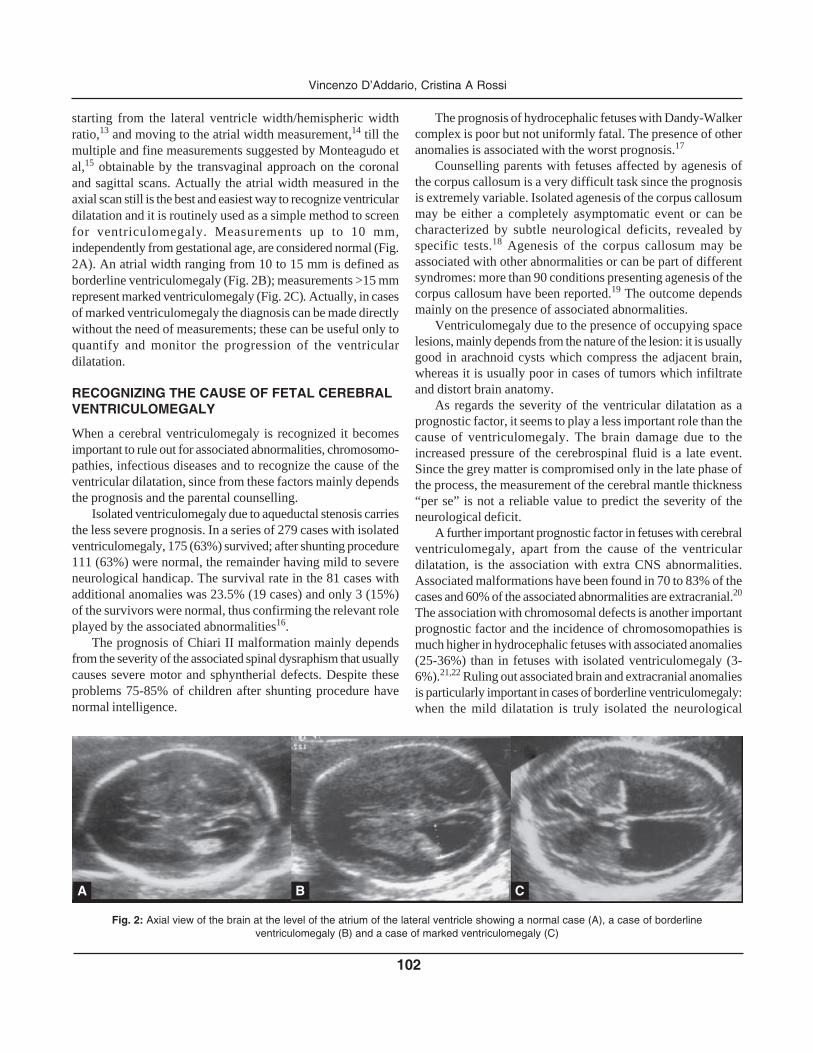
starting from the lateral ventricle width/hemispheric widthratio,13 and moving to the atrial width measurement,14 till themultiple and fine measurements suggested by Monteagudo etal,15 obtainable by the transvaginal approach on the coronaland sagittal scans. Actually the atrial width measured in theaxial scan still is the best and easiest way to recognize ventriculardilatation and it is routinely used as a simple method to screenfor ventriculomegaly. Measurements up to 10 mm,independently from gestational age, are considered normal (Fig.2A). An atrial width ranging from 10 to 15 mm is defined asborderline ventriculomegaly (Fig. 2B); measurements >15 mmrepresent marked ventriculomegaly (Fig. 2C). Actually, in casesof marked ventriculomegaly the diagnosis can be made directlywithout the need of measurements; these can be useful only toquantify and monitor the progression of the ventriculardilatation.
RECOGNIZING THE CAUSE OF FETAL CEREBRALVENTRICULOMEGALY
When a cerebral ventriculomegaly is recognized it becomesimportant to rule out for associated abnormalities, chromosomo-pathies, infectious diseases and to recognize the cause of theventricular dilatation, since from these factors mainly dependsthe prognosis and the parental counselling.
Isolated ventriculomegaly due to aqueductal stenosis carriesthe less severe prognosis. In a series of 279 cases with isolatedventriculomegaly, 175 (63%) survived; after shunting procedure111 (63%) were normal, the remainder having mild to severeneurological handicap. The survival rate in the 81 cases withadditional anomalies was 23.5% (19 cases) and only 3 (15%)of the survivors were normal, thus confirming the relevant roleplayed by the associated abnormalities16.
The prognosis of Chiari II malformation mainly dependsfrom the severity of the associated spinal dysraphism that usuallycauses severe motor and sphyntherial defects. Despite theseproblems 75-85% of children after shunting procedure havenormal intelligence.
The prognosis of hydrocephalic fetuses with Dandy-Walkercomplex is poor but not uniformly fatal. The presence of otheranomalies is associated with the worst prognosis.17
Counselling parents with fetuses affected by agenesis ofthe corpus callosum is a very difficult task since the prognosisis extremely variable. Isolated agenesis of the corpus callosummay be either a completely asymptomatic event or can becharacterized by subtle neurological deficits, revealed byspecific tests.18 Agenesis of the corpus callosum may beassociated with other abnormalities or can be part of differentsyndromes: more than 90 conditions presenting agenesis of thecorpus callosum have been reported.19 The outcome dependsmainly on the presence of associated abnormalities.
Ventriculomegaly due to the presence of occupying spacelesions, mainly depends from the nature of the lesion: it is usuallygood in arachnoid cysts which compress the adjacent brain,whereas it is usually poor in cases of tumors which infiltrateand distort brain anatomy.
As regards the severity of the ventricular dilatation as aprognostic factor, it seems to play a less important role than thecause of ventriculomegaly. The brain damage due to theincreased pressure of the cerebrospinal fluid is a late event.Since the grey matter is compromised only in the late phase ofthe process, the measurement of the cerebral mantle thickness“per se” is not a reliable value to predict the severity of theneurological deficit.
A further important prognostic factor in fetuses with cerebralventriculomegaly, apart from the cause of the ventriculardilatation, is the association with extra CNS abnormalities.Associated malformations have been found in 70 to 83% of thecases and 60% of the associated abnormalities are extracranial.20
The association with chromosomal defects is another importantprognostic factor and the incidence of chromosomopathies ismuch higher in hydrocephalic fetuses with associated anomalies(25-36%) than in fetuses with isolated ventriculomegaly (3-6%).21,22 Ruling out associated brain and extracranial anomaliesis particularly important in cases of borderline ventriculomegaly:when the mild dilatation is truly isolated the neurological
Fig. 2: Axial view of the brain at the level of the atrium of the lateral ventricle showing a normal case (A), a case of borderlineventriculomegaly (B) and a case of marked ventriculomegaly (C)
A B C

Fetal Cerebral Ventriculomegaly
103
handicap is good in 90-92% of the cases;23,24 the smallpercentage of bad outcome mainly affects the subgroup withatrial width between 12 and 15 mm25.
Considering the role that the cause of ventriculomegaly playsin influencing the prognosis, it appears evident how crucial isits prenatal recognition. An accurate evaluation of the shape ofthe dilated ventricles as well as the presence of associatedintracranial abnormalities allows to obtain this goal in mostcases.
The sonographic feature of aqueductal stenosis is that oftri-ventricular dilatation affecting the lateral and third ventricleswith no other abnormality of the brain structures: for this reasonit is also defined as isolated hydrocephalus (Fig. 3). Thedilatation of the third ventricle is sometimes of little degreeand the dilated lateral ventricles appear as the prominent feature.In cases of huge dilatation of the lateral ventricles, fenestrationof the septum pellucid may occur (Fig. 4). Prenatal sonography
does not permit to recognize the stenotic aqueduct, but theabsence of any other abnormality in the brain authorizes tosuspect this entity as the cause of the ventriculomegaly.Sonographic demonstration of abducted thumbs should promptthe diagnosis of X-linked hydrocephalus spectrum.26,27
In the Chiari II malformation, ventriculomegaly is theconsequence of an altered flow of the cerebrospinal fluid at thelevel of a small and funnelling posterior fossa associated with aspinal defect. The ultrasonographic appearance of theventriculomegaly is not specific, but the axial scan on theposterior fossa shows that it is small, with cisterna magna beingeffaced and the cerebellum assuming the typical banana shape,secondary to its herniation in the foramen magnum28 (Fig. 5).In order to evaluate the shape and quantify the size of theposterior fossa, our group suggested to measure the anglebetween the clivus and the supraocciput obtained in a sagittalscan of the skull (Fig. 6); this angle has a value of 79.3°+6°independently from gestational age and fetuses affected byChiari II malformation show values below 5° centile (<72°).29
Ventriculomegaly is a common finding in fetuses affectedby Dandy-Walker complex. In a series of 99 cases (50 Dandy-Walker and 49 Dandy-Walker variant) Ecker et al. reported anassociation with ventriculomegaly in 32% and 27%respectively.17 Once again the sonographic appearance of theventricular dilatation “per se” is not specific, but the presenceof a “cyst” of variable size in the posterior fossa is highlycharacteristic: it is secondary to the complete or partial agenesisof the cerebellar vermis and to the cystic dilatation of the fourthventricle (Fig. 7).17,30
The proportion of fetuses with agenesis of the corpuscallosum that have also ventriculomegaly is uncertain. Whenpresent, the ventricular dilatation is usually mild to moderate,although severe forms of ventriculomegaly may also develop.The diagnosis is usually late since the development of the corpuscallosum is a late event that is complete only by midgestation.Bennet et al. affirm that the diagnosis is not possible before 22weeks,31 but available experience suggests that agenesis of thecorpus callosum can be suspected at 18-20 weeks. Theventriculomegaly is the consequence of an altered developmentof the intracranial architecture and derives from the distortedarray of the white matter tracts in the occipital lobes: for thisreason it is usually moderate and limited to the posterior horns,so that the lateral ventricles assume the typical “tear-drop”appearance. The corpus callosum cannot be demonstrated bythe axial scans. But indirect signs useful for the diagnosis are;32
• the enlargement of the interhemispheric fissure where threeparallel lines can be recognized (the falx and the medialborders of the separated hemispheres): this sign is due tothe altered architecture of the sulci and gyri of the medialaspect of the hemispheres.
Figs 3A and B: Ventriculomegaly in a case of aqueductal stenosis. Amarked dilatation of the lateral ventricles and a mild dilatation of thethird ventricle (arrow) are visible. The remaining brain anatomy is normal
B
A

Vincenzo D’Addario, Cristina A Rossi
104
Fig. 4: 3D tomographic ultrasound imaging in the axial plane in a case of huge ventriculomegaly, showingthe fenestration of the septum pellucidum (dots)
Figs 5A and B: Axial views of the fetal brain in a case of ventriculomegaly due to Chiari II malformation. (A)The ventriculomegaly and the typical “lemon” shaped skull are visible (B) The posterior fossa is small witheffaced cisterna magna and with small and “banana” shaped cerebellum
Figs 6A and B: Sagittal view of the brain in a normal case (A) and in a fetus affected by Chiari II malformation(B). The funnelling appearance of the posterior fossa is pointed out by the smaller clivus-supraocciput angleas compared to the normal case
A B
A B

Fetal Cerebral Ventriculomegaly
105
Figs 7A and B: (A) Axial scan of the brain in a fetus with ventriculomegaly due to Dandy-Walker malformation:the “cyst” in the posterior fossa and the absence of the cerebellar vermis are visible (arrow). (B) Sagittal viewof the brain in a fetus affected by Dandy-walker malformation. The upward rotation of the hypoplastic vermisand the cystic dilatation of the fourth ventricle are clearly visible (curved arrow)
• the lateral displacement and enlargement of the bodies ofthe lateral ventricles, due to the medial compression of thewhite fibres failing to cross the midline.
• the upward displacement of the third ventricle (present onlyin 50% of the cases), sometimes communicating with aninterhemispheric cyst, as a consequence of the absence ofthe corpus callosum (Fig. 8).
The definitive diagnosis relies on the demonstration of theabsence of the complex formed by the corpus callosum and thecavum septi pellucidi. This sign can be achieved by the sagittaland coronal sections. The midsagittal section will demonstratethe complete or partial absence of the corpus callosum (Fig. 9).In cases of partial agenesis the missing part is the caudal one.In complete agenesis the gyri and sulci of the medial hemisphericsurface show an atypical radiate appearance, due to the absenceof the gyrus cinguli. With the use of color-doppler the absenceof the pericallosal artery can be noticed. The coronal section atthe level of the frontal horns will show their typical “bull-shape”appearance due to the medial compression by the white fibresthat fail to cross the hemispheres (bundles of Probst) (Fig. 10).
Considering the sonographic findings described so far thatcharacterize the different causes of ventriculomegaly, it appearsevident that the differential diagnosis mainly relies on theevaluation of the posterior fossa and the corpus callosum. Bothstructures can be evaluated in the midsagittal scan of the fetalbrain. By combining the sonographic findings at the level ofthe posterior fossa and the corpus callosum it is possible to identifythe main causes of fetal ventriculomegaly described so far.
When the posterior fossa is normal and the corpus callosumis present, the third ventricle is dilated and no other brainanomaly can be demonstrated, the ventriculomegaly is isolatedand the most probable cause is aqueductal stenosis (Fig. 11B).
Figs 8A and B: Indirect signs of agenesis of the corpus callosum in theaxial view: (A) enlargement of the interhemispheric fissure whit threeparallel lines referring to the falx and the medial borders of the separatedhemispheres. (B) Lateral displacement and enlargement of the bodiesof the lateral ventricles
A B
A
B

Vincenzo D’Addario, Cristina A Rossi
106
Figs 9A to C: Midsagittal view of the brain: (A) Complete agenesis of the corpus callosum(B) Partial agenesis of the corpus callosum (C) Normal corpus callosum
When the posterior fossa is small with a clivus-supraocciputangle <72° and the cisterna magna is effaced, Chiari IImalformation should be suspected as the cause of the ventriculardilatation (Fig. 11C) and a spinal defect should be carefullysearched for.
When the posterior fossa shows a complete or partialagenesis of the cerebellar vermis with a cystic dilatation of thefourth ventricle, Dandy-Walker complex is the cause of theventriculomegaly (Fig. 11D).
When the posterior fossa is normal and the corpus callosumis completely or partially absent the ventriculomegaly is due tothe failed development of this structure (Fig. 11F).
A combinations of signs can of course be occasionallyfound; a typical association is that of agenesis of the corpuscallosum with Dandy-Walker complex: both sonographicfeatures can be achieved in the same sagittal scan (Fig. 11E).
3D facility and particularly the multiplanar view mode isextremely useful in “reconstructing” the midsagittal view oncethe volume from the fetal bain has been extracted.
A B C
Figs 10A to C: 3D multiplanar view of the brain in a fetus affected by agenesis of the corpus callosum: thecoronal view (A) shows the typical “bull shape” appearance of the frontal horns (arrows); the sagittal view (B)shows the absence of the corpus callosum; the mild dilatation of the occipital horns (arrow) can be seen in theaxial view (C)
A B
C

Fetal Cerebral Ventriculomegaly
107
The accuracy of midsagittal scan in the diagnosis of thecause of ventriculomegaly is higher in cases with abnormalposterior fossa, lower in cases of isolated ventriculomegaly,due to the fact that the visualization of the corpus callosum issometimes doubtful. In a series of 58 cases of ventriculomegalyexamined by our group, the midsagittal view of the fetal brainallowed to recognize accurately the cause of the ventriculardilatation in 8: 93.1.% of the cases. The only uncorrecteddiagnoses referred to four cases of partial agenesis of the corpuscallosum, thus confirming the difficulties in recognizing thisstructure.33 The use of 3D multiplanar view helps in by-passingthese technical problems.
Even though the midsagittal view of the brain is a powerfultool in the differential diagnosis of fetuses withventriculomegaly, the diagnostic work-up should include alsoa careful evaluation of the finest anatomical details of the brainin order to recognize even rare cases of ventricular dilatation.Huge occupying space lesions, such as tumors or cysts, are easilyrecognizable (Fig. 12), but also small and subtle anomalies,such as cavum veli interpositi cyst (Fig. 13) or subependymalcysts (Fig. 14) may cause mild ventricular dilatation.34 The innerwalls of the lateral ventricles should also be carefully evaluatedin order to recognize signs of intra- or extra-ventricularhemorrhage (Fig 15 and 16).
Figs 11A to F: Midsagittal view of the brain in different causes of ventriculomegaly: (A) normal brain: the normal corpus callosumand cerebellar vermis with fourth ventricle can be recognized. (B) Aqueductal stenosis: the posterior fossa is normal; the thirdventricle is dilated; the corpus callosum is present. (C) Chiari II malformation : the posterior fossa is small with a clivus-supraocciputangle <72° and the cisterna magna is effaced. (D) Dandy-Walker complex : the posterior fossa shows a partial agenesis of thecerebellar vermis with a cystic dilatation of the fourth ventricle. (E) Agenesis of the corpus callosum: the posterior fossa is normalbut corpus callosum is absent. (F) Combined agenesis of the corpus callosum and Dandy-Walker complex: the corpus callosum isabsent, the third and the fourth ventricle are dilated
Fig. 12: Ventriculomegaly associated with brain teratoma. The tumor(T) is easily recognizable as an echogenic mass distorting the brainanatomy and causing asymmetrical ventriculomegaly (*)
A B C
FED

Vincenzo D’Addario, Cristina A Rossi
108
Fig. 13: Borderline ventriculomegaly associated with cavum veli interpositi cyst. The 3D multiplanarview clearly demonstrate the small cyst (arrow) located below the splenium of the corpus callosum
and posterior to the cavum septi pellucidi
Fig. 14: Borderline ventriculomegaly associated with multiple subependymal cysts. The 3D multiplanarview clearly demonstrate the cysts located below the frontal horns of the lateral ventricles

Fetal Cerebral Ventriculomegaly
109
Fig. 15: Marked triventricular dilatation with signs of intraventricularhemorrhage, floating blood clots and hyperechogenic ventricular wallsare visible
Fig. 18: Mild ventriculomegaly in a case of lissencephaly at 26 weeksof gestation. The brain surface is smooth and the arachnoidal space islarge
Fig. 17: Asymmetrical ventriculomegaly due to choroid plexus papilloma
The choroid plexuses should also be evaluated. In case ofhypertensive ventriculomegaly they are usually squeezed by theincreased cerebrospinal fluid pressure. The finding of an enlargedchoroids plexus can be the sign of a papilloma (Fig. 17).
A careful examination of the brain surface should also beperfomed in order to recognize neuronal proliferation anddifferentiation disorders, such as lissencephaly (Fig. 18).
Finally, in cases of intrauterine infections, smallperiventricular echogenic areas may be seen (Fig. 19).
Figs 16A and B: Periventricular hemorrhage (black arrow) at 21 weeksgestation (A), evolving in unilateral ventriculomegaly at 24 weeks (B);the hyperechogenicity of the ventricular wall is still visible(white arrow)
BA
Fig 19: Mild ventriculomegaly in a case of toxoplasmosis infection.Small periventricular echogenic spots (arrows) can be visualized
CONCLUSIONSThe prenatal sonographic diagnosis of fetal ventriculomegalyis relatively easy and in cases of marked ventricular dilatationit can be achieved “at a glance” on the classical axial scans onthe fetal skull. However, the recognition of the cause of theventricular dilatation may represent a challenging problem evenin the hands of experienced sonologists. The accurate anddetailed evaluation of the brain anatomy is a crucial point inthe differential diagnosis. The sagittal scan of the fetal brain isa powerful source of information since it allows thecontemporary view of both the corpus callosum and the posteriorfossa, where typical sonographic findings are present in thedifferent causes of ventricular dilatation. The sagittal scan of

Vincenzo D’Addario, Cristina A Rossi
110
13. Pooh RK, Pooh KH. The assessment of fetal brain morphologyand circulation by transvaginal 3D sonography and powerDoppler. J Perinat Med 2002;30:48-56.
14. Pilu G, Reece EA, Goldstein I, Hobbins JC, Bovicelli L.Sonographic evaluation of the normal developmental anatomyof the cerebral ventricles: II. The atria. Obstet Gynecol1989;73:250-255.
15. Monteagudo A, Timor-Tritsch IE, Moomij M. Nomograms ofthe fetal lateral ventricles using transvaginal sonography. JUltrasound Med 1993;5:265-69.
16. Gupta JK, Bryce FC, Lilford RJ. Management of apparentlyisolated fetal ventriculomegaly. Obstet Gynecol Surv1994;49:716-21.
17. Ecker JL, Shipp TD, Bromley B, Benacerraf C. The sonographicdiagnosis of Dandy-Walker and Dandy-Walker variant:associated findings and outcomes. Prenat Diagn 2000;20:32.
18. Goodyear PW, Bannister CM, Russel S, Rimmer S. Outcome inprenatally diagnosed fetal agenesis of the corpus callosum. FetalDiagn Ther 2001;16:139-45.
19. Davila-Gutierrez G. Agenesis and dysgenesis of the corpuscallosum. Sem Pediatr Neurol 2002;9:292-93.
20. Nyberg DA, Mack LA, Hirsch J, Pagon RO, Shepard TH. Fetalhydrocephalus: sonographic detection and clinical significanceof associated anomalies. Radiology 1987;163:187-91.
21. Nicolaides KH, Berry S, Snijders RJ. Fetal lateral cerebralventriculomegaly: associated malformations and chromosomaldefects. Fetal Diagn Ther 1990;5:5-14.
22. Schwanitz G, Schuler H, Gembruch U, Zerres K. Chromosomalfindings in fetuses with ultrasonographically diagnosedventriculomegaly. Ann Genet 1993;36:150-53.
23. Pilu G, Falco P, Gabrielli S, Perolo A, Sandri F, Bovicelli LThe clinical significance of fetal isolated cerebral borderlineventriculomegaly: report of 31 cases and review of theliterature.Ultrasound Obstet Gynecol. 1999; 14:320-26.
24. Gaglioti P, Danelon D, Bontempo S, Mombrò M, CardaropoliS, Todros T. Fetal cerebral ventriculomegaly: outcome in 176cases. Ultrasound Obstet Gynecol 2005; 25:372-77.
25. Signorelli M, Tiberti A, Valseriati D, Molin E, Cerri V, GroliC, Bianchi UA.: Width of the lateral ventricular atrium between10 and 12 mm: a simple variation of the norm? Ultrasound ObstetGynecol. 2004; 23:14-18
26. Senat MV, Bernard JP, Delezoide A, Saugier-Veber P, HillionY, Roume J. Prenatal diagnosis of hydrocephalus-stenosis ofthe aqueduct of Sylvius by ultrasound in the first trimester ofpregnancy. Report of two cases. Prenat Diagn 2001;21:1129-32.
27. Timor-Tritsch IE, Monteagudo A, Haratz-Rubinstein N, LevineRU. Transvaginal sonographic detection of adducted thumbs,hydrocephalus, and agenesis of the corpus callosum at 22postmenstrual weeks: the masa spectrum or L1 spectrum. A casereport and review of the literature. Prenat Diagn 1996;16:543-45.
28. Nicolaides KH, Campbell S, Gabbe SG, Guidetti R. Ultrasoundscreening for spina bifida: cranial and cerebellar signs. Lancet1986;328:72-76.
the fetal brain is not looked for routinely; however the sonologistshould be familiar with such a scan in cases of ventriculomegalyin order to obtain a more comprehensive view of the brainmorphology. Furthermore he can better communicate hisprenatal findings to the neonatologist, neuroradiologist andpediatric neurosurgeon, who usually evaluate the neonatal brainimaging based on the sagittal and coronal sections and cantherefore better understand the prenatal sonographic features,with the final goal to offer the parents a correct counselling.
REFERENCES
1. Myrianthopoulos NC. Epidemiology of central nervousmalformations. In: Vinken PJ, Bruyn GW, (Eds). Handbook ofclinical neurology. Amsterdam: Elsevier 1977; pp 139-171.
2. Milhorat TH. Hydrocephaly. In: Vinken PJ, Bruyn GW, (Eds).Handbook of clinical neurology. Amsterdam: Elsevier 1987; pp285-300.
3. Fransen E, Vits L, Van Camp G, Willems PJ. The clinicalspectrum of mutations in L1, a neuronal cell adhesion molecule.Am J Med Genet 1996;64:73-77.
4. McLone DG, Naidich TP. Development morphology of thesubarachnoid space, brain vasculature and continuous structures,and the cause of the Chiari II malformation. Am J Neuroradiol1992;13:463-83.
5. Barkovich AJ, Kjos BO, Normal D. Revised classification ofthe posterior fossa cysts and cystic-like malformations based onthe results of multiplanar MR imaging. Am J Neuroradiol1990;10:977-82.
6. Young ID. Genetics of neurodevelopmental abnormalities. In:Levene MI, Lilford RJ, Bennet MJ, Punt F, editors. Fetal andneonatal neurology and neurosurgery. London: ChurchillLivingstone 1995; pp 256-261.
7. Hadlock FP, Deter RL, Park K. Real time sonography:ventricular and vascular anatomy of the fetal brain in utero. AJR1981;136:133-37.
8. D’Addario V, Kurjak A. Ultrasound investigation of the fetalcerebral ventricles. J Perinat Med 1985;13:67-77.
9. Monteagudo A, Reuss ML, Timor-Tritsch IE. Imaging the fetalbrain in the second and third trimesters using transvaginalsonography. Obstet Gynecol 1991;77:27-32.
10. Timor-Tritsch IE, Monteagudo A, Warren WB. Transvaginalultrasonic definition of the central nervous system in the firstand early second trimester. Am J Obstet Gynecol 1991;164:747-53.
11. Jeanty P, Dramix-Wilmet M, Delbeke D. Ultrasonic evaluationof fetal ventricular growth. Neuroradiology 1981;21:127-131.
12. Hata T, Yanagihara T, Matsumoto M, Hanaoka U, Ueta M,Tanaka Y. Three-dimensional sonographic features of fetalcentral nervous system anomaly. Acta Obstet Gynecol Scand2000;79:635-39.
Fetal Cerebral Ventriculomegaly
111
29. D’Addario V, Pinto V, Del Bianco A, Di Naro E, Tartagni M,Miniello G. The clivus-supraocciput angle: a useful measurementto evaluate the posterior fossa and to diagnose Chiari IImalformation. Ultrasound Obstet Gynecol 2001;18:146-149.
30. Pilu G, Romero R, De Palma L. Antenatal diagnosis andobstetrical management of Dandy-Walker syndrome. J ReprodMed 1986;31:1017-22.
31. Bennet GL, Bromley B, Benacerraf BR. Agenesis of the corpuscallosum: prenatal detection usually is not possible before 22weeks of gestation. Radiology 1996;99:447-50.
32. Pilu G, Sandri F, Perolo A, Pittalis MC, Grisolia G, Cocchi G.Sonography of fetal agenesis of the corpus callosum: a surveyof 35 cases. Ultrasound Obstet Gynecol 1993;3:318-329.
33. D’Addario V, Pinto V, Di Cagno L, Pintucci A. The midsagittalview of the fetal brain: a useful landmark in recognizing thecause of fetal cerebral ventriculomegaly. J Perinat Med2005;33:423-24
34. D’Addario V, Selvaggio S, Pinto V, Resta M, Di Cagno L, FamàA: Fetal subependymal cysts with normal neonatal outcome.Fetal Diag Ther 2003;18:170-73