Dominant Mutations in GRHL3 Cause Van der Woude Syndrome ... · dependent pathway of periderm...
10
ARTICLE Dominant Mutations in GRHL3 Cause Van der Woude Syndrome and Disrupt Oral Periderm Development Myriam Peyrard-Janvid, 1,16, * Elizabeth J. Leslie, 2,16 Youssef A. Kousa, 3,16 Tiffany L. Smith, 4,16 Martine Dunnwald, 2,16 Ma ˚ns Magnusson, 5 Brian A. Lentz, 2 Per Unneberg, 6 Ingegerd Fransson, 1 Hannele K. Koillinen, 7 Jorma Rautio, 8 Marie Pegelow, 9 Agneta Karsten, 9 Lina Basel-Vanagaite, 10,11,12 William Gordon, 13 Bogi Andersen, 13 Thomas Svensson, 5 Jeffrey C. Murray, 2 Robert A. Cornell, 4 Juha Kere, 1,5,14, * and Brian C. Schutte 15 Mutations in interferon regulatory factor 6 (IRF6) account for ~70% of cases of Van der Woude syndrome (VWS), the most common syn- dromic form of cleft lip and palate. In 8 of 45 VWS-affected families lacking a mutation in IRF6, we found coding mutations in grainy- head-like 3 (GRHL3). According to a zebrafish-based assay, the disease-associated GRHL3 mutations abrogated periderm development and were consistent with a dominant-negative effect, in contrast to haploinsufficiency seen in most VWS cases caused by IRF6 muta- tions. In mouse, all embryos lacking Grhl3 exhibited abnormal oral periderm and 17% developed a cleft palate. Analysis of the oral phenotype of double heterozygote (Irf6 þ/ ;Grhl3 þ/ ) murine embryos failed to detect epistasis between the two genes, suggesting that they function in separate but convergent pathways during palatogenesis. Taken together, our data demonstrated that mutations in two genes, IRF6 and GRHL3, can lead to nearly identical phenotypes of orofacial cleft. They supported the hypotheses that both genes are essential for the presence of a functional oral periderm and that failure of this process contributes to VWS. Introduction Grainyhead-like 3 (Drosophila)(GRHL3 [MIM 608317]) belongs to a family of three human genes that encode tran- scription factor orthologs of the Drosophila gene grainy head (grh). Among multiple conserved roles, this gene family is required for the development and repair of the epidermal barrier layer. 1–3 In zebrafish, grhl1 and grhl3 were shown to be required for the development of the peri- derm, 4 the transient layer of squamous epithelial cells located on the surface of developing embryos. Interferon regulatory factor 6 (irf6) is also required for periderm devel- opment in zebrafish 5 and directly regulates the expression of grhl3. 4,6 In addition, overexpression of Grhl3 partially rescued periderm development in zebrafish embryos that expressed a dominant-negative mutant form of irf6. 4 These data suggest that Grhl3 is an important player in the Irf6- dependent pathway of periderm development. IRF6 belongs to the IRF family of transcription factors that are known best for their roles in immune function. 7 However, IRF6 (MIM 607199) is required for skin, limb, and craniofacial development. 8–10 In mice, embryos that lack Irf6 expression fail to develop the epidermal barrier. 9,10 Although reminiscent of embryos that lack Grhl3, 2 the cutaneous phenotype of Irf6 mutant embryos appears to be more severe macroscopically. In addition, Irf6 mutant embryos have extensive oral epithelial adhe- sions, 9,10 a phenotype not reported in the Grhl3 mutant. The oral epithelial adhesions in Irf6 knockout embryos lead to cleft palate 9,10 and appear to stem from periderm dysfunction. 4,11 In humans, mutations in IRF6 cause Van der Woude syndrome (VWS [MIM 119300]), the most common syn- dromic form of orofacial clefting, or popliteal pterygium syndrome (PPS [MIM 119500]). Individuals with VWS can have cleft lip (CL), cleft palate (CP), or cleft lip and pal- ate (CLP). In addition, 85% of affected individuals have pits in their lower lip. 12 To date, mutations in IRF6 have been identified in 70% of families with VWS. 8,13,14 The possibility that locus heterogeneity accounts for some of the remaining 30% of VWS mutations is underscored by linkage in one large pedigree from Finland to a locus on 1p33–p36 rather than to IRF6 at 1q32–q41. 15 In this fam- ily, most affected individuals have an orofacial cleft and 1 Department of Biosciences and Nutrition, Karolinska Institutet, and Center for Biotechnology, 14183 Huddinge, Sweden; 2 Department of Pediatrics and Interdisciplinary Program in Genetics, University of Iowa, Iowa City, IA 52242, USA; 3 Department of Biochemistry and Molecular Biology, Michigan State University, East Lansing, MI 48824, USA; 4 Department of Anatomy and Cell Biology, University of Iowa, Iowa City, IA 52242, USA; 5 Department of Bio- sciences and Nutrition, Science for Life Laboratory, Karolinska Institutet, 17121 Solna, Sweden; 6 Department of Biochemistry and Biophysics Science for Life Laboratory, Stockholm University, 17121 Solna, Sweden; 7 Department of Clinical Genetics, Helsinki University Hospital, 00029 Helsinki, Finland; 8 Cleft Palate and Craniofacial Center, Department of Plastic Surgery, Helsinki University Hospital, 00029 Helsinki, Finland; 9 Department of Orthodontics, Stockholm Craniofacial Team, Institute of Odontology, Karolinska Institutet, 17177 Stockholm, Sweden; 10 Pediatric Genetics Unit, Schneider Children’s Medical Center of Israel and Raphael Recanati Genetic Institute, Rabin Medical Center, Petah Tikva 49100, Israel; 11 Sackler Faculty of Medicine, Tel Aviv University, Tel Aviv 69978, Israel; 12 Felsenstein Medical Research Center, Petah Tikva 49100, Israel; 13 Department of Biological Chemistry, University of California Irvine, Irvine, CA 92697, USA; 14 Research Programs Unit, University of Helsinki, and Folkha ¨lsan Institute of Genetics, 00014 Helsinki, Finland; 15 Department of Microbiology and Molecular Genetics, Michigan State University, East Lansing, MI 48824, USA 16 These authors contributed equally to this work *Correspondence: [email protected] (M.P.-J.), [email protected] (J.K.) http://dx.doi.org/10.1016/j.ajhg.2013.11.009. Ó2014 by The American Society of Human Genetics. The American Journal of Human Genetics 94, 23–32, January 2, 2014 23 Open access under CC BY-NC-ND license.
Transcript of Dominant Mutations in GRHL3 Cause Van der Woude Syndrome ... · dependent pathway of periderm...
ARTICLE
Dominant Mutations in GRHL3Cause Van der Woude Syndromeand Disrupt Oral Periderm Development
Myriam Peyrard-Janvid,1,16,* Elizabeth J. Leslie,2,16 Youssef A. Kousa,3,16 Tiffany L. Smith,4,16
Martine Dunnwald,2,16 Mans Magnusson,5 Brian A. Lentz,2 Per Unneberg,6 Ingegerd Fransson,1
Hannele K. Koillinen,7 Jorma Rautio,8 Marie Pegelow,9 Agneta Karsten,9 Lina Basel-Vanagaite,10,11,12
William Gordon,13 Bogi Andersen,13 Thomas Svensson,5 Jeffrey C. Murray,2 Robert A. Cornell,4
Juha Kere,1,5,14,* and Brian C. Schutte15
Mutations in interferon regulatory factor 6 (IRF6) account for ~70% of cases of Van derWoude syndrome (VWS), the most common syn-
dromic form of cleft lip and palate. In 8 of 45 VWS-affected families lacking a mutation in IRF6, we found coding mutations in grainy-
head-like 3 (GRHL3). According to a zebrafish-based assay, the disease-associated GRHL3 mutations abrogated periderm development
and were consistent with a dominant-negative effect, in contrast to haploinsufficiency seen in most VWS cases caused by IRF6 muta-
tions. In mouse, all embryos lacking Grhl3 exhibited abnormal oral periderm and 17% developed a cleft palate. Analysis of the oral
phenotype of double heterozygote (Irf6þ/�;Grhl3þ/�) murine embryos failed to detect epistasis between the two genes, suggesting
that they function in separate but convergent pathways during palatogenesis. Taken together, our data demonstrated that mutations
in two genes, IRF6 andGRHL3, can lead to nearly identical phenotypes of orofacial cleft. They supported the hypotheses that both genes
are essential for the presence of a functional oral periderm and that failure of this process contributes to VWS.
Introduction
Grainyhead-like 3 (Drosophila) (GRHL3 [MIM 608317])
belongs to a family of three human genes that encode tran-
scription factor orthologs of the Drosophila gene grainy
head (grh). Among multiple conserved roles, this gene
family is required for the development and repair of the
epidermal barrier layer.1–3 In zebrafish, grhl1 and grhl3
were shown to be required for the development of the peri-
derm,4 the transient layer of squamous epithelial cells
located on the surface of developing embryos. Interferon
regulatory factor 6 (irf6) is also required for periderm devel-
opment in zebrafish5 and directly regulates the expression
of grhl3.4,6 In addition, overexpression of Grhl3 partially
rescued periderm development in zebrafish embryos that
expressed a dominant-negative mutant form of irf6.4 These
data suggest that Grhl3 is an important player in the Irf6-
dependent pathway of periderm development.
IRF6 belongs to the IRF family of transcription factors
that are known best for their roles in immune function.7
However, IRF6 (MIM 607199) is required for skin, limb,
and craniofacial development.8–10 In mice, embryos that
1Department of Biosciences and Nutrition, Karolinska Institutet, and Center fo
Interdisciplinary Program in Genetics, University of Iowa, Iowa City, IA 52242
University, East Lansing, MI 48824, USA; 4Department of Anatomy and Cell B
sciences and Nutrition, Science for Life Laboratory, Karolinska Institutet, 17
for Life Laboratory, Stockholm University, 17121 Solna, Sweden; 7Department8Cleft Palate and Craniofacial Center, Department of Plastic Surgery, Helsinki U
Stockholm Craniofacial Team, Institute of Odontology, Karolinska Institutet,
Medical Center of Israel and Raphael Recanati Genetic Institute, Rabin Med
Aviv University, Tel Aviv 69978, Israel; 12Felsenstein Medical Research Center,
of California Irvine, Irvine, CA 92697, USA; 14Research ProgramsUnit, Universi15Department of Microbiology and Molecular Genetics, Michigan State Unive16These authors contributed equally to this work
*Correspondence: [email protected] (M.P.-J.), [email protected] (J.K.)
http://dx.doi.org/10.1016/j.ajhg.2013.11.009. �2014 by The American Societ
The A
lack Irf6 expression fail to develop the epidermal
barrier.9,10 Although reminiscent of embryos that lack
Grhl3,2 the cutaneous phenotype of Irf6 mutant embryos
appears to be more severe macroscopically. In addition,
Irf6 mutant embryos have extensive oral epithelial adhe-
sions,9,10 a phenotype not reported in the Grhl3 mutant.
The oral epithelial adhesions in Irf6 knockout embryos
lead to cleft palate9,10 and appear to stem from periderm
dysfunction.4,11
In humans, mutations in IRF6 cause Van der Woude
syndrome (VWS [MIM 119300]), the most common syn-
dromic form of orofacial clefting, or popliteal pterygium
syndrome (PPS [MIM 119500]). Individuals with VWS
can have cleft lip (CL), cleft palate (CP), or cleft lip and pal-
ate (CLP). In addition, 85% of affected individuals have
pits in their lower lip.12 To date, mutations in IRF6 have
been identified in 70% of families with VWS.8,13,14 The
possibility that locus heterogeneity accounts for some of
the remaining 30% of VWS mutations is underscored by
linkage in one large pedigree from Finland to a locus on
1p33–p36 rather than to IRF6 at 1q32–q41.15 In this fam-
ily, most affected individuals have an orofacial cleft and
r Biotechnology, 14183 Huddinge, Sweden; 2Department of Pediatrics and
, USA; 3Department of Biochemistry and Molecular Biology, Michigan State
iology, University of Iowa, Iowa City, IA 52242, USA; 5Department of Bio-
121 Solna, Sweden; 6Department of Biochemistry and Biophysics Science
of Clinical Genetics, Helsinki University Hospital, 00029 Helsinki, Finland;
niversity Hospital, 00029 Helsinki, Finland; 9Department of Orthodontics,
17177 Stockholm, Sweden; 10Pediatric Genetics Unit, Schneider Children’s
ical Center, Petah Tikva 49100, Israel; 11Sackler Faculty of Medicine, Tel
Petah Tikva 49100, Israel; 13Department of Biological Chemistry, University
ty of Helsinki, and Folkhalsan Institute of Genetics, 00014 Helsinki, Finland;
rsity, East Lansing, MI 48824, USA
y of Human Genetics.
merican Journal of Human Genetics 94, 23–32, January 2, 2014 23
Open access under CC BY-NC-ND license.
the proband has lip pits, the hallmark of VWS. Because of
the autosomal-dominant inheritance pattern and the pres-
ence of the lip pits, this family was diagnosed with VWS
and the linked region was named the VWS2 locus.15
Here we report disease-causing mutations in GRHL3 in
the above-mentioned original Finnish family as well as in
seven additional families with VWS, thereby demon-
strating that GRHL3 is the second gene for which muta-
tions lead to VWS. Although we observed no consistently
unique phenotypes in these families, individuals with a
GRHL3 mutation are more likely to have CP and less likely
to have CL or lip pits than individuals with an IRF6 muta-
tion. In addition, we used zebrafish and murine models to
show that Grhl3, like Irf6, has a conserved role in the devel-
opment of the periderm. Our observations from all three
species support the conclusion that a functional oral peri-
derm is essential for the proper palatogenesis.
Material and Methods
Human DNA SamplesDNA samples from 45 families of multiple ethnicities and who
were completely sequenced for IRF6 without identifying a causa-
tive mutation were used in this study. All subjects were examined
by clinical geneticists or genetic counselors who made diagnoses
as described previously.15–17 Written informed consent was ob-
tained for all subjects and all protocols were approved by the local
ethical boards in Helsinki (Finland) or in Stockholm (Sweden) or
by the Institutional Review Boards at the University of Iowa
(USA). A total of 360 unrelated individuals without a history of
oral cleft from the Philippines were used as controls for the
GRHL3 (c.1171C>T) Filipino variant and 561 unrelated Finnish
individuals (blood donors) were used as controls for the GRHL3
(c.969_970insTG), the PHACTR4 (c.1615G>A; rs200581707),
and the KTI12 (c.337_363delCCGATCGCGGGACCTCAGGTGG
CGGGC; ss836732090) Finnish variants.
Targeted Exome SequencingGenomic DNA from eight affected and three healthy individuals
from the VWS2 Finnish family underwent SureSelect Target
Enrichment (Agilent Technologies) in order to perform sequence
capture of the exome. Enriched samples were sequenced on an
Illumina HiSeq instrument. Reads were aligned to reference
sequence with the bwa read mapper.18 A high-quality variant
call set was generated based on a best-practice workflow,19 in
which we utilized the Picard and Genome analysis toolkit
(GATK) for data processing and analysis.
GenotypingGenotyping of the GRHL3 c.969_970insTG (Finnish) and
c.1171C>T (Filippino) variants and the PHACTR4 (c.1615G>A;
rs200581707) Finnish variant was performed with TaqMan SNP
Genotyping Assays (Life Technologies) on the ABI Prism 7900HT
or ABI 7500 and analyzed with SDS 2.3 or SDS 1.4 software
(Applied Biosystems). Family relationships for apparently de
novo variants (c.1171C>T and c.1559_1562delGGAG) were
confirmed by genotyping 16 markers distributed across the
genome (Table S2 available online). The KTI12 (c.337_363delCC
GATCGCGGGACCTCAGGTGGCGGGC; ss836732090) variant
24 The American Journal of Human Genetics 94, 23–32, January 2, 20
was genotyped by PCR amplification with SYBR green labeling
of the wild-type (100 bp) and the deleted (73 bp) alleles and
checked for their respective melting temperatures/curves.
Mutation Screening by Sanger SequencingPrimers for GRHL3 were designed to amplify the exons of all iso-
forms of GRHL3 via Primer3. The exons of all four GRHL3 tran-
script variants were screened in a total of 13 PCR amplicons (Table
S1). PCR reactions were incubated at 94�C for 5 min followed by
35 amplification cycles (45 s at 94�C, 45 s at 60�C, 45 s at 72�C)and a final extension at 72�C for 7 min. PCR products were sent
for sequencing on an ABI 3730XL (Functional Biosciences). Chro-
matograms were transferred to a UNIX workstation, base-called
with PHRED (v.0.961028), assembled with PHRAP (v.0.960731),
scanned by POLYPHRED (v.0.970312), and viewed with the
CONSED program (v.4.0). The effects of missense variants were
predicted with the Variant Effect Predictor program,20 which
generates scores from PolyPhen2 and SIFT.
Phenotype AnalysisAffected individuals withGRHL3mutations (n¼ 27) were assigned
a phenotype classification of cleft lip with or without cleft palate
(CL/P includes CL and CLP cases), cleft palate (CP), lip pits only,
CL/P with lip pits, or CP with lip pits based on the clinical diagno-
ses. Additional phenotypic classifications described the presence
of dental anomalies (hypodontia, dental aplasia, or malocclusion),
limb anomalies (syndactyly, polydactyly, club foot, or contrac-
tures), or popliteal pterygia. From the set of families positive for
IRF6 mutations,8,13,14,17,21 affected individuals were also assigned
to the same phenotype classifications (n ¼ 632). Exclusion criteria
for this analysis were individuals with a cleft but without identi-
fied familial mutation (i.e., potential phenocopies) and individ-
uals diagnosed with VWS without a known IRF6 or GRHL3
mutation.
Transfection of Human GRHL3 Mutation Variants
into Zebrafish EmbryosFull-length, wild-type human GRHL3 cDNA variant 4 (v4) was
obtained as a cDNA clone from Open Biosystems (MHS1010-
9204655) and shuttled by Gateway cloning into the CS2þ destina-
tion vector (kindly provided by Dave Turner, University of Michi-
gan). This construct was used for in vitro synthesis of wild-type
GRHL3 mRNA. Specific mutations from VWS-affected individuals
were generated in the GRHL3mRNA (v4) via PCR-mediated muta-
genesis and the resulting cDNAs engineered into CS2þ, resulting
in the truncation of the first 6 bp of 50 UTR and the last 70 bp of
30 UTR from mutant variants. These constructs were further used
for in vitro synthesis of mutant variants of GRHL3. These trunca-
tions (the first 6 bp of 50 UTR and the last 70 bp of 30 UTR from
mutant variants) had no functional consequence, as shown by
the fact that we tested a similarly truncated and cloned wild-
type GRHL3, and GRHL3 mRNA synthesized from this construct
behaved equivalently to full-length GRHL3 in the zebrafish-based
functional assay.
Capped mRNA was synthesized in vitro (mMESSAGE
mMACHINE SP6 kit, Ambion) and purified with the MEGAclear
kit (Ambion) and approximately 1 ng of mRNA was injected into
wild-type zebrafish embryos (Scientific Hatcheries outbred strain)
at the 1-cell or, for mosaic injections, at the 16-cell stage. Embryos
were fixed at 50% epiboly or corresponding time-point (5–6 hpf),
and whole-mount in situ hybridization for krt4 was performed as
14
previously described.22 Plasmids used for probe synthesis are avail-
able upon request. Embryos were injected with biotinylated-
dextran (Invitrogen, D-1956) and processed for visualization as
previously described.4 Animal use protocols were approved by
the Public Health Service Assurance.
Murine CrossesWe crossed mice heterozygous for the Irf6 genetrap allele (Irf6þ/gt;
here referred to as Irf6þ/�)9 with mice heterozygous for the Grhl3
knockout allele (Grhl3þ/�)3 to generate wild-type, Irf6þ/�, Grhl3þ/�,and Irf6þ/�;Grhl3þ/� double heterozygous embryos. Grhl3
knockout embryos were obtained by crossing Grhl3þ/� mice. Pres-
ence of a copulation plug was denoted as E0.5. Pregnant dams
were injected intraperitoneally with BrdU (Sigma) 2 hr before
euthanization at a dose of 100 mg per gram pregnant dam body
weight. Embryos were collected at indicated time points and geno-
typed for Irf6 andGrhl3 null alleles as described previously.3,9 Both
allelesweremaintained on aC57BL/6 background. Animal use pro-
tocols were approved by the Institutional Animal Care and Use
Committees at Michigan State University and the University of
California, Irvine.
Morphological, Histological, and Molecular Analyses
of MiceGross morphological analysis of the Irf6þ/� by Grhl3þ/� cross was
done at E13.5, E17.5, P0, and P21. Embryos were then fixed in 4%
paraformaldehyde, embedded in paraffin, and sectioned at 7 mm
intervals. Haematoxylin and eosin staining was performed as
described.9 For immunostaining, antigen retrieval was performed
in sodium citrate, followed by blocking steps in BSA and a goat
anti-mouse Fab fragment (Jackson ImmunoResearch Laboratories,
115-007-003). Primary antibody was incubated overnight at 4�Cand secondary antibody was incubated for 1.5 hr at room temper-
ature. We used primary antibodies against Keratin 6 (Covance,
PRB-190 169P), tumor protein p63 (Santa Cruz, 4A4, SC-8431),
Irf6 (Sigma-Aldrich, SAB2102995), and Activated Notch1 (Act
N1, Cell Signaling, Val1744, D3B8, 4147S). We used the following
secondary antibodies: goat anti-rabbit (Molecular Probes,
A21429), goat anti-mouse (Molecular Probes, A11029), and goat
anti-rat (Molecular Probes, A11006). Nuclei were stained with
DAPI (Invitrogen, D3571) followed by slide mounting in ProLong
Gold Antifade Reagent (Invitrogen, P36930).
ImagingHistological and immunostained sections were imaged with a
Nikon Eclipse 90i upright microscope with a Plan APO 103/0.45
DIC, a CFI Plan Apo Lambda 203/0.75, and a Plan APO 403/
0.95 DIXM/N2 objectives. A Nikon DS-Fi1 high-definition camera
head and a DigitalSight PC-use control unit were used for haema-
toxylin and eosin imaging. A X-Cite Series 120Q laser and a
CoolSnap HQ2 photometric camera were used to obtain immuno-
fluorescent images. NIS Elements Advanced Research v.3.10 was
used for RAW image deconvolution and Adobe Photoshop
Elements v.9.0 was used for figure formation.
Statistical AnalysisFisher’s exact test in STATA (v.12.1) was used to compare the
frequencies of VWS-associated phenotypes between individuals
with GRHL3 mutations and those with IRF6 mutations. The
threshold p value for this analysis was calculated with a Bonfer-
roni correction (p ¼ 0.05; 8 phenotypes ¼ 0.006). We used
The A
chi-square analysis to compare the observed genotype distribu-
tions of mice with the predicted Mendelian frequencies. Previ-
ous reports show that resorption rates in C57BL/6 mice range
between 1% and 3%. We used a two-tailed Fisher’s exact test
to compare the upper limit of this range with the observed
resorption rates.
Results
GRHL3 Is the VWS2 Gene
A single large VWS-affected family of Finnish origin
(Figure S1) showed linkage to a ~40 cM region on 1p33–36,
pointing to a second VWS locus,15 i.e., VWS2 (MIM
606713). From this family, we selected eight affected
individuals, including the proband who is the only one
with lip pits, and three healthy individuals for whole-exome
sequencing. We searched the ~700 genes contained in the
entire linkage region (~46 Mb) for variants common to all
eight affected family members but not seen in any of the
three healthy members. This resulted in three segregating
exonic variants in GRHL3 (chr1: 24,666,175; RefSeq acces-
sion number NM_198174.2; c.969_970insTG), PHACTR4
(chr1: 28,806,971; rs200581707; RefSeq NM_001048183.1;
c.1615G>A), and KTI12 (chr1: 52,499,097–52,499,071;
ss836732090; RefSeq NM_138417.2; c.337_363delCCGA
TCGCGGGACCTCAGGTGGCGGGC). The GRHL3 and
PHACTR4 variants were confirmed by TaqMan genotyping
and the KTI12 variant by allelic discrimination based on dif-
ferential melting temperature. The PHACTR4 variant was
found in 2 out of 8,252 European American chromosomes
in the NHLBI/ESP database and is therefore unlikely to be
the causative variant for VWS. In a set of 561 Finnish con-
trols, the KTI12 variant was found at a frequency of 12.4%
and is therefore a common, noncausative variant. The
GRHL3 variant was not found in any of the Finnish controls
nor inNHLBI/ESP,makingGRHL3 a strongcandidate gene in
the VWS2 locus.
To test whether mutations in GRHL3 accounted for VWS
in other families, we screened 44 families of variable
ethnicity where no causative IRF6mutations had been pre-
viously detected. We identified GRHL3 variants in seven
families, including four protein-truncating mutations
and four missense mutations (Figure 1). All mutations
except c.1661A>G (coding for the p.Asn554Ser missense
alteration) were predicted by PolyPhen2 and SIFT to be
damaging/deleterious and two were confirmed de novo
events (Table 1). In one of the seven families (VWS-III),
we found two variants located in trans. Variant
c.268_278delTACTACCATGG was inherited from the pro-
band’s affected father and from the healthy paternal
grandfather, and variant c.1661A>G was inherited from
the proband’s healthy mother (Figure S1). In addition,
one family (VWS-IV) was previously determined to have
a novel IRF6 missense variant (c.239A>G) that was not
conclusively determined to be causative for VWS,23 raising
the possibility that variants in both IRF6 and GRHL3 could
contribute to VWS in one family (Figure S1).
merican Journal of Human Genetics 94, 23–32, January 2, 2014 25
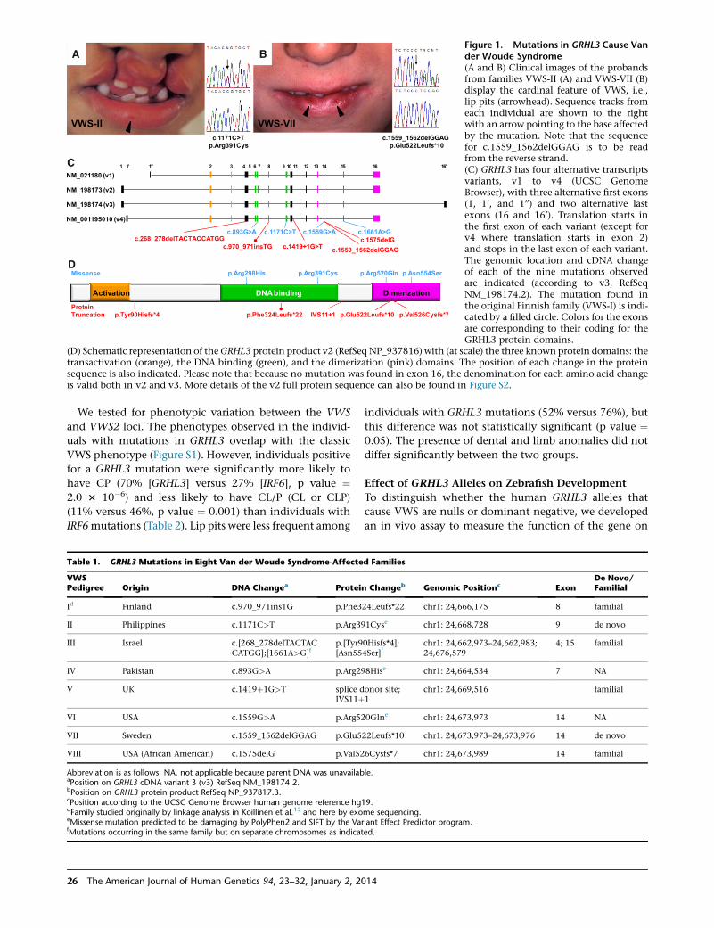
A B
C
D
Figure 1. Mutations in GRHL3 Cause Vander Woude Syndrome(A and B) Clinical images of the probandsfrom families VWS-II (A) and VWS-VII (B)display the cardinal feature of VWS, i.e.,lip pits (arrowhead). Sequence tracks fromeach individual are shown to the rightwith an arrow pointing to the base affectedby the mutation. Note that the sequencefor c.1559_1562delGGAG is to be readfrom the reverse strand.(C) GRHL3 has four alternative transcriptsvariants, v1 to v4 (UCSC GenomeBrowser), with three alternative first exons(1, 1’, and 1’’) and two alternative lastexons (16 and 16’). Translation starts inthe first exon of each variant (except forv4 where translation starts in exon 2)and stops in the last exon of each variant.The genomic location and cDNA changeof each of the nine mutations observedare indicated (according to v3, RefSeqNM_198174.2). The mutation found inthe original Finnish family (VWS-I) is indi-cated by a filled circle. Colors for the exonsare corresponding to their coding for theGRHL3 protein domains.
(D) Schematic representation of theGRHL3 protein product v2 (RefSeqNP_937816) with (at scale) the three known protein domains: thetransactivation (orange), the DNA binding (green), and the dimerization (pink) domains. The position of each change in the proteinsequence is also indicated. Please note that because no mutation was found in exon 16, the denomination for each amino acid changeis valid both in v2 and v3. More details of the v2 full protein sequence can also be found in Figure S2.
We tested for phenotypic variation between the VWS
and VWS2 loci. The phenotypes observed in the individ-
uals with mutations in GRHL3 overlap with the classic
VWS phenotype (Figure S1). However, individuals positive
for a GRHL3 mutation were significantly more likely to
have CP (70% [GRHL3] versus 27% [IRF6], p value ¼2.0 3 10�6) and less likely to have CL/P (CL or CLP)
(11% versus 46%, p value ¼ 0.001) than individuals with
IRF6mutations (Table 2). Lip pits were less frequent among
Table 1. GRHL3 Mutations in Eight Van der Woude Syndrome-Affecte
VWSPedigree Origin DNA Changea Protein
Id Finland c.970_971insTG p.Phe3
II Philippines c.1171C>T p.Arg39
III Israel c.[268_278delTACTACCATGG];[1661A>G]f
p.[Tyr9[Asn55
IV Pakistan c.893G>A p.Arg29
V UK c.1419þ1G>T splice dIVS11þ
VI USA c.1559G>A p.Arg52
VII Sweden c.1559_1562delGGAG p.Glu5
VIII USA (African American) c.1575delG p.Val52
Abbreviation is as follows: NA, not applicable because parent DNA was unavailabaPosition on GRHL3 cDNA variant 3 (v3) RefSeq NM_198174.2.bPosition on GRHL3 protein product RefSeq NP_937817.3.cPosition according to the UCSC Genome Browser human genome reference hg1dFamily studied originally by linkage analysis in Koillinen et al.15 and here by exoeMissense mutation predicted to be damaging by PolyPhen2 and SIFT by the VarfMutations occurring in the same family but on separate chromosomes as indicat
26 The American Journal of Human Genetics 94, 23–32, January 2, 20
individuals with GRHL3 mutations (52% versus 76%), but
this difference was not statistically significant (p value ¼0.05). The presence of dental and limb anomalies did not
differ significantly between the two groups.
Effect of GRHL3 Alleles on Zebrafish Development
To distinguish whether the human GRHL3 alleles that
cause VWS are nulls or dominant negative, we developed
an in vivo assay to measure the function of the gene on
d Families
Changeb Genomic Positionc ExonDe Novo/Familial
24Leufs*22 chr1: 24,666,175 8 familial
1Cyse chr1: 24,668,728 9 de novo
0Hisfs*4];4Ser]f
chr1: 24,662,973–24,662,983;24,676,579
4; 15 familial
8Hise chr1: 24,664,534 7 NA
onor site;1
chr1: 24,669,516 familial
0Glne chr1: 24,673,973 14 NA
22Leufs*10 chr1: 24,673,973–24,673,976 14 de novo
6Cysfs*7 chr1: 24,673,989 14 familial
le.
9.me sequencing.iant Effect Predictor program.ed.
14
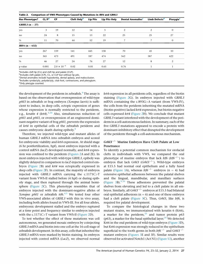
Table 2. Comparison of VWS Phenotypes Caused by Mutations in IRF6 and GRHL3
Has Phenotype? CL/Pa CP Cleft Onlyb Lip Pits Lip Pits Only Dental Anomaliesc Limb Defectsd Pterygiae
GRHL3 (n ¼ 27)
yes 3 19 12 14 5 2 2 0
no 24 8 15 13 22 25 25 27
% 11 70 44 52 19 7 7 0
IRF6 (n ¼ 632)
yes 267 159 141 445 158 70 45 10
no 365 473 491 187 474 562 587 622
% 46 27 24 76 27 12 8 2
p value 0.001 2.0 3 10�6 0.02 0.05 0.65 0.76 1 1
aIncludes cleft lip (CL) and cleft lip and palate (CLP).bIncludes cleft palate (CP), CL, or CLP but without lip pits.cDental anomalies include hypodontia, dental aplasia, and malocclusion.dIncludes syndactyly, polydactyly, club foot, contractures, and pterygium.eOnly pterygia counted.
the development of the periderm in zebrafish.4 The assay is
based on the observation that overexpression of wild-type
grhl3 in zebrafish or frog embryos (Xenopus laevis) is suffi-
cient to induce, in deep cells, ectopic expression of genes
whose expression is normally restricted to the periderm,
e.g., keratin 4 (krt4).4,24 Also, simultaneous reduction of
grhl1 and grhl3, or overexpression of an engineered domi-
nant-negative variant of frog grhl1, prevents the expression
of krt4 in epithelial cells of the zebrafish periderm and
causes embryonic death during epiboly.4
Therefore, we injected wild-type and mutant alleles of
human GRHL3 mRNA into zebrafish embryos and scored
for embryonic viability and krt4 expression. At shield stage
(6 hr postfertilization, hpf), most embryos injected with a
control mRNA (lacZ) developed normally, and krt4 expres-
sion was confined to the periderm (Figures 2A and 2E). In
most embryos injected with wild-type GRHL3, epiboly was
slightly delayed in comparison to lacZ-injected control em-
bryos (Figure 2B) and krt4 was ectopically expressed in
deep cells (Figure 2F). In contrast, the majority of embryos
injected with GRHL3 mRNA carrying the c.1171C>T
variant from VWS-II stalled before (4 hpf) or during epib-
oly stage, and then ruptured through the animal hemi-
sphere (Figure 2C). This phenotype resembles that of
embryos injected with the dominant-negative alleles of
Xenopus grhl1 or zebrafish irf6.4,5 We tested four other
VWS-associated alleles of GRHL3 with this in vivo assay,
including both alleles found in VWS-III. For all four alleles,
embryonic development stalled and the embryo ruptured
at a time point and frequency similar to embryos injected
with the c.1171C>T variant from VWS-II (Figure 2D).
To test whether the effect of these mutations was cell
autonomous, we generated mosaic embryos by coinjecting
GRHL3mRNA and biotin into one cell at the 16-cell stage of
zebrafish development. In this assay, cells that inherited the
GRHL3 mRNAwere marked by biotin staining. In embryos
injected with control mRNA (LacZ), we observed normal
The A
krt4 expression in all periderm cells, regardless of the biotin
staining (Figure 2G). In embryos injected with GRHL3
mRNA containing the c.893G>A variant (from VWS-IV),
the cells from the periderm inheriting the mutated mRNA
(biotin-positive) lacked krt4 expression, but biotin-negative
cells expressed krt4 (Figure 2H). We conclude that mutant
GRHL3 variant interferedwith the development of the peri-
derminacell-autonomous fashion. In summary, eachof the
five GRHL3 mutations appeared to encode a protein with
dominant-inhibitory effect that disrupted the development
of the periderm through a cell-autonomous mechanism.
Grhl3�/� Murine Embryos Have Cleft Palate at Low
Penetrance
To identify a potential common mechanism for orofacial
clefts in individuals with VWS, we compared the oral
phenotype of murine embryos that lack Irf6 (Irf6�/�) to
embryos that lack Grhl3 (Grhl3�/�). Wild-type embryos
at E15.5 had normal oral epithelium and a fully fused
palate (Figure 3A), whereas Irf6�/� embryos (n ¼ 4) had
extensive epithelial adhesions between the palatal shelves
and the lingual, mandibular, and maxillary surfaces
(Figure 3B).9,10 These adhesions prevented the palatal
shelves from elevating and led to a cleft palate in all em-
bryos. Similarly, allGrhl3�/� embryos at E15.5 had bilateral
oral epithelial adhesions (n ¼ 6) and one of these embryos
had a cleft palate (Figure 3C). Thus, Grhl3, like Irf6, is
required for palatal development.
To compare the histological changes in these two
mutant strains, we immunostained with keratin 6 (Krt6),
a marker for the periderm,25 and tumor protein p63
(p63), a marker for the basal epithelial layer.26 We detected
Krt6 in the oral periderm of wild-type embryos (Figure 3D),
but Krt6 expression was strongly reduced in the epithelium
superficial to the tooth germs in both Irf6�/� and Grhl3�/�
mutant embryos (Figures 3E and 3F). Similar results were
observed for activated Notch1 (Act N1) (Figure S3), another
merican Journal of Human Genetics 94, 23–32, January 2, 2014 27
Figure 2. VWS-Associated Alleles ofGRHL3 Disrupt the Development of thePeriderm when Expressed in ZebrafishEmbryos(A–C) Lateral views of live sibling embryosinjected with control (A), GRHL3 (B), orGRHL3 (c.1171C>T) (C) mRNA. Embryoshown in (C), injected with the GRHL3mRNA carrying the c.1171C>T mutation,ruptured through the animal hemisphereshortly after the image was taken (67%[n ¼ 48] of wild-type GRHL3-injected em-bryos reached at least 50% epiboly stage,whereas 76% [n ¼ 115] of mutant-injectedembryos burst without initiating epiboly).(D) Histogram showing fraction of em-bryos that ruptured when injected with
indicated mRNA. Percentage is the average from 3–4 separate experiments of 20–40 embryos each. Error bars represent standarderror.(E and F) Animal pole views of embryos injected with indicated mRNA and processed to detect krt4 expression. Insets, cross sec-tions of the same embryos showing (E) krt4 expression confined to the periderm and (F) ectopically in deep cells.(G and H) Animal pole views of mosaic embryos injected with mRNA and biotinylated-dextran at 16-cell stage, fixed at shieldstage, and processed for krt4 expression (blue) and biotin distribution (brown). Periderm cells possessed (black arrowhead) or lacked(white arrowhead) biotin stain, demonstrating that they were, or were not, derived from an RNA-injected cell, respectively.Daughter cells derived from the cell injected with the c.893G>A mutant variant of GRHL3 lack krt4 expression.Scale bars represent 500 mm (A–C, E, F), 100 mm (inset in E and F), and 20 mm (G, H).
protein expressed in the periderm.11 Thus, we concluded
that both Irf6 and Grhl3 were required for proper develop-
ment of the oral periderm in the mouse.
In addition to its potential role in the periderm, Irf6 regu-
lates the differentiation of the keratinocytes in the
epidermis9,10 and the oral cavity.11 In the oral cavity, wild-
type embryos had a uniform, single layer of basal epithe-
lium (Figure 3D), whereas the basal layer in Irf6�/� embryos
was disorganized and thicker, and p63 was ectopically ex-
pressed in the cells of the suprabasal layer (Figure 3E). In
Grhl3�/� embryos, the basal epithelial layer appeared
grossly normal with normal expression of p63 (Figure 3F).
We also looked at the medial edge epithelium (MEE), the
epithelium located at themedial edge of the palatal shelves
that must dissolve for proper palatal fusion. In wild-type
(Figure 3G) and Grhl3�/� (Figure 3I) embryos, the MEE dis-
solved to form a confluent bridge of mesenchymal cells
across the palate as shown by the loss of expression of
p63. In contrast, although we do not know the exact loca-
tion of the MEE in Irf6�/� embryos, expression of p63 per-
sisted throughout the epithelium of the palatal shelves
(Figure 3H).11 Thus, Irf6�/� embryos have at least two prob-
lems during palatal development: the presence of oral
epithelial adhesions and the failure of the MEE to dissolve.
In contrast, Grhl3�/� embryos have only oral epithelial ad-
hesions because of the loss of periderm. Because mutations
in both these genes cause VWS, these results are consistent
with the hypothesis that abnormal periderm function con-
tributes to CL/P in humans.
The Oral Phenotypes of Irf6 and Grhl3 Heterozygous
Murine Mutants Are Independent
Based on ChIP-seq experiments on a human keratinocyte
cell line and epistasis experiments in zebrafish embryos,
we hypothesized that Irf6 andGrhl3 function in a common
28 The American Journal of Human Genetics 94, 23–32, January 2, 20
pathway.4,6 To test for epistasis during murine palatogene-
sis, we generated embryos that were heterozygous for both
Irf6 and Grhl3 (Irf6þ/�;Grhl3þ/�). As expected, we did not
observe any oral epithelial adhesions in wild-type embryos
(Figures 4Aand4D). In Irf6þ/� embryoswedetectedbilateral
oral adhesions at the tooth germ sites (Figure 4B). We also
observed bilateral epithelial abnormalities in Grhl3þ/� em-
bryos (Figure 4E), but they differed from those seen in the
Irf6þ/� embryos in three respects. First, whereas oral adhe-
sions in Irf6þ/� embryos were more prominent at the tooth
germ sites (Figure 4B), epithelial abnormalities in Grhl3þ/�
embryos were located throughout the oral cavity and most
frequently posterior to the tooth germs (Figure 4E). Second,
epithelial abnormalities included oral fusions (Figure 4E),
which donot occur in Irf6þ/� embryos.Here,we distinguish
oral epithelial adhesions from oral fusions histologically.
Whereas adhesions have a loss of periderm that allows cell
interactions between two adjacent epithelial layers, fusions
have a loss of both the periderm and the basal epithelial
layers that allows cell interactions between the underlying
mesenchymal cells from adjacent tissues. Finally, whereas
oral adhesions in Irf6þ/� occurredmost frequently between
themandible andmaxilla, oral fusions inGrhl3þ/� embryos
occurred between themandible and either the palate or the
maxilla. In the Irf6þ/�;Grhl3þ/� double heterozygous em-
bryos, we found oral adhesions at areas superficial to the
tooth germ (Figure 4C), similar to Irf6þ/� embryos, as well
as oral adhesions and fusions posterior to the tooth germ
(Figure 4F), similar to Grhl3þ/� embryos. Thus, the oral his-
topathology of the Irf6þ/�;Grhl3þ/� double heterozygote
embryos provides no evidence for epistasis and suggests
that Irf6 andGrhl3 function in independent but converging
pathways during oral periderm development.
As previously observed in the single knockout Irf6�/�
and Grhl3�/� embryos, we detected a reduction in
14
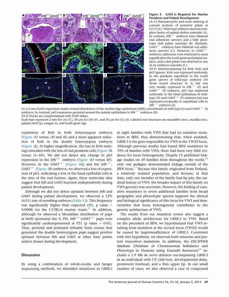
Figure 3. Grhl3 Is Required for MurinePeriderm and Palatal Development(A–C) Haematoxylin and eosin staining ofcoronal sections of posterior palate atE15.5 (A0).Wild-typeembryos showedcom-plete fusion of palatal shelves (asterisk) (A).In contrast, Irf6�/� embryos have bilateraloral adhesions (arrows) and a fully pene-trant cleft palate (asterisk) (B). Similarly,Grhl3�/� embryos have bilateral oral adhe-sions (arrows) (C). However, in Grhl3�/�
embryos, adhesions were restricted to areassuperficial to the toothgermandpalatal sur-faces, and a cleft palate was observed in oneof six embryos (asterisk) (C).(D–F) Immunostaining for Krt6 (red) andp63 (green). Krt6 was expressed uniformlyin the periderm superficial to the toothgerm (arrow) of wild-type embryos (D)(from boxed structure in A) but wasvery weakly expressed in Irf6�/� (E) andGrhl3�/� (F) embryos. p63 was expresseduniformly in the basal epithelium of wild-type (D) and Grhl3�/� (F) embryos but wasexpressed ectopically in suprabasal cells inIrf6�/� embryos (E).
(G–I) Loss of p63 expression marks normal dissolution of the medial edge epithelium (MEE) (arrowhead) in wild-type (G) andGrhl3�/� (I)embryos. In contrast, p63 expression persisted around the palatal epithelium in Irf6�/� embryos (H).(D–I) Nuclei are counterstained with DAPI (blue).Scale bars represent 2 mm for (A)–(C), 20 mm for (D)–(F), and 50 mm for (G)–(I). Labeled oral structures are mandible (mn), maxilla (mx),palatal shelf (p), tongue (t), and tooth germ (tg).
expression of Krt6 in both heterozygous embryos
(Figure 4G versus 4H and 4I) and a more apparent reduc-
tion of Krt6 in the double heterozygous embryos
(Figure 4J). At higher magnification, the loss in Krt6 stain-
ing coincided with the loss of oral periderm cells (Figure 4K
versus 4L–4N). We did not detect any change in p63
expression in the Irf6þ/� embryos (Figure 4O versus 4P).
However, in the Grhl3þ/� (Figure 4Q) and the Irf6þ/�;Grhl3þ/� (Figure 4R) embryos, we observed a loss of expres-
sion of p63, indicating a loss of the basal epithelial cells at
the sites of the oral fusions. Again, these molecular data
suggest that Irf6 and Grhl3 function independently during
palatal development.
Although we did not detect epistasis between Irf6 and
Grhl3 during palatal development, we observed a 12%
(6/51) rate of resorbing embryos (Table S3). This frequency
was significantly higher than expected (3%, p value ¼0.0008) for the C57BL/6 murine strain.27 In addition,
although we observed a Mendelian distribution of pups
at birth (postnatal day 0, P0), Irf6þ/�;Grhl3þ/� pups were
significantly underrepresented at P21 (p value ¼ 0.01).
Thus, prenatal and postnatal lethality from crosses that
generated the double heterozygous pups suggest positive
epistasis between Irf6 and Grhl3 at other time points
and/or tissues during development.
Discussion
By using a combination of whole-exome and Sanger
sequencing methods, we identified mutations in GRHL3
The A
in eight families with VWS that had no causative muta-
tions in IRF6, thus demonstrating that, when mutated,
GRHL3 is the gene responsible for VWS at the VWS2 locus.
Although previous studies had found IRF6 mutations in
70% of families with VWS, there had been very little evi-
dence for locus heterogeneity. Despite 15 published link-
age studies on 49 families from throughout the world,28
only one pedigree demonstrated linkage outside of the
IRF6 locus.15 Because this family originated from Finland,
a relatively isolated population, and because, at that
time, only one member of the family had lip pits, the car-
dinal feature of VWS, the broader impact of this family on
VWS genetics was uncertain. However, the finding of caus-
ative mutations in seven additional families from broad
geographic and phenotypic spectra supports the clinical
and biological significance of this locus for VWS and dem-
onstrates that locus heterogeneity contributes to the
genetic architecture of VWS.
The results from our mutation screen also suggest a
complex allelic architecture for GRHL3 in VWS. Based
on the precedent of IRF6, we hypothesized that VWS re-
sulting from mutation at the second locus (VWS2) would
be caused by haploinsufficiency of GRHL3. Consistent
with this hypothesis, we observed both missense and pro-
tein truncation mutations. In addition, the DECIPHER
database (Database of Chromosomal Imbalance and
Phenotype in Humans using Ensembl Resources)29 in-
cludes a 1.9 Mb de novo deletion encompassing GRHL3
in an individual with CP, club foot, developmental delay,
prominent forehead, and a thin upper lip. In our small
number of cases, we also observed a case of compound
merican Journal of Human Genetics 94, 23–32, January 2, 2014 29
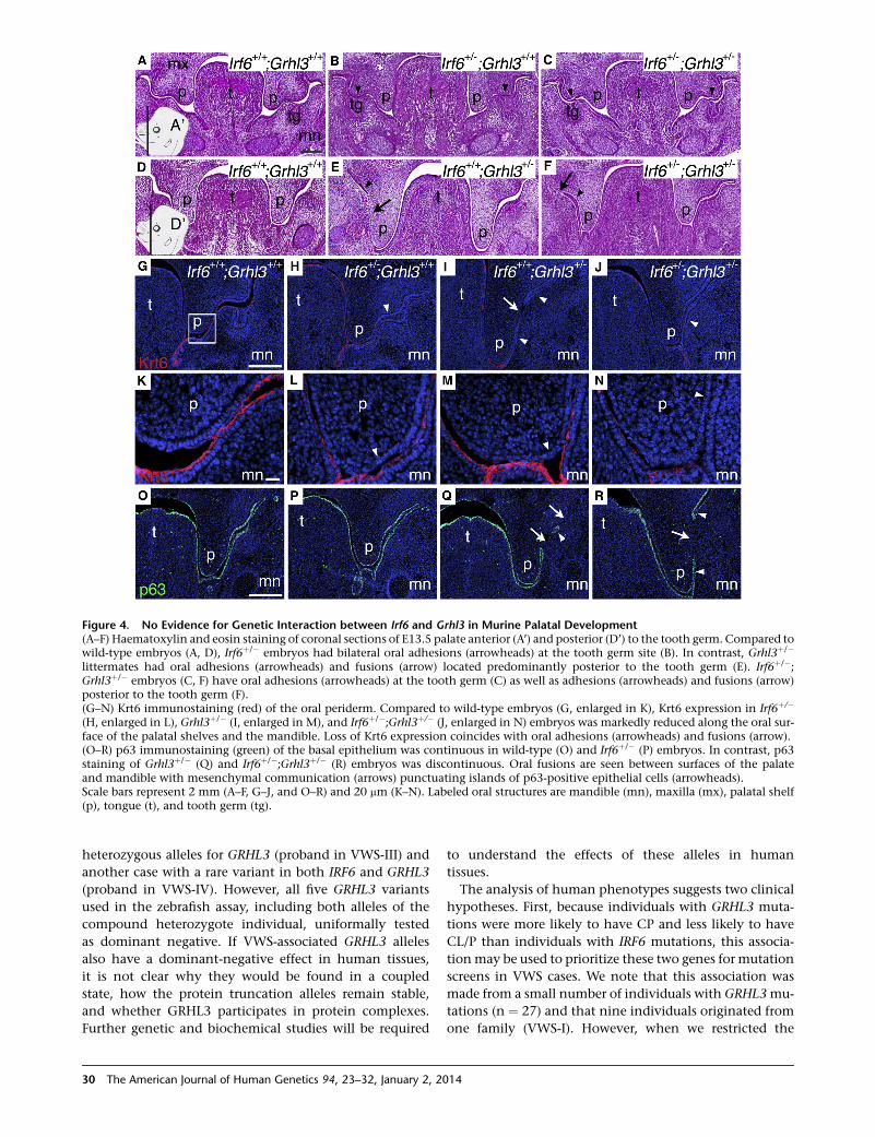
Figure 4. No Evidence for Genetic Interaction between Irf6 and Grhl3 in Murine Palatal Development(A–F) Haematoxylin and eosin staining of coronal sections of E13.5 palate anterior (A’) and posterior (D’) to the tooth germ. Compared towild-type embryos (A, D), Irf6þ/� embryos had bilateral oral adhesions (arrowheads) at the tooth germ site (B). In contrast, Grhl3þ/�
littermates had oral adhesions (arrowheads) and fusions (arrow) located predominantly posterior to the tooth germ (E). Irf6þ/�;Grhl3þ/� embryos (C, F) have oral adhesions (arrowheads) at the tooth germ (C) as well as adhesions (arrowheads) and fusions (arrow)posterior to the tooth germ (F).(G–N) Krt6 immunostaining (red) of the oral periderm. Compared to wild-type embryos (G, enlarged in K), Krt6 expression in Irf6þ/�
(H, enlarged in L), Grhl3þ/� (I, enlarged in M), and Irf6þ/�;Grhl3þ/� (J, enlarged in N) embryos was markedly reduced along the oral sur-face of the palatal shelves and the mandible. Loss of Krt6 expression coincides with oral adhesions (arrowheads) and fusions (arrow).(O–R) p63 immunostaining (green) of the basal epithelium was continuous in wild-type (O) and Irf6þ/� (P) embryos. In contrast, p63staining of Grhl3þ/� (Q) and Irf6þ/�;Grhl3þ/� (R) embryos was discontinuous. Oral fusions are seen between surfaces of the palateand mandible with mesenchymal communication (arrows) punctuating islands of p63-positive epithelial cells (arrowheads).Scale bars represent 2 mm (A–F, G–J, and O–R) and 20 mm (K–N). Labeled oral structures are mandible (mn), maxilla (mx), palatal shelf(p), tongue (t), and tooth germ (tg).
heterozygous alleles for GRHL3 (proband in VWS-III) and
another case with a rare variant in both IRF6 and GRHL3
(proband in VWS-IV). However, all five GRHL3 variants
used in the zebrafish assay, including both alleles of the
compound heterozygote individual, uniformally tested
as dominant negative. If VWS-associated GRHL3 alleles
also have a dominant-negative effect in human tissues,
it is not clear why they would be found in a coupled
state, how the protein truncation alleles remain stable,
and whether GRHL3 participates in protein complexes.
Further genetic and biochemical studies will be required
30 The American Journal of Human Genetics 94, 23–32, January 2, 20
to understand the effects of these alleles in human
tissues.
The analysis of human phenotypes suggests two clinical
hypotheses. First, because individuals with GRHL3 muta-
tions were more likely to have CP and less likely to have
CL/P than individuals with IRF6 mutations, this associa-
tionmay be used to prioritize these two genes for mutation
screens in VWS cases. We note that this association was
made from a small number of individuals with GRHL3mu-
tations (n ¼ 27) and that nine individuals originated from
one family (VWS-I). However, when we restricted the
14
analysis to a family-based phenotype (n ¼ 8), we observed
the same trends, although not achieving statistical signifi-
cance because of low power. Second, like IRF6, common
DNA variants in GRHL3 may also be associated with iso-
lated forms of orofacial clefting,30 especially for CP, given
the increased likelihood of CP in individuals with a muta-
tion in GRHL3. However, multiple genome-wide associa-
tion studies for CL/P31 and one for CP32 have not provided
strong evidence for common variants at the GRHL3 locus.
Although these studies suggest that commonDNA variants
inGRHL3 do not account for significant risk for CL/P or CP,
GRHL3 remains an excellent candidate gene for isolated
orofacial clefts.
Finally, our analysis of phenotypes in Irf6 and Grhl3
mutant mice identified common and distinct oral abnor-
malities. Previous studies revealed that Irf6 deficiency in
mice could lead to an orofacial cleft by at least two patho-
physiological mechanisms: abnormal periderm differentia-
tion and failure of the medial edge epithelium (MEE) to
dissolve.11,33 The MEE was able to dissolve normally in
embryos that lack Grhl3, so the common feature of Irf6
and Grhl3 mutants is failed periderm differentiation,
strengthening the previously hypothesized role of peri-
derm in development of the lip and palate.
In conclusion, these studies identifyGRHL3 as the second
gene thatwhenmutated leads toVanderWoude syndrome,
thus confirming locus heterogeneity for this syndrome.
Further, they strengthen the connection between cleft pal-
ate and abnormal periderm development. We anticipate
that these findings will improve the molecular diagnostic
for VWS and other forms of orofacial clefting.
Supplemental Data
Supplemental Data include three figures and three tables and can
be found with this article online at http://www.cell.com/AJHG/.
Acknowledgments
Wegreatly appreciate themany individuals affectedwithVWS, their
family members, and clinicians for participating in this study. We
would like to thank Arianna L. Smith andMager Scientific for tech-
nical assistance; Nicole Patel for the artistic renderings of murine
embryos at E13.5 and E15.5; Paivi Lahermo for providing the
Finnish controls and Pat Venta for critiques. Financial support for
this research was provided by the Swedish Research Council 521-
2007-3133 (M.P.-J.) and 2009-5091 (J.K.), by National Institutes of
Health grants DE021071 (R.A.C.), DE13513 (B.C.S.), F31DE022696
(Y.A.K.), DE08559 (J.C.M.), GM008629 (E.J.L.), AR061586 (M.D.),
and AR44882 (B.A.), and by the Sigrid Juselius Foundation (J.K.).
Received: September 9, 2013
Accepted: November 14, 2013
Published: December 19, 2013
Web Resources
The URLs for data presented herein are as follows:
GATK, http://www.broadinstitute.org/gatk/
The A
NHLBI Exome Sequencing Project (ESP) Exome Variant Server,
http://evs.gs.washington.edu/EVS/
Online Mendelian Inheritance in Man (OMIM), http://www.
omim.org/
Picard, http://picard.sourceforge.net/
Primer3, http://bioinfo.ut.ee/primer3-0.4.0/primer3/
UCSC Human Genome Browser, http://genome.ucsc.edu/cgi-bin/
hgGateway
Accession Numbers
The dbSNP accession number for the KTI12 (c.337_363delCCGA
TCGCGGGACCTCAGGTGGCGGGC) variant reported in this
paper is ss836732090.
References
1. Mace, K.A., Pearson, J.C., and McGinnis, W. (2005). An
epidermal barrier wound repair pathway inDrosophila is medi-
ated by grainy head. Science 308, 381–385.
2. Ting, S.B., Caddy, J., Hislop, N., Wilanowski, T., Auden, A.,
Zhao, L.L., Ellis, S., Kaur, P., Uchida, Y., Holleran, W.M., et al.
(2005). A homolog of Drosophila grainy head is essential for
epidermal integrity in mice. Science 308, 411–413.
3. Yu, Z., Lin, K.K., Bhandari, A., Spencer, J.A., Xu, X., Wang, N.,
Lu, Z., Gill, G.N., Roop, D.R., Wertz, P., and Andersen, B.
(2006). The Grainyhead-like epithelial transactivator Get-1/
Grhl3 regulates epidermal terminal differentiation and inter-
acts functionally with LMO4. Dev. Biol. 299, 122–136.
4. de la Garza, G., Schleiffarth, J.R., Dunnwald, M., Mankad, A.,
Weirather, J.L., Bonde, G., Butcher, S., Mansour, T.A., Kousa,
Y.A., Fukazawa, C.F., et al. (2013). Interferon regulatory factor
6promotes differentiationof theperidermbyactivatingexpres-
sion of Grainyhead-like 3. J. Invest. Dermatol. 133, 68–77.
5. Sabel, J.L., d’Alencon, C., O’Brien, E.K., Van Otterloo, E., Lutz,
K., Cuykendall, T.N., Schutte, B.C., Houston, D.W., and Cor-
nell, R.A. (2009). Maternal Interferon Regulatory Factor 6 is
required for the differentiation of primary superficial epithelia
in Danio and Xenopus embryos. Dev. Biol. 325, 249–262.
6. Botti, E., Spallone, G., Moretti, F., Marinari, B., Pinetti, V., Gal-
anti, S., De Meo, P.D., De Nicola, F., Ganci, F., Castrignano, T.,
et al. (2011). Developmental factor IRF6 exhibits tumor sup-
pressor activity in squamous cell carcinomas. Proc. Natl.
Acad. Sci. USA 108, 13710–13715.
7. Tamura, T., Yanai, H., Savitsky, D., and Taniguchi, T. (2008).
The IRF family transcription factors in immunity and onco-
genesis. Annu. Rev. Immunol. 26, 535–584.
8. Kondo, S., Schutte, B.C., Richardson, R.J., Bjork, B.C., Knight,
A.S., Watanabe, Y., Howard, E., de Lima, R.L., Daack-Hirsch, S.,
Sander, A., et al. (2002).Mutations in IRF6 causeVanderWoude
and popliteal pterygium syndromes. Nat. Genet. 32, 285–289.
9. Ingraham, C.R., Kinoshita, A., Kondo, S., Yang, B., Sajan, S.,
Trout, K.J., Malik, M.I., Dunnwald, M., Goudy, S.L., Lovett,
M., et al. (2006). Abnormal skin, limb and craniofacial
morphogenesis in mice deficient for interferon regulatory
factor 6 (Irf6). Nat. Genet. 38, 1335–1340.
10. Richardson, R.J., Dixon, J., Malhotra, S., Hardman, M.J.,
Knowles, L., Boot-Handford, R.P., Shore, P., Whitmarsh, A., and
Dixon, M.J. (2006). Irf6 is a key determinant of the keratinocyte
proliferation-differentiation switch. Nat. Genet. 38, 1329–1334.
11. Richardson, R.J., Dixon, J., Jiang, R., and Dixon, M.J. (2009).
Integration of IRF6 and Jagged2 signalling is essential for
merican Journal of Human Genetics 94, 23–32, January 2, 2014 31
controlling palatal adhesion and fusion competence. Hum.
Mol. Genet. 18, 2632–2642.
12. Burdick, A.B., Bixler, D., and Puckett, C.L. (1985). Genetic
analysis in families with van der Woude syndrome.
J. Craniofac. Genet. Dev. Biol. 5, 181–208.
13. de Lima, R.L., Hoper, S.A., Ghassibe, M., Cooper, M.E., Rorick,
N.K., Kondo, S., Katz, L., Marazita, M.L., Compton, J., Bale, S.,
et al. (2009). Prevalence and nonrandom distribution of
exonic mutations in interferon regulatory factor 6 in 307 fam-
ilies with Van der Woude syndrome and 37 families with
popliteal pterygium syndrome. Genet. Med. 11, 241–247.
14. Leslie, E.J., Standley, J., Compton, J., Bale, S., Schutte, B.C.,
andMurray, J.C. (2013). Comparative analysis of IRF6 variants
in families with Van der Woude syndrome and popliteal pte-
rygium syndrome using public whole-exome databases.
Genet. Med. 15, 338–344.
15. Koillinen, H.,Wong, F.K., Rautio, J., Ollikainen, V., Karsten, A.,
Larson, O., Teh, B.T., Huggare, J., Lahermo, P., Larsson, C., and
Kere, J. (2001). Mapping of the second locus for the Van der
Woude syndrome to chromosome 1p34. Eur. J. Hum. Genet.
9, 747–752.
16. Schutte, B.C., Bjork, B.C., Coppage, K.B., Malik, M.I., Gregory,
S.G., Scott, D.J., Brentzell, L.M., Watanabe, Y., Dixon, M.J.,
and Murray, J.C. (2000). A preliminary gene map for the
Van der Woude syndrome critical region derived from 900
kb of genomic sequence at 1q32-q41. Genome Res. 10, 81–94.
17. Peyrard-Janvid, M., Pegelow, M., Koillinen, H., Larsson, C.,
Fransson, I., Rautio, J., Hukki, J., Larson, O., Karsten, A.L.,
and Kere, J. (2005). Novel and de novo mutations of the
IRF6 gene detected in patients with Van der Woude or popli-
teal pterygium syndrome. Eur. J. Hum. Genet. 13, 1261–1267.
18. Li, H., and Durbin, R. (2010). Fast and accurate long-read
alignment with Burrows-Wheeler transform. Bioinformatics
26, 589–595.
19. DePristo, M.A., Banks, E., Poplin, R., Garimella, K.V., Maguire,
J.R., Hartl, C., Philippakis, A.A., del Angel, G., Rivas, M.A.,
Hanna, M., et al. (2011). A framework for variation discovery
and genotyping using next-generation DNA sequencing data.
Nat. Genet. 43, 491–498.
20. McLaren, W., Pritchard, B., Rios, D., Chen, Y., Flicek, P., and
Cunningham, F. (2010). Deriving the consequences of
genomic variants with the Ensembl API and SNP Effect Predic-
tor. Bioinformatics 26, 2069–2070.
21. Pegelow, M., Koillinen, H., Magnusson, M., Fransson, I.,
Unneberg, P., Kere, J., Karsten, A., and Peyrard-Janvid, M.
(2013). Association and mutation analyses of the IRF6 gene
in families with non-syndromic and syndromic cleft lip
and/or cleft palate. Cleft Palate Craniofac. J. Published online
February 8, 2013. http://dx.doi.org/10.1597/11-220.
22. Thisse, C., and Thisse, B. (2008). High-resolution in situ hybridi-
zation towhole-mount zebrafish embryos.Nat. Protoc. 3, 59–69.
32 The American Journal of Human Genetics 94, 23–32, January 2, 20
23. Malik, S., Kakar, N., Hasnain, S., Ahmad, J., Wilcox, E.R., and
Naz, S. (2010). Epidemiology of Van der Woude syndrome
from mutational analyses in affected patients from Pakistan.
Clin. Genet. 78, 247–256.
24. Chalmers, A.D., Lachani, K., Shin, Y., Sherwood, V., Cho,
K.W., and Papalopulu, N. (2006). Grainyhead-like 3, a tran-
scription factor identified in a microarray screen, promotes
the specification of the superficial layer of the embryonic
epidermis. Mech. Dev. 123, 702–718.
25. Mazzalupo, S., and Coulombe, P.A. (2001). A reporter trans-
gene based on a human keratin 6 gene promoter is specifically
expressed in the periderm of mouse embryos. Mech. Dev. 100,
65–69.
26. Koster, M.I., Kim, S., Mills, A.A., DeMayo, F.J., and Roop, D.R.
(2004). p63 is the molecular switch for initiation of an epithe-
lial stratification program. Genes Dev. 18, 126–131.
27. Krishnan, L., Guilbert, L.J., Wegmann, T.G., Belosevic, M., and
Mosmann, T.R. (1996). T helper 1 response against Leishmania
major in pregnant C57BL/6 mice increases implantation fail-
ure and fetal resorptions. Correlation with increased IFN-
gamma and TNF and reduced IL-10 production by placental
cells. J. Immunol. 156, 653–662.
28. Schutte, B.C., Dixon, M.J., and Murray, J.C. (2008). IRF6 and
the Van der Woude popliteal pterygium syndromes and the
risk for non-syndromic cleft lip and palate. In Inborn Errors
of Development, C. Epstein, ed. (New York: Oxford University
Press), pp. 1069–1072.
29. Firth, H.V., Richards, S.M., Bevan, A.P., Clayton, S., Corpas,
M., Rajan, D., Van Vooren, S., Moreau, Y., Pettett, R.M., and
Carter, N.P. (2009). DECIPHER: Database of Chromosomal
Imbalance and Phenotype in Humans Using Ensembl Re-
sources. Am. J. Hum. Genet. 84, 524–533.
30. Zucchero, T.M., Cooper, M.E., Maher, B.S., Daack-Hirsch, S.,
Nepomuceno, B., Ribeiro, L., Caprau, D., Christensen, K., Su-
zuki, Y., Machida, J., et al. (2004). Interferon regulatory factor
6 (IRF6) gene variants and the risk of isolated cleft lip or palate.
N. Engl. J. Med. 351, 769–780.
31. Ludwig, K.U., Mangold, E., Herms, S., Nowak, S., Reutter, H.,
Paul, A., Becker, J., Herberz, R., AlChawa, T., Nasser, E., et al.
(2012). Genome-wide meta-analyses of nonsyndromic cleft
lip with or without cleft palate identify six new risk loci.
Nat. Genet. 44, 968–971.
32. Beaty, T.H., Ruczinski, I., Murray, J.C., Marazita, M.L., Munger,
R.G., Hetmanski, J.B., Murray, T., Redett, R.J., Fallin, M.D.,
Liang, K.Y., et al. (2011). Evidence for gene-environment
interaction in a genome wide study of nonsyndromic cleft
palate. Genet. Epidemiol. 35, 469–478.
33. Knight, A.S., Schutte, B.C., Jiang, R., and Dixon, M.J. (2006).
Developmental expression analysis of the mouse and chick
orthologues of IRF6: the gene mutated in Van der Woude
syndrome. Dev. Dyn. 235, 1441–1447.
14